Article

Characteristics of the Dental Pulp and Periodontal Ligament Stem Cells of the Yucatan Miniature Pig

1
College of Veterinary Medicine, Chungbuk National University, Cheongju 28644, Korea
2
Onnuri Animal Medical Center, Cheongju 28320, Korea
*
Author to whom correspondence should be addressed.
These two authors contributed equally to this work.
Appl. Sci. 2021, 11(20), 9461; https://doi.org/10.3390/app11209461
Submission received: 20 August 2021 / Revised: 8 October 2021 / Accepted: 9 October 2021 / Published: 12 October 2021
(This article belongs to the Topic State-of-the-Art Dentistry and Oral Health)

Abstract

:
Miniature pigs have been considered as a recommended large animal model for biomedical research. Mesenchymal stem cells offer promising potential for tissue regeneration. Recent studies have suggested that dental pulp stem cells (DPSCs) and periodontal ligament stem cells (PDLSCs) may provide more reliable strategies for the treatment of dental diseases using a cell-based tissue engineering approach. The aim of this study was to isolate and compare the characteristics of the DPSCs and PDLSCs of a miniature pig breed to the DPSCs and PDLSCs of a domestic farm pig breed. Stem cells of the DP and PDL were obtained from a male Yucatan miniature pig (nine months old) and a male domestic farm pig breed (six months old). The cell morphology, surface stem cell marker expression, proliferation, and osteogenic differentiation ability were evaluated. Under a light microscope, the DPSCs and PDLSCs of the miniature pig breed had morphologies similar to those of the domestic farm pig breed. The proliferation of PDLSCs in both animals showed no significant differences, except on day five, whereas the proliferation of DPSCs was significantly higher in the miniature pig breed. However, the osteogenic abilities of the DPSCs and PDLSCs from the miniature pig breed were significantly lower compared to the domestic farm pig breed. This observation emphasizes the need for the breed-specific optimization of an osteogenic differentiation culture protocol for Yucatan miniature pig DPSCs and PDLSCs before application to cell-based therapy for tissue engineering and regenerative medicine.

1. Introduction

Pigs have recently been considered one of the major animal models in biomedical research, as an alternative to dogs and primates, due to their own unique anatomy and physiology [1,2]. Pigs and humans share a number of important anatomic and physiologic characteristics [3,4], which potentially makes the pig a better model for biomedical research compared to other large animal species. Several strains of miniature pigs have been developed for biomedical research [5]. Miniature pig breeds require less space and less food, tend to be more tractable, and require less compound to be tested than domestic farm pig breeds [6]. These advantages of miniature pig breeds have contributed to a wide range of biomedical research.
Stem cell research with miniature pigs has developed rapidly. Recent successes suggest that stem cell research using miniature pigs will be promising in the future [7]. Mesenchymal stem cells (MSCs) have been found in the dental pulp and periodontal ligament. Dental pulp stem cells (DPSCs) and periodontal ligament stem cells (PDLSCs) have a variety of benefits, including a rapid proliferation rate, multi-differentiation ability, easy accessibility, and high viability. The most prominent feature of DPSCs and PDLSCs is the capacity to regenerate tissues, such as pulp and periodontal ligament, in vivo. These stem cells have broader clinical applications in bone and cartilage, adipose formation, muscle, and nerve cell production [8,9,10,11]. They are also simple when attempting to create distinct cell lineages [12]. In addition, these stem cells have characteristics that suppress the immune reaction, as do other stem cells [13,14]. On the basis of these features, many studies have utilized these cells from pigs, mainly in a dental disease model. Recently, many studies have suggested that dental stem cells, such as dental pulp (DP) and periodontal ligament (PDL) stem cells, may be useful in the treatment of dental diseases that require tissue regeneration [15,16,17,18]. However, it has been unclear as to whether studies using DPSCs and PDLSCs from miniature pig breeds are more efficient than those using domestic farm pig breeds. Furthermore, knowledge of the characteristics of DPSCs and PDLSCs of miniature pig breeds is scant compared to the published work on domestic farm pig breeds. Therefore, the present study aims to investigate and compare the characteristics of the DPSCs and PDLSCs of a miniature pig breed to those of a domestic farm pig breed by evaluating the cell morphology, surface marker expression levels, proliferation rates, and osteogenic differentiation abilities.

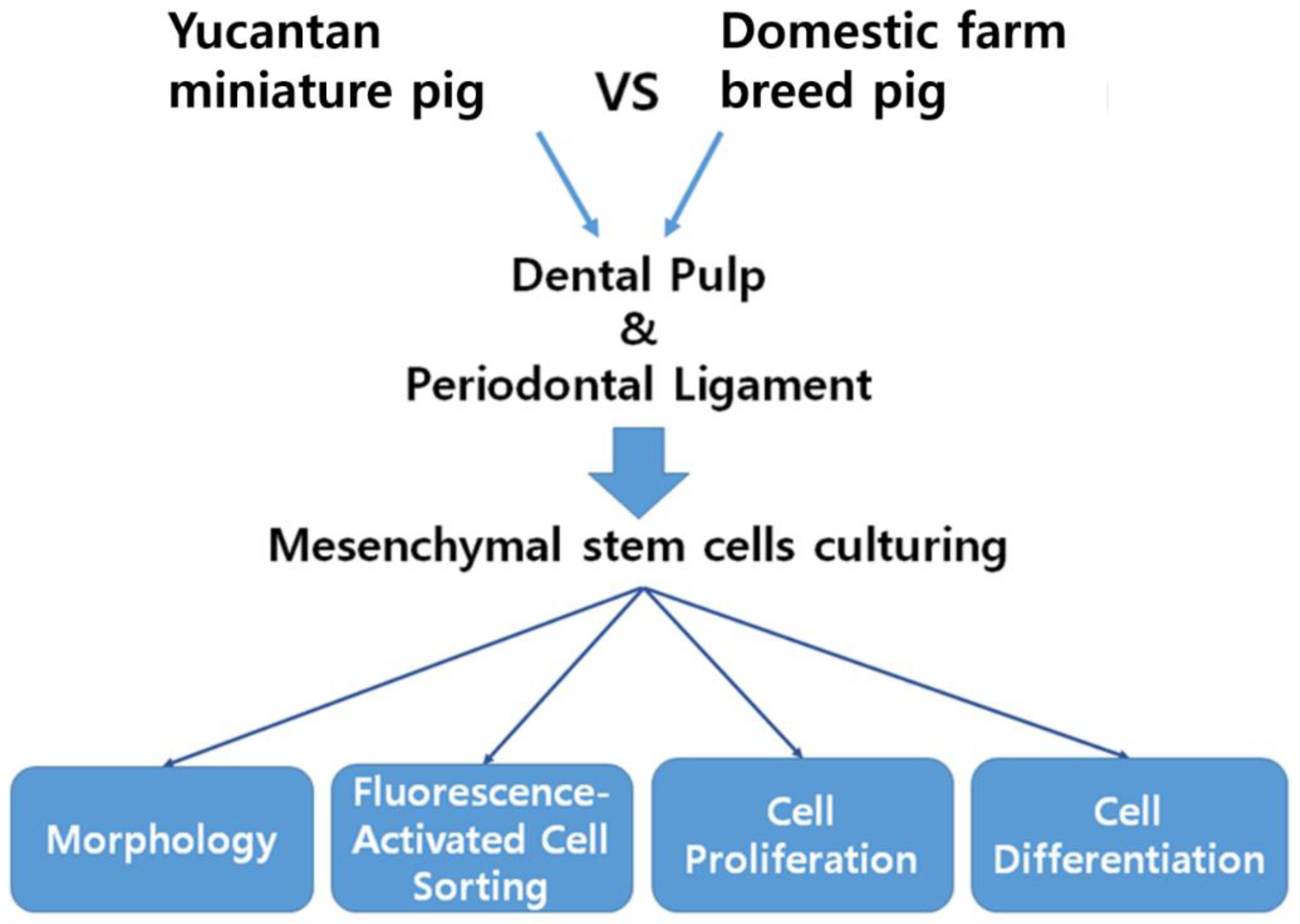
2. Materials and Methods

2.1. Animal and Cell Preparation

Incisor teeth were extracted from a male domestic farm pig breed (age 170 days, 115 kg weight) and a male Yucatan miniature pig breed (age 270 days, 25 kg weight) within two hours of death. PDL tissues were scraped from the middle third of the root surface. After the PDL tissue was collected, the pulp cavity was exposed using a hammer and chisel, and the pulp tissue was separated from the crown and root. The DP and PDL tissues were washed three times in PBS supplemented with penicillin-streptomycin (Gibco, Thermo Fisher Scientific, Waltham, MA, USA) and minced and digested in a solution of 3 mg/mL collagenase type I (Worthington Biochemical Products, Lakewood, NJ, USA) for 2 h at 37 °C. Then, the cell-digested solutions were filtered through a 40 μm cell strainer (Falcon, Thermo Fisher Scientific, Waltham, MA, USA) to obtain the single-cell suspensions of the PDLSCs and DPSCs. The single-cell suspensions were then cultured in α-modified Eagle medium (Hyclone, Global Life Sciences Solutions, Marlborough, MA, USA) containing 10% fetal bovine serum (Hyclone, Global Life Sciences Solutions, Marlborough, MA, USA) and 1% penicillin-streptomycin (Gibco, Thermo Fisher Scientific, Waltham, MA, USA) at 37 °C in a 5% CO2-humidified incubator. The culture medium was changed every three days. The scheme of the present study is presented in Figure 1. Cell morphology was evaluated at the passage 3 confluent cell monolayer using light microscopy (Figure 2).

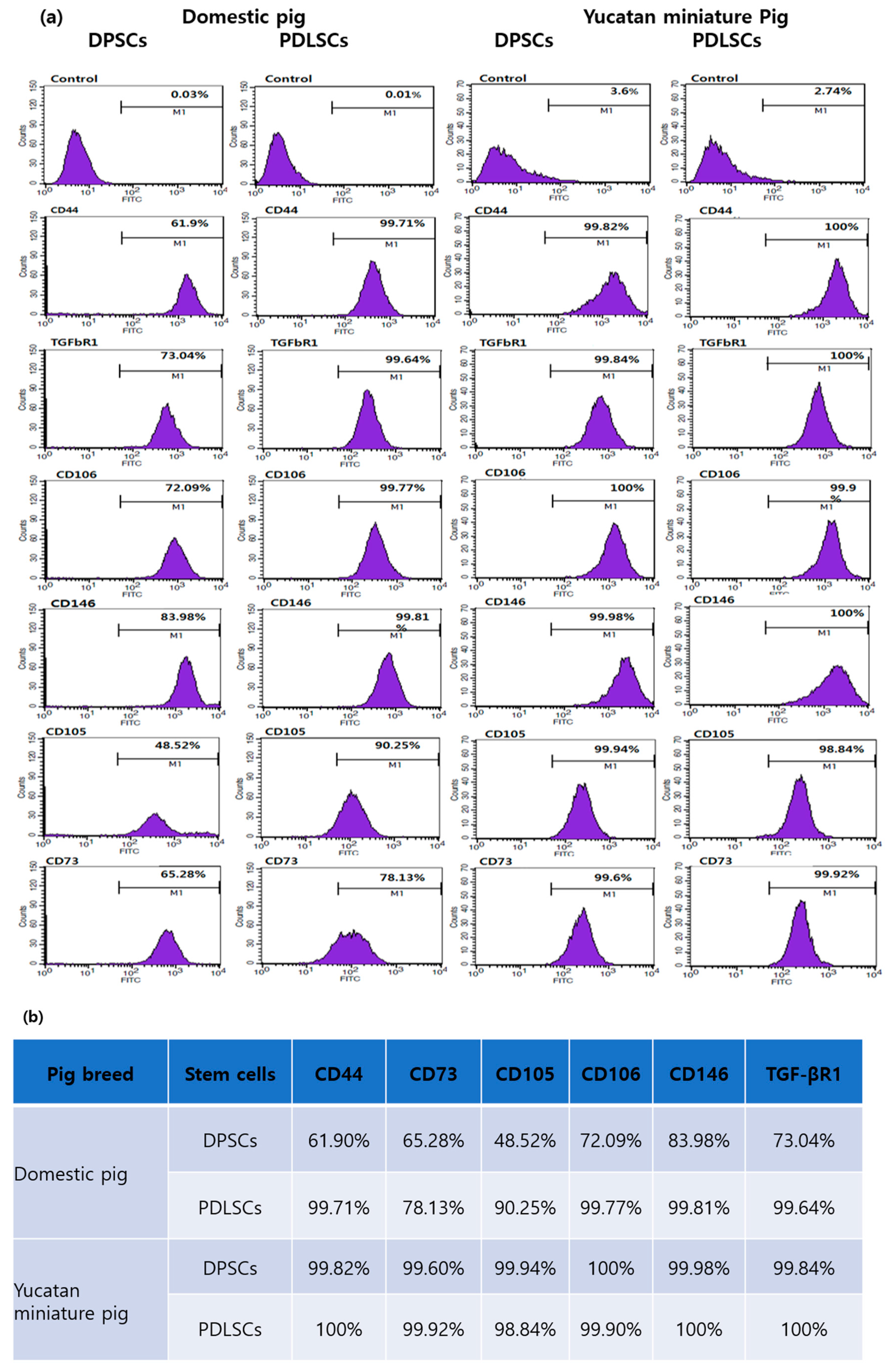
2.2. Fluorescence-Activated Cell Sorting (FACS) Analysis for Cell Surface Markers

Passage 3 cells of the DPSCs and PDLSCs of the domestic farm pig breed and the miniature pig breed were digested with trypsin/EDTA to obtain single-cell suspensions. Individual tubes containing the stem cells were incubated with the surface markers for multipotent mesenchymal stem cells—in this case, CD44 (Santa Cruz Biotechnology, Dallas, Texas, USA), TGF-β receptor1 (TGF-βR1) (AbCam, Cambridge MA, USA), CD106 (Santa Cruz Biotechnology), CD146 (AbCam), CD105 (Santa Cruz Biotechnology), and CD73 (Santa Cruz Biotechnology) for positive signals—and a FITC-labeled second antibody was added. After being washed in PBS, the cells were analyzed using a fluorescence-activated cell sorter (FACSCalibur, BD Biosciences, San Jose, CA, USA).

2.3. Cell Proliferation

The passage 3 cells of the DPSCs and PDLSCs of the domestic farm pig breed and the miniature pig breed were plated at a density of 104 cells/well in 24-well plates, and the proliferation outcomes of the DPSCs and PDLSCs were measured using a cell counting kit (CCK)-8 (Dojindo, Rockville, MD, USA) on days 1, 3, 5, and 7. The stem cell-seeded plates, at the desired time points, were rinsed with PBS and incubated in a CCK-8 solution in a 5% CO2 incubator at 37 °C for two hours. The absorbance was measured at 450 nm using a microplate reader. The cell number was correlated with the optical density (OD). These procedures were repeated three times for reliable results.

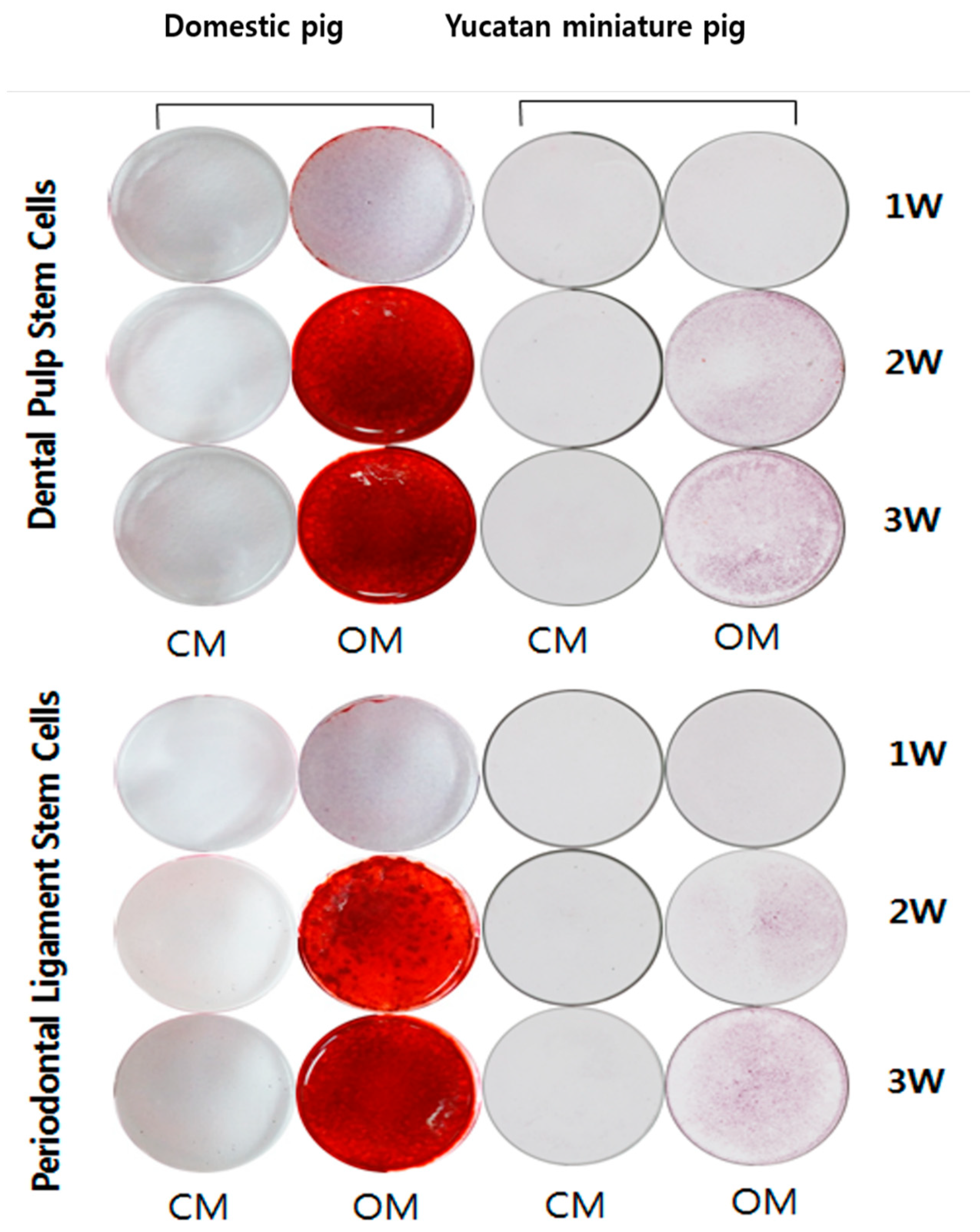
2.4. Alkaline Phosphatase Assay and Alizarin Red Staining of Osteogenic Differentiation and Mineralization

An alkaline phosphatase (ALP) assay and Alizarin red staining were performed, as previously reported [19]. Briefly, to test the ALP activity (osteogenic differentiation), cells were harvested using collagenase I and trypsin, centrifuged at 1500 rpm for 5 min, resuspended in 1 mL of cold PBS (4 °C), and counted. Next, the Alizarin red stain was quantified by measuring the absorbance of the eluted stain at 570 nm using a spectrophotometer and normalizing it by the number of cells (mineralization).

2.5. Statistical Analysis

Data (mean ± SD) were tested for normal distributions and equal variances. Paired t-tests were used for comparisons between the DPSCs and PDLSCs within the same animal, and independent t-tests were used when comparing the properties of the DPSCs and PDLSCs between the animals. The value of p < 0.05 was considered significant in all cases.

3. Results

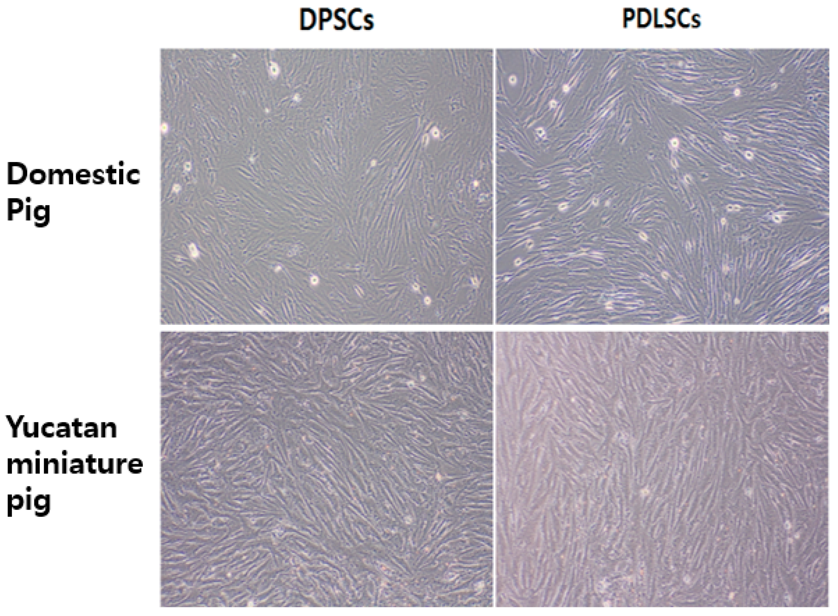
3.1. Cell Morphology

Isolated DPSCs and PDLSCs of the domestic farm pig breed and the miniature pig breed are presented in Figure 2. The passage 3 cells of the DPSCs and PDLSCs had a fibroblast-like cell morphology, which has a close resemblance to MSCs.

3.2. Cell Surface Marker Expression

A FACS analysis was conducted to characterize the phenotypes of the DPSCs and PDLSCs. MSC markers, including CD44, CD73, CD105, CD106, CD146, and TGF-βR1, were positively expressed in the DPSCs and PDLSCs (Figure 3a). The two stem cell populations exhibited high expression patterns for CD44, CD73, CD105, CD106, CD146, and TGF-βR1 (Figure 3b), which indicates that the DPSCs and PDLSCs have the characteristics of multipotent MSCs.

3.3. Cell Proliferation

A CCK-8 assay was used to evaluate the cell proliferation outcomes of the DPSCs and PDLSCs (Figure 4). The DPSCs of the miniature pig breed were found to have a higher cell proliferation rate than the DPSCs of the domestic farm pig breed. The DPSCs of the miniature pig breed showed significantly higher absorbance than those of the domestic farm pig breed upon five days of culturing (p < 0.05). The results at seven and nine days of culturing in the miniature pig breed also showed significantly higher absorbance (p < 0.05). On the other hand, the results from the PDLSCs showed no significant difference between the two pigs, except on day five. The proliferation capacity of the PDLSCs of the miniature pig breed was found to be similar to that of the PDLSCs of the domestic farm pig breed.

3.4. Osteogenic Differentiation

The presence of calcium deposits, an indicator of osteogenic differentiation, was determined through Alizarin red S staining. Mineralization was observed from 14 days of culturing in both the DPSCs and PDLSCs of the domestic farm pig breed, while weak mineral depositions were observed in both the DPSCs and PDLSCs of the miniature pig breed (in the osteogenic media of Figure 5). The levels of alkaline phosphatase activity, which is an independent indicator of osteogenic differentiation, were significantly lower (p < 0.05) in the miniature pig breed than in the domestic farm pig breed for both the DPSCs and the PDLSCs. The results of the ALP activity in the domestic farm pig breed showed a peak at two weeks, but the ALP activity of the miniature pig breed continued to increase at three weeks (Figure 6).

4. Discussion

Research on mesenchymal stem cell-based therapies for tissue regeneration has grown rapidly. Stem cells have been actively studied, as have their characteristics, as important research topics [20,21,22]. Comparative studies between stem cells have been active, such as comparing the characteristics of several stem cells in the same species [20,21,22,23,24,25] or comparing interspecific differences [26,27]. The present study is the first attempt to focus on a comparison of the characteristics between the DPSCs and PDLSCs of a Yucatan miniature pig breed and a domestic farm pig breed. Although the two pig breeds are in the same species, comparative studies of mesenchymal stem cells are necessary because they have different phenotypical characteristics, such as weight, size, skin, and physiological mechanisms [28].
The scheme used here to compare the characteristics of the dental pulp stem cells and the periodontal ligament stem cells of the Yucatan miniature pig breed to those of the domestic farm pig breed is presented in Figure 1. In a morphology evaluation, the DPSCs and PDLSCs of the miniature pig breed showed fibroblast-like cell morphologies at the passage 3 confluent monolayer under a light microscope, similar to those of the DPSCs and PDLSCs of the domestic farm pig breed. A fibroblast-like cell shape is one of the features found in mesenchymal stem cells [29]. This result demonstrates that mesenchymal stem cells can be separated from the dental pulp and periodontal ligament tissues of a domestic farm pig breed as well as a miniature pig breed, and successful cultivation was demonstrated in this study. The successful cultivation of mesenchymal stem cells from dental pulp and periodontal ligament tissues was also reported in previous studies using human subjects [30,31]. The results of the high expression patterns of the mesenchymal stem cell-specific surface markers in the FACS analysis support the contention that the cultured cells from the DP and PDL of the miniature pig breed and the domestic farm pig breed in the present study have the characteristics of multipotent MSCs.
Lee et al. [32] conducted a comparison of the proliferation of stem cells from various tissues—in that case, the adipose, bone marrow, ear skin, abdominal skin, and lung tissues of Yucatan miniature pigs—but there has been no report of the DPSCs and the PDLSCs of Yucatan miniature pigs. In the present study, it was found that the proliferation rates of the PDLSCs of both the domestic farm pig breed and the miniature pig breed have no significant differences, except for the day five results, whereas the proliferation of DPSCs was significantly higher in the miniature pig breed.
Osteogenic differentiation is an important capability because DPSCs and PDLSCs are widely used in dental disease models. An ALP assay and ARS staining showed that the DPSCs and PDLSCs of the miniature pig breed were significantly less able to undergo osteogenic differentiation compared to those of the domestic farm pig breed. In a comparison of the differentiation ability of the MSCs of the miniature pig breed to those of other species’ MSCs, Heino et al. [26] reported that the osteogenic differentiation capacity of miniature pig bone-marrow-derived MSCs was also lower compared to that of human bone-marrow-derived MSCs.
One of the limitations of this study is that it is difficult to establish a comparable age for the miniature pig breed and the domestic farm pig breed because they are different breeds. In this study, comparable ages were set at 170 days old for the domestic farm pig breed and 270 days old for the miniature pig breed, considering that the lifespans of the domestic farm pig breed and the miniature pig breed used in this study are estimated to be 10 years and 16 years, respectively. Furthermore, the weights of the pigs from two different breeds were not matched. However, miniature breed pigs are more mature than domestic pigs at the same body weight, and matching miniature and domestic pigs by body weight for comparison could be misleading because of the potential age differences [33]. Based on the estimated age comparison, proliferation analyses demonstrated that the DPSC proliferation rates of both pigs were significantly different, whereas the PDLSC proliferation rates of both pigs were not significantly different, except on day five. Another limitation of this study is that osteogenic differentiation alone was carried out to assess the multipotency of stem cells. More assays, including chondrogenic differentiation, adipogenic differentiation, and colony-forming unit-fibroblast (CFU-F) assays, are necessary to demonstrate stem cell characteristics and stem cell stemness fully. However, the expression of cell surface markers, including CD44, CD73, CD105, CD106, and CD146, may support their multipotency [34,35,36]. The third limitation of this study is that cell surface markers that express positively on MSCs alone, such as CD105 and CD73, were analyzed without cell surface markers that express negatively on MSCs, such as CD45 and CD34. Adding more cell surface markers, such as CD90, CD45, and CD34, in further studies would make the results more solid.
In conclusion, different characteristics of the DPSCs and PDLSCs of a Yucatan miniature pig from those of a domestic farm pig were identified in this study. This observation emphasizes the need for the breed-specific optimization of an osteogenic differentiation culture protocol for the DPSCs and PDLSCs of the Yucatan miniature pig before applying cell-based therapy for tissue engineering and regenerative medicine. Additional studies of the differences in the characteristics of DPSCs and PDLSCs of the Yucatan miniature pig and the domestic farm pig breed at matched ages should be carried out. In addition, the breed-specific optimization of an osteogenic differentiation culture protocol for the DPSCs and PDLSCs of the Yucatan miniature pig must be established before the application of stem cell-based tissue engineering.

Author Contributions

Writing—original draft, S.-J.S.; investigation, S.-J.S. and S.J.; data curation, S.-J.S. and H.R.; methodology, S.-J.S. and S.J.; supervision, S.C.; resources, S.C.; validation, H.R. and S.C.; writing—review and editing, H.R. and S.C. All authors have read and agreed to the published version of the manuscript.

Funding

This research was funded by the “Cooperative Research Program for Agriculture Science and Technology Development (Project No. PJ016208012021)”, Rural Development Administration, Republic of Korea.

Institutional Review Board Statement

The protocols for the animal experiments were reviewed and approved by the Institutional Animal Care and Use Committee of Chungbuk National University (CBNU-14–11).

Informed Consent Statement

Not applicable.

Conflicts of Interest

The authors declare no conflict of interest.

References

  1. Swindle, M.M.; Makin, A.; Herron, A.J.; Clubb, F.J., Jr.; Frazier, K.S. Swine as models in biomedical research and toxicology testing. Vet. Pathol. 2012, 49, 344–356. [Google Scholar] [CrossRef]
  2. Vodička, P.; Smetana Jr, K.; Dvořánková, B.; Emerick, T.; Xu, Y.Z.; Ourednik, J.; Ourednik, V.; MotlÍk, J. The miniature pig as an animal model in biomedical research. Ann. N. Y. Acad. Sci. 2005, 1049, 161–171. [Google Scholar] [CrossRef]
  3. Kararli, T.T. Comparison of the gastrointestinal anatomy, physiology, and biochemistry of humans and commonly used laboratory animals. Biopharm. Drug Dispos. 1995, 16, 351–380. [Google Scholar] [CrossRef]
  4. Swindle, M.M.; Smith, A.C.; Laber-Laird, K.; Dungan, L. Swine in biomedical research: Management and models. ILAR J. 1994, 36, 1–5. [Google Scholar] [CrossRef] [Green Version]
  5. Panepinto, L.M. Miniature swine breeds used worldwide in research. In Advances in Swine in Biomedical Research; Springer: Berlin/Heidelberg, Germany, 1996; pp. 681–691. [Google Scholar]
  6. McAnulty, P.A.; Dayan, A.D.; Ganderup, N.-C.; Hastings, K.L. The Minipig in Biomedical Research; CRC Press: Boca Raton, FL, USA, 2011. [Google Scholar]
  7. Kobayashi, H.; Tai, T.; Nagao, K.; Nakajima, K. The Minipig—A New Tool in Stem Cell Research. Pluripotent Stem Cell Biol. Adv. Mech. Methods Models 2014, 197, 431–444. [Google Scholar]
  8. Lei, M.; Li, K.; Li, B.; Gao, L.-N.; Chen, F.-M.; Jin, Y. Mesenchymal stem cell characteristics of dental pulp and periodontal ligament stem cells after in vivo transplantation. Biomaterials 2014, 35, 6332–6343. [Google Scholar] [CrossRef]
  9. Huang, G.-J.; Gronthos, S.; Shi, S. Mesenchymal stem cells derived from dental tissues vs. those from other sources: Their biology and role in regenerative medicine. J. Dent. Res. 2009, 88, 792–806. [Google Scholar] [CrossRef]
  10. Mantesso, A.; Sharpe, P. Dental stem cells for tooth regeneration and repair. Expert Opin. Biol. Ther. 2009, 9, 1143–1154. [Google Scholar] [CrossRef] [PubMed]
  11. Chen, F.-M.; Sun, H.-H.; Lu, H.; Yu, Q. Stem cell-delivery therapeutics for periodontal tissue regeneration. Biomaterials 2012, 33, 6320–6344. [Google Scholar] [CrossRef] [PubMed]
  12. Estrela, C.; Alencar, A.H.G.d.; Kitten, G.T.; Vencio, E.F.; Gava, E. Mesenchymal stem cells in the dental tissues: Perspectives for tissue regeneration. Braz. Dent. J. 2011, 22, 91–98. [Google Scholar] [CrossRef] [PubMed] [Green Version]
  13. Tang, R.; Ding, G. Swine dental pulp stem cells inhibit T-cell proliferation. Transplant. Proc. 2011, 43, 3955–3959. [Google Scholar] [CrossRef]
  14. Wada, N.; Menicanin, D.; Shi, S.; Bartold, P.M.; Gronthos, S. Immunomodulatory properties of human periodontal ligament stem cells. J. Cell. Physiol. 2009, 219, 667–676. [Google Scholar] [CrossRef]
  15. Ding, G.; Liu, Y.; Wang, W.; Wei, F.; Liu, D.; Fan, Z.; An, Y.; Zhang, C.; Wang, S. Allogeneic periodontal ligament stem cell therapy for periodontitis in swine. Stem Cells 2010, 28, 1829–1838. [Google Scholar] [CrossRef] [Green Version]
  16. Liu, Y.; Zheng, Y.; Ding, G.; Fang, D.; Zhang, C.; Bartold, P.M.; Gronthos, S.; Shi, S.; Wang, S. Periodontal ligament stem cell-mediated treatment for periodontitis in miniature swine. Stem Cells 2008, 26, 1065–1073. [Google Scholar] [CrossRef] [Green Version]
  17. Park, J.-Y.; Jeon, S.H.; Choung, P.-H. Efficacy of periodontal stem cell transplantation in the treatment of advanced periodontitis. Cell Transplant. 2011, 20, 271–286. [Google Scholar] [CrossRef] [PubMed] [Green Version]
  18. Sonoyama, W.; Liu, Y.; Fang, D.; Yamaza, T.; Seo, B.-M.; Zhang, C.; Liu, H.; Gronthos, S.; Wang, C.-Y.; Shi, S. Mesenchymal stem cell-mediated functional tooth regeneration in swine. PLoS ONE 2006, 1, e79. [Google Scholar] [CrossRef] [PubMed] [Green Version]
  19. Hanna, H.; Mir, L.M.; Andre, F.M. In vitro osteoblastic differentiation of mesenchymal stem cells generates cell layers with distinct properties. Stem Cell Res. Ther. 2018, 9, 203. [Google Scholar] [CrossRef] [PubMed]
  20. Bobis, S.; Jarocha, D.; Majka, M. Mesenchymal stem cells: Characteristics and clinical applications. Folia Histochem. Et Cytobiol. 2006, 44, 215–230. [Google Scholar]
  21. Osipova, E.; Shamanskaya, T.; Kurakina, O.; Nikitina, V.; Purbueva, B.; Ustugov, A.; Kachanov, D.; Skorobogatova, E.; Dishlevaya, Z.; Roumiantsev, S. Biological characteristics of mesenchymal stem cells during ex vivo expansion. J. Adv. Med. Med. Res. 2011, 1, 85–95. [Google Scholar] [CrossRef]
  22. Wagner, W.; Wein, F.; Seckinger, A.; Frankhauser, M.; Wirkner, U.; Krause, U.; Blake, J.; Schwager, C.; Eckstein, V.; Ansorge, W. Comparative characteristics of mesenchymal stem cells from human bone marrow, adipose tissue, and umbilical cord blood. Exp. Hematol. 2005, 33, 1402–1416. [Google Scholar] [CrossRef]
  23. Holan, V.; Trosan, P.; Cejka, C.; Javorkova, E.; Zajicova, A.; Hermankova, B.; Chudickova, M.; Cejkova, J. A comparative study of the therapeutic potential of mesenchymal stem cells and limbal epithelial stem cells for ocular surface reconstruction. Stem Cells Transl. Med. 2015, 4, 1052–1063. [Google Scholar] [CrossRef]
  24. Hui, J.H.; Li, L.; Teo, Y.-H.; Ouyang, H.-W.; Lee, E.-H. Comparative study of the ability of mesenchymal stem cells derived from bone marrow, periosteum, and adipose tissue in treatment of partial growth arrest in rabbit. Tissue Eng. 2005, 11, 904–912. [Google Scholar] [CrossRef]
  25. Johnstone, S.A. A Comparative Study of the Biological and Molecular Properties of Mesenchymal Stem Cells Isolated from Bone Marrow and the Olfactory System. Ph.D. Thesis, University of Glasgow, Scotland, UK, 2015. [Google Scholar]
  26. Heino, T.J.; Alm, J.J.; Moritz, N.; Aro, H.T. Comparison of the osteogenic capacity of minipig and human bone marrow-derived mesenchymal stem cells. J. Orthop. Res. 2012, 30, 1019–1025. [Google Scholar] [CrossRef] [PubMed] [Green Version]
  27. Li, Y.; Zhang, C.; Xiong, F.; Yu, M.-J.; Peng, F.-L.; Shang, Y.-C.; Zhao, C.-P.; Xu, Y.-F.; Liu, Z.-S.; Zhou, C. Comparative study of mesenchymal stem cells from C57BL/10 and mdx mice. BMC Cell Biol. 2008, 9, 24. [Google Scholar] [CrossRef] [Green Version]
  28. Gupta, R. (Ed.) Biomarkers in Toxicology, 1st ed.; Academic Press: New York, NY, USA, 2014. [Google Scholar]
  29. Pittenger, M.F.; Mackay, A.M.; Beck, S.C.; Jaiswal, R.K.; Douglas, R.; Mosca, J.D.; Moorman, M.A.; Simonetti, D.W.; Craig, S.; Marshak, D.R. Multilineage potential of adult human mesenchymal stem cells. Science 1999, 284, 143–147. [Google Scholar] [CrossRef] [Green Version]
  30. Gay, I.C.; Chen, S.; MacDougall, M. Isolation and characterization of multipotent human periodontal ligament stem cells. Orthod. Craniofac. Res. 2007, 10, 149–160. [Google Scholar] [CrossRef] [PubMed]
  31. Pierdomenico, L.; Bonsi, L.; Calvitti, M.; Rondelli, D.; Arpinati, M.; Chirumbolo, G.; Becchetti, E.; Marchionni, C.; Alviano, F.; Fossati, V. Multipotent mesenchymal stem cells with immunosuppressive activity can be easily isolated from dental pulp. Transplantation 2005, 80, 836–842. [Google Scholar] [CrossRef] [PubMed] [Green Version]
  32. Lee, A.-Y.; Lee, J.; Kim, C.-L.; Lee, K.S.; Lee, S.-H.; Gu, N.-Y.; Kim, J.-M.; Lee, B.C.; Koo, O.J.; Song, J.-Y. Comparative studies on proliferation, molecular markers and differentiation potential of mesenchymal stem cells from various tissues (adipose, bone marrow, ear skin, abdominal skin, and lung) and maintenance of multipotency during serial passages in miniature pig. Res. Vet. Sci. 2015, 100, 115–124. [Google Scholar] [PubMed]
  33. Fish, R.; Danneman, P.J.; Brown, M.; Karas, A. Anesthesia and Analgesia in Laboratory Animals; Academic Press: New York, NY, USA, 2011. [Google Scholar]
  34. Maleki, M.; Ghanbarvand, F.; Behvarz, M.R.; Ejtemaei, M.; Ghadirkhomi, E. Comparison of mesenchymal stem cell markers in multiple human adult stem cells. Int. J. Stem Cells 2014, 7, 118. [Google Scholar] [CrossRef] [Green Version]
  35. Lv, F.-J.; Tuan, R.S.; Cheung, K.M.; Leung, V.Y. Concise review: The surface markers and identity of human mesenchymal stem cells. Stem Cells 2014, 32, 1408–1419. [Google Scholar] [CrossRef]
  36. Harkness, L.; Zaher, W.; Ditzel, N.; Isa, A.; Kassem, M. CD146/MCAM defines functionality of human bone marrow stromal stem cell populations. Stem Cell Res. Ther. 2016, 7, 4. [Google Scholar] [CrossRef] [PubMed] [Green Version]
Figure 1. Scheme to compare the characteristics of the dental pulp stem cells (DPSCs) and periodontal ligament stem cells (PDLSCs) of the Yucatan miniature pig breed to those of the domestic farm pig breed.
Applsci 11 09461 g001
Figure 2. Morphology of the dental pulp stem cells (DPSCs) and periodontal ligament stem cells (PDLSCs) of the miniature pig breed and the domestic farm pig breed at the passage 3 confluent cell monolayer under a light microscope. The passage 3 cells of the DPSCs and PDLSCs of the miniature pig breed and the domestic farm pig breed had a fibroblast-like cell morphology, which is one of the hallmarks of stem cells.
Applsci 11 09461 g002
Figure 3. Fluorescence-activated cell sorting (FACS) data of the dental pulp stem cells (DPSCs) and periodontal ligament stem cells (PDLSCs) of the miniature pig breed and the domestic farm pig breed: (a) representative figures of the cytometric flow test; (b) percentages of positive expression (mean data). The stem cell populations of the pigs in this study exhibited high expression patterns for CD44, TGF-βR1, CD106, CD146, CD105, and CD73 for positive signals.
Applsci 11 09461 g003
Figure 4. The proliferation of the dental pulp stem cells (DPSCs) and periodontal ligament stem cells (PDLSCs) of the miniature pig breed and the domestic farm pig breed after 1, 3, 5, 7, and 9 days of culturing in the CCK-8 assay. The DPSCs of the miniature pig breed showed significantly higher absorbance than those of the domestic farm pig breed at five days of culturing (p < 0.05). On the other hand, the PDLSC results showed no significant difference between the pigs in the study, except after five days of culturing (* p < 0.05).
Applsci 11 09461 g004
Figure 5. Alizarin red S staining of the dental pulp stem cells (DPSCs) and periodontal ligament stem cells (PDLSCs) of the miniature pig breed and the domestic farm pig breed, cultured in controlled media (CM) and osteogenic media (OM), after 7, 14, and 21 days of culturing. Mineralization was observed from 14 days of culturing in the DPSCs and PDLSCs of the domestic farm pig breed, whereas weak mineral depositions were observed in the DPSCs and PDLSCs of the miniature pig breed.
Applsci 11 09461 g005
Figure 6. The alkaline phosphatase (ALP) activity of the dental pulp stem cells (DPSCs) and periodontal ligament stem cells (PDLSCs) of the miniature pig breed and the domestic farm pig breed, cultured in osteogenic media at one, two, and three weeks. The ALP activity was significantly lower in the DPSCs and PDLSCs of the miniature pig breed than in the DPSCs and PDLSCs of the domestic farm pig breed (* p < 0.05).
Applsci 11 09461 g006
Publisher’s Note: MDPI stays neutral with regard to jurisdictional claims in published maps and institutional affiliations.

