Article

The Conformity of Rehabilitation Protocols Used for Different Cartilage Repairs of the Knee Joint—A Review on Rehabilitation Standards in German Speaking Countries

1
Department of Paediatric Surgery and Orthopaedics, Clinic St. Hedwig, Barmherzige Brueder Regensburg, KUNO Paediatric University Medical Centre and FIFA Medical Centre of Excellence, University Medical Centre Regensburg, 93053 Regensburg, Germany
2
FIFA Medical Centre of Excellence, Department of Trauma Surgery, University Medical Centre Regensburg, 93053 Regensburg, Germany
3
Centre of Clinical Studies, University Medical Centre Regensburg, 93053 Regensburg, Germany
*
Authors to whom correspondence should be addressed.
Correspondence (for review).
Correspondence (for publication).
Appl. Sci. 2021, 11(19), 8873; https://doi.org/10.3390/app11198873
Submission received: 26 August 2021 / Revised: 19 September 2021 / Accepted: 21 September 2021 / Published: 24 September 2021

Abstract

:
The present study analysed current rehabilitation protocols to investigate whether there is a standard for early rehabilitation after microfracturing (MFX), matrix-assisted cartilage transplantation (MACT), and osteochondral autograft transfer (OATS) of the knee joint in clinical routine, and if rehabilitation protocols differ in the repair technique used or the localization of the cartilage defect. The evaluation included rehabilitation criteria such as weight-bearing, range of motion, use of an orthosis, motion therapy, and rehabilitation training during the early rehabilitation phase after MFX, MACT, and OATS of the femorotibial and retropatellar joint space. We analysed 153 rehabilitation protocols after cartilage repair of the knee joint, including 137 protocols for after repair of the main weight-bearing (femorotibial) area and 16 for after retropatellar cartilage repair. Most of the protocols differed significantly according to the location of the repair and the procedure performed. Our findings indicate that full weight-bearing can be achieved significantly faster after MFX (5.6 weeks) and OATS (5.3 weeks) than after MACT of the main weight-bearing zone (6.6 weeks, p < 0.001). In addition, after retropatellar cartilage repair, patients are allowed full weight-bearing after 2.1 weeks compared to the main weight-bearing zone (5.3–6.6 weeks; p < 0.001). No standardized rehabilitation recommendations have been established. The present study shows that rehabilitation needs to be adjusted to the surgical technique and the location of the defect zone, and further investigation is warranted to establish standardized rehabilitation protocols after cartilage repair of the knee joint.

1. Introduction

Due to the extensive exposure of the knee joint to high mechanical loads, injuries and chronic degenerative processes of the cartilage, especially the hyaline cartilage, are very common [1,2]. Defects can be caused by trauma or due to degenerative processes such as osteoarthritis. They lead to pain, immobilization, and reduced quality of life [3]. The choice of surgical treatment depends on the dimension and grade of the defect, the patient’s preoperative activity level, and the cause of the defect (e.g., trauma, osteoarthritis), as well as numerous co-factors. In German-speaking countries, bone marrow stimulation techniques such as MFX and MACT are currently the most commonly performed surgical techniques to restore these defects [4,5].
Weber et al. (2018) [6] showed an improvement in all patient-reported outcomes after microfracturing of the knee joint after a mean follow-up of 5.7 years. However, according to Kon et al. (2009) [7] the improvement worsens after 5 years when compared to the 2 year mark for MACT. This indicates a comparative advantage of MACT, especially regarding long-term outcome. It is therefore better suited for younger patients [8]. Brittberg et al. (2018) [9] also reported that improvements in daily life activities lasted significantly longer in MACI (using autologous cultured chondrocytes on porcine collagen membrane) than MFX. Lim et al. (2012) [10] compared all three repair techniques (MFX, MACT, OATS) for similar defect size and location and showed that all three procedures led to similar improvement in functional outcome scores at 5 year follow-up. For osteochondral allograft, Assenmacher et al. (2016) [11] reported an improvement in all scores (Knee Society Function Score, Knee Society Knee Score, Lysholm Score) in 75% of patients after a long-term follow-up 12.3 years post-surgery.
Cartilage repair surgery is well-established. It is commonly performed and yields good clinical results. However, the early stages of post-surgical rehabilitation have not been thoroughly investigated. Evidence-based rehabilitation protocols are needed because the regeneration of cartilaginous tissue is challenging and limited. On the one hand, early weight-bearing and shear forces from a progressive release of ROM might endanger the outcome of the surgical procedure by inducing apoptosis of the chondrocytes [12,13]. On the other hand in vitro studies have demonstrated that physiological contact forces produced by, for example, continuous passive motion therapy (CPM) or partial weight-bearing enable cartilaginous tissue to enhance chondrogenic differentiation [14,15]. Additionally, even healthy joint cartilage reacts to the restriction of weight bearing by atrophy [16]. Therefore, rehabilitation after cartilage repair of the knee joint must be performed with caution and must be balanced between restriction and progression of rehabilitation criteria such as weight bearing and ROM.
This study presents the current status of rehabilitation strategies after cartilage repair of the knee joint in German-speaking countries. It presents a comparative analysis of different rehabilitation approaches after various surgical techniques (MFX, MACT, OATS) and different defect localizations (femorotibial vs. retropatellar).

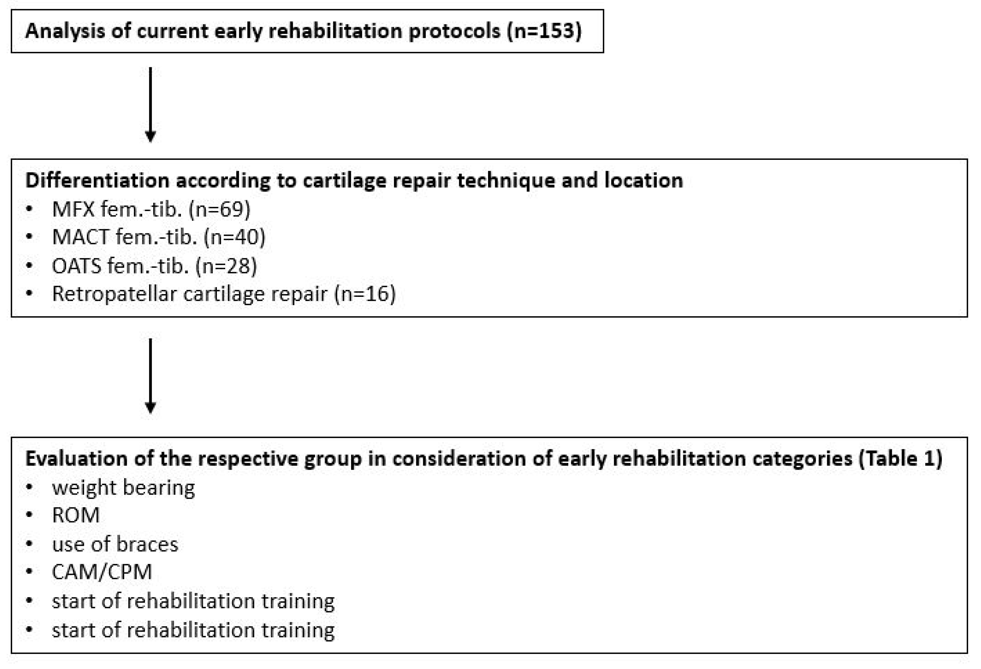
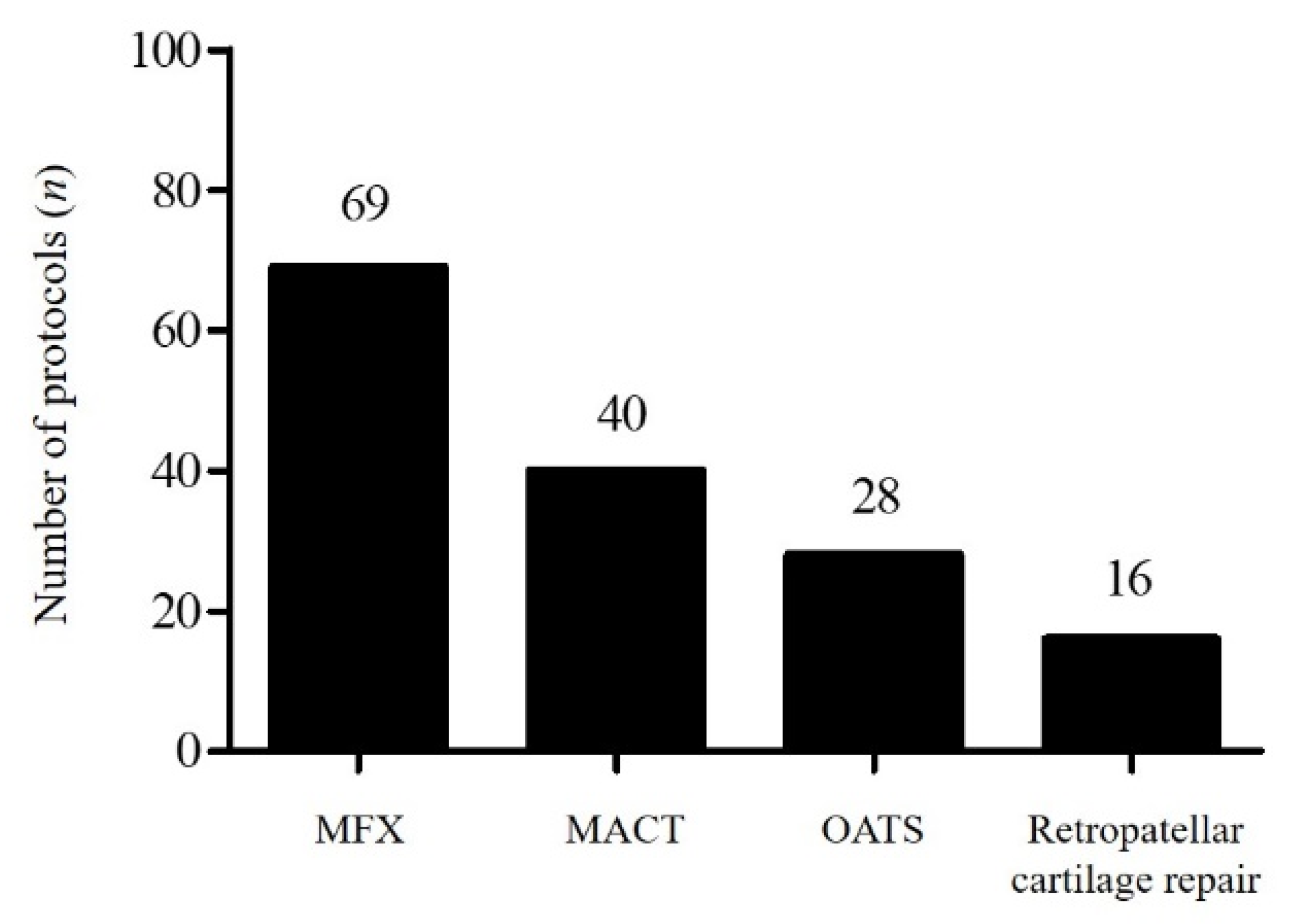
2. Materials and Methods

This study is an analysis of rehabilitation protocols currently used in Germany, Austria, and Switzerland that are designed to instruct patients and describe the rehabilitative steps in the early rehabilitation phase a minimum of the first 3 months after knee surgery. The protocols are standardized and set based on the professional experience of the institution’s orthopaedic surgeons and physiotherapists.

2.1. Measurement Criteria

All the analysed protocols contained information on the progression of weight-bearing activities and range of motion (ROM) as well as the use of motion therapy such as CPM. Additionally, most of the protocols provided detailed recommendations for rehabilitation training (e.g., use of ergometers, jogging, sports-specific training). The recommendations given by the protocols were categorized as shown in Table 1.

2.2. Participants

The rehabilitation protocols were provided by OPED (GmbH/oapl., Valley, Germany), which offered to create and design free protocols for orthopaedic institutions according to the categories and criteria described above. All rehabilitation protocols originated in German, Austrian, and Swiss orthopaedic institutions, and were blinded for analysis.

2.3. Statistical Analysis
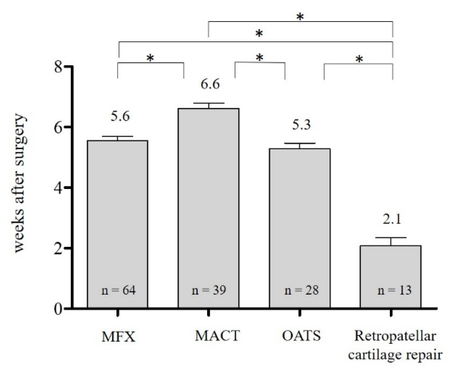
Statistical analysis was performed using SPSS® (Version 25, IBM, Armonk, NY, USA). Data are presented as mean ± SD or absolute and relative frequencies. Continuous data between two or more groups was compared by analysis of variance (ANOVA). Categorical data were compared using the chi-square test of independence. In case of significance, additional post-hoc tests were run according to Fisher’s least significant difference. A probability (p) value of ≤0.05 was considered significant for each test. Graphical illustrations were generated with GraphPad Prism® (Version 5.01, GraphPad Software, La Jolla, CA, USA) and Microsoft PowerPoint 2013® (Microsoft Corporation, Redmond, WA, USA).

3. Results

3.1. Evaluation Algorithm

In this retrospective study, 620 protocols for rehabilitation following surgery of the knee joint from 120 orthopaedic and trauma surgery institutions in Germany, Austria, and Switzerland were included. Sixty-three were categorized as ambulatory medical departments and fifty-seven as clinical medical departments, including four university medical centres. One hundred fifty-three of the six hundred twenty protocols described techniques for the early rehabilitation phase after cartilage repair (Figure 1). Among those, there were 69 protocols for after MFX of the femorotibial area, 40 protocols for after MACT, 28 for after OATS, and 16 protocols for after retropatellar cartilage repair (Figure 2).

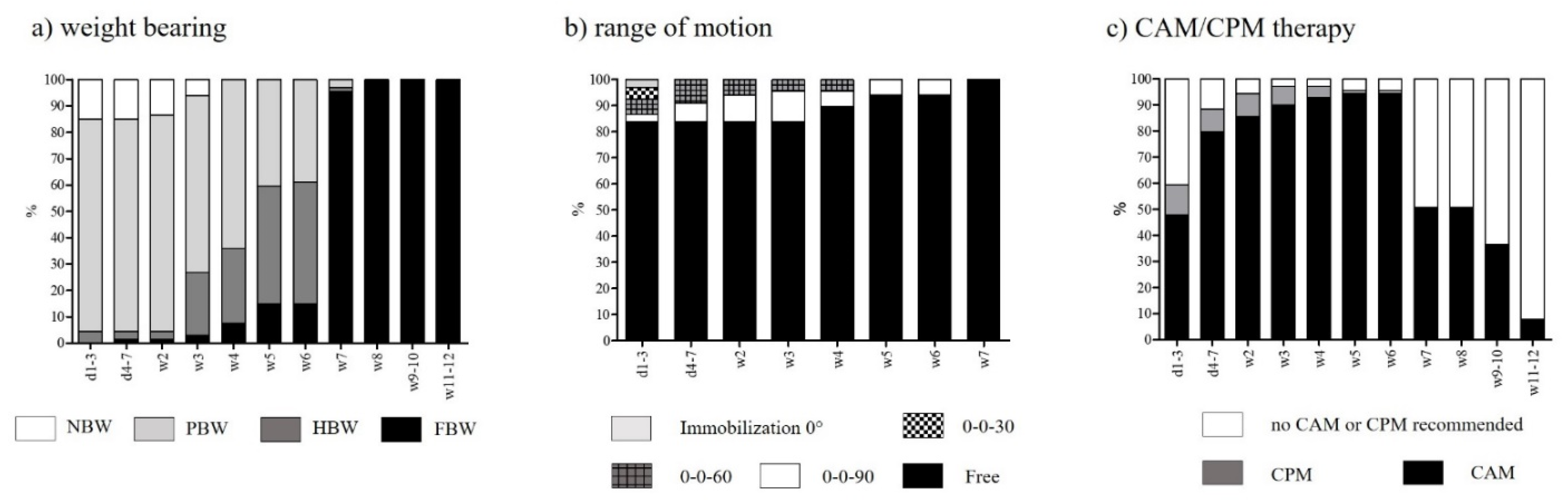
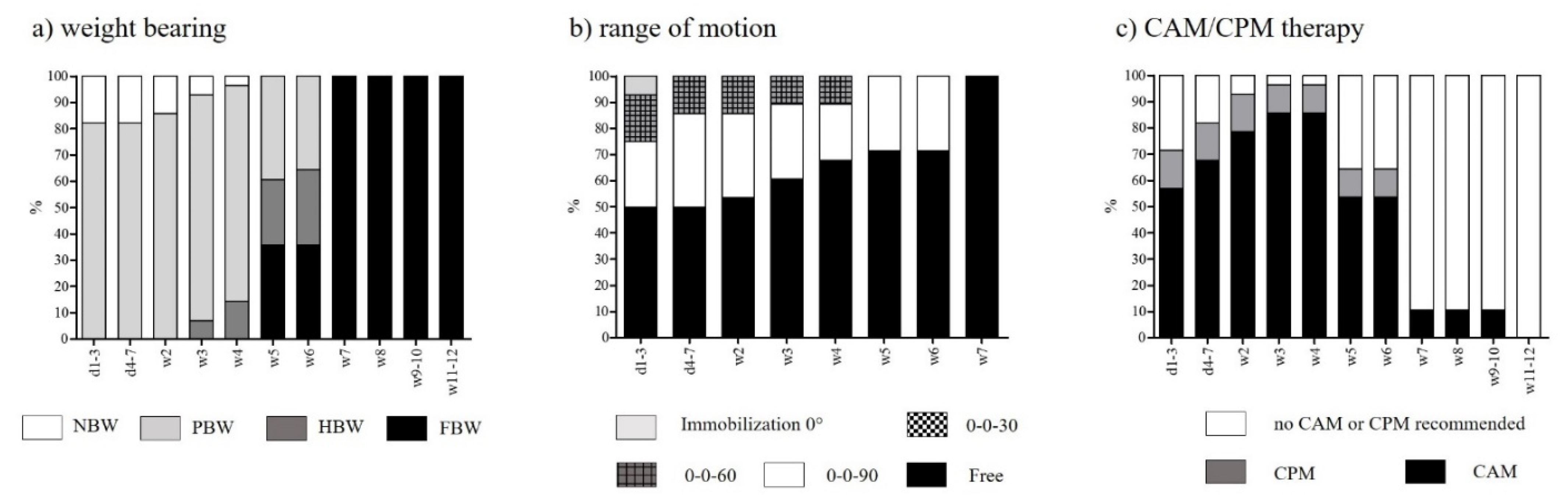
3.2. Rehabilitation after Femorotibial MFX

The recommendations on weight-bearing in the early rehabilitation phase are characterised by 3–4 weeks of partial weight-bearing (PWB) and a consistent progression to full weight-bearing (FWB), which can be reached after 5.6 ± 1.1 weeks. Restriction of ROM is recommended by only a few institutions. A proportion of 83.8% of orthopaedic surgeons release full range of motion after MFX of the femorotibial compartment directly after surgery. Motion therapy is recommended immediately or 3 to 7 days at the latest after surgery, and is applied for a period of 7.8 ± 2.1 weeks (Figure 3). Rehabilitation training is recommended after 5.5 weeks ± 1.8 weeks. Most training programs start with low-intensity exercises such as ergometer training or aqua-jogging to increase mobility with minimal loading. More specifically targeted training starts after 14.3 ± 5.7 weeks.

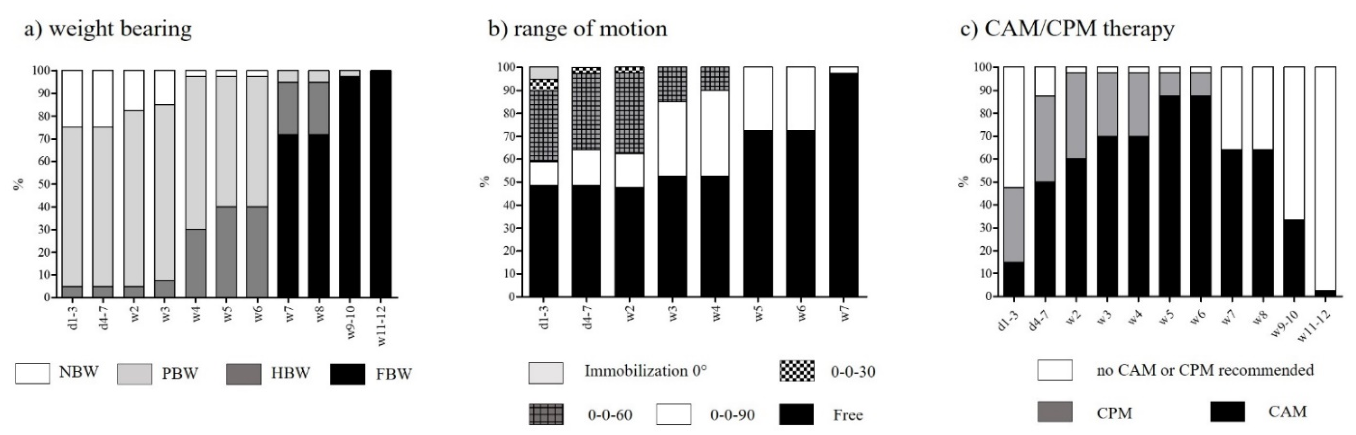
3.3. Rehabilitation after Femorotibial MACT

In contrast to MFX, orthopaedic surgeons performing MACT tend to delay the release of FWB until 6.6 ± 1.0 weeks after surgery (p < 0.001). Restriction of ROM is not handled uniformly. Of the protocols, 48.7% do not recommend any restriction of ROM, and the rest release full ROM in steps of 30° flexion every two weeks until full release in week 5 or 7 (Figure 4). The release of full ROM is significantly delayed in MACT compared to MFX (MFX: 0.7 ± 1.7 weeks; MACT: 2.4 ± 2.6 weeks; p < 0.001). There is no standardized protocol concerning ROM after MACT, but there are two vastly different approaches to this matter. Motion therapy is applied for 8.2 ± 1.8 weeks. Rehab training begins after 6.6 ± 2.1 weeks, and specific training after 12.9 ± 5.1 weeks.

3.4. Rehabilitation after Femorotibial OATS

Due to the nature of the bone-to-bone healing process, orthopaedic surgeons tend to release FWB earlier for OATS patients than for patients after MACT (OATS: 5.3 ± 1.0 weeks; MACT: 6.6 ± 1.0 weeks; p < 0.001). The release of ROM is similar to the previously described approaches after MACT, though its is slightly more progressive (Figure 5). Nevertheless, there is no observable standardized protocol. Motion therapy is recommended for a shorter postoperative period than with MFX or MACT (OATS: 5.8 ± 1.8 weeks; p < 0.001). Moreover, the start of rehabilitation training is set earlier than after MACT (OATS: 4.9 ± 1.0 weeks; p < 0.001).

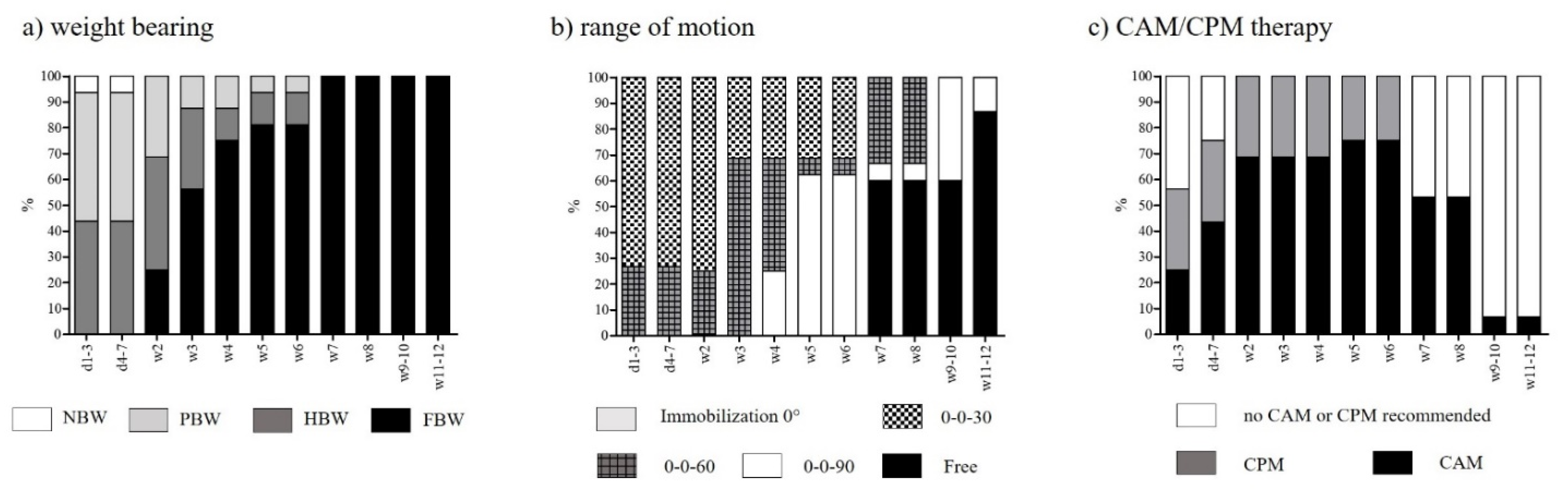
3.5. Rehabilitation after Retropatellar Cartilage Repair

According to most rehabilitation criteria, protocols after retropatellar cartilage repair differ from those after surgical therapy of a main load-carrying area, with the release of axial loading of the knee joint being much more progressive. FWB is released after 2.1 ± 1.0 weeks (p < 0.001, see Figure 6). ROM is restricted in the early rehabilitation phase to avoid shear forces to the retropatellar cartilage induced by increased flexion of the knee joint (Figure 7b). Release of full ROM is therefore much more delayed compared to femorotibial cartilage repair. It can be reached after 7.2 ± 1.9 weeks (p < 0.001). More than half of the protocols (56.3%) recommend the use of a brace to ensure the restriction of ROM. Whereas bracing is recommended for a period of 8.9 ± 2.5 weeks after retropatellar cartilage repair, the utilization of orthoses after femorotibial cartilage repair is only recommended sporadically. Passive motion therapy is applied for a period of 7.3 ± 1.6 weeks. Rehabilitation training begins after 7.2 ± 1.7 weeks, and specific training after 12.6 ± 2.0 weeks.

4. Discussion

This study presents the current status of rehabilitation after surgical cartilage repair of the knee joint in German-speaking countries, and offers insights into daily routine for rehabilitation. Overall, our findings indicate that there is no standardised rehabilitation protocol. Representative of this fact are the different approaches to releasing ROM after femorotibial MACT and OATS (Figure 4b and Figure 5b). Rehabilitation protocols tend to reflect surgeons’ empirical values rather than resulting from an evidence-based approach.

4.1. Rehabilitation after Femorotibial MFX

Although there was no single standardized approach, consensus could be ascertained, especially regarding the protocol for early rehabilitation after femorotibial MFX. Inter alia, the usual release of FWB six weeks after MFX as well as the release of ROM immediately after surgery could be seen as milestones that most of the institutions consider to be safe and appropriate. Several studies chose a more restrictive approach to the progression of weight-bearing after femorotibial MFX, maintaining NWB at no more than toe contact for 6 to 8 weeks, followed by stepwise increases to FWB [17,18,19,20]. However, Mithoefer et al. (2005) [21] released FWB after only 6 weeks, which seems to be the standard procedure and indicates consensus among the surveyed institutions. Using a rabbit model, Salter et al. (1980) [22] showed that cartilaginous defects of the knee joint healed more often and faster with hyaline cartilage after the application of CPM. This is often used in the early rehabilitation phase after MFX, MACT, and OATS, even though the improvement in clinical outcome has not been proven sufficiently and lacks scientific evidence obtained in clinical trials [23,24,25]. Generally speaking, there was institutional consensus concerning the rehabilitation criteria for weight-bearing and ROM. Additionally, motion therapy is an established procedure, and is considered a supportive treatment, but its use is not evidence-based.

4.2. Rehabilitation after Femorotibial MACT

The aim of restricting FWB for the early rehabilitation phase is, on the one hand, to protect the new transplant by avoiding shear forces and decreasing contact forces, and on the other hand, to allow physiological contact forces (physiological in both extent and direction) and therefore enable the transplant to differentiate and support the healing process [14,26]. This might explain the delayed release of weight-bearing in MACT compared to MFX and OATS (Figure 6). Due to the special requirements of transplants, an extended rehabilitation phase after MACT is required. The approaches regarding the release of ROM after MACT showed a surprising diversity. Although several studies and approximately half of the surveyed protocols use a stepwise release until full ROM is reached [14,26,27], the remaining institutions do not recommend any restriction of ROM after surgery. The question of whether restriction of ROM after femorotibial MACT of the knee joint is needed should be answered by controlled clinical studies in order to develop the best possible rehabilitation strategy. Richardson et al. (2014) [28] considered CPM to play an essential role in the early rehabilitation phase after MACT. This is also widely accepted and performed, as shown in several studies [19,26,28,29,30]. However, there is no consensus concerning the start of motion therapy, the intensity and length of training per day, or the duration of postoperative CPM.

4.3. Rehabilitation after Femorotibial OATS

In comparison to MACT, the early rehabilitation phase after OATS is more progressive and leads to faster recovery due to the greater stability of the osteochondral transplant. This was indicated by a significantly earlier progression to FWB (Figure 6 and Figure 7a) and a significantly earlier start of rehab training. This can also be explained by the bone-to-bone healing process, which is considered to be more resilient in OATS than in the case of MACT [5]. In addition, patients who have undergone OATS of the knee joint are more likely to return to full pre-injury activity levels sooner than patients who have had cartilage repair using MFX or MACT [21,31]. The findings of the current study support this conclusion.
As the results of our analysis of the rehabilitation protocols for post-surgical femorotibial cartilage repair show, the early rehabilitation phase is adapted to the surgical procedure. There are no standardised, generally accepted rehabilitation programs, so multiple approaches are needed to gain recovery.

4.4. Rehabilitation after Retropatellar Cartilage Repair

Rehabilitation after retropatellar cartilage repair differs from rehabilitation after femorotibial repair in every criterion, proving that rehabilitation is strongly adapted not only to the surgical technique, but to the anatomical location of the defect area. Weight-bearing is allowed more rapidly and progressively, and ROM is therefore released much more restrictively. The utilization of orthoses is recommended more often to enhance the compliance of restricting ROM.
However, there is again no standard in rehabilitation. The review of Hambly et al. (2006) [14] showed a great diversity in the start of FWB after retropatellar MACT, with it starting as soon as 6 h postoperatively and ending as late as 12 weeks post-surgery. In contrast, our results showed that the latest release of FWB is most commonly recommended after 6 weeks. Some studies set the release of FWB between the 6th and 8th weeks of the postsurgical period, while others wait until after 9 weeks [19,27]. Compared to elsewhere in the literature, our results show a more progressive release of FWB. All studies included in this survey have in common a restrictive release of ROM. Steadman et al. (2001) [17] released FWB immediately after surgery if tolerated, but only in extension and up to 20° of flexion, guaranteed by the use of an orthosis for 8 weeks. The surveyed institutions recommended the utilization of bracing for 8.9 weeks on average. As we can see from the recommendations for postsurgical bracing, the restriction and release of ROM plays an essential part in protecting the surgical effort during rehabilitation after retropatellar cartilage repair.

5. Limitations

The present study has some limitations. Due to the retrospective design, the results lack information about modification of the rehabilitation protocol by orthopaedists, clinical outcomes, and patient compliance. Furthermore, technical details such as defect size, degree of the cartilage defect, and concomitant lesions of the meniscus are not available. In addition, the evaluated rehabilitation protocols only cover the early period of rehabilitation up to 12 weeks post-surgery. Regeneration and returning to work and sports activities require a longer period of rehabilitation and are therefore not covered by this data sample.

6. Conclusions

This study shows detailed information on the early rehabilitation phase after surgical cartilage repair techniques of the knee joint in German-speaking countries. Although the surgical procedures are well-established, the analysed protocols differ in approach, especially concerning the progression of weight-bearing activities or the start of rehabilitation training. Even though the study shows that rehabilitation is adapted to the surgical technique (MFX, MACT, OATS) and the location of the defect zone (femorotibial, retropatellar), further investigation is needed to establish standardized rehabilitation protocols following cartilage repair of the knee joint. Multiple obstacles have to be overcome to gain evidence-based recommendations. This study may serve as the foundation for further clinically controlled trials to improve clinical outcomes.

Author Contributions

Conceptualization, C.M. and C.P.; methodology, W.K.; software, C.P. and F.Z.; validation: W.K. and F.Z.; formal analysis: C.M. and M.K.; investigation, C.M.; resources, C.P.; data curation, C.M. and C.P.; writing—original draft, C.M.; writing—review and editing, C.P., W.K. and M.K.; visualization, F.Z., L.H. and M.R.; supervision, V.A. and C.K.; project administration, C.P. and W.K. All authors have read and agreed to the published version of the manuscript.

Funding

This research received no external funding.

Institutional Review Board Statement

Not applicable.

Data Availability Statement

The data presented in this study are available on request from the corresponding author. The data are not publicly available due to data protection reasons.

Conflicts of Interest

The authors declare no conflict of interest.

References

  1. Hjelle, K.; Solheim, E.; Strand, T.; Muri, R.; Brittberg, M. Articular cartilage defects in 1000 knee arthroscopies. Arthroscopy 2002, 18, 730–734. [Google Scholar] [CrossRef] [PubMed]
  2. Widuchowski, W.; Widuchowski, J.; Trzaska, T. Articular cartilage defects: Study of 25,124 knee arthroscopies. Knee 2007, 14, 177–182. [Google Scholar] [CrossRef] [PubMed]
  3. Heir, S.; Nerhus, T.K.; Rotterud, J.H.; Loken, S.; Ekeland, A.; Engebretsen, L.; Aroen, A. Focal cartilage defects in the knee impair quality of life as much as severe osteoarthritis: A comparison of knee injury and osteoarthritis outcome score in 4 patient categories scheduled for knee surgery. Am. J. Sports Med. 2010, 38, 231–237. [Google Scholar] [CrossRef] [PubMed] [Green Version]
  4. Niemeyer, P.; Feucht, M.J.; Fritz, J.; Albrecht, D.; Spahn, G.; Angele, P. Cartilage repair surgery for full-thickness defects of the knee in Germany: Indications and epidemiological data from the German Cartilage Registry (KnorpelRegister DGOU). Arch. Orthop. Trauma Surg. 2016, 136, 891–897. [Google Scholar] [CrossRef] [PubMed]
  5. Zellner, J.; Krutsch, W.; Pfeifer, C.; Koch, M.; Nerlich, M.; Angele, P. Autologous chondrocyte implantation for cartilage repair. Orthop. Res. Rev. 2015, 7, 149–158. [Google Scholar]
  6. Weber, A.E.; Locker, P.H.; Mayer, E.N.; Cvetanovich, G.L.; Tilton, A.K.; Erickson, B.J.; Yanke, A.B.; Cole, B.J. Clinical Outcomes After Microfracture of the Knee: Midterm Follow-up. Orthop. J. Sports Med. 2018, 6, 1–7. [Google Scholar] [CrossRef]
  7. Kon, E.; Gobbi, A.; Filardo, G.; Delcogliano, M.; Zaffagnini, S.; Marcacci, M. Arthroscopic second-generation autologous chondrocyte implantation compared with microfracture for chondral lesions of the knee: Prospective nonrandomized study at 5 years. Am. J. Sports Med. 2009, 37, 33–41. [Google Scholar] [CrossRef]
  8. Na, Y.; Shi, Y.; Liu, W.; Jia, Y.; Kong, L.; Zhang, T.; Han, C.; Ren, Y. Is implantation of autologous chondrocyte superior to microfracture for articular-cartilage defects of the knee? A systematic review of 5-year follow-up data. Int. J. Surg. 2019, 68, 56–62. [Google Scholar] [CrossRef]
  9. Brittberg, M.; Recker, D.; Ilgenfritz, J.; Saris, D.B.F. Matrix-Applied Characterized Autologous Cultured Chondrocytes Versus Microfracture: Five-Year Follow-up of a Prospective Randomized Trial. Am. J. Sports Med. 2018, 46, 1343–1351. [Google Scholar] [CrossRef]
  10. Lim, H.-C.; Bae, J.-H.; Song, S.-H.; Park, Y.-E.; Kim, S.-J. Current treatments of isolated articular cartilage lesions of the knee achieve similar outcomes. Clin. Orthop. Relat. Res. 2012, 470, 2261–2267. [Google Scholar] [CrossRef] [Green Version]
  11. Assenmacher, A.T.; Pareek, A.; Reardon, P.J.; Macalena, J.A.; Stuart, M.J.; Krych, A.J. Long-term Outcomes After Osteochondral Allograft: A Systematic Review at Long-term Follow-up of 12.3 Years. Arthroscopy 2016, 32, 2160–2168. [Google Scholar] [CrossRef]
  12. Martin, J.A.; Brown, T.; Heiner, A.; Buckwalter, J.A. Post-traumatic osteoarthritis: The role of accelerated chondrocyte senescence. Biorheology 2004, 41, 479–491. [Google Scholar]
  13. Martin, J.A.; Buckwalter, J.A. Post-traumatic osteoarthritis: The role of stress induced chondrocyte damage. Biorheology 2006, 43, 517–521. [Google Scholar] [PubMed]
  14. Hambly, K.; Bobic, V.; Wondrasch, B.; van Assche, D.; Marlovits, S. Autologous chondrocyte implantation postoperative care and rehabilitation: Science and practice. Am. J. Sports Med. 2006, 34, 1020–1038. [Google Scholar] [CrossRef]
  15. Knapik, D.M.; Harris, J.D.; Pangrazzi, G.; Griesser, M.J.; Siston, R.A.; Agarwal, S.; Flanigan, D.C. The basic science of continuous passive motion in promoting knee health: A systematic review of studies in a rabbit model. Arthroscopy 2013, 29, 1722–1731. [Google Scholar] [CrossRef] [PubMed] [Green Version]
  16. Hinterwimmer, S.; Krammer, M.; Krotz, M.; Glaser, C.; Baumgart, R.; Reiser, M.; Eckstein, F. Cartilage atrophy in the knees of patients after seven weeks of partial load bearing. Arthritis Rheum. 2004, 50, 2516–2520. [Google Scholar] [CrossRef] [PubMed]
  17. Steadman, J.R.; Rodkey, W.G.; Rodrigo, J.J. Microfracture: Surgical technique and rehabilitation to treat chondral defects. Clin. Orthop. Relat. Res. 2001, 391, 362–369. [Google Scholar] [CrossRef]
  18. Braun, S.; Steadman, J.R.; Rodkey, W.G.; Briggs, K.K. Microfracture and specific rehabilitation for treating osteoarthritis of the knee. Indications, surgical technique, and rehabilitation protocol. Z. Rheumatol. 2009, 68, 811–818. [Google Scholar]
  19. Tyler, T.F.; Lung, J.Y. Rehabilitation following osteochondral injury to the knee. Curr Rev Musculoskelet Med 2012, 5, 72–81. [Google Scholar] [CrossRef] [Green Version]
  20. Erggelet, C. Stimulation Thechniques: Microfracturing, Drilling. In Techniques in Cartilage Repair Surgery; Springer: Berlin/Heidelberg, Germany, 2014; pp. 35–47. [Google Scholar]
  21. Mithoefer, K.; Williams, R.J., III; Warren, R.F.; Potter, H.G.; Spock, C.R.; Jones, E.C.; Wickiewicz, T.L.; Marx, R.G. The microfracture technique for the treatment of articular cartilage lesions in the knee. A prospective cohort study. J. Bone Jt. Surg. Am. 2005, 87, 1911–1920. [Google Scholar] [CrossRef]
  22. Salter, R.B.; Simmonds, D.F.; Malcolm, B.W.; Rumble, E.J.; MacMichael, D.; Clements, N.D. The biological effect of continuous passive motion on the healing of full-thickness defects in articular cartilage. An experimental investigation in the rabbit. J. Bone Jt. Surg. Am. 1980, 62, 1232–1251. [Google Scholar] [CrossRef]
  23. Fazalare, J.A.; Griesser, M.J.; Siston, R.A.; Flanigan, D.C. The use of continuous passive motion following knee cartilage defect surgery: A systematic review. Orthopedics 2010, 33, 878. [Google Scholar] [CrossRef]
  24. Howard, J.S.; Mattacola, C.G.; Romine, S.E.; Lattermann, C. Continuous Passive Motion, Early Weight Bearing, and Active Motion following Knee Articular Cartilage Repair; Evidence for Clinical Practice. Cartilage 2010, 1, 276–286. [Google Scholar] [CrossRef] [PubMed] [Green Version]
  25. Rogan, S.; Taeymans, J.; Hirschmuller, A.; Niemeyer, P.; Baur, H. Effect of continuous passive motion for cartilage regenerative surgery—A systematic literature review. Z. Orthop. Unfall. 2013, 151, 468–474. [Google Scholar] [PubMed]
  26. Hirschmuller, A.; Baur, H.; Braun, S.; Kreuz, P.C.; Sudkamp, N.P.; Niemeyer, P. Rehabilitation after autologous chondrocyte implantation for isolated cartilage defects of the knee. Am. J. Sports Med. 2011, 39, 2686–2696. [Google Scholar] [CrossRef] [Green Version]
  27. Gillogly, S.D.; Myers, T.H.; Reinold, M.M. Treatment of full-thickness chondral defects in the knee with autologous chondrocyte implantation. J. Orthop. Sports Phys. Ther. 2006, 36, 751–764. [Google Scholar] [CrossRef] [PubMed] [Green Version]
  28. Richardson, J.B. Classical ACI for Chondral and Ostechondral Defects. In Techniques in Cartilage Repair Surgery; Springer: Berlin/Heidelberg, Germany, 2014; pp. 165–174. [Google Scholar]
  29. Pestka, J.M.; Feucht, M.J.; Porichis, S.; Bode, G.; Sudkamp, N.P.; Niemeyer, P. Return to Sports Activity and Work after Autologous Chondrocyte Implantation of the Knee: Which Factors Influence Outcomes? Am. J. Sports Med. 2016, 44, 370–377. [Google Scholar] [CrossRef]
  30. Riegger-Krugh, C.L.; McCarthy, E.C.; Robinson, M.S.; Wegzyn, D.A. Autologous chondrocyte implantation: Current surgery and rehabilitation. Med. Sci. Sports Exerc. 2008, 40, 206–214. [Google Scholar] [CrossRef] [Green Version]
  31. Campbell, A.B.; Pineda, M.; Harris, J.D.; Flanigan, D.C. Return to Sport After Articular Cartilage Repair in Athletes’ Knees: A Systematic Review. Arthroscopy 2016, 32, 651–668.e1. [Google Scholar] [CrossRef]
Figure 1. Flow of evaluation. MFX: microfracturing; fem./tib.: femorotibial compartment/main load-carrying area; MACT: matrix-assisted cartilage transplantation; OATS: osteochondral autograft transfer system; ROM: range of motion; CAM/CPM: continuous active/passive motion.
Applsci 11 08873 g001
Figure 2. Number of rehabilitation protocols of orthopaedic institutions concerning cartilage repair techniques of the knee joint. MFX: microfracturing; MACT: matrix-assisted cartilage transplantation; OATS: osteochondral autograft transfer system.
Applsci 11 08873 g002
Figure 3. Early rehabilitation phase after femorotibial MFX concerning (a) weight-bearing, (b) range of motion, and (c) motion therapy, expressed as the percentage of protocols. NBW: no body weight; PBW: partial body weight; HBW: half body weight; FBW: full body weight; CAM: continuous active motion; CPM: continuous passive motion; d: day; w: week.
Applsci 11 08873 g003
Figure 4. Early rehabilitation phase after femorotibial MACT concerning (a) weight-bearing, (b) range of motion, and (c) motion therapy, expressed as the percentage of protocols. NBW: no body weight; PBW: partial body weight; HBW: half body weight; FBW: full body weight; CAM: continuous active motion; CPM: continuous passive motion; d: day; w: week.
Applsci 11 08873 g004
Figure 5. Early rehabilitation phase after femorotibial OATS concerning (a) weight-bearing, (b) range of motion, and (c) motion therapy, expressed as the percentage of protocols. NBW: no body weight; PBW: partial body weight; HBW: half body weight; FBW: full body weight; CAM: continuous active motion; CPM: continuous passive motion; d: day; w: week.
Applsci 11 08873 g005
Figure 6. Weeks until release of FWB after surgical cartilage repair of the knee joint. MFX: microfracturing; MACT: matrix-assisted cartilage transplantation; OATS: osteochondral autograft transfer system. * p ≤ 0.001.
Applsci 11 08873 g006
Figure 7. Early rehabilitation phase after retropatellar cartilage repair concerning (a) weight-bearing, (b) range of motion, and (c) motion therapy, expressed in percentage of protocols using them. NBW: no body weight; PBW: partial body weight; HBW: half body weight; FBW: full body weight; CAM: continuous active motion; CPM: continuous passive motion; d: day; w: week.
Applsci 11 08873 g007
Table 1. Categories of early rehabilitation.
Rehabilitation CriterionCategories
Weight-bearingNo body weight (NBW): no loading
Partial body weight (PBW): ≤20 kg
Half body weight (HBW): >20 kg
Full body weight (FBW): no restriction
Range of motion (extension/flexion)Immobilization 0°
0-0-30
0-0-60
0-0-90
0-0-free
Use of bracesYes/No
Recommended wearing time (weeks)
Continuous active/
passive motion (CAM/CPM)
CAM recommended
CPM recommended
No CAM/CPM recommended
Start of rehabilitation trainingWeeks after surgery
Start of specific trainingWeeks after surgery
Publisher’s Note: MDPI stays neutral with regard to jurisdictional claims in published maps and institutional affiliations.

