Article

Genome Mining Associated with Analysis of Structure, Antioxidant Activity Reveals the Potential Production of Levan-Rich Exopolysaccharides by Food-Derived Bacillus velezensis VTX20

1
Institute of Biotechnology, Vietnam Academy of Science and Technology, Hanoi 100000, Vietnam
2
Graduate University of Science and Technology, Vietnam Academy of Science and Technology, Hanoi 100000, Vietnam
3
Vietnam-Russia Tropical Centre, Hanoi 100000, Vietnam
4
Center for Research and Technology Transfer, Vietnam Academy of Science and Technology, Hanoi 100000, Vietnam
5
School of Biotechnology and Food Technology, Hanoi University of Science and Technology, Hanoi 100000, Vietnam
*
Author to whom correspondence should be addressed.
These authors have contributed equally to this work and share the first authorship.
Appl. Sci. 2021, 11(15), 7055; https://doi.org/10.3390/app11157055
Submission received: 9 July 2021 / Revised: 28 July 2021 / Accepted: 29 July 2021 / Published: 30 July 2021

Abstract

:
Exopolysaccharides (EPSs) produced by Bacillus species have recently emerged as promising commercial antioxidants in various industries, such as pharmaceutics and biomedicine. However, little is known about EPS production and function from Bacillus velezensis so far. In the present study, the effect of sugar sources on EPS production by B. velezensis VTX20 and the genetic biosynthesis, characteristics, and antioxidant activity of the resulting EPS were evaluated. The strain VTX20 produced the maximum EPS yield of 75.5 ± 4.8 g/L from an initial 200 g/L of sucrose after a 48-h cultivation. Through genomic analysis, ls-levB operon was found, for the first time, to be responsible for the levan-type EPS production in B. velezensis. Biochemical and structural characterization further confirmed the majority of levan, followed by an extremely low level of dextran biopolymer. The water solubility index and water holding capacity of the EPSs were 81.9 ± 3.4% and 100.2 ± 3.4%, respectively. In vitro antioxidant activity analyses showed strong scavenging activity for 1,1-diphenyl-2-picrylhydrazyl and hydroxyl radical values of 40.1–64.0% and 16.0–40%, respectively. These findings shed light on the EPS biosynthesis of B. velezensis at both structural and genetic levels and the potential application of EPS as a natural antioxidant for pharmaceutical and biomedical industries.

1. Introduction

Exopolysaccharides (EPSs) represent the most significant group of biopolymers found outside the microbial cell wall. EPS components are predominantly carbohydrates produced by bacteria, cyanobacteria, and fungi [1,2,3], which possess many valuable biological activities, such as antimicrobial, immunoregulatory, and antioxidant activities [4,5]. In recent years, EPSs have been receiving greater attention from the pharmaceutical industry as immune-stimulators, antitumor agents, and blood cholesterol-lowering agents. Moreover, EPSs are used as biomaterials for drug delivery due to their ability to conjugate to hydrophobic molecules and drugs [6,7]. EPSs also have outstanding applications in the food industry as natural thickeners, stabilizers, emulsifiers, and texturizers [3]. Moreover, EPSs play important role in producing food additives and functional food ingredients [8,9].
Recently, there has been increasing interest in the exploitation of bacterial EPSs. Bioactive EPSs are well-studied in many strains belonging to the genus Lactobacillus, Bacillus, Lactococcus, and Streptococcus [3,5,10]. Among them, EPSs produced by Bacillus species have been recognized as safe, cost-effective, and stable. EPS extracted from a sponge-associated B. licheniformis reduced the biofilm development of pathogenic bacteria, such as Escherichia coli and Pseudomonas fluorescens [11]. Endophytic B. amyloliquefaciens derived from the medicinal plant Ophiopogon japonicas is a producer of EPS that showed great antitumoral activity against gastric cancers [12]. Of note, members of the genus Bacillus produce complex EPSs. Different types of EPS, such as glucan, γ-polyglutamic acid (γ-PGA), levan, and fructan are found in B. subtilis, but their productivities rely on a substrate supplemented in the medium [13,14]. Bacillus subtilis EPS synthesized by a 15-gene operon espA-O is found in the supernatant, which is rich in glucose and galactose [15]. EPS biosynthesis by the eps gene cluster is a complex process, requiring genes responsible for the regulation, determination of chain length, synthesis, polymerization, and exportation of the repeating sugar units. While this EPS is produced by various sugar sources, levan is only synthesized when sucrose is supplied [3,16]. Levan is classified as a homopolysaccharide that is mainly composed of β-(2→6) fructan linkages with occasional β-(2→1) branching, which is catalyzed by levansucrase in sucrose-rich environments [17,18]. The levan produced by B. licheniformis exhibited strong hypoglycemic and antioxidant activities, protecting the main organs in alloxan-induced diabetic rats [19]. B. subtilis strains derived from honey were reported to produce levan that exhibited antiviral activity on avian influenza and type 40 adenovirus [20]. However, levan production capacity remains low and the production cost high due to the small amounts of levansucrase expressed in the wild-type strain. Thus, screening for Bacillus species with high EPS production rates is required.
One of the important therapeutic properties of EPSs is its antioxidant capacity. Excess of oxygen-derived hydroxyl radical, superoxide-free radical, and reactive oxygen species are the main factors contributing to various disorders including inflammation, lung injury, cancer, aging, and atherosclerosis [21,22]. To prevent the generated oxidative stress, it is an urgent need to develop natural antioxidant agents for applications in the pharmaceutical and food industries, since synthetic antioxidants are suspected to be involved in carcinogenesis and liver damage. L. reuteri and L. vaginalis isolated from the gut cecum of healthy poultry birds produced low EPS yields that showed potent in vitro antioxidant activity [4]. EPSs extracted from Leuconostoc mesenteroides present during early kimchi fermentation displayed DPPH radical-scavenging activity, a lipid peroxidation inhibition effect, and a ferrous chelating ability. Thus far, few investigations have been performed to demonstrate antioxidant activities of EPSs from Bacillus spp.
Bacillus velezensis has been emerging as a probiotic for plants and animals. There have been a few studies indicating the presence of EPS in B. velezensis [23,24]; however, the structure and gene cluster involved in the biosynthetic pathways and antioxidant properties of EPS remain poorly characterized. This study aimed to isolate and evaluate the EPS production of bacteria associated with traditional soybean fermented paste. The genome of the highest EPS-producing bacterium—B. velezensis VTX20—was sequenced for elucidation of EPS biosynthesis and involvement of sugar sources in EPS yield. Furthermore, functional characterization and antioxidant activity were analyzed to highlight the biotechnological potential of EPS.

2. Materials and Methods

2.1. Isolation of Bacteria Producing Exopolysaccharide

Six samples of traditional soybean fermented pastes were collected from Phu Tho and Quang Ninh provinces, Vietnam. The bacterial isolates were obtained by serial dilution plating on EPS agar medium (KH2PO4 0.2 g; K2HPO4 1.5 g; MgSO4·7H2O 0.2 g; CaSO4·2H2O 0.1 g; FeCl3 0.002 g; yeast extract 0.5; glucose 100 g; pH 7.0 in a liter) at 37 °C for 2 days [25]. The exopolymer-producing bacteria were screened on the basis of colony morphology, which formed thick slime or mucoid. In addition, sterilized toothpicks were used to observe mucinous strands formed when the sterilized toothpicks were put into contact with potential colonies, as previously described [26]. All potential isolates were picked up and streaked out on EPS agar plates and then stored in 30% glycerol at −80 °C.

2.2. Exopolysaccharide Production and Quantification

The strain VTX20 most capable of producing EPS was grown overnight in LB medium at 37 °C with vigorous shaking. The overnight culture was transferred to EPS medium (yeast extract 2.0 g, (NH4)2SO4 1.5 g, K2HPO4 2.5 g, pH 7.0 per liter) supplemented with different glucose and sucrose concentrations (100 g/L, 150 g/L, 200 g/L) and adjusted to an optical density at 600 nm (OD600) of 0.1 with rotary shaking at 180 rpm. At intervals, samples were taken for the determination of EPS yield [27]. Briefly, the cultures were heated at 100 °C for 20 min in order to inactivate enzymes involved in EPS degradation. After centrifugation at 10,000 rpm for 20 min, the collected cell-free supernatant was mixed with three volumes of cold absolute ethanol and the solution was kept at 4 °C overnight. The resulting precipitate was centrifuged at 10,000 rpm for 20 min and washed two times with ethanol to remove the residual sugars and impurities of low molecular weight. To remove salts, the raw EPS was re-dissolved in distilled water and dialyzed overnight at 4 °C. The purified EPS samples were dried in a speed vacuum concentrator and then freeze-dried in a lyophilizer for further studies. The EPS was calculated as g/L.

2.3. Genome Sequencing, De Novo Assembly, and Annotation

High-quality genomic DNA was extracted with a G-spin™ Total DNA Extraction Mini Kit as per the manufacturer’s instructions. The genomic DNA from strain VTX20 was sequenced using the Pacific Bioscience SEQUEL platform (Menlo Park, CA, USA) with single-molecule real-time (SMRT) sequencing by the Institute of Biotechnology, Vietnam Academy of Science and Technology. A de novo assembly method was employed to carry out the assembly of quality-filtered reads using CLC Genomics Workbench version 7.5.1 (CLC Bio, Aarhus, Denmark) with the hierarchical genome-assembly process (HGAP) algorithm in SMRT analysis. Genome annotation of B. velezensis VTX20 was accomplished using Prokaryotic Genomes Annotation Pipeline (PGAP) at NCBI and the fully-automated service RAST (Rapid Annotation using Subsystem Technology) [28,29]. Clusters of orthologous genes (COGs) were used to predict functional genes [30].

2.4. Genome Comparisons

The average nucleotide identity (ANI) was calculated using the orthologous average nucleotide identity (OrthoANI) [31], taking the complete available genomes from Bacillus subtilis subsp. subtilis 168 (ABQK01000001.1), B. velezensis FZB42 (NC_009725.2), B. velezensis KMU01 (NZ_CP063768.1), B. velezensis L-S60 (NZ_CP011278.1), B. velezensis LPL061(CP042271.1) as reference. Genes involved in EPS and levan biosynthesis were determined using BLAST of NCBI and RAST [28].

2.5. Water Solubility Index and Water-Holding Capacity

The water solubility index was determined to assess the degree of solubility of EPS in water, as described previously [32]. About 200 mg EPS was dissolved in 5 mL sterile water and stirred for 60 min in a water bath at 40 °C to obtain a uniform suspension, followed by centrifugation at 6000 rpm for 15 min. The supernatant was put on a Petri dish and then dried at 105 °C for 4 h to attain a dry solid weight. The water solubility was calculated as:
Index   ( % ) = Dry   weight   of   solids   in   supernatant Weight   of   dry   sample × 100
Water-holding capacity was detected by using a suspension of 200 mg EPS in 10 mL sterile water that was kept at 40 °C for 10 min [33]. The sample was centrifuged at 12,000 rpm for 30 min, and the supernatant was discarded. The pellets were harvested and then put on pre-weighed filter paper to drain water. The precipitated EPS was weighed to calculate the water-holding capacity as:
Water   holding   capacity   ( % ) = Total   sample   weight   after   absorption Total   dry   sample   weight × 100

2.6. Monosaccharide and NMR Analysis of EPS

The monosaccharide present in EPS was hydrolyzed using sulfuric acid, followed by chromatographic analysis [34]. This procedure was performed using an HPLC (DGU-20A, Shimadzu, Tokyo Japan) equipped with a column InertSustain (4.6 mm × 250 mm). A UV detector at 245 nm was used to detect the EPS.
NMR spectra of EPS in D2O was measured on a Bruker Avance 500 Hz spectrometer (Bruker Corporation, Karlsruhe, Germany) using trimethylsilane as an internal reference. Chemical shift (δ) was represented as parts per million (ppm).

2.7. Anti-Oxidant Activity

The hydroxyl radical scavenging activity of purified EPS was evaluated using the assay reported previously with slight modification [22]. The hydroxyl radical reaction includes 1 mL of 0.75 mM 1,10-phenanthroline, 1 mL of 0.75 mM FeSO4, 1 mL of H2O2 (0.01%, v/v) and 1.5 mL of 0.15 M sodium phosphate buffer (pH 7.4). The reaction was started by the addition of 1.0 mL EPS solution (0.5; 1; 1.5; 2 mg/mL), followed by incubation at 37 °C for 30 min. The absorbance was measured at 536 nm and hydroxyl radical scavenging activity was calculated as follows:
Scavenging   activity   ( % )   =   ( A sample   A blank ) ( A 0   A blank )   ×   100
where Asample is the absorbance of the sample, A0 is the absorbance of the control, and Ablank is the absorbance of the blank.
The 2-diphenyl-1- picrylhydrazyl (DPPH) method was performed based on the capture of the DPPH radical for antioxidants that leads to a decrease in absorbance at 517 nm [35]. About 0.2 mL DPPH dissolved in methyl alcohol was mixed with 1.0 mL EPS solution (0.5; 1; 1.5; 2 mg/mL) and then 2.0 mL of deionized water. The mixture was shaken and protected from light at room temperature for 30 min. The percentage of scavenging activity for DPPH radical was expressed as:
Scavenging   activity   ( % )   = [ 1   ( A sample   A 0 ) A blank ]   ×   100
where Asample is the absorbance of the sample, A0 is the absorbance of the sample under identical condition as Asample with water instead of DPPH radical solution, and Ablank is the absorbance of the blank.

3. Results

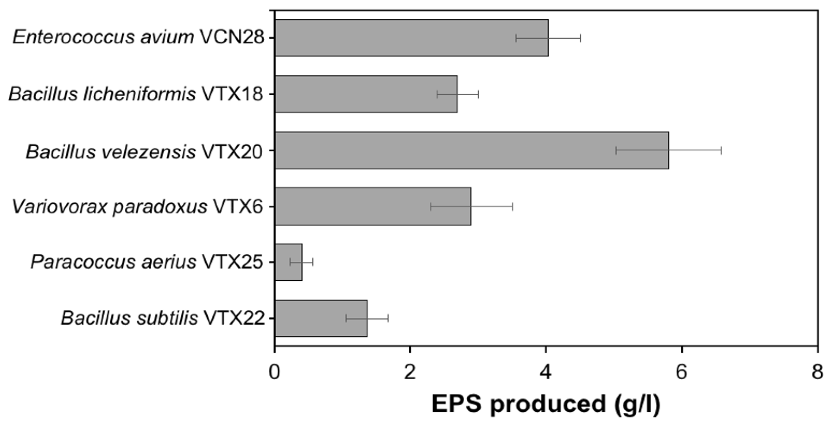
3.1. Isolation and Identification of High EPS-Producing Strain

A total of 65 bacteria were isolated from traditional fermented soybean paste samples collected from Phu Tho Province, Vietnam. After 48 h of incubation, six potent isolates with distinct morphologies with the highest mucoid, slimy, and string-forming appearance were selected. Further, 16S rRNA was used to presumably classify isolates as Enterococcus avium VCN28, Bacillus licheniformis VTX18, Bacillus velezensis VTX20, Variovorax paradoxus VTX6, Paracoccus aerius VTX25, or Bacillus subtilis VTX22 (Table S1). Of six tested isolates, the strain B. velezensis VTX20 yielded the highest EPS of 5.8 ± 0.78 g/L (Figure 1). Combining the fast-growing features of this strain on EPS medium, EPS-producing B. velezensis VTX20 was chosen for further studies.

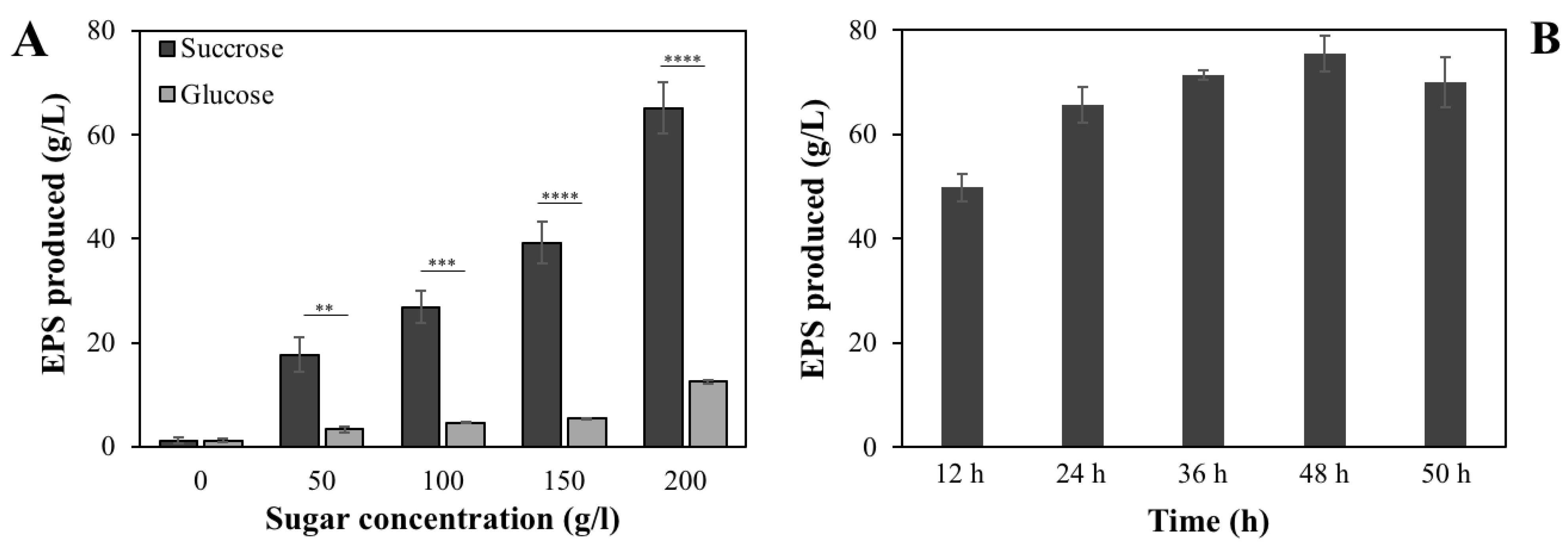
3.2. Effects of Different Sugars on EPS Production of B. velezensis VTX20

One of the important factors that significantly contributes to EPS production is sugar concentration. EPS production by VTX20 was first assessed by altering glucose concentrations in a range of 0–200 g/L. This revealed that the absence of glucose did not result in EPS production (Figure 2A). The EPS yield was in the range of 3.3–12.5 g/L after 24 h when glucose was supplied from 50–200 g/L into the medium. The highest yield of EPS was 12.5 ± 0.25 g/L in the medium with 200 g/L glucose.
Given that the sugar source is important for EPS production [36], different sucrose concentrations were used to obtain a higher yield of EPS for VTX20. As shown in Figure 2A, the yield of EPS experienced a significant and steady upward trend with 0–200 g/L of sucrose. The EPS yield obtained with 50 g/L sucrose was 17.7 ± 3.4 g/L and was the lowest EPS production by VTX20. Differing from glucose, increasing sucrose concentrations significantly enhanced EPS productivity. The highest EPS yield of 65.1 ± 4.9 g/L was achieved when 200 g/L sucrose was utilized, which was 5.2-fold higher than that of glucose. In addition, incubation time was also taken into consideration, choosing 200 g/L sucrose as the supplement to the medium. B. velezensis VTX20 grew well and rapidly produced EPS after 12 h (49.8 ± 2.6 g/L) (Figure 2B). Observed EPS production slightly increased within 12 to 48 h of incubation time. The highest EPS yield was recorded in the stationary phase reaching a maximum (75.5 ± 4.8 g/L) at 48 h. As the incubation time increased, the production of EPS decreased slightly. These results proved that EPS secreted by B. velezensis VTX20 was sugar- and dose-dependent. Sucrose is an efficient carbon source, significantly promoting EPS yields.

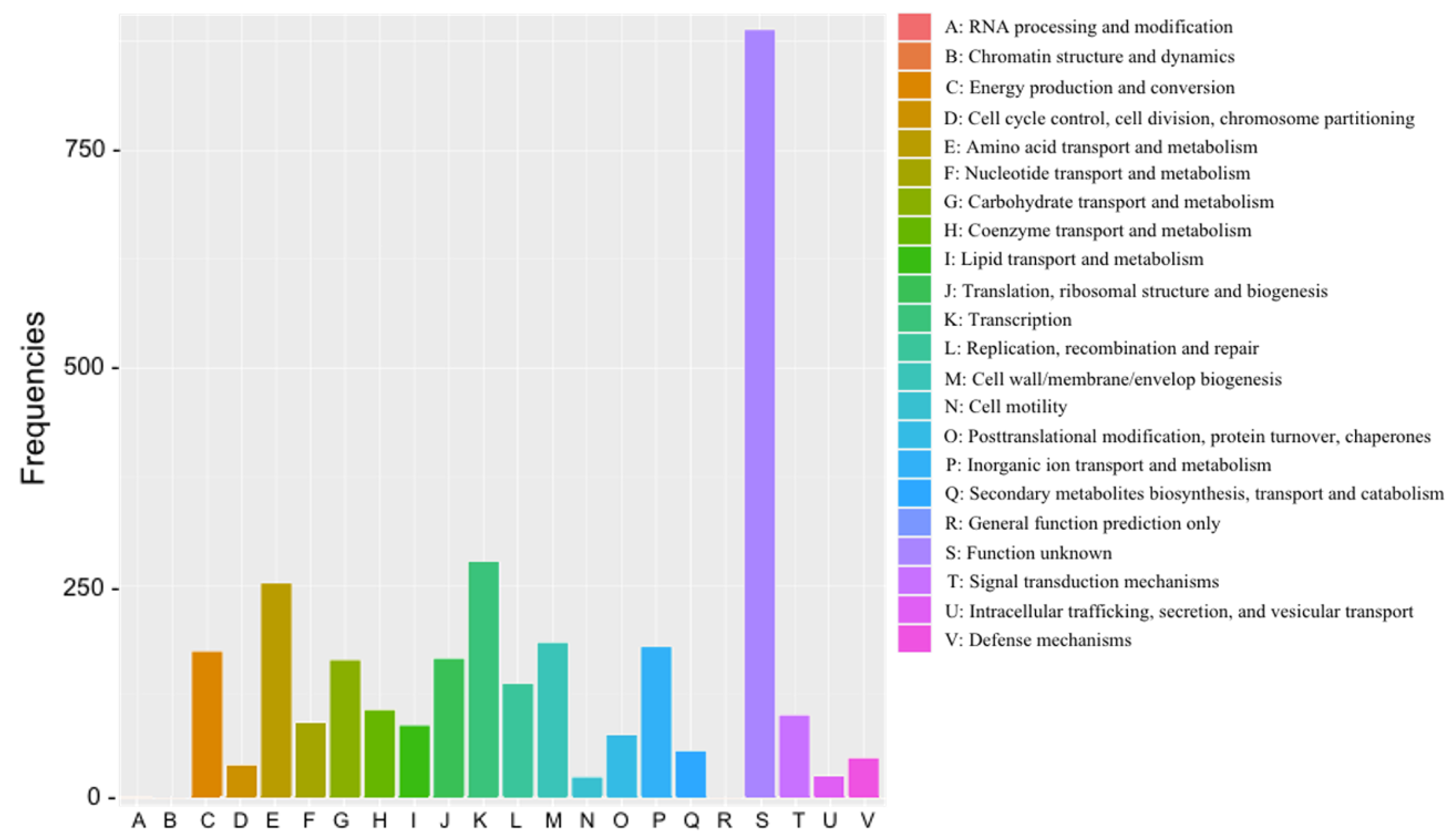
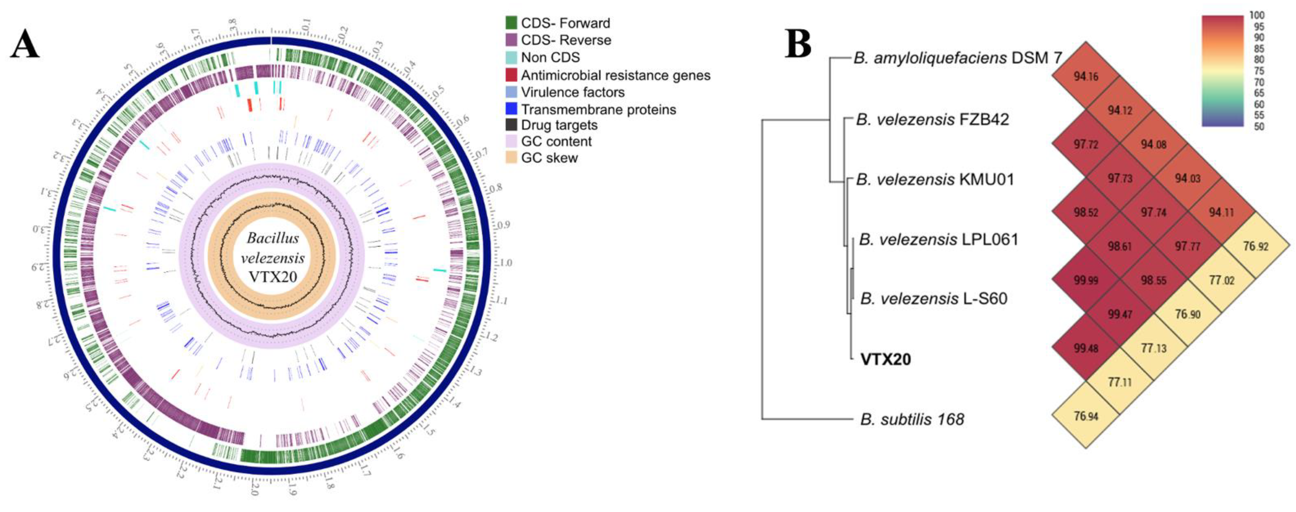
3.3. Genome Feature of EPS-Producing B. velezensis VTX20

The complete genome of strain VTX20 was obtained and submitted to GenBank under the accession number CP075054. Briefly, the complete genome comprised a single and circular chromosome of 3,891,321 bp with a G + C content of 46.1% (Table S2). Moreover, the strain comprised a total of 118 RNAs including 86 tRNA genes, 27 rRNAs, and 5 ncRNAs. The VTX20 genome did not contain a plasmid sequence and habored one CRISPR sequence. Based on the genome annotation, the primary virulence factors in Bacillus species, including hemolysin BL, nonhemolytic, emetic toxin, and one-component cytotoxin K, were not found in the genome (Figure 3A), confirming the safety of this strain.
In support of 16s rRNA analysis, OrthoANI software was utilized to determine the ANI values between isolate VTX20 and six closely related Bacillus species. The values indicated that isolate VTX20 had 76.94–99.48% genome sequence identities with other strains (Figure 3B). VTX20 showed the highest OrthoANI value of 99.48% with B. velezensis L-S60 and 99.47% with B. velezensis LPL061, which was statistically significant enough for species demarcation. This result further confirmed the classification of VTX20 as belonging to B. velezensis species. This strain was then deposited at VAST-Culture Collection of Microorganisms (VCCM) under accession number 14,175.
Out of 3834 genes predicted, a total of 3124 proteins were classified into 19 cluster of orthologous groups (COG) categories. Of note, the cluster for “function unknown” was found to be the largest functional group (Figure 4). Among the other functional groups, the clusters for “transcription” (278 genes) and “amino acid transport and metabolism” (253 genes) were the most represented functional categories. EPS genes were reported to be contained within “cell wall/membrane/envelop biogenesis” (185 genes) and “secondary metabolites biosynthesis, transport and catabolism” (61 genes). This indicated that EPS biosynthetic pathways might be present in the strain VTX20.

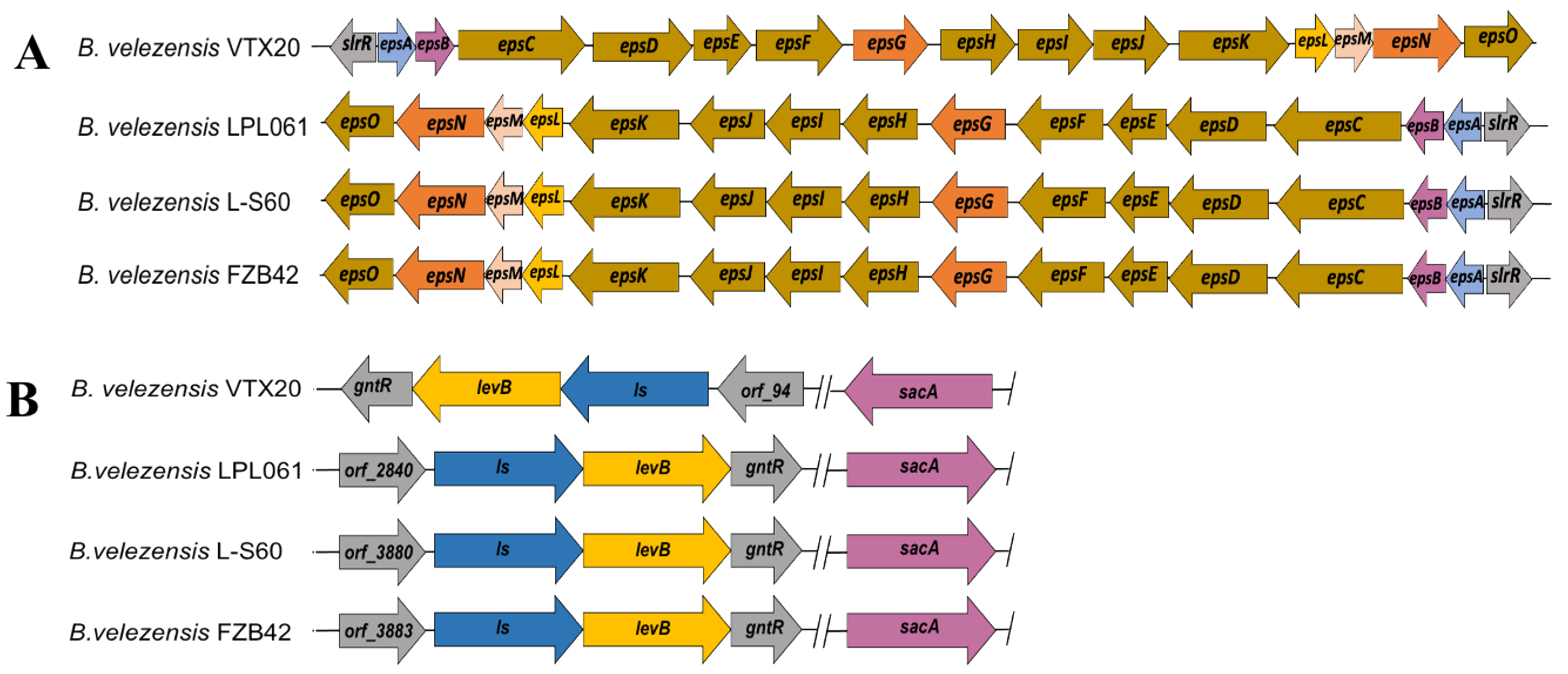
3.4. In Silico Analysis of EPS Biosynthetic Pathways

In silico genome analysis revealed that B. velezensis VTX20 contains two EPS clusters an EPS cluster and a levan cluster. The eps cluster consists of 16 open reading frames, epsA, epsB, epsC, epsD, epsE, epsF, epsG, epsH, epsI, epsJ, epsK, epsL, epsN, epsM, epsO, and slrR. All eps genes are located in the same orientation with a size of 16,414 bp. B. velezensis VTX20 presented around 99% sequence similarity to B. velezensis LPL061, B. velezensis FZB42, B. velezensis L-S60, indicating that the eps cluster is conserved across B. velezensis strains (Figure 5A). Surprisingly, eps genes observed in B. velezensis VTX20 are located on the plus strands, holding an opposite orientation compared to that of other compared strains.
The epsA-O operon is shown to be negatively regulated by the transcriptional regulation factor slrR (orf_720) (Figure 5A). Both epsA (orf_721) and epsB (orf_722) function in determining polysaccharide chain length; however, epsA is also responsible for the export of polysaccharides [37]. The epsL (orf_732), epsD (orf_724), epsE (orf_725), epsF (orf_726), epsH (orf_728), and epsJ (orf_730) genes all encoded glycosyltransferases, participating in the biosynthesis of the repeating units. epsL transfers UDP-glucose or UDP-galactose onto lipid carriers, while epsD (orf_724) functions in transporting UDP-GlcA. Both epsE (orf_725) and epsF (orf_726) are responsible for UDP-Glc transport, and epsH (orf_728) and epsJ (orf_730) are responsible for amino sugars transport. epsG (orf_727) functions in the polymerization of EPS repeating units, and epsK (orf_731) transports polysaccharide repeating units into periplasmic space [10]. Moreover, the epsC (orf_723), epsI (orf_729) and epsO (orf_725), epsM (orf_734), and epsN (orf_733) are involved in the modification of polysaccharide repeating units.
Other bacterial EPSs, such as levan, the fructan polysaccharide, were found in the genome of VTX20. Levan-type fructooligosaccharide biosynthesis includes levansucrase ls (EC 2.4.1.10), 2,6-beta-fructan 6-levanbiohydrolase levB (EC 3.2.1.64), and levanase sacA (EC 3.2.1.26) (Figure 5B). ls and levB genes are clustered together in an operon, while sacA is located on a different operon. Although these genes are conserved among B. velezensis strains, the direction of all three genes in the VTX20 genome is opposite to those of the compared strains (Figure 5B). Levan is synthesized outside cells by an action Ls enzyme that converts disaccharide sucrose into levan, leading to the release of glucose [14]. The released glucose is then employed for metabolic purposes and partly inhibits levan biosynthesis [18]. Moreover, levan is hydrolyzed to levan-type fructooligosaccharides, for example, levanbiose, by LevB and SacA. In addition, SacA is also responsible for cleaving sucrose into glucose and fructose [38].

3.5. Characterization of EPSs Produced by B. velezensis VTX20

Moisture indicators such as water solubility index and water-holding capacity of B. velezensis levan were determined. EPS was completely soluble in distilled water at 25 °C. High EPS concentrations resulted in appearance change from clear to turbid. EPS exhibited a high water solubility index (81.9 ± 3.4%) with water capacity (100.2 ± 3.4). The water molecules might be retained by the hydrophilic fructose residues of levan, leading to gelling and water retention capacities [39].
The structure of EPS was elucidated by NMR experiments. The 1H NMR spectrum revealed the peaks at δH 4.09 (d, J = 8.5 Hz, H-3), 4.00 (t, J = 8.0 Hz, H-4), 3.85 (m, H-5), 3.80 (m, H-6a), 3.68 (d, J = 12.0 Hz, H-1a), 3.56 (m, H-1b), and 3.45 (t, J = 9.0 Hz, H-6b) (Figure S1). The 13C HMBC spectrum showed six signals at δC 104.3 (C-2), 80.2 (C-5), 76.6 (C-3), 75.4 (C-4), 63.4 (C-6), and 59.9 (C-1) (Figure S2), which are almost identical with those of β-(2→ 6)-fructofuranoside [40]. In support of this result, chromatographic analysis revealed that two peaks subjected to fructose and glucose were detected, in which fructose is the predominant sugar (data not shown). The high content of fructose in the EPS corresponded to the occurrence of levan, and an extremly low amount of glucose identified is attributed to dextran, a glucose polymer. Our results confirmed that levan occupied predominantly, followed by an extremely low level of dextran polymer. The monosaccharide composition of EPS from Bacillus strains varies dramatically depending on strains and cultivation parameters. The constitution of EPS from B. velezensis VTX20 is not comparable with EPS from B. mojavensis MT012152 (Table 1). B. amyloliquefaciens C-1 produced an EPS composed of glucose, mannose, galactose, arabinose [25], while B. tequilensis GM was reported to produce only fructose [41].

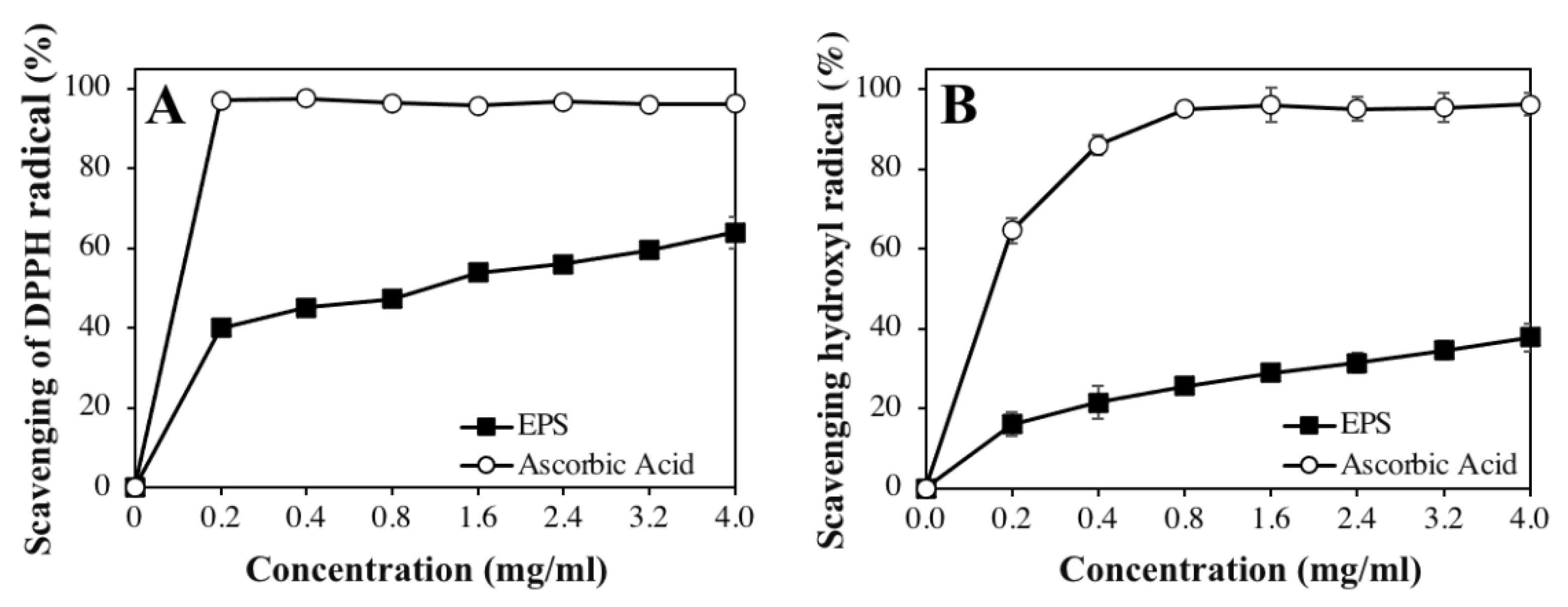
3.6. In Vitro Antioxidant Activity

DPPH is widely used to assess the free-radical-scavenging capacities of antioxidants. The high radical-scavenging activity of antioxidants is subjected to donating a hydrogen atom to DPPH radical [45]. The DPPH free radical scavenging activity of EPS was evaluated in concentrations ranging from 0.2 to 4 mg/mL. The results showed that EPS produced by B. velezensis VTX20 has a strong DPPH radical-scavenging activity that is dose dependent. At 0.2 mg/mL, the EPS reached the lowest DPPH free radical scavenging activity (40.1 ± 0.7%) (Figure 6A). Increasing the concentration of EPS to 4 mg/mL resulted in the highest DPPH radical scavenging of 64.0 ± 4.0%. As the positive control, the DPPH scavenging activity of ascorbic acid always reached maximum activity. This indicated that the scavenging activity of EPS on the inhibition of DPPH free radicals is dose dependent. Proteins, amino acids, and microelements present in the EPS might contribute mainly to the free-radical-scavenging activity.
The scavenging activity of EPS and ascorbic acid on hydroxyl radical is shown in Figure 6B. The EPS produced by B. velezensis VTX20 had moderate inhibitory effects in scavenging hydroxyl radicals. The lowest scavenging activity was observed at 0.2 mg/mL EPS (16.0 ± 3.0%). At 4 mg/mL concentration, the scavenging activity of EPS and ascorbic acid were 40.0 ± 3.5% and 96.2 ± 2.9%, respectively. According to these results, the scavenging activity of EPS on the inhibition of hydroxyl radicals increased as the concentrations increased.

4. Discussion

Bacterial EPSs have received a great deal of attention in recent years due to wide applications in various industries, such as pharmacy, cosmetics, and food. Among them, only dextran extracted from bacteria is used commercially in the food and pharmaceutical industries [46]. Commercial applications of EPS are limited due to the production costs, including fermentation productivity and purification process [47]. Until now, there have been few reports on the production and biological function of EPS from B. velezensis. In this study, we demonstrated for the first time that B. velezensis isolated from traditional fermented soybean paste is a producer of levan at both genotypic and phenotypic levels, which showed great antioxidant activity, offering the potential for use as medical compounds and functional additives.
The EPS biosynthesis capabilities rely on the bacterial strain and its medium composition. A low yield (112.1 mg/L) of EPS was observed in B. tequilensis PS21 when grown in the medium supplemented with 80 g/L lactose [43]. Conversely, B. mojavensis MT012152 isolated from soil produced a high levan amount (22 g/L) using a 5% sucrose concentration [42]. Under an optimized condition that contains 120 g/L molasses and 6 g/L yeast extract, and 30 °C for cultivation temperature, 7.6 g/L EPS consisting of glucose, mannose, and galactose was produced by B. velezensis KY471306 derived from soil [23]. Comparing to our study, it is 9.9-fold lower than levan harvested from B. velezensis VTX20 using 20% sucrose as the main carbon source. To the best of our knowledge, it was the highest Bacillus EPS recorded to date. However, when using a supplement of glucose (200 g/L), B. velezensis VTX20 only produced 12.5 g/L EPS. These results indicated that the type and concentration of carbon source significantly influence EPS production in Bacillus strains. Optimization of levan production by B. velezensis VTX20 using response surface methodology will be an interesting subject for further studies, which will help to reduce production costs.
Using complete genome sequencing of B. velezensis VTX20, the genome of B. velezensis VTX20 contains genes involved in EPS and levan-type EPS biosynthesis. Surprisingly, they are in the opposite strand in comparison to other compared B. velezensis (Figure 5). The slrR-epsAO operon is around 16.4 kb that includes the genes encoding for the regulation, determination of chain length, synthesis, polymerization, and exportation of the repeating sugar units concerned. Excessive glucose might lead to inactivation of the SlrR repressor activity, dissociation of SlrR from its promoter, and derepression of epsA-O genes. Deletion of transcriptional factor SlrR could be an effective strategy for overproduction of EPS encoded by epsA-O operon in the medium supplemented with glucose. By contrast, under excessive sucrose, the ls encoding for levansucrase hydrolyzes sucrose to yield levan and release glucose as a by-product [48]. Levan-degrading enzymes including levB and sacA confer to the rapid hydrolysis of levan to levan-type fructooligosaccharides [20], which are supposed to be inhibited due to high sucrose concentration as described previously in B. subtilis [49]. Biochemical and structural analysis of EPS confirmed that EPS produced by B. velezensis VTX20 contained predominantly levan, accompanied by an extremly low level of dextran polymer. These results led to a hypothesis that ls is highly expressed in presence of high sucrose concentration, yielding levan and releasing free glucose. Released glucose activated transcription of the epsA-O operon, resulting in the production of dextran polymer. A previous study proved that the presence of glucose strongly inhibits the enzymatic activity of levansucrase, causing a decrease in levan production [50].
Our result proved that obtained EPS is a functional biopolymer. The EPS from B. velezensis VTX20 showed great solubility and water-holding capacity, which are in accordance with the presence of fructose residues. It is consistent with levan produced by other strains [42,51]. In terms of bioactivities, EPS obtained in this study exhibited 64.0 ± 4.0% of DPPH radical scavenging activity at 4 mg/mL, which was significantly higher than that of EPS from B. tequilensis PS21 [43], Lactobacillus helveticus MB2-1 [52], and Leuconostoc mesenteroides WiKim32 [45]. This indicated that B. velezensis EPS had strong scavenging activity on DPPH. Since the DPPH free radical scavenging activity of antioxidants is based on the ability to transfer either an electron or a hydrogen atom to DPPH, neutralizing its free radical character [53], the different antioxidant activity could be attributed to chemical features discrepancy. On the other hand, hydroxyl radical is the most reactive and toxic reactive oxygen species, which is generated via Fenton reaction and iron-catalyzed Haber-Wiss reaction, causing aging, cancer, and other diseases [22,53]. Thus, it is of great importance to characterize the hydroxyl radical scavenging potential of antioxidant agents. EPS from B. velezensis VTX20 showed moderate scavenging activity on hydroxyl radicals, which was nearly similar to that of B. amyloliquefaciens C-1 [25]. EPS is able to scavenge hydroxyl radical because of the hydrogen and electron abstraction mechanism. These findings concluded that B. velezensis EPS could be considered as a promising candidate that has potential uses in the biomedical and food industries, such as drug delivery, food hydrocolloids, and as bioabsorbents.

5. Conclusions

In this study, the production, genome mining, characterization, and antioxidant activities of EPS produced by B. velezensis VTX20 were exploited for the first time. The maximum yield of EPS (75.5 ± 4.8 g/L) was observed after 48 h, which was the highest EPS production observed to date. The complete genome sequencing of B. velezensis VTX20 brings new insights into the biosynthesis of EPS encoded by slrR-epsAO and ls-levB operons. Characterization of the EPS demonstrated that it was predominantly levan occupied, followed by a non-significant level of dextran. Additionally, EPS had strong scavenging activities on DPPH and hydroxyl radicals. These findings, for the first time, proved that B. velezensis VTX20 holds potential for more economic EPS production and as a natural alternative to commercial antioxidants in the pharmaceutical and biomedical industries.

Supplementary Materials

The following are available online at https://www.mdpi.com/article/10.3390/app11157055/s1, Figure S1: 1H NMR of the dialyzed EPS from B. velezensis VTX20, Figure S2: 13C HMBC spectrum of the dialyzed EPS, Table S1: Closest species, by 16S rRNA gene similarity, of EPS-producing bacteria isolated from traditional fermented soybean pastes. Table S2: Genomic feature of the strain VTX20.

Author Contributions

Conceptualization, T.H.N.V. and N.T.Q.; methodology, T.H.N.V., N.T.Q., N.A.N., T.D.N. and H.T.N.; investigation, P.-H.H., C.C.N. and H.H.; writing—original draft preparation, T.H.N.V. and N.T.Q.; writing—review and editing, H.H.C. and Q.-T.P.; project administration, Q.-T.P. All authors have read and agreed to the published version of the manuscript.

Funding

This research was funded by the Vietnam Academy of Science and Technology, grant number CN40SH.01/18-21 and Ministry of Science and Technology, grant number ĐTĐL.CN-71/19.

Institutional Review Board Statement

Not applicable.

Informed Consent Statement

Not applicable.

Data Availability Statement

Data sharing is not applicable to this article.

Acknowledgments

The authors would like to acknowledge the support of the VAST Culture Collection of Microorganisms, Institute of Biotechnology, Vietnam Academy of Science and Technology (www.vccm.vast.vn). We are grateful to Tung Lam Le for support with whole genome sequencing.

Conflicts of Interest

The authors declare no conflict of interest.

References

  1. Freitas, F.; Alves, V.D.; Pais, J.; Carvalheira, M.; Costa, N.; Oliveira, R.; Reis, M.A.M. Production of a new exopolysaccharide (EPS) by Pseudomonas oleovorans NRRL B-14682 grown on glycerol. Process Biochem. 2010, 45, 297–305. [Google Scholar] [CrossRef]
  2. Parikh, A.; Madamwar, D. Partial characterization of extracellular polysaccharides from cyanobacteria. Bioresour. Technol. 2006, 97, 1822–1827. [Google Scholar] [CrossRef] [PubMed]
  3. Barcelos, M.C.S.; Vespermann, K.A.C.; Pelissari, F.M.; Molina, G. Current status of biotechnological production and applications of microbial exopolysaccharides. Crit. Rev. Food Sci. Nutr. 2020, 60, 1475–1495. [Google Scholar] [CrossRef] [PubMed]
  4. Rajoka, M.S.R.; Mehwish, H.M.; Hayat, H.F.; Hussain, N.; Sarwar, S.; Aslam, H.; Nadeem, A.; Shi, J. Characterization, the antioxidant and antimicrobial activity of exopolysaccharide Isolated from poultry origin Lactobacilli. Probiotics Antimicrob. Proteins 2019, 11, 1132–1142. [Google Scholar] [CrossRef] [PubMed]
  5. Liu, C.T.; Chu, F.J.; Chou, C.C.; Yu, R.C. Antiproliferative and anticytotoxic effects of cell fractions and exopolysaccharides from Lactobacillus casei 01. Mutat. Res./Genet. Toxicol. Environ. Mutagenesis 2011, 721, 157–162. [Google Scholar] [CrossRef]
  6. Kavita, K.; Singh, V.K.; Mishra, A.; Jha, B. Characterisation and anti-biofilm activity of extracellular polymeric substances from Oceanobacillus iheyensis. Carbohydr. Polym. 2014, 101, 29–35. [Google Scholar] [CrossRef]
  7. Ismail, B.; Nampoothiri, K.M. Production, purification and structural characterization of an exopolysaccharide produced by a probiotic Lactobacillus plantarum MTCC 9510. Arch. Microbiol. 2010, 192, 1049–1057. [Google Scholar] [CrossRef]
  8. Jolly, L.; Vincent, S.J.; Duboc, P.; Neeser, J.R. Exploiting expolysaccharides from lactic acid bacteria. Antonie Van Leeuwenhoek 2002, 82, 367–374. [Google Scholar] [CrossRef] [PubMed]
  9. Welman, A.D.; Maddox, I.S. Exopolysaccharides from lactic acid bacteria: Perspectives and challenges. Trends Biotechnol. 2003, 21, 269–274. [Google Scholar] [CrossRef]
  10. Sayem, S.M.A.; Manzo, E.; Ciavatta, L.; Tramice, A.; Cordone, A.; Zanfardino, A.; De Felice, M.; Varcamonti, M. Anti-biofilm activity of an exopolysaccharide from a sponge-associated strain of Bacillus licheniformis. Microb. Cell Factories 2011, 10, 74. [Google Scholar] [CrossRef] [Green Version]
  11. Chen, Y.-T.; Yuan, Q.; Shan, L.E.T.; Lin, M.-A.; Cheng, D.-Q.; Li, C.-Y. Antitumor activity of bacterial exopolysaccharides from the endophyte Bacillus amyloliquefaciens sp. isolated from Ophiopogon japonicus. Oncol. Lett. 2013, 5, 1787–1792. [Google Scholar] [CrossRef]
  12. Wu, J.-Y.; Ye, H.-F. Characterization and flocculating properties of an extracellular biopolymer produced from a Bacillus subtilis DYU1 isolate. Process Biochem. 2007, 42, 1114–1123. [Google Scholar] [CrossRef]
  13. Mu, D.; Zhou, Y.; Wu, X.; Montalban-Lopez, M.; Wang, L.; Li, X.; Zheng, Z. Secretion of Bacillus amyloliquefaciens levansucrase from Bacillus subtilis and Its application in the enzymatic synthesis of levan. ACS Food Sci. Technol. 2021, 1, 249–259. [Google Scholar] [CrossRef]
  14. Chai, Y.; Beauregard, P.B.; Vlamakis, H.; Losick, R.; Kolter, R. Galactose metabolism plays a crucial role in biofilm formation by Bacillus subtilis. mBio 2012, 3, e00184. [Google Scholar] [CrossRef] [Green Version]
  15. Srikanth, R.; Reddy, C.H.S.S.S.; Siddartha, G.; Ramaiah, M.J.; Uppuluri, K.B. Review on production, characterization and applications of microbial levan. Carbohydr. Polym. 2015, 120, 102–114. [Google Scholar] [CrossRef]
  16. Shih, I.-L.; Yu, Y.-T.; Shieh, C.-J.; Hsieh, C.-Y. Selective production and characterization of levan by Bacillus subtilis (Natto) Takahashi. J. Agric. Food Chem. 2005, 53, 8211–8215. [Google Scholar] [CrossRef]
  17. Jakob, F.; Gebrande, C.; Bichler, R.M.; Vogel, R.F. Insights into the pH-dependent, extracellular sucrose utilization and concomitant levan formation by Gluconobacter albidus TMW 2.1191. Antonie Van Leeuwenhoek 2020, 113, 863–873. [Google Scholar] [CrossRef] [PubMed] [Green Version]
  18. Dahech, I.; Belghith, K.S.; Hamden, K.; Feki, A.; Belghith, H.; Mejdoub, H. Antidiabetic activity of levan polysaccharide in alloxan-induced diabetic rats. Int. J. Biol. Macromol. 2011, 49, 742–746. [Google Scholar] [CrossRef]
  19. Esawy, M.A.; Ahmed, E.F.; Helmy, W.A.; Mansour, N.M.; El-Senousy, W.M.; El-Safty, M.M. Production of levansucrase from novel honey Bacillus subtilis isolates capable of producing antiviral levans. Carbohydr. Polym. 2011, 86, 823–830. [Google Scholar] [CrossRef]
  20. Zhang, Z.; Liu, Z.; Tao, X.; Wei, H. Characterization and sulfated modification of an exopolysaccharide from Lactobacillus plantarum ZDY2013 and its biological activities. Carbohydr. Polym. 2016, 153, 25–33. [Google Scholar] [CrossRef]
  21. Liu, J.; Luo, J.; Ye, H.; Sun, Y.; Lu, Z.; Zeng, X. Production, characterization and antioxidant activities in vitro of exopolysaccharides from endophytic bacterium Paenibacillus polymyxa EJS-3. Carbohydr. Polym. 2009, 78, 275–281. [Google Scholar] [CrossRef]
  22. Moghannem, S.A.M.; Farag, M.M.S.; Shehab, A.M.; Azab, M.S. Exopolysaccharide production from Bacillus velezensis KY471306 using statistical experimental design. Braz. J. Microbiol. 2018, 49, 452–462. [Google Scholar] [CrossRef] [PubMed]
  23. Xu, M.; Zhang, L.; Zhao, F.; Wang, J.; Zhao, B.; Zhou, Z.; Han, Y. Cloning and expression of levansucrase gene of Bacillus velezensis BM-2 and enzymatic synthesis of levan. Processes 2021, 9, 317. [Google Scholar] [CrossRef]
  24. Yang, H.; Deng, J.; Yuan, Y.; Fan, D.; Zhang, Y.; Zhang, R.; Han, B. Two novel exopolysaccharides from Bacillus amyloliquefaciens C-1: Antioxidation and effect on oxidative stress. Curr. Microbiol. 2015, 70, 298–306. [Google Scholar] [CrossRef]
  25. Ruas-Madiedo, P.; de los Reyes-Gavilán, C.G. Invited review: Methods for the screening, isolation, and characterization of exopolysaccharides produced by lactic acid bacteria. J. Dairy Sci. 2005, 88, 843–856. [Google Scholar] [CrossRef] [Green Version]
  26. Bikric, S.; Aslim, B.; Dincer, İ.; Yuksekdag, Z.; Ulusoy, S.; Yavuz, S. Characterization of exopolysaccharides (EPSs) obtained from Ligilactobacillus salivarius strains and investigation at the prebiotic potential as an alternative to plant prebiotics at poultry. Probiotics Antimicrob. Proteins 2021. [Google Scholar] [CrossRef]
  27. Overbeek, R.; Olson, R.; Pusch, G.D.; Olsen, G.J.; Davis, J.J.; Disz, T.; Edwards, R.A.; Gerdes, S.; Parrello, B.; Shukla, M.; et al. The SEED and the Rapid Annotation of microbial genomes using Subsystems Technology (RAST). Nucleic Acids Res. 2014, 42, D206–D214. [Google Scholar] [CrossRef]
  28. Tatusova, T.; DiCuccio, M.; Badretdin, A.; Chetvernin, V.; Nawrocki, E.P.; Zaslavsky, L.; Lomsadze, A.; Pruitt, K.D.; Borodovsky, M.; Ostell, J. NCBI prokaryotic genome annotation pipeline. Nucleic Acids Res. 2016, 44, 6614–6624. [Google Scholar] [CrossRef]
  29. Galperin, M.Y.; Makarova, K.S.; Wolf, Y.I.; Koonin, E.V. Expanded microbial genome coverage and improved protein family annotation in the COG database. Nucleic Acids Res. 2015, 43, D261–D269. [Google Scholar] [CrossRef] [PubMed]
  30. Lee, I.; Ouk Kim, Y.; Park, S.C.; Chun, J. OrthoANI: An improved algorithm and software for calculating average nucleotide identity. Int. J. Syst. Evol. Microbiol. 2016, 66, 1100–1103. [Google Scholar] [CrossRef]
  31. Anderson, R.A.; Conway, H.F.; Peplinski, A.J. Gelatinization of corn grits by roll cooking, extrusion cooking and steaming. Starch Stärke 1970, 22, 130–135. [Google Scholar] [CrossRef]
  32. Feng, F.; Zhou, Q.; Yang, Y.; Zhao, F.; Du, R.; Han, Y.; Xiao, H.; Zhou, Z. Characterization of highly branched dextran produced by Leuconostoc citreum B-2 from pineapple fermented product. Int. J. Biol. Macromol. 2018, 113, 45–50. [Google Scholar] [CrossRef] [PubMed]
  33. Lv, Y.; Yang, X.; Zhao, Y.; Ruan, Y.; Yang, Y.; Wang, Z. Separation and quantification of component monosaccharides of the tea polysaccharides from Gynostemma pentaphyllum by HPLC with indirect UV detection. Food Chem. 2009, 112, 742–746. [Google Scholar] [CrossRef]
  34. Kadaikunnan, S.; Rejiniemon, T.; Khaled, J.M.; Alharbi, N.S.; Mothana, R. In-vitro antibacterial, antifungal, antioxidant and functional properties of Bacillus amyloliquefaciens. Ann. Clin. Microbiol. Antimicrob. 2015, 14, 9. [Google Scholar] [CrossRef] [PubMed] [Green Version]
  35. Xu, C.; Yu, J.; Zhao, S.; Wu, S.; He, P.; Jia, X.; Liu, Y.; Mao, D. Effect of carbon source on production, characterization and bioactivity of exopolysaccharide produced by Phellinus vaninii Ljup. An. Acad. Bras. Ciências 2017, 89, 2033–2041. [Google Scholar] [CrossRef] [Green Version]
  36. Güvener, Z.T.; McCarter, L.L. Multiple regulators control capsular polysaccharide production in Vibrio parahaemolyticus. J. Bacteriol. 2003, 185, 5431–5441. [Google Scholar] [CrossRef] [PubMed] [Green Version]
  37. Marvasi, M.; Visscher, P.T.; Casillas Martinez, L. Exopolymeric substances (EPS) from Bacillus subtilis: Polymers and genes encoding their synthesis. FEMS Microbiol. Lett. 2010, 313, 1–9. [Google Scholar] [CrossRef] [Green Version]
  38. Velázquez-Hernández, M.L.; Baizabal-Aguirre, V.M.; Bravo-Patiño, A.; Cajero-Juárez, M.; Chávez-Moctezuma, M.P.; Valdez-Alarcón, J.J. Microbial fructosyltransferases and the role of fructans. J. Appl. Microbiol. 2009, 106, 1763–1778. [Google Scholar] [CrossRef]
  39. Niknezhad, S.V.; Najafpour-Darzi, G.; Morowvat, M.H.; Ghasemi, Y. Eexopolysaccharide production of Pantoea sp. BCCS 001 GH: Physical characterizations, emulsification, and antioxidant activities. Int. J. Biol. Macromol. 2018, 118, 1103–1111. [Google Scholar] [CrossRef]
  40. Han, J.; Xu, X.; Gao, C.; Liu, Z.; Wu, Z.; Elkins, C.A. Levan-producing Leuconostoc citreum strain BD1707 and its growth in tomato juice supplemented with sucrose. Appl. Environ. Microbiol. 2016, 82, 1383–1390. [Google Scholar] [CrossRef] [Green Version]
  41. Abid, Y.; Azabou, S.; Casillo, A.; Gharsallah, H.; Jemil, N.; Lanzetta, R.; Attia, H.; Corsaro, M.M. Isolation and structural characterization of levan produced by probiotic Bacillus tequilensis-GM from Tunisian fermented goat milk. Int. J. Biol. Macromol. 2019, 133, 786–794. [Google Scholar] [CrossRef] [PubMed]
  42. Haddar, A.; Hamed, M.; Bouallegue, A.; Bastos, R.; Coelho, E.; Coimbra, M.A. Structural elucidation and interfacial properties of a levan isolated from Bacillus mojavensis. Food Chem. 2021, 343, 128456. [Google Scholar] [CrossRef]
  43. Vijitra, L.-I.; Worachot, S. Characterization and bioactivities of a novel exopolysaccharide produced from lactose by Bacillus tequilensis PS21 isolated from Thai Milk Kefir. Microbiol. Biotechnol. Lett. 2018, 46, 9–17. [Google Scholar] [CrossRef] [Green Version]
  44. Mohamed, S.S.; Amer, S.K.; Selim, M.S.; Rifaat, H.M. Characterization and applications of exopolysaccharide produced by marine Bacillus altitudinis MSH2014 from Ras Mohamed, Sinai, Egypt. Egypt. J. Basic Appl. Sci. 2018, 5, 204–209. [Google Scholar] [CrossRef] [Green Version]
  45. Choi, I.S.; Ko, S.H.; Lee, M.E.; Kim, H.M.; Yang, J.E.; Jeong, S.G.; Lee, K.H.; Chang, J.Y.; Kim, J.C.; Park, H.W. Production, characterization, and antioxidant activities of an exopolysaccharide extracted from spent media wastewater after Leuconostoc mesenteroides WiKim32 fermentation. ACS Omega 2021, 6, 8171–8178. [Google Scholar] [CrossRef]
  46. Kodali, V.P.; Sen, R. Antioxidant and free radical scavenging activities of an exopolysaccharide from a probiotic bacterium. Biotechnol. J. 2008, 3, 245–251. [Google Scholar] [CrossRef] [PubMed]
  47. Freitas, F.; Alves, V.D.; Reis, M.A. Advances in bacterial exopolysaccharides: From production to biotechnological applications. Trends Biotechnol. 2011, 29, 388–398. [Google Scholar] [CrossRef]
  48. Pabst, M.J. Levan and levansucrase of Actinomyces viscosus. Infect. Immun. 1977, 15, 518–526. [Google Scholar] [CrossRef] [Green Version]
  49. Jensen, S.L.; Diemer, M.B.; Lundmark, M.; Larsen, F.H.; Blennow, A.; Mogensen, H.K.; Nielsen, T.H. Levanase from Bacillus subtilis hydrolyses β-2,6 fructosyl bonds in bacterial levans and in grass fructans. Int. J. Biol. Macromol. 2016, 85, 514–521. [Google Scholar] [CrossRef]
  50. Ullah, M.W.; Ul-Islam, M.; Khan, T.; Park, J.K. Chapter 31—Recent developments in the synthesis, properties, and applications of various microbial polysaccharides. In Handbook of Hydrocolloids, 3rd ed.; Phillips, G.O., Williams, P.A., Eds.; Woodhead Publishing: Sawston, UK, 2021; pp. 975–1015. [Google Scholar] [CrossRef]
  51. Pantelić, I.; Lukić, M.; Gojgić-Cvijović, G.; Jakovljević, D.; Nikolić, I.; Lunter, D.J.; Daniels, R.; Savić, S. Bacillus licheniformis levan as a functional biopolymer in topical drug dosage forms: From basic colloidal considerations to actual pharmaceutical application. Eur. J. Pharm. Sci. 2020, 142, 105109. [Google Scholar] [CrossRef]
  52. Li, W.; Ji, J.; Chen, X.; Jiang, M.; Rui, X.; Dong, M. Structural elucidation and antioxidant activities of exopolysaccharides from Lactobacillus helveticus MB2-1. Carbohydr. Polym. 2014, 102, 351–359. [Google Scholar] [CrossRef]
  53. Liu, C.F.; Tseng, K.C.; Chiang, S.S.; Lee, B.H.; Hsu, W.H.; Pan, T.M. Immunomodulatory and antioxidant potential of Lactobacillus exopolysaccharides. J. Sci. Food Agric. 2011, 91, 2284–2291. [Google Scholar] [CrossRef]
Figure 1. EPS production observed in six selected bacterial strains.
Applsci 11 07055 g001
Figure 2. EPS production by B. velezensis VTX20. (A) Effect of glucose and sucrose concentrations on the production of EPSs. (B) Time course of EPS production in the medium supplemented with 200 g/L sucrose. The statistics were calculated using a Student’s unpaired two-tailed t-test using the GraphPad Prism software. Symbols are: ** p ≤ 0.01, *** p ≤ 0.001, and **** p ˂ 0.0001.
Applsci 11 07055 g002
Figure 3. Circular map (A) and heat map (B) based on OrthoANI values determined for B. velezensis VTX20 and related strains.
Applsci 11 07055 g003
Figure 4. The cluster of orthologous group (COG) categories of the strain VTX20.
Applsci 11 07055 g004
Figure 5. The EPS (A) and levan biosynthetic gene cluster (B) identified in B. velezensis VTX20.
Applsci 11 07055 g005
Figure 6. Antioxidant activities of EPS produced by B. velezensis VTX20. Scavenging activity on DPPH free radicals (A) and hydroxyl radicals (B).
Applsci 11 07055 g006
Table 1. Monosaccharide composition of EPS produced by Bacillus strains and their antioxidant activities.
EPS-Producing BacteriaMonosaccharidesAntioxidant ActivityReferences
B. mojavensis MT012152FructoseND[42]
B. amyloliquefaciens C-1Glucose, mannose, galactose, arabinoseSuperoxide, hydroxyl radicals[25]
B. tequilensis PS21Glucose, xylose, ribose, rhamnose, galactoseDPPH, ABTS[43]
B. altitudinis MSH2014GlucoseDPPH[44]
B. tequilensis GMFructoseND[41]
B. velezensis VTX20Fructose, glucoseDPPH, hydroxyl radicalsThis study
Abbreviation: ND, Not determined; ABTS: 2,2′-azino-bis(3-ethylbenzothiazoline-6-sulfonic acid); DPPH, 1,1-diphenyl-2-picrylhydrazyl.
Publisher’s Note: MDPI stays neutral with regard to jurisdictional claims in published maps and institutional affiliations.

