High-Resolution Measurement of Molecular Internal Polarization Structure by Photoinduced Force Microscopy
Abstract
:1. Introduction
2. Theory and Calculation Model
2.1. Discrete Dipole Approximation
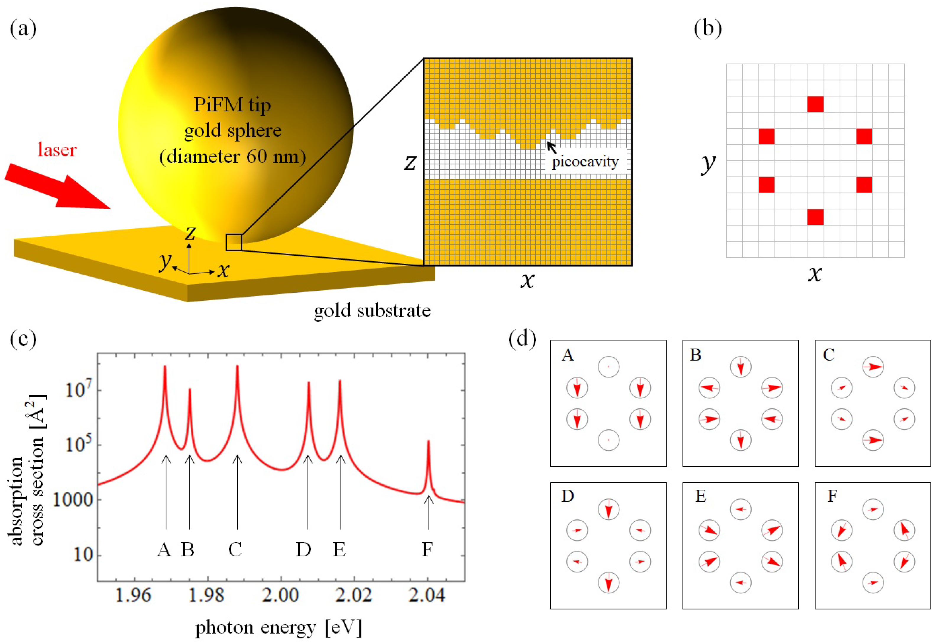
2.2. Model Parameters
3. PiFM Measurement for a Single-Molecule
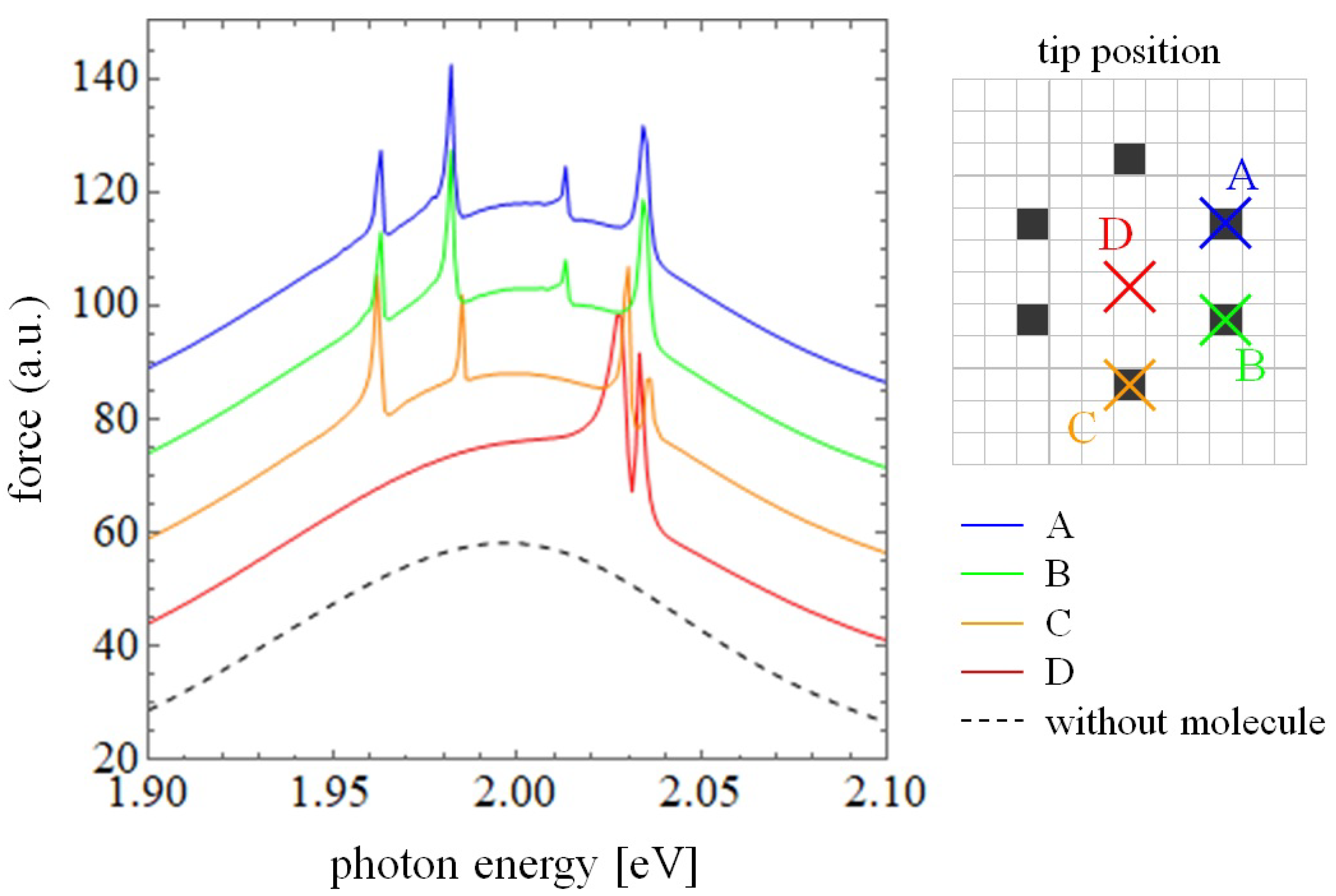
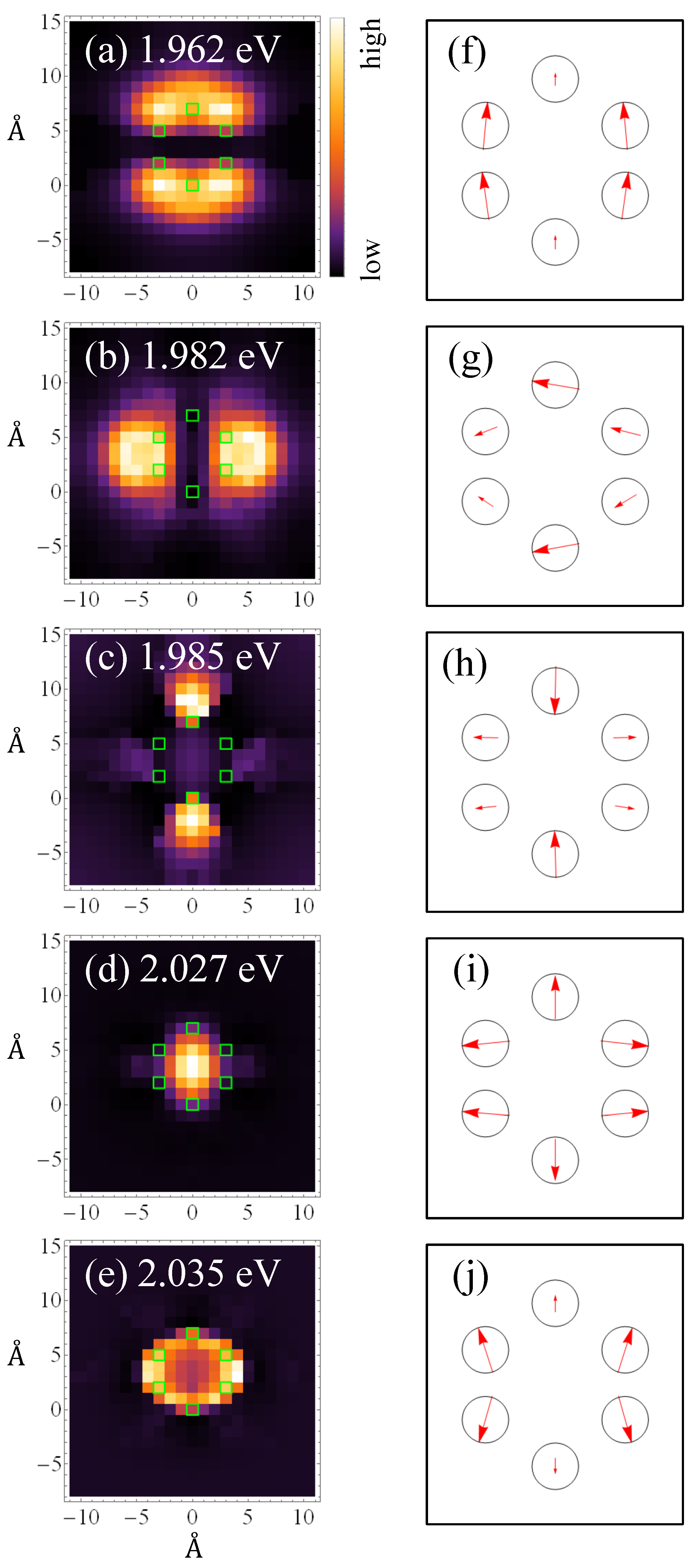
3.1. Microscopic Interaction between Molecule and Plasmon
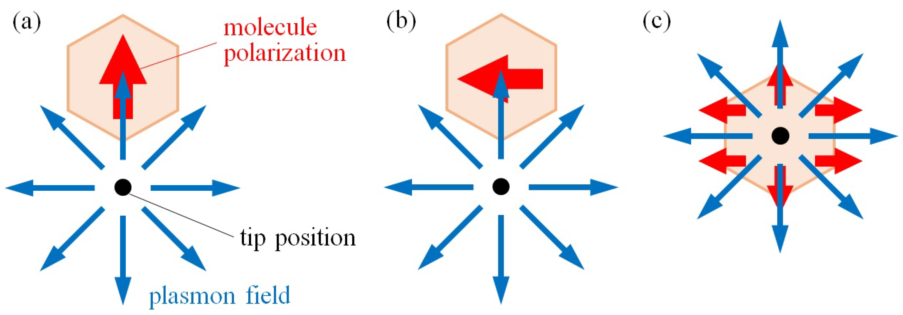
3.2. Critical Role of Picocavity in Resolution
4. Conclusions
Author Contributions
Funding
Institutional Review Board Statement
Informed Consent Statement
Data Availability Statement
Acknowledgments
Conflicts of Interest
References
- Papakostas, A.; Potts, A.; Bagnall, D.; Prosvirnin, S.; Coles, H.; Zheludev, N. Optical manifestations of planar chirality. Phys. Rev. Lett. 2003, 90, 107404. [Google Scholar] [CrossRef] [PubMed]
- Vallius, T.; Jefimovs, K.; Turunen, J.; Vahimaa, P.; Svirko, Y. Optical activity in subwavelength-period arrays of chiral metallic particles. Appl. Phys. Lett. 2003, 83, 234–236. [Google Scholar] [CrossRef]
- Kuwata-Gonokami, M.; Saito, N.; Ino, Y.; Kauranen, M.; Jefimovs, K.; Vallius, T.; Turunen, J.; Svirko, Y. Giant optical activity in quasi-two-dimensional planar nanostructures. Phys. Rev. Lett. 2005, 95, 227401. [Google Scholar] [CrossRef] [PubMed]
- Bai, B.; Svirko, Y.; Turunen, J.; Vallius, T. Optical activity in planar chiral metamaterials: Theoretical study. Phys. Rev. A 2007, 76, 023811. [Google Scholar] [CrossRef]
- Hendry, E.; Carpy, T.; Johnston, J.; Popland, M.; Mikhaylovskiy, R.; Lapthorn, A.; Kelly, S.; Barron, L.; Gadegaard, N.; Kadodwala, M. Ultrasensitive detection and characterization of biomolecules using superchiral fields. Nat. Nanotech. 2010, 5, 783. [Google Scholar] [CrossRef] [Green Version]
- Kuzyk, A.; Schreiber, R.; Fan, Z.; Pardatscher, G.; Roller, E.M.; Högele, A.; Simmel, F.C.; Govorov, A.O.; Liedl, T. DNA-based self-assembly of chiral plasmonic nanostructures with tailored optical response. Nature 2012, 483, 311. [Google Scholar] [CrossRef] [Green Version]
- Kumar, J.; Eraña, H.; López-Martínez, E.; Claes, N.; Martín, V.F.; Solís, D.M.; Bals, S.; Cortajarena, A.L.; Castilla, J.; Liz-Marzán, L.M. Detection of amyloid fibrils in Parkinson’s disease using plasmonic chirality. Proc. Natl. Acad. Sci. USA 2018, 115, 3225–3230. [Google Scholar] [CrossRef] [Green Version]
- Mohammadi, E.; Tsakmakidis, K.L.; Askarpour, A.N.; Dehkhoda, P.; Tavakoli, A.; Altug, H. Nanophotonic Platforms for Enhanced Chiral Sensing. ACS Photon. 2018, 5, 2669–2675. [Google Scholar] [CrossRef]
- Rajapaksa, I.; Uenal, K.; Wickramasinghe, H.K. Image force microscopy of molecular resonance: A microscope principle. Appl. Phys. Lett. 2010, 97, 073121. [Google Scholar] [CrossRef] [Green Version]
- Rajapaksa, I.; Kumar Wickramasinghe, H. Raman spectroscopy and microscopy based on mechanical force detection. Appl. Phys. Lett. 2011, 99, 161103. [Google Scholar] [CrossRef] [Green Version]
- Imada, H.; Miwa, K.; Imai-Imada, M.; Kawahara, S.; Kimura, K.; Kim, Y. Real-space investigation of energy transfer in heterogeneous molecular dimers. Nature 2016, 538, 364–367. [Google Scholar] [CrossRef] [PubMed]
- Imada, H.; Miwa, K.; Imai-Imada, M.; Kawahara, S.; Kimura, K.; Kim, Y. Single-molecule investigation of energy dynamics in a coupled plasmon-exciton system. Phys. Rev. Lett. 2017, 119, 013901. [Google Scholar] [CrossRef] [Green Version]
- Zhang, Y.; Luo, Y.; Zhang, Y.; Yu, Y.J.; Kuang, Y.M.; Zhang, L.; Meng, Q.S.; Luo, Y.; Yang, J.L.; Dong, Z.C.; et al. Visualizing coherent intermolecular dipole–dipole coupling in real space. Nature 2016, 531, 623–627. [Google Scholar] [CrossRef] [PubMed]
- Liu, P.; Chulhai, D.V.; Jensen, L. Single-molecule imaging using atomistic near-field tip-enhanced Raman spectroscopy. ACS Nano 2017, 11, 5094–5102. [Google Scholar] [CrossRef] [PubMed] [Green Version]
- Gross, L.; Mohn, F.; Moll, N.; Liljeroth, P.; Meyer, G. The chemical structure of a molecule resolved by atomic force microscopy. Science 2009, 325, 1110–1114. [Google Scholar] [CrossRef] [Green Version]
- Almajhadi, M.; Wickramasinghe, H.K. Contrast and imaging performance in photo induced force microscopy. Opt. Express 2017, 25, 26923–26938. [Google Scholar] [CrossRef]
- Rajaei, M.; Almajhadi, M.A.; Zeng, J.; Wickramasinghe, H.K. Near-field nanoprobing using Si tip-Au nanoparticle photoinduced force microscopy with 120: 1 signal-to-noise ratio, sub-6-nm resolution. Opt. Express 2018, 26, 26365–26376. [Google Scholar] [CrossRef] [Green Version]
- Yamane, H.; Yamanishi, J.; Yokoshi, N.; Sugawara, Y.; Ishihara, H. Theoretical analysis of optically selective imaging in photoinduced force microscopy. Opt. Express 2020, 28, 34787–34803. [Google Scholar] [CrossRef]
- Yamanishi, J.; Yamane, H.; Naitoh, Y.; Li, Y.; Yokoshi, N.; Masuoka, K.; Kameyama, T.; Torimoto, T.; Ishihara, H.; Sugawara, Y. Optical Force Mapping at the Single-Nanometre Scale. Nat. Commun. 2021, 12, 3865. [Google Scholar] [CrossRef]
- Yamanishi, J.; Naitoh, Y.; Li, Y.; Sugawara, Y. Heterodyne technique in photoinduced force microscopy with photothermal effect. Appl. Phys. Lett. 2017, 110, 123102. [Google Scholar] [CrossRef]
- Yamanishi, J.; Naitoh, Y.; Li, Y.; Sugawara, Y. Heterodyne Frequency Modulation in Photoinduced Force Microscopy. Phys. Rev. Appl. 2018, 9, 024031. [Google Scholar] [CrossRef]
- Benz, F.; Schmidt, M.K.; Dreismann, A.; Chikkaraddy, R.; Zhang, Y.; Demetriadou, A.; Carnegie, C.; Ohadi, H.; De Nijs, B.; Esteban, R.; et al. Single-molecule optomechanics in “picocavities”. Science 2016, 354, 726–729. [Google Scholar] [CrossRef] [PubMed] [Green Version]
- Takase, M.; Ajiki, H.; Mizumoto, Y.; Komeda, K.; Nara, M.; Nabika, H.; Yasuda, S.; Ishihara, H.; Murakoshi, K. Selection-rule breakdown in plasmon-induced electronic excitation of an isolated single-walled carbon nanotube. Nat. Photonics 2013, 7, 550. [Google Scholar] [CrossRef]
- Iida, T.; Ishihara, H. Theory of resonant radiation force exerted on nanostructures by optical excitation of their quantum states: From microscopic to macroscopic descriptions. Phys. Rev. B 2008, 77, 245319. [Google Scholar] [CrossRef]
- Johnson, P.B.; Christy, R.W. Optical constants of the noble metals. Phys. Rev. B 1972, 6, 4370. [Google Scholar] [CrossRef]
- Antoine, R.; Brevet, P.F.; Girault, H.H.; Bethell, D.; Schiffrin, D.J. Surface plasmon enhanced non-linear optical response of gold nanoparticles at the air/toluene interface. Chem. Commun. 1997, 1901–1902. [Google Scholar] [CrossRef]
- Tsuda, A.; Osuka, A. Fully conjugated porphyrin tapes with electronic absorption bands that reach into infrared. Science 2001, 293, 79–82. [Google Scholar] [CrossRef]
- Mukai, K.; Abe, S. Effects of positional disorder on optical absorption spectra of light-harvesting antenna complexes in photosynthetic bacteria. Chem. Phys. Lett. 2001, 336, 445–450. [Google Scholar] [CrossRef]
- Carnegie, C.; Griffiths, J.; de Nijs, B.; Readman, C.; Chikkaraddy, R.; Deacon, W.M.; Zhang, Y.; Szabó, I.; Rosta, E.; Aizpurua, J.; et al. Room-temperature optical picocavities below 1 nm3 accessing single-atom geometries. J. Phys. Chem. Lett. 2018, 9, 7146–7151. [Google Scholar] [CrossRef] [PubMed]
- Ishihara, H.; Hoshina, M.; Yamane, H.; Yokoshi, N. Light-Nanomatter Chiral Interaction in Optical-Force Effects. In Chirality, Magnetism and Magnetoelectricity: Separate Phenomena and Joint Effects in Metamaterial Structures; Kamenetskii, E., Ed.; Springer: Cham, Switzerland, 2021; Chapter 5; pp. 105–126. [Google Scholar]
- Hashiyada, S.; Narushima, T.; Okamoto, H. Imaging chirality of optical fields near achiral metal nanostructures excited with linearly polarized light. ACS Photonics 2018, 5, 1486–1492. [Google Scholar] [CrossRef]
Publisher’s Note: MDPI stays neutral with regard to jurisdictional claims in published maps and institutional affiliations. |
© 2021 by the authors. Licensee MDPI, Basel, Switzerland. This article is an open access article distributed under the terms and conditions of the Creative Commons Attribution (CC BY) license (https://creativecommons.org/licenses/by/4.0/).
Share and Cite
Yamane, H.; Yokoshi, N.; Ishihara, H. High-Resolution Measurement of Molecular Internal Polarization Structure by Photoinduced Force Microscopy. Appl. Sci. 2021, 11, 6937. https://doi.org/10.3390/app11156937
Yamane H, Yokoshi N, Ishihara H. High-Resolution Measurement of Molecular Internal Polarization Structure by Photoinduced Force Microscopy. Applied Sciences. 2021; 11(15):6937. https://doi.org/10.3390/app11156937
Chicago/Turabian StyleYamane, Hidemasa, Nobuhiko Yokoshi, and Hajime Ishihara. 2021. "High-Resolution Measurement of Molecular Internal Polarization Structure by Photoinduced Force Microscopy" Applied Sciences 11, no. 15: 6937. https://doi.org/10.3390/app11156937
APA StyleYamane, H., Yokoshi, N., & Ishihara, H. (2021). High-Resolution Measurement of Molecular Internal Polarization Structure by Photoinduced Force Microscopy. Applied Sciences, 11(15), 6937. https://doi.org/10.3390/app11156937


