Article

Energy Absorption Strategies in the Lower Extremities during Double-Leg Landings in Knee Valgus Alignment

1
Department of Physical Therapy, School of Health Sciences at Narita, International University of Health and Welfare, Narita, Chiba 286-8686, Japan
2
Graduate School of Medicine, Saitama Medical University, Moroyama, Saitama 350-0496, Japan
3
School of Physical Therapy, Saitama Medical University, Moroyama, Saitama 350-0496, Japan
*
Author to whom correspondence should be addressed.
Appl. Sci. 2020, 10(23), 8742; https://doi.org/10.3390/app10238742
Submission received: 28 October 2020 / Revised: 1 December 2020 / Accepted: 2 December 2020 / Published: 7 December 2020
(This article belongs to the Special Issue Biomechanical and Biomedical Factors of Knee Osteoarthritis)

Abstract

:
Landing with the knee in a valgus position may alter energy absorption strategies in the lower extremities and increase mechanical stress on the knee joint. We compared the energy absorption strategies in the lower extremities during valgus and varus landings. Seventeen females were divided into valgus and varus groups. Lower extremity kinetic data were obtained during drop jumps, using a three-dimensional motion analysis system. Negative mechanical work in the lower extremities were calculated during landing. The valgus group exhibited significantly more negative mechanical work at the knee, and less negative mechanical work at the hip, compared with the varus group. However, there was no difference in the negative mechanical work at the ankle between the two groups. Findings suggest that an increased valgus landing reduces the contribution of the hip to energy absorption and is associated with a reciprocal increased contribution by the knee. Hence a knee valgus landing position may be a key biomechanical factor that increases energy absorption in the knee, thereby increasing the risk of injury. Results further indicate that this can be prevented by adopting a knee varus position on landing, which facilitates absorption of the mechanical load at the hip, rather than at the knee.

1. Introduction

Studies suggest that deep knee flexion before landing is important for reducing kinetic energy in the lower extremities during a soft landing [1,2,3]. DeVita and Skelly reported that a soft landing, defined as >90° knee flexion after landing from a vertical height, could absorb 19% more kinetic energy than a stiff landing [1]. In addition, hip and knee extensors could absorb more energy during a soft landing than a stiff landing [1]. Therefore, the stress exerted on the body when landing from a fall or jump can be absorbed by landing softly in flexion [1,4]. Research also indicates that soft landings play an important role in the energy absorption capacity of the knee joint [5,6,7]. In addition to changes in energy absorption contribution by the knee joint, soft landings also result in a difference in energy distribution in the lower extremities, including in the hip and ankle joints, compared with stiff landings. Soft landings may therefore reduce the impact imposed on the joints of the lower extremities by increasing the energy absorption in the knee joint, and altering the energy distribution in the lower extremities when landing.
Knee injuries during sporting activities typically result from large mechanical stress applied to the joint on impact with the ground during landing [8,9]. Among the most common knee injuries in many sports (e.g., soccer and basketball) are those involving the anterior cruciate ligament (ACL) [10,11,12]. Recent studies indicate that this injury typically occurs in non-contact situations, such as landing from a jump, cutting, and pivoting [11,12,13,14]. Furthermore, previous studies have reported that landing with a decreased knee flexion angle may increase ACL loading by increasing quadriceps muscle activations and knee extensor moments [15,16]. This finding is supported by some cadaver studies, which revealed that quadriceps loading, with only a slight knee flexion angle, produces significant anterior tibial translation and higher tension on the ACL [17,18,19]. Knee compression force has been shown to be lower in soft landings than in stiff landings [1,20], and there is a potential reduction in mechanical stress on bone and cartilage in the knee joint associated with soft landings. In terms of frontal plane motion, it has been reported that the biomechanical components of non-contact ACL injury mechanisms are lateral trunk and knee abduction motion [21]. A forceful valgus collapse with the knee close to full extension, combined with knee external or internal rotation at the instant of injury, have also been shown to be key factors in ACL injury mechanisms [22]. Some researchers have indicated that an increased knee valgus moment [23] and medial knee displacement, evaluated by the difference between the medial knee position at initial foot contact and the peak value [14], is associated with increased ACL injury risk. Landing posture at the instant of non-contact ACL injury is characterized by an alignment that includes knee valgus, combined with other biomechanical components, such as the lateral trunk position and knee alignment in the sagittal or horizontal plane [14,22,23]. Hence, lower extremity alignment during landing appears to be an important biomechanical factor in the prevention of non-contact ACL injuries. In addition, increased knee loading is associated with progression of cartilage changes and onset of knee osteoarthritis [24,25]. Therefore, large mechanical stress applied to the knee on impact with the ground during landing may affect the progression of knee osteoarthritis, with cartilage changes on application of continuous and strong mechanical stress.
Some researchers have employed three-dimensional motion systems to investigate characteristic biomechanics of the lower extremities in subjects with knee valgus landings during cutting maneuvers and squatting tasks [26,27,28]. Previous studies have noted an association between knee extensor-flexor muscle (e.g., quadriceps and hamstrings) activation and knee valgus/varus landings [29,30,31]. Palmieri-Smith et al. reported that the co-contraction balance of quadricep and hamstring activations may help reduce the abduction load on the knee and thus decrease the risk of ACL injuries [31]. These reports also indicate that an increased knee valgus during landing may affect landing alignment in the sagittal plane and increase mechanical stress applied to the knee joint. Therefore, knee valgus or varus alignment may affect mechanical stress on the lower extremity joints, and alter energetic parameters in the sagittal plane resulting in a change of lower extremity muscle (e.g., quadriceps and hamstrings) activation. Regarding the negative effects of increased mechanical stress, it is well-known that ACL injuries and knee cartilage changes occur when sudden or continuous mechanical stresses are applied to the knee joint during landing. However, there has been no research around how knee valgus/varus influences the landing strategy in the lower extremities and causes an increase in the mechanical load on the knee joint. The purpose of this study was to investigate energy absorption strategies in the lower extremities in subjects with knee valgus landing compared to those with knee varus landing. We hypothesized that knee valgus landings would be associated with a larger mechanical load on the knee joint, and a different energy absorption strategy in the lower extremity joints, compared to knee varus landings.

2. Materials and Methods

2.1. Participants

Seventeen healthy (age: (21.2 ± 2.1) year; height: (160.5 ± 5.0) cm; weight: (52.9 ± 7.1) kg) subjects agreed to participate in this prospective study. We excluded participants with current pain; recent injuries to the lower extremity joints; and histories of orthopedic surgeries in the trunk and lower extremities. The study was carried out in accordance with the Declaration of Helsinki and was approved by the Ethics Committee of the Saitama Medical University (M-,54). Informed consent was obtained from all participants before testing.

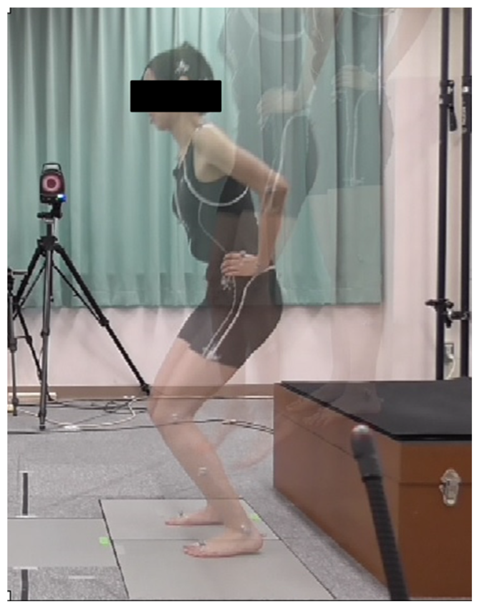
2.2. Experimental Design

A three-dimensional motion analysis system (Vicon MX, Vicon Motion Systems, Oxford, UK), sampling at 240 Hz, was used to record lower extremity kinematic data during testing. Kinematic and kinetic data were computed with low-pass filtering at 16 Hz using a fourth-order zero-lag Butterworth filter. Two AMTI force plates with amplifier (Mini-Amp MSA-6, Watertown, MA, USA), sampling at 1200 Hz, were used to record the vertical ground reaction force (vGRF) during testing. Thirty-five reflective anatomical and technical markers were positioned in various places on the body, based on Vicon’s Plug-in-Gait full body marker set, and used to derive the bilateral lower limbs and trunk model during testing [32].
The participants wore close-fitting dark shorts to aid data collection. They performed drop vertical jumps (DVJs) from a box measuring 40 cm in height (Figure 1). Before testing, the trial sequence was demonstrated by research assistants and all participants were allowed to perform several practice jumps to become familiar with the trial procedure. DVJ trials were repeated and recorded until data were obtained from five successful trials. Trials were excluded if the subject lost their balance during the landing stages. All variables were calculated as the mean value of the three trials from the five trials. The first and fifth of the five trials were excluded to eliminate larger variations due to lack of experience and fatigue, respectively.
A drop vertical jump consisted of two stages: first, landing after dropping down on two force plates from a 40 cm box; and second landing after a maximal vertical jump rebounding from first drop.

2.3. Data Processing

All data were calculated for the dominant leg during the first landing of the DVJ. The dominant leg was defined by the foot which was used to kick a ball [22,33]. The landing phase was defined as the duration from initial contact on the ground to toe-off from the force plates [34]. The initial contact on the ground was defined as the instant when the vGRF first reached more than 10 N [35,36].
The valgus or varus angles of the participants’ knees were determined using the anatomical knee neutral position as the reference point, and calculating the Euler angles, derived from the relative orientations of femur and tibia segments.
Participants were divided into valgus and varus groups according to the knee valgus or varus angle at the instant of their maximum knee flexion, on the basis of the anatomical position of the knee joint in the frontal plane. Within-subject coefficients of variation, and standard deviations for the three successful trials, were calculated for each of the knee valgus and varus angles.
vGRF impulses were normalized to each participant’s body weight (kg). The vGRF impulses were calculated by integrating the vGRF curve over time during the deceleration phase (DP) of DVJ. The latter was defined as the duration from initial contact on the ground to the instant when the maximum angle of knee flexion was recorded during the first DVJ landing [1,37].
The angular impulses of the lower extremity joints were estimated in the sagittal plane by integrating the respective joint moment curves over time during DP of DVJ. These variables were normalized to the product of each participant’s body weight (kg) and height (m).
The mechanical work of the lower extremity joints was estimated by integrating the joint power curves over time during DP of DVJ.
The vGRF, joint angles, and joint moments (hip, knee, and ankle) in the sagittal plane were logged at the instant when maximum knee flexion was recorded during landing.

2.4. Statistical Analysis

A Kolmogorov–Smirnov test was used to test for normal distribution (p < 0.05). The data were found to be normal for all parameters. Unpaired student t-tests were used to determine if significant differences existed between the valgus and varus groups in peak vGRF, vGRF impulse, joint angular impulses, and mechanical work of the lower extremity joints (p < 0.05). In order to mitigate the effects of the low numbers of participants in each group, effect sizes (ES) were calculated for all the analyses to illustrate the magnitude of the differences between the groups. Pearson correlation coefficients were calculated for comparisons between the lower extremity joints negative mechanical work, and the knee valgus or varus angles of each participant. All statistical analyses were carried out using SPSS Version 22.0 (IBM Corp., Armonk, NY, USA). Means and standard deviations are reported, unless otherwise stated.

3. Results

The valgus group comprised 10 subjects (age: (20.7 ± 1.6) year.; height: (154.9 ± 5.4) cm; weight: (54.1 ± 8.6) kg), and the varus group comprised seven subjects (age: (22.0 ± 2.5) year; height: (161.8 ± 4.6) cm; weight: (51.7 ± 4.5) kg). Maximum knee valgus angles in the valgus group were 11.5 ± 6.1°, and −17.8 ± 9.5° in the varus group. The within-subject coefficient of variation was 23.5% and the standard deviation for the knee valgus and varus angles was 1.6°.
The average vGRF impulse, joint angular impulse, and mechanical work values of the lower extremities in the valgus and varus groups are shown in Table 1. The ratios (expressed as percentages) of negative mechanical work at the lower extremity joints to total negative mechanical work (i.e., the sum of hip, knee, and ankle joint negative mechanical work) were 5.9%, 47.1%, and 47.0%, respectively, in the valgus group, and 17.6%, 36.1%, and 46.6%, respectively, in the varus group.
The average kinetic and kinematic results at the instant of the maximum knee flexion indicated that the vGRF in the valgus group was significantly greater than that in the varus group ((1.65 ± 0.36) N/kg vs. (1.24 ± 0.46) N/kg, p = 0.049, ES = 0.23). Hip flexion ((51.4 ± 12.6)° vs. (64.7 ± 19.5)°, p = 0.10, ES = 0.06), knee flexion ((72.8 ± 9.2)° vs. (83.5 ± 16.3)°, p = 0.10, ES = 0.17), and ankle dorsiflexion angles, at the instant of the maximum knee flexion ((35.1 ± 1.60° vs. (37.8 ± 8.8)°, p = 0.36, ES = 0.16), did not differ significantly between the valgus and varus groups. Moment variables in joints at the instant of the maximum knee flexion showed significantly larger hip extensor moments ((0.49 ± 0.23) N·m/kg·m vs. (0.79 ± 0.27) N·m/kg·m, p = 0.03, ES = 0.28) and smaller knee extensor moments ((1.17 ± 0.28) N·m/kg·m vs. (0.72 ± 0.34) N·m/kg·m, p = 0.01, ES = 0.37) in the valgus group than those in the varus group. There was no significant difference in the ankle plantar flexor moment ((1.27 ± 0.3) N·m/kg·m 1 vs. (1.04 ± 0.45) N·m/kg·m, p = 0.22, ES = 0.09). Moments and power curves over time, in lower extremity joints of the valgus and varus groups during DVJ landings, are represented in Figure 2 and Figure 3. The scatter plots in Figure 4 show the negative mechanical work in lower extremity joints and knee valgus and varus angles for each participant. There was a significant positive correlation between negative mechanical work at the hip and knee valgus angles (R = 0.59, p = 0.01). By contrast, negative mechanical work at the knee had a significant, but negative, correlation with knee valgus angles (R = −0.55, p = 0.02). However, there was no correlation between negative mechanical work at the ankle and the knee valgus angles (R = −0.36, p = 0.15).

4. Discussion

We showed that, during deceleration, participants landing in the knee valgus position experienced greater negative mechanical work at the knee joint, compared with those landing in the knee varus position. Similarly, they had greater knee extensor moments at the instant of the maximum knee flexion, and greater knee extensor angular impulses. This explains the net joint moments experienced by the lower extremity joints during DP of landing. When landing in the knee valgus position, participants experienced less negative mechanical work at the hip, compared with those landing in the knee varus position. They also had lower hip extensor angular impulses during DP. Negative mechanical work represents energy absorption through eccentric muscular contraction [38]. Norcross et al. reported that biomechanical factors, related to ACL injury are influenced by the magnitude and timing of energy absorption at the lower extremity joints during landing [39]. Therefore, changes in the lower extremity energy absorption capacity, brought on by altering knee alignment during landing, are likely to be effective in eliminating biomechanical components of the ACL injury mechanism. This study demonstrated that landings in the knee valgus position induced a characteristic energy absorption strategy with less energy absorption at the hip, and greater energy absorption at the knee during DP of landing. Thus, knee valgus alignment may increase the load on the knee joint and decrease the load on the hip joint during landing. Because a higher load applied to the knee joint can cause knee injuries during sporting activities [8,9], greater energy absorption capacity at the knee should be avoided by adopting knee varus alignment during landing. Previous reports have indicated that the knee joint is the most important impact absorber within the lower extremities during landing [5,6,7]. Decker et al. reported that the hip extensor, knee extensor, and ankle plantar flexor muscles in females contributed 18%, 47%, and 35%, respectively, to total energy absorption during natural landing [5]. By contrast, the present study showed contribution ratios to total negative mechanical work in the hip, knee, and ankle joints of 5.8 ± 5.6%, 46.8 ± 6.8%, and 47.5 ± 7.4%, respectively, in the valgus group, and 17.2 ± 6.3%, 36.1 ± 9.0%, and 46.7 ± 11.7%, respectively, in the varus group. Thus, the energy contributions in the valgus group were smaller in our study than those reported in the previous study, while the energy contributions in the varus group were larger. Nonetheless, the results indicate that landing with knee valgus may lead to poor energy absorption contribution by the hip joint. The results also demonstrate that the knee joint in the valgus group was the most important contributor of the energy absorption of the lower extremity joints. By comparison, the varus group showed a smaller contribution toward energy absorption by the knee joint and an increased contribution for energy absorption by the hip joints. These findings supported the notion that part of the knee joint’s role as energy absorber could be translated to the hip joint in the participants who land in the knee varus position.
The present study was carried out using the drop vertical jump test, which included both a drop landing and a second jump, as an experimental method, whereas a drop landing completed without a second jump after landing was used in a study by DeVita and Skelly [1]. Despite the different landing methods, the two studies agree that both stiff landing [1] and landing in the valgus position tend to lead to increased energy absorption contributions by the knee joint. Therefore, as with stiff landings [1], landing in the knee valgus position appears to result in greater energy absorption in the knee joint.
In addition to demonstrating that the knee experienced the greatest impact load in the valgus position, we also showed that negative mechanical work at the hip and knee had positive and negative correlations with the knee valgus angle, respectively. This suggested that an increase in the knee valgus angle during landing may reduce the contribution of the hip joint to energy absorption and that there is an associated reciprocal increased contribution by the knee joint. Hollman et al. revealed a relationship between increased knee valgus and decreased gluteus maximus engagement, which is related to sagittal plane hip joint motion, during single-leg squat maneuvers [40,41]. They indicated that gluteus maximus recruitment may be associated with an improvement of knee valgus alignment in females. Furthermore, Ford et al. and Russell et al. indicated that the hip joint of females, who have larger knee valgus than males during landings [42,43], was a poor contributor to energy absorption compared with the knee and ankle joints. We therefore conclude that landing in a knee valgus position is a key biomechanical factor that increases the contribution of the knee joint to energy absorption, and that this can be prevented by adopting a knee varus position during landing, thereby absorbing the impact in hip extensors, such as the gluteus maximus. Furthermore, reduced impact in hip extensors during landing in the knee varus position can prevent the progression of knee cartilage change caused by sustained mechanical stress for a long period of time.
During DP of the DVJ, the angles in the lower extremities were not affected by the difference in the frontal knee alignment. Thus, landing in the valgus position influenced the kinetics and energetics in the lower extremities (i.e., angular impulses in the lower extremities, the hip and knee negative mechanical works, and the joint moments) without altering alignment in the sagittal plane. These findings suggest that it is not sufficient to evaluate lower extremity kinematics alone when assessing impact attenuation during landing, but that combining kinetic and energetic evaluations of the hip and knee joints could help when assessing the mechanical load in the knee joint and energy absorption in the lower extremity joints. When evaluating the landing strategy and energetic characteristics in subjects who land in the valgus, these considerations may be useful.
Some limitations to this study should be noted. First, we focused on the energy absorption strategy in the lower extremities in the sagittal plane because motion in this plane has been shown to make a greater contribution to energy absorption than that in the frontal plane. An area for future study exists in assessing the energy absorption strategy in the frontal plane during landing in the knee valgus position. Second, we had a small sample size per group after group categorization, therefore, it could be argued that the results of this study might not provide an accurate measure of the difference in characteristics related to energy absorption between the two knee alignments during landing. In order to mitigate the effects of sample size, we calculated the ES for all the analyses. This enabled us to illustrate the magnitude of the differences between the groups more meaningfully. Analysis of the hip and knee angular impulses and mechanical work resulted in a medium ES of more than 0.3, which indicates that, despite the small sample size, our results provide a useful comparison of the differences in energy absorption strategies in lower extremity joints between the two landing postures.

5. Conclusions

Our study showed that landing in the knee valgus position involved significant negative mechanical work at the knee, and less negative mechanical work at the hip, compared with landing in the knee varus position. The findings of this study indicate that landings in the knee valgus position induced a characteristic energy absorption strategy in which, during DP of landings, less energy absorption occurs at the hip, and greater energy absorption occurs at the knee. We therefore conclude that landing in the knee valgus position is a key biomechanical factor that increases the energy absorption contribution of the knee joint. However, our results indicate that this can be prevented by adopting a knee varus position during landing, which engages the hip extensors to absorb the impact. These findings indicate that altering the landing strategy during motor learning tasks could be effective in decreasing the energy absorption at the knee, thereby minimizing the risk of knee injuries (e.g., ACL injuries).

Author Contributions

Conceptualization, A.T. and K.A.; methodology, A.T. and K.A.; software, A.T.; validation, A.T., K.A.; formal analysis, A.T.; investigation, A.T.; resources, K.A. and T.O.; data curation, A.T.; writing—original draft preparation, A.T.; writing—review and editing, K.A. and T.O.; visualization, K.A. and T.O.; supervision, K.A.; project administration, K.A.; funding acquisition, none. All authors have read and agreed to the published version of the manuscript.

Funding

This research received no external funding.

Acknowledgments

The authors would like to thank all participants and assistants in this experiment.

Conflicts of Interest

The authors declare no conflict of interest.

References

  1. DeVita, P.; Skelly, W.A. Effect of landing stiffness on joint kinetics and energetics in the lower extremity. Med. Sci. Sports Exerc. 1992, 24, 108–115. [Google Scholar] [CrossRef] [PubMed] [Green Version]
  2. Pollard, C.D.; Sigward, S.M.; Powers, C.M. Limited hip and knee flexion during landing is associated with increased frontal plane knee motion and moments. Clin. Biomech. 2010, 25, 142–146. [Google Scholar] [CrossRef] [PubMed] [Green Version]
  3. Dai, B.; Garrett, W.E.; Gross, M.T.; Padua, D.A.; Queen, R.M.; Yu, B. The effects of 2 landing techniques on knee kinematics, kinetics, and performance during stop-jump and side-cutting tasks. Am. J. Sports Med. 2015, 43, 466–474. [Google Scholar] [CrossRef] [PubMed]
  4. Konow, N.; Azizi, E.; Roberts, T.J. Muscle power attenuation by tendon during energy dissipation. Proc. Biol. Sci. 2012, 279, 1108–1113. [Google Scholar] [CrossRef] [Green Version]
  5. Decker, M.J.; Torry, M.R.; Wyland, D.J.; Sterett, W.I.; Richard Steadman, J. Gender differences in lower extremity kinematics, kinetics and energy absorption during landing. Clin. Biomech. 2003, 18, 662–669. [Google Scholar] [CrossRef]
  6. Derrick, T.R. The effects of knee contact angle on impact forces and accelerations. Med. Sci. Sports Exerc. 2004, 36, 832–837. [Google Scholar] [CrossRef]
  7. Brown, T.N.; O’Donovan, M.; Hasselquist, L.; Corner, B.; Schiffman, J.M. Lower limb flexion posture relates to energy absorption during drop landings with soldier-relevant body borne loads. Appl. Ergon. 2016, 52, 54–61. [Google Scholar] [CrossRef]
  8. Dargel, J.; Gotter, M.; Mader, K.; Pennig, D.; Koebke, J.; Schmidt-Wiethoff, R. Biomechanics of the anterior cruciate ligament and implications for surgical reconstruction. Strateg. Trauma Limb Reconstr. 2007, 2, 1–12. [Google Scholar]
  9. Buckwalter, J.A.; Anderson, D.D.; Brown, T.D.; Tochigi, Y.; Martin, J.A. The roles of mechanical stresses in the pathogenesis of osteoarthritis implications for treatment of joint injuries. Cartilage 2013, 4, 286–294. [Google Scholar] [CrossRef]
  10. Arendt, E.A.; Agel, J.; Dick, R. Anterior cruciate ligament injury patterns among collegiate men and women. J. Athl. Train. 1999, 34, 86–92. [Google Scholar]
  11. Agel, J.; Arendt, E.A.; Bershadsky, B. Anterior cruciate ligament injury in national collegiate athletic association basketball and soccer: A 13-year review. Am. J. Sports Med. 2005, 33, 524–530. [Google Scholar] [CrossRef] [PubMed]
  12. Kobayashi, H.; Kanamura, T.; Koshida, S.; Miyashita, K.; Okado, T.; Shimizu, T.; Yokoe, K. Mechanisms of the anterior cruciate ligament injury in sports activities: A twenty-year clinical research of 1700 athletes. J. Sports Sci. Med. 2010, 9, 669–675. [Google Scholar] [PubMed]
  13. Boden, B.P.; Dean, G.S.; Feagin, J.A.; Garrett, W.E., Jr. Mechanisms of anterior cruciate ligament injury. Orthopedics 2000, 23, 573–578. [Google Scholar] [CrossRef] [PubMed]
  14. Krosshaug, T.; Nakamae, A.; Boden, B.P.; Engebretsen, L.; Smith, G.; Slauterbeck, J.R.; Hewett, T.E.; Bahr, R. Mechanisms of anterior cruciate ligament injury in basketball, Video analysis of 39 cases. Am. J. Sports Med. 2007, 35, 359–367. [Google Scholar] [CrossRef]
  15. Chappell, J.D.; Creighton, R.A.; Giuliani, C.; Yu, B.; Garrett, W.E. Kinematics and electromyography of landing preparation in vertical stop-jump: Risks for noncontact anterior cruciate ligament injury. Am. J. Sports Med. 2007, 35, 235–241. [Google Scholar] [CrossRef]
  16. Ameer, M.A.; Muaidi, Q.I. Relation between peak knee flexion angle and knee ankle kinetics in single-leg jump landing from running: A pilot study on male handball players to prevent ACL injury. Phys. Sportsmed. 2017, 45, 337–343. [Google Scholar] [CrossRef]
  17. DeMorat, G.; Weinhold, P.; Blackburn, T.; Chudik, S.; Garrett, W. Aggressive quadriceps loading can induce noncontact anterior cruciate ligament injury. Am. J. Sports Med. 2004, 32, 477–483. [Google Scholar] [CrossRef]
  18. Boden, B.P.; Sheehan, F.T.; Torg, J.S.; Hewett, T.E. Non-contact ACL injuries, mechanisms and risk factors. J. Am. Acad. Orthop. Surg. 2010, 18, 520–527. [Google Scholar] [CrossRef] [Green Version]
  19. Berns, G.S.; Hull, M.L.; Patterson, H.A. Strain in the anteromedial bundle of the anterior cruciate ligament under combination loading. J. Orthop. Res. 1992, 10, 167–176. [Google Scholar] [CrossRef]
  20. Verniba, D.; Vescovi, J.D.; Hood, D.A.; Gage, W.H. The analysis of knee joint loading during drop landing from different heights and under different instruction sets in healthy males. Sports Med. Open 2017, 3, 6. [Google Scholar] [CrossRef] [Green Version]
  21. Hewett, T.E.; Torg, J.S.; Boden, B.P. Video analysis of trunk and knee motion during noncontact anterior cruciate ligament injury in female athletes: Lateral trunk and knee abduction motion are combined components of the injury mechanism. Br. J. Sports Med. 2009, 43, 417–422. [Google Scholar] [CrossRef] [PubMed] [Green Version]
  22. Hewett, T.E.; Myer, G.D.; Ford, K.R.; Heidt, R.S.; Colosimo, A.J.; McLean, S.G.; van den Bogert, A.J.; Paterno, M.V.; Succop, P. Biomechanical measures of neuromuscular control and valgus loading of the knee predict anterior cruciate ligament injury risk in female athletes: A prospective study. Am. J. Sports Med. 2005, 33, 492–501. [Google Scholar] [CrossRef] [PubMed] [Green Version]
  23. Olsen, O.E.; Myklebust, G.; Engebretsen, L.; Bahr, R. Injury mechanisms for anterior cruciate ligament injuries in team handball: A systematic video analysis. Am. J. Sports Med. 2004, 32, 1002–1012. [Google Scholar] [CrossRef] [PubMed]
  24. Imwalle, L.E.; Myer, G.D.; Ford, K.R.; Hewett, T.E. Relationship between hip and knee kinematics in athletic women during cutting maneuvers: A possible link to noncontact anterior cruciate ligament injury and prevention. J. Strength Cond. Res. 2009, 23, 2223–2230. [Google Scholar] [CrossRef] [Green Version]
  25. Miyazaki, T.; Wada, M.; Kawahara, H.; Sato, M.; Baba, H.; Shimada, S. Dynamic load at baseline can predict radiographic diseaseprogression in medial compartment knee osteoarthritis. Ann. Rheum. Dis. 2002, 61, 617–622. [Google Scholar] [CrossRef]
  26. Bennell, K.L.; Bowles, K.; Wang, Y.; Cicuttini, F.; Davies-Tuck, M.; Hinman, R.S. Higher dynamic medial knee load predicts greater cartilage loss over 12 months in medial knee osteoarthritis. Ann. Rheum. Dis. 2011, 70, 1770–1774. [Google Scholar] [CrossRef]
  27. Itoh, H.; Takiguchi, K.; Shibata, Y.; Okubo, S.; Yoshiya, S.; Kuroda, R. Correlation between hip function and knee kinematics evaluated by three-dimensional motion analysis during lateral and medial side-hopping. J. Phys. Ther. Sci. 2016, 28, 2461–2467. [Google Scholar] [CrossRef] [Green Version]
  28. Paz, G.A.; Maia Mde, F.; Farias, D.; Santana, H.; Miranda, H.; Lima, V.; Herrington, L. Kinematic analysis of knee valgus during drop vertical jump and forward step-up in young basketball players. Int. J. Sports Phys. Ther. 2016, 11, 212–219. [Google Scholar]
  29. Sharma, L.; Dunlop, D.D.; Cahue, S.; Song, J.; Hayes, K.W. Quadriceps strength and osteoarthritis progression in malaligned and lax knees. Ann. Intern. Med. 2003, 138, 613–619. [Google Scholar] [CrossRef]
  30. Palmieri-Smith, R.M.; Wojtys, E.M.; Ashton-Miller, J.A. Association between preparatory muscle activation and peak valgus knee angle. J. Electromyogr. Kinesiol. 2008, 18, 973–979. [Google Scholar] [CrossRef]
  31. Palmieri-Smith, R.M.; McLean, S.G.; Ashton-Miller, J.A.; Wojtys, E.M. Association of quadriceps and hamstrings cocontraction patterns with knee joint loading. J. Athl. Train. 2009, 44, 256–263. [Google Scholar] [CrossRef] [PubMed]
  32. Wernli, K.; Ng, L.; Phan, X.; Davey, P.; Grisbrook, T. The relationship between landing sound, vertical ground reaction force, and kinematics of the lower limb during drop landings in healthy men. J. Orthop. Sports Phys. Ther. 2016, 46, 194–199. [Google Scholar] [CrossRef] [PubMed]
  33. Urabe, Y.; Kobayashi, R.; Sumida, S.; Tanaka, K.; Yoshida, N.; Nishiwaki, G.A.; Tsutsumi, E.; Ochi, M. Electromyographic analysis of the knee during jump landing in male and female athletes. Knee 2005, 12, 129–134. [Google Scholar] [CrossRef] [PubMed]
  34. McLean, S.G.; Huang, X.; van den Bogert, A.J. Association between lower extremity posture at contact and peak knee valgus moment during sidestepping: Implications for ACL injury. Clin. Biomech. 2005, 20, 863–870. [Google Scholar] [CrossRef] [PubMed] [Green Version]
  35. Paterno, M.V.; Schmitt, L.C.; Ford, K.R.; Rauh, M.J.; Myer, G.D.; Huang, B.; Hewett, T.E. Biomechanical measures during landing and postural stability predict second anterior cruciate ligament injury after anterior cruciate ligament reconstruction and return to sport. Am. J. Sports Med. 2010, 38, 1968–1978. [Google Scholar] [CrossRef] [PubMed]
  36. Lucci, S.; Cortes, N.; Van Lunen, B.; Ringleb, S.; Onate, J. Knee and hip sagittal and transverse plane changes after two fatigue protocols. Med. Sci. Sports Exerc. 2011, 14, 453–459. [Google Scholar] [CrossRef] [Green Version]
  37. Yu, B.; Lin, C.F.; Garrett, W.E. Lower extremity biomechanics during the landing of a stop-jump task. Clin. Biomech. 2006, 21, 297–305. [Google Scholar] [CrossRef]
  38. Winter, D.A. Biomechanics and Motor Control of Human Movement, 4th ed.; John Wiley & Sons, Inc.: Hoboken, NJ, USA, 2009; Volume 2009. [Google Scholar]
  39. Hollman, J.H.; Galardi, C.M.; Lin, I.H.; Voth, B.C.; Whitmarsh, C.L. Frontal and transverse plane hip kinematics and gluteus maximus recruitment correlate with frontal plane knee kinematics during single-leg squat tests in women. Clin. Biomech. 2014, 29, 468–474. [Google Scholar] [CrossRef]
  40. Norcross, M.F.; Blackburn, J.T.; Goerger, B.M.; Padua, D.A. The association between lower extremity energy absorption and biomechanical factors related to anterior cruciate ligament injury. Clin. Biomech. 2010, 25, 1031–1036. [Google Scholar] [CrossRef]
  41. Hollman, J.H.; Ginos, B.E.; Kozuchowski, J.; Vaughn, A.S.; Krause, D.A.; Youdas, J.W. Relationships between knee valgus, hip-muscle strength, and hip-muscle recruitment during a single-limb step-down. J. Sport Rehabil. 2009, 18, 104–117. [Google Scholar] [CrossRef]
  42. Ford, K.R.; Myer, G.D.; Hewett, T.E. Valgus knee motion during landing in high school female and male basketball players. Med. Sci. Sports Exerc. 2003, 35, 1745–1750. [Google Scholar] [CrossRef] [PubMed] [Green Version]
  43. Russell, K.A.; Palmieri, R.M.; Zinder, S.M.; Ingersoll, C.D. Sex differences in valgus knee angle during a single-leg drop jump. J. Athl. Train. 2006, 41, 166–171. [Google Scholar] [PubMed]
Figure 1. A drop vertical jump and the experimental setup.
Applsci 10 08742 g001
Figure 2. Moments curves over time, in lower extremity joints of the valgus and varus groups during drop vertical jumps (DVJ) landings. Means of the hip extensor (a), knee extensor (b), and ankle plantar flexor (c) moments, and the standard deviations of the valgus (solid black lines) and varus groups (broken gray lines) during landings of DVJ.
Applsci 10 08742 g002
Figure 3. Power curves over time, in lower extremity joints of the valgus and varus groups during DVJ landings. Means of the hip extensor (a), knee extensor (b), and ankle plantar flexor (c) powers, and standard deviations of the valgus (solid lines) and knee varus groups (broken lines) during landings of DVJ.
Applsci 10 08742 g003
Figure 4. The negative mechanical work in lower extremity joints and knee valgus and varus angles. The mutual relations between negative mechanical work done at the hip (a), knee (b), and ankle (c) and the knee valgus/varus angles of each participant.
Applsci 10 08742 g004
Table 1. The vGRF impulse, joint angular impulse, and mechanical work of the lower extremities (mean ± standard deviation) by the valgus and varus knee positions.
Valgus GroupVarus GroupPES
n = 10n = 7
vGRF impulse (N/kg) a0.111 (0.020)0.110 (0.018)0.93 0.00
Hip joint
Extensor angular impulse (Nm∙s/kg∙m)0.030 (0.026)0.082 (0.032)<0.01 a0.48
Negative mechanical work (J/kg)−0.128 (0.128)−0.364 (0.194)<0.01 a0.38
Knee joint
Extensor angular impulse (Nm∙s/kg∙m)0.119 (0.020)0.099 (0.025)0.04 b0.35
Negative mechanical work (J/kg)−1.025 (0.233)−0.745 (0.209)0.02 b0.30
Ankle joint
Extensor angular impulse (Nm∙s/kg∙m)0.139 (0.019)0.165 (0.025)0.02 b0.20
Negative mechanical work (J/kg)−1.022 (0.139)−0.957 (0.260)0.510.03
a Statistically significant at p < 0.01. b Statistically significant at p < 0.05. Abbreviations: vGRF, vertical ground reaction force; ES, effect size.
Publisher’s Note: MDPI stays neutral with regard to jurisdictional claims in published maps and institutional affiliations.

