Article

Hydroquinone-Based Fabrication of Gold Nanorods with a High Aspect Ratio and LSPR Greater than 850 nm to Be Used as a Surface Plasmon Resonance Platform for Rapid Detection of Thiophanate Methyl

by
Hang Nguyen Thi Nhat
1,
Ngoc Thuy Trang Le
2,
Nguyen Thi Phuong Phong
3,
Dai Hai Nguyen
4,5 and
Minh-Tri Nguyen-Le
6,7,*
1
Faculty of Natural Sciences, Thu Dau Mot University, Thu Dau Mot City 820000, Binh Duong Province, Vietnam
2
Institute of Research and Development, Duy Tan University, Danang 550000, Vietnam
3
Faculty of Chemistry, University of Science, Vietnam National University Ho Chi Minh City, 227 Nguyen Van Cu Street, District 5, Ho Chi Minh City 70000, Vietnam
4
Institute of Applied Materials Science, Vietnam Academy of Science and Technology, 01 TL29, District 12, Ho Chi Minh City 700000, Vietnam
5
Graduate University of Science and Technology, Vietnam Academy of Science and Technology, 18 Hoang Quoc Viet, Cau Giay, Hanoi 100000, Vietnam
6
Laboratory of Advanced Materials Chemistry, Advanced Institute of Materials Science, Ton Duc Thang University, Ho Chi Minh City 758307, Vietnam
7
Faculty of Applied Sciences, Ton Duc Thang University, Ho Chi Minh City 758307, Vietnam
*
Author to whom correspondence should be addressed.
Appl. Sci. 2020, 10(10), 3654; https://doi.org/10.3390/app10103654
Submission received: 4 April 2020 / Revised: 17 May 2020 / Accepted: 21 May 2020 / Published: 25 May 2020

Abstract

:
The use of gold nanorods (AuNRs) as surface-enhanced Raman scattering (SERS) substrates has gained much attraction due to their remarkably aspect-ratio-dependent plasmonic properties. In this report, we described the development of AuNRs with a high aspect ratio and longitudinal surface plasmon resonance (LSPR) >850 nm through a hydroquinone-based fabrication with minor modifications. The synthesis started with the reduction of chloroauric acid (HAuCl4) by sodium borohydride (NaBH4) to make gold nanoseeds from which AuNRs were grown with the aid of silver nitrate (AgNO3), HAuCl4, cetyltrimethylammonium bromide (CTAB), and hydroquinone (HQ). Scanning electron microscopy coupled with energy-dispersive X-ray (SEM-EDX), Transmission electron microscope (TEM), X-ray diffraction (XRD) and Ultra-violet-Visible spectroscopy (UV-Vis) were performed to study the shape, size, and structural and optical properties of AuNRs, respectively. The results showed that AuNRs with high aspect ratios (AR > 3) were single crystals with a heterogenous size distribution, and that the growth of Au nanoseeds into AuNRs took place along the [001] direction. AuNRs exhibited two plasmon resonance peaks at 520 nm and 903 nm, while gold nanoseeds had only a plasmon resonance peak at 521 nm. The as-synthesized AuNRs also showed SERS effects for thiophanate methyl, a broad-spectrum fungicide, with the limit of detection down to 5 mg/L of the fungicide. AuNR-coated glass can serve as a SERS-based sensing platform for rapid detection of thiophanate methyl with high sensitivity and reproducibility.

1. Introduction

Recently, nanomaterials have drawn much attention due to their advanced physical and chemical properties over bulk materials, and variety of applications in the biomedical, environmental, industrial, and food agriculture industries [1]. Among nanomaterials, gold nanoparticles with nontoxicity and high bioactivity are the most stable metal nanoparticles, and they present a wide range of applications in modern medical and biological studies such as drug, deoxyribonucleic acid (DNA), and antigens delivery; biosensorics, genomics, immunoanalysis, and detection of microorganisms and cancer cells; and bioimaging [2,3]. The nanoparticles present their unique physical and chemical properties, particularly optical surface plasmon resonance (SPR), which is related to collective excitations of conductive localized electrons on surface metals under visible and near infra-red (VIS-NIR) irradiation [4,5,6,7]. In fact, SPR effects have been used to enhance the surface sensitivity of several spectroscopic analyses including infra-red/ultra-violet-visible (IR/UV-Vis) light absorption or deflection spectroscopy, hyper-Rayleigh, differential static, and dynamic light scattering, as well as surface-enhanced Raman scattering (SERS) [8,9,10,11,12]. Recently, gold nanoparticles have been studied for application in SERS-based analytical chemistry due to their remarkable SPR [13]. In conventional Raman-supported analysis methods, the intensity of Raman scattering is relatively weak despite high intensity of the applied incident laser beam, resulting in low sensitivity of the method [14]. However, in the presence of gold nanoparticles, when the analyte molecules are adsorbed onto the surface of gold nanoparticles, the enhancement of the electromagnetic field on the surface of the nanoparticles due to SPR will increase the Raman scattering intensity, thus significantly improving the sensitivity of the method [15,16]. It has been argued that the shape, size, and structure of gold nanoparticles determine their chemical and physical properties [17,18]. To date, possible wet synthesis methods have been used to fabricate gold nanoparticles of various controlled sizes, shapes, and structures such as nanospheres, nanorods, nanoshells, nanocages, nanostars, and so forth [19]. Among different anisotropic shapes of gold nanoparticles, the use of gold nanorods (AuNRs) as SERS substrates has gained much attraction due to their remarkably aspect-ratio-dependent plasmonic properties [20]. In addition to their transverse plasmon absorption, the longitudinal absorption of AuNRs, which is related to the oscillation of electrons along longitudinal axis, can be tuned easily by changing their aspect ratio (AR, length/width ratio) [21]. Then, the surface Raman scattering enhancement from AuNRs can be optimized by adjusting the shape and size of nanorods. The most popular protocol to synthesize AuNRs with uniform size and aspect ratio is a seeded growth route pioneered by Murphy and coworkers [22]. The method was performed with the aid of cetyltrimethylammonium bromide (CTAB) and ascorbic acid as a surfactant and a reducing agent, respectively, during the nanorod growth in the presence of silver ions. However, the obtained AuNRs usually have poor quality, poor reproducibility, and low aspect ratios with LSPR <850 nm [23]. In order to reproducibly achieve AuNRs with a high aspect ratio and high quality, Leonid Vigderman et al. replaced ascorbic acid with hydroquinone, an acromatic molecule with a weak reducing power [24]. The use of hydroquinone could result in a mild reaction condition of nanorod synthesis which is less dependent on reagent concentration, therefore increasing the reliability of the method [24]. By varying the hydroquinone, gold, silver, and seed concentration in the growth solution, it is possible for them to fabricate AuNRs with LSPR up to 1230 nm, and with an aspect ratio of 6–7. Later on, another research group led by C. Morasso also reported a successfully controlled hydroquinone-based synthesis of AuNRs. However, the obtained AuNRs were relatively short, having lower aspect ratio (2–4) and LSPR (ca. 800 nm) [20]. Although the synthesis of AuNRs has gained much attraction in synthesis of SERS-related nanomaterials, hydroquinone-based fabrication of AuNRs with a high aspect ratio and LSPR >850 nm as a SERS platform for rapid detection of fungicides has not been tapped yet.
Thiophanate methyl is well known as a member of the benzimidazole group of fungicides. It is a broad-spectrum systemic fungicide that has been employed to control pathogens recently. However, according to the Office of Environmental Health Hazard Assessment (OEHHA), long exposure to residual thiophanate methyl may cause serious effects on human health [25,26]. Therefore, it is essential to develop a sensitive method to detect residual thiophanate methyl in agricultural products. Until now, various techniques have been developed to detect thiophanate methyl such as high-performance liquid chromatography (HPLC), fluorescence assay (FL), high-performance liquid chromatography–mass spectrometry/mass spectrometry (HPLC–MS/MS), electrochemical assay (EC), and gas chromatography/mass spectrometry (GC/MS) [27,28,29]. Although thiophanate methyl can be analyzed by those methods with high accuracy, it has many limitations such as time-consuming operation, complicated sample preparation, and so forth, leading to a low ability to be applied for portable analysis where high accuracy and quick response are required. Meanwhile, SERS is a method that has a short measurement time and simpler sample preparation process. Therefore, the analysis of residual thiophanate methyl by SERS has the potential to be an accurate portable analytical method.
For all these reasons, in this article, AuNRs with a high aspect ratio and LSPR > 850 nm were synthesized via the reduction of HAuCl4 by NaBH4 to make gold nanoseeds from which AuNRs were grown with the aid of AgNO3, HAuCl4, CTAB, and HQ. In this seed-mediated method, NaBH4 and CTAB acted as primary reducing and stabilizing agents during the nucleation stage and HQ and AgNO3 as secondary reducing and aspect tuning agents, respectively. The as-synthesized AuNRs were used as a SERS-based sensing platform for rapid detection of thiophanate methyl (Figure 1).

2. Materials and Methods

2.1. Chemicals and Characterization

Tetrachloroauric (III) acid trihydrate (HAuCl4.3H2O, ≥99.5%), sodium borohydride (NaBH4, ≥96.0%), hydroquinone (HQ, ≥99.0%), silver nitrate (AgNO3, ≥99.0%), cetyltrimethylammonium bromide (CTAB, ≥99.0%), sulfuric acid (H2SO4, 98%), and hydrogen peroxide (H2O2, 30%) were purchased from Merck. Deionized (DI) water (>18 MW, Millipore) was used during the synthesis of the gold nanorods. A commercial fungicide product, called TOPSIN M 70WP (containing 70% of thiophanate methyl), was purchased from Nippon Soda Company, Ltd. (Tokyo, Japan). All reagents were used without further purification.
Scanning electron microscopy (Hitachi, Horiba S-4300, Japan), coupled with energy-dispersive X-ray (SEM-EDX) spectroscopy operated at 20 KV of the incident electron beam energy, was performed to study the morphologies of the as-synthesized AuNRs. Transmission electron microscope (TEM) and high-resolution TEM (HRTEM) images were obtained with a JEOL JEM (JEOL Ltd., Tokyo, Japan, 120 kV). X-ray diffractometer (XRD, Bruker D8 advance powder diffractometer model) with Cu Kα radiation (λ = 1.54056 Å) operated at 40 kV and 30 mA with a scanning rate of 0.02° per step in the 2θ range of 10° ≤ 2θ ≤ 90° has been carried out to characterize the crystal structure of AuNRs. The plasmon resonance spectra of AuNRs were recorded on UV-Vis spectroscopy (UV-Vis-NIR-V670, JASCO). A LabRam HR micro-Raman instrument with a 532 nm Ar+ ion laser was utilized to obtain Raman spectra of AuNR samples at room temperature.

2.2. Synthesis of Au Nanoseeds and AuNRs

Au nanoseeds were synthesized by mixing 100 µL of 25 mM HAuCl4 with 10 mL of 0.1 M CTAB under vigorous stirring at 400 rpm. A total of 460 µL of 0.01 M NaBH4 (at 4 °C) was then quickly added into the mixture. The color of the solution changed from yellow to brown. The solution was kept in the dark for 30 min for stabilization.
For the synthesis of AuNRs, 0.01 M AgNO3 and 25 mM HAuCl4 were firstly added into 10 mL of 0.1 M CTAB under vigorous stirring. The 0.1 M HQ solution was added drop by drop into the solution. The color of the solution changed from yellow to colorless. Finally, 80 µL of the above solution containing Au nanoseeds was then added to this growth solution, and the obtained solution was kept in the dark for 24 h. The growth of Au nanoseeds into AuNRs is illustrated in Figure 2.

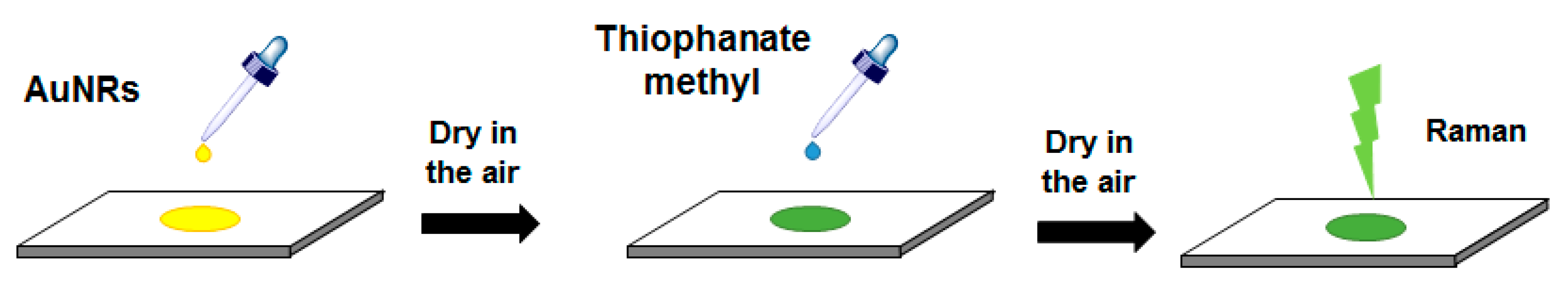
2.3. SERS Measurement

The detection of thiophanate methyl by SERS effects was performed on an AuNR-coated glass substrate which acted as a SERS-based sensing platform. Scheme 1 illustrates the fabrication of the platform. The platform was prepared by firstly rinsing the glass substrate with a 3:1 (v/v) mixture of concentrated H2SO4 and 30% H2O2 for 15 min, subsequently washing it with DI water and drying in the air. A portion of AuNRs solution was gently dropped onto the clean substrate homogeneously, followed by drying it in the air. Next, 20 µL of thiophanate methyl solution (in DI water) was gently dropped onto the platform and subsequently dried in the air to be ready for SERS measurements. For comparison, Raman spectra of the clean substrate, AuNRs, and thiophanate methyl solution were also recorded.

3. Results and Discussion

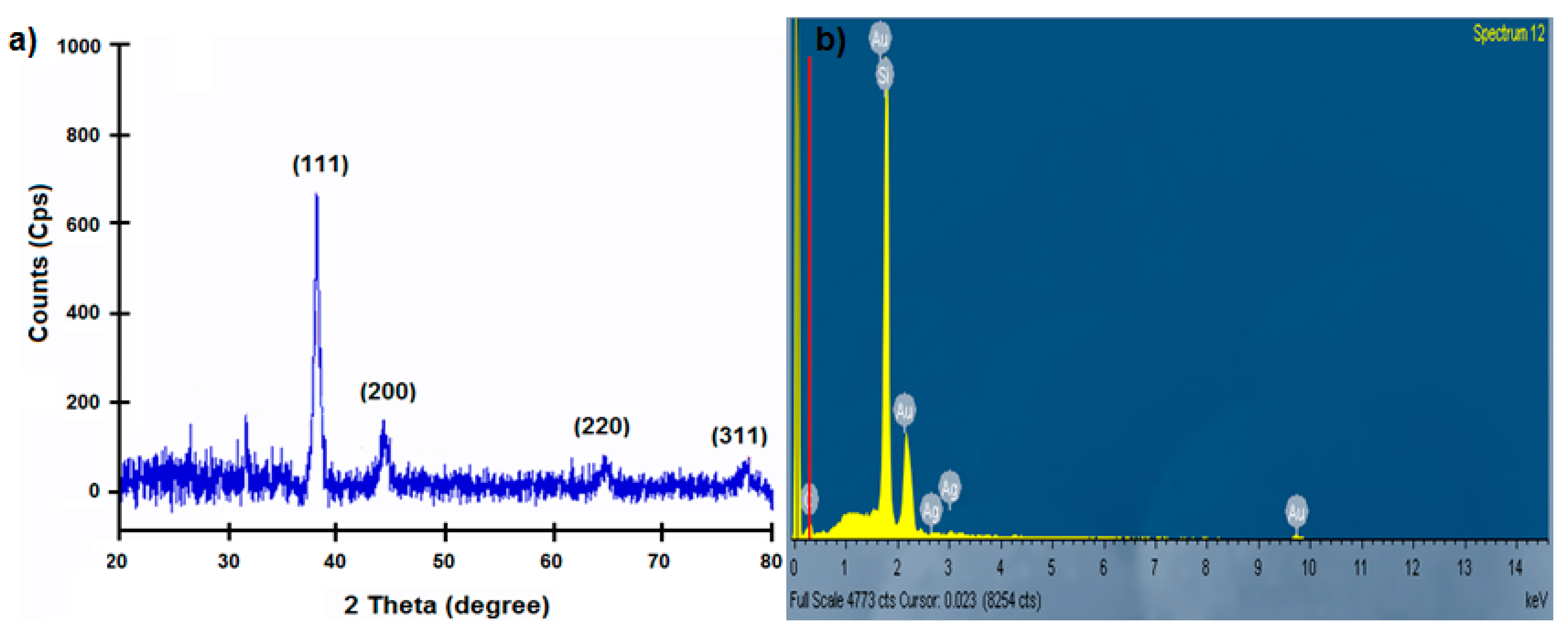
3.1. Growth of Au Nanoseeds into AuNRs

Plasmon resonance properties of AuNRs and Au nanoseeds were studied through UV-Vis spectroscopy. As shown in Figure 3, the absorption spectra of Au nanoseeds exhibited a peak at 521 nm, while those of AuNRs showed two peaks at 520 nm and 903 nm. It is widely accepted that only a single surface plasmon resonance (SPR) peak is expected from the absorption spectra of spherical Au nanoparticles [30]. In contrast, two observed plasmon resonance peaks of AuNRs are ascribed to transverse and longitudinal SPRs caused by the width and the length of the nanorods, respectively [31].
The crystalline structures of Au nanoseeds and AuNRs were investigated through TEM. As shown in Figure 4a,c, the solution of Au nanoseeds consisted of clusters of Au nano-sized particles with the average diameter of 15.37 ± 0.61 nm (N = 30), and with the crystalline lattices of 0.23 nm, corresponding to the (111) crystal plane of Au (Figure 4a, the inset) [32,33]. Meanwhile, the TEM images of the AuNRs with high aspect ratios (AR >3) revealed the presence of rod-like nanostructures with a heterogenous size distribution of 96.05 ± 10.89 nm (N = 122) in length and of 16.44 ± 1.11 nm (N = 122) in width (Figure 4b–f) [34]. Interestingly, the AR of AuNRs estimated from the discrete dipole approximation (DDA), λLSPR = 96AR + 418 where λLSPR is the longitudinal SPR wavelength of AuNRs, was 5.1, which is close to the average AR of AuNRs obtained from the TEM images (AR = 5.7 ± 0.8) and other studies [35,36]. Moreover, the FFT pattern of the AuNRs along [110] zone axis (Figure 4b, the inset) proved that AuNRs were single crystals with a face-centered cubic (fcc) close packing [37]. It is obvious that the existence of a few nano-sized particles indicates the growth of Au nanoseeds into AuNRs along the [001] direction. It is due to the fact that silver ions are selectively bonding to the high-energy (110) facets of Au nanocrystals, which slows down the growth rate of Au atoms on these facets during the synthesis [38]. It is also noteworthy that the AuNR solution is relatively stable at room temperature due to the presence of CTAB molecules, which prevent AuNRs from aggregation.
The XRD pattern of AuNRs with characteristic peaks of Au at 2θ = 38.1° (111), 44.3° (200), 64.5° (220), and 77.7° (311) in Figure 5a revealed high crystallinity of AuNRs, which is consistent with the TEM results [39]. Furthermore, the EDX spectra in Figure 5b represented that Au and Si are the major elements of AuNR-coated glass substrate, indicating successful coating of glass substrate with AuNRs. The presence of Ag+ ions was also observed in the EDX, indicating the deposition of Ag+ ions (pairing with Br− from CTAB) on the AuNR-coated glass substrate.

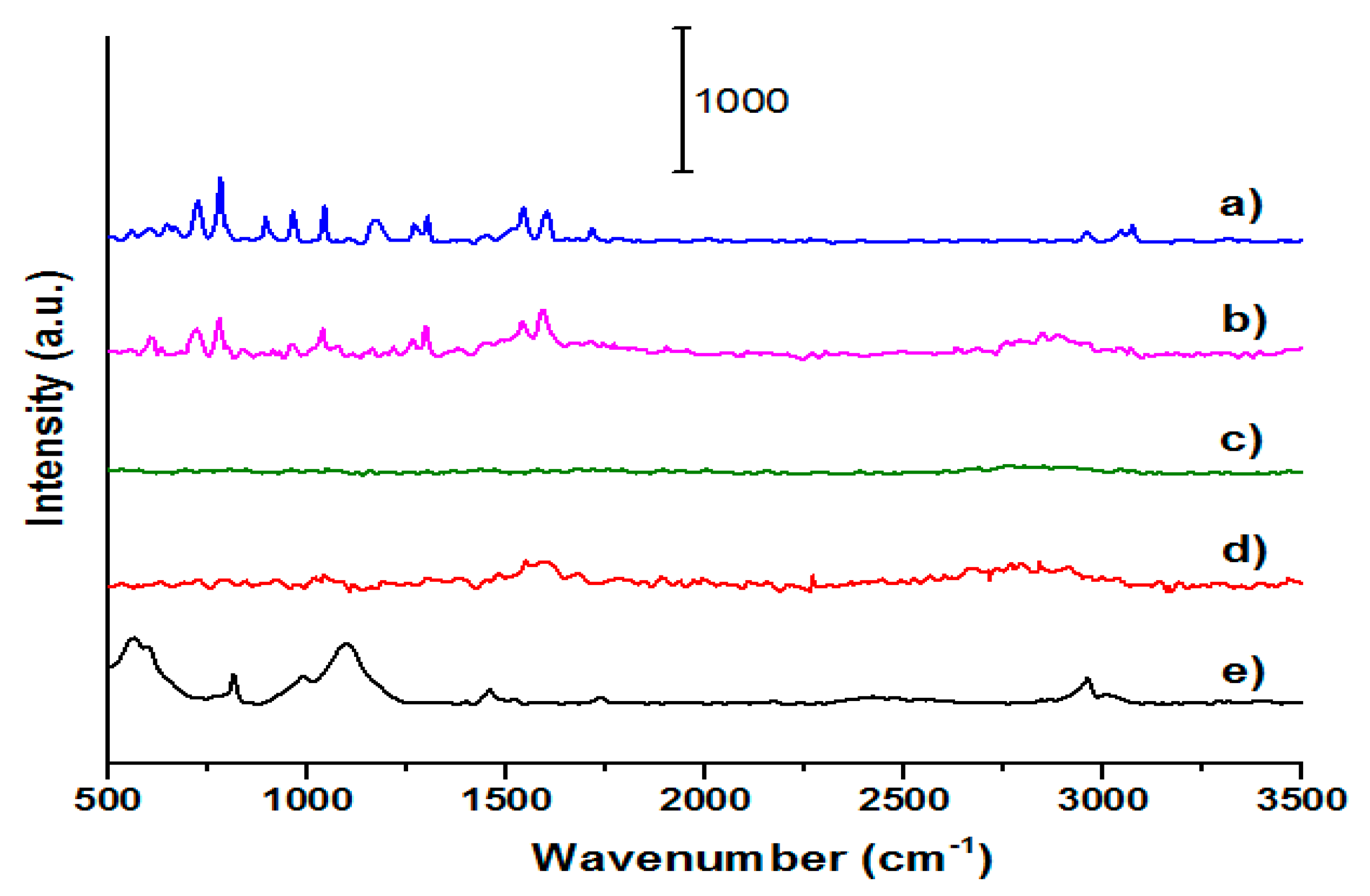
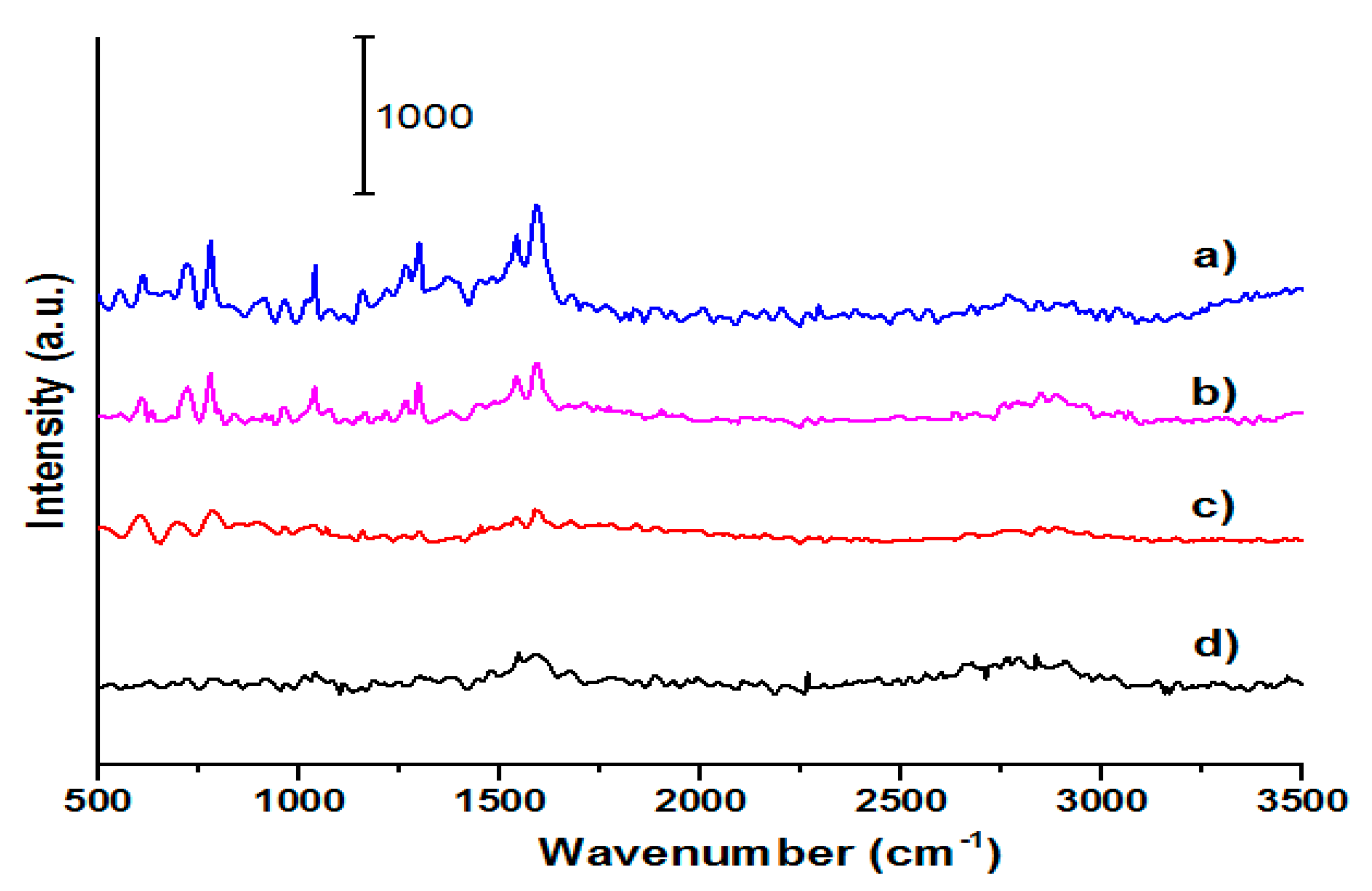
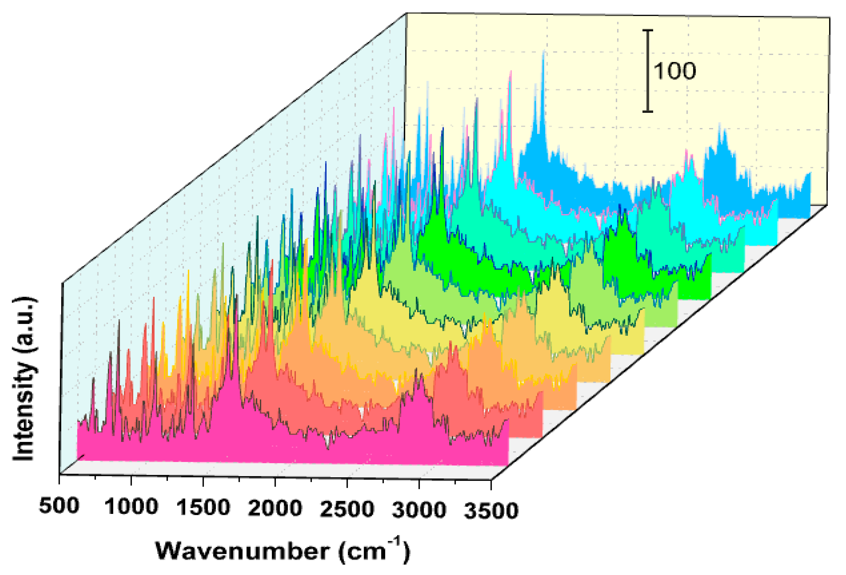
3.2. Detection of Fungicides on a SERS-Based Sensing Platform

In this section, AuNR-coated glass was used as a SERS-based sensing platform for detection of thiophanate methyl. No Raman signals of thiophanate methyl can be observed for glass or AuNR-coated glass, indicating thiophanate methyl-free surfaces (Figure 6d,e). However, after deposition of 100 mg/L of the fungicide, all of its Raman characteristic peaks can be observed at 613 cm−1, 726 cm−1, 779 cm−1, 898 cm−1, 959 cm−1, 1039 cm−1, 1154 cm−1, 1267 cm−1, 1298 cm−1, 1538 cm−1, 1601 cm−1, 1708 cm−1, which are ascribed to –N–C=S deformation, N–H wagging of –(C=O)–NH–(C=S)–, C=S stretching, C–S stretching, C=S, =C–H deformation, N–C–N asymmetric stretching, C–O, C=C, C–N, C=O and N–H, respectively (Figure 6a) [40]. It should be noted that even in the presence of thiophanate methyl, those signals cannot be observed as the concentration of thiophanate methyl is as low as 5 mg/L (Figure 6c). Interestingly, at the same concentration of thiophanate methyl, most of the characteristic Raman peaks of thiophanate methyl were clearly observed after deposition of thiophanate methyl onto the AuNR-coated glass substrate (Figure 6b). These results indicated that AuNR-coated glass can be used as a SERS-based sensing platform for detection of fungicides.
The sensitivity of the method was further evaluated through the corresponding Raman spectra of thiophanate methyl deposited onto AuNR-coated glass substrates with concentrations ranging from 1 mg/L to 7 mg/L. It can be seen that the characteristic peaks of thiophanate methyl could be clearly identified in all Raman spectra (Figure 7). Typically, the SERS intensity of thiophanate methyl gradually decreases with the decreasing concentration. The limit of detection (LOD) was determined as low as 5 mg/L (S/N = 3 for the peak 779 cm−1). The limit of quantification (LOQ), therefore, was estimated as ~17 mg/L.
In order to prove the reproducibility of the measurement, SERS spectra of 5 mg/L of thiophanate methyl were obtained at 10 randomly selected locations on the platform. No significant difference was observed between those positions (Figure 8). The relative standard deviation (RSD) was found to be 5.7%, which indicates that the platform is able to be used for routine SERS analysis with a good reproducibility.

4. Conclusions

AuNRs with a high aspect ratio and LSPR >850 nm have been successfully grown from Au nanoseeds through a facile seed-mediated method with the aid of cetyltrimethylammonium bromide and hydroquinone as a surfactant and a reducing agent, respectively, during the nanorod growth in the presence of silver ions. TEM and XRD results showed that AuNRs are single crystals with a heterogenous size distribution, and that the growth of Au nanoseeds into AuNRs took place along the [001] direction. The as-synthesized AuNRs showed SERS effects for thiophanate methyl with low LOD of thiophanate methyl down to 5 mg/L, indicating that AuNR-coated glass can serve as a SERS-based sensing platform for rapid detection of fungicides with high sensitivity and reproducibility.

Author Contributions

Conceptualization, N.T.P.P. and D.H.N.; Formal analysis, H.N.T.N., N.T.T.L., and M.-T.N.-L.; Funding acquisition, D.H.N.; Investigation, H.N.T.N. and N.T.T.L.; Methodology, N.T.P.P. and D.H.N.; Project administration, D.H.N.; Supervision, M.-T.N.-L.; Writing—original draft, H.N.T.N.; Writing—review & editing, M.-T.N.-L. All authors have read and agreed to the published version of the manuscript.

Funding

This research was funded by the Thu Dau Mot University.

Acknowledgments

The authors would like to thank Cuu Khoa Nguyen and Ngoc Quyen Tran—Board of Directors of Institute of Applied Materials Science, Vietnam Academy of Science and Technology, Ho Chi Minh City—for their helpful advice and technical support for this study.

Conflicts of Interest

The authors declare no conflict of interest.

References

  1. Pyrzynska, K. Nanomaterials in speciation analysis of metals and metalloids. Talanta 2020, 212, 120784. [Google Scholar] [CrossRef] [PubMed]
  2. Daniel, M.-C.; Astruc, D. Gold nanoparticles: Assembly, supramolecular chemistry, quantum-size-related properties, and applications toward biology, catalysis, and nanotechnology. Chem. Rev. 2004, 104, 293–346. [Google Scholar] [CrossRef] [PubMed]
  3. Dykman, L.A.; Khlebtsov, N.G. Gold nanoparticles in biology and medicine: Recent advances and prospects. Acta Nat. (англoязычная версия) 2011, 3, 34–35. [Google Scholar]
  4. Pacheco-Londono, L.C.; Aparicio-Bolano, J.; Primera-Pedrozo, O.M.; Hernandez-Rivera, S.P. Growth of Ag, Au, Cu, and Pt nanostructures on surfaces by micropatterned laser-image formations. Appl. Opt. 2011, 50, 4161–4169. [Google Scholar] [CrossRef] [PubMed]
  5. Li, X.; Zhang, S.; Yu, Z.; Yang, T. Surface-enhanced Raman spectroscopic analysis of phorate and fenthion pesticide in apple skin using silver nanoparticles. Appl. Spectrosc. 2014, 68, 483–487. [Google Scholar] [CrossRef]
  6. Huong Nguyen, T.; Dai Hai, N.; Minh Thanh, V.; Huynh Nhu, T.; Linh Phuong Pham, T.; Ngoc-Tram, N.-T.; Thuy Trang Le, N.; Minh-Tri, N.-L. Fabrication Process and Characterization of AgNPs/PVA/ Cellulose as a SERS Platform for In-situ Detection of Residual Pesticides in Fruit. Mater. Res. Express 2020, 7, 035019. [Google Scholar] [CrossRef]
  7. Tuan Anh, M.N.; Nguyen, D.T.D.; Ke Thanh, N.V.; Phuong Phong, N.T.; Nguyen, D.H.; Nguyen-Le, M.-T. Photochemical Synthesis of Silver Nanodecahedrons under Blue LED Irradiation and Their SERS Activity. Processes 2020, 8, 292. [Google Scholar] [CrossRef] [Green Version]
  8. Liu, X.; Dai, Q.; Austin, L.; Coutts, J.; Knowles, G.; Zou, J.; Chen, H.; Huo, Q. A One-Step Homogeneous Immunoassay for Cancer Biomarker Detection Using Gold Nanoparticle Probes Coupled with Dynamic Light Scattering. J. Am. Chem. Soc. 2008, 130, 2780–2782. [Google Scholar] [CrossRef]
  9. Khlebtsov, N.G.; Melnikov, A.G.; Dykman, L.A.; Bogatyrev, V.A. Optical Properties and Biomedical Applications of Nanostructures Based on Gold and Silver Bioconjugates. In Proceedings of the Photopolarimetry in Remote Sensing, Dordrecht, The Netherlands, 20 September–4 October 2003; pp. 265–308. [Google Scholar]
  10. Russier-Antoine, I.; Huang, J.; Benichou, E.; Bachelier, G.; Jonin, C.; Brevet, P.-F. Hyper Rayleigh scattering of protein-mediated gold nanoparticles aggregates. Chem. Phys. Lett. 2008, 450, 345–349. [Google Scholar] [CrossRef]
  11. Kamnev, A.A.; Dykman, L.A.; Tarantilis, P.A.; Polissiou, M.G. Spectroimmunochemistry Using Colloidal Gold Bioconjugates. Biosci. Rep. 2002, 22, 541–547. [Google Scholar] [CrossRef]
  12. Lin, C.-C.; Yang, Y.-M.; Chen, Y.-F.; Yang, T.-S.; Chang, H.-C. A new protein A assay based on Raman reporter labeled immunogold nanoparticles. Biosens. Bioelectron. 2008, 24, 178–183. [Google Scholar] [CrossRef] [PubMed]
  13. Kim, J.E.; Choi, J.H.; Colas, M.; Kim, D.H.; Lee, H. Gold-based hybrid nanomaterials for biosensing and molecular diagnostic applications. Biosens. Bioelectron. 2016, 80, 543–559. [Google Scholar] [CrossRef] [PubMed]
  14. Ye, K.; Li, K.; Lu, Y.; Guo, Z.; Ni, N.; Liu, H.; Huang, Y.; Ji, H.; Wang, P. An overview of advanced methods for the characterization of oxygen vacancies in materials. TrAC Trends Anal. Chem. 2019, 116, 102–108. [Google Scholar] [CrossRef]
  15. Thygesen, L.G.; Jorgensen, K.; Moller, B.L.; Engelsen, S.B. Raman spectroscopic analysis of cyanogenic glucosides in plants: Development of a flow injection surface-enhanced Raman scatter (FI-SERS) method for determination of cyanide. Appl. Spectrosc. 2004, 58, 212–217. [Google Scholar] [CrossRef] [PubMed]
  16. Baia, L.; Baia, M.; Popp, J.; Astilean, S. Gold films deposited over regular arrays of polystyrene nanospheres as highly effective SERS substrates from visible to NIR. J. Phys. Chem. B 2006, 110, 23982–23986. [Google Scholar] [CrossRef]
  17. Luty-Błocho, M.; Fitzner, K.; Hessel, V.; Löb, P.; Maskos, M.; Metzke, D.; Pacławski, K.; Wojnicki, M. Synthesis of gold nanoparticles in an interdigital micromixer using ascorbic acid and sodium borohydride as reducers. Chem. Eng. J. 2011, 171, 279–290. [Google Scholar] [CrossRef]
  18. Dozol, H.; Mériguet, G.; Ancian, B.; Cabuil, V.r.; Xu, H.; Wang, D.; Abou-Hassan, A. On the synthesis of Au nanoparticles using EDTA as a reducing agent. J. Phys. Chem. C 2013, 117, 20958–20966. [Google Scholar] [CrossRef]
  19. Huang, X.; El-Sayed, I.H.; Qian, W.; El-Sayed, M.A. Cancer cell imaging and photothermal therapy in the near-infrared region by using gold nanorods. J. Am. Chem. Soc. 2006, 128, 2115–2120. [Google Scholar] [CrossRef]
  20. Morasso, C.; Picciolini, S.; Schiumarini, D.; Mehn, D.; Ojea-Jiménez, I.; Zanchetta, G.; Vanna, R.; Bedoni, M.; Prosperi, D.; Gramatica, F. Control of size and aspect ratio in hydroquinone-based synthesis of gold nanorods. J. Nanopart. Res. 2015, 17, 330. [Google Scholar] [CrossRef]
  21. Orendorff, C.J.; Gearheart, L.; Jana, N.R.; Murphy, C.J. Aspect ratio dependence on surface enhanced Raman scattering using silver and gold nanorod substrates. Phys. Chem. Chem. Phys. 2006, 8, 165–170. [Google Scholar] [CrossRef] [PubMed]
  22. Jana, N.R.; Gearheart, L.; Murphy, C.J. Evidence for Seed-Mediated Nucleation in the Chemical Reduction of Gold Salts to Gold Nanoparticles. Chem. Mater. 2001, 13, 2313–2322. [Google Scholar] [CrossRef]
  23. Jiang, X.C.; Brioude, A.; Pileni, M.P. Gold nanorods: Limitations on their synthesis and optical properties. Colloids Surf. A Physicochem. Eng. Asp. 2006, 277, 201–206. [Google Scholar] [CrossRef]
  24. Vigderman, L.; Zubarev, E.R. High-Yield Synthesis of Gold Nanorods with Longitudinal SPR Peak Greater than 1200 nm Using Hydroquinone as a Reducing Agent. Chem. Mater. 2013, 25, 1450–1457. [Google Scholar] [CrossRef]
  25. OEHHA. Proposition 65 Maximum Allowable Dose Level (madl) for Reproductive Toxicity for Thiophanate-Methyl for the Oral Route of Exposure; OEHHA: Sacramento, CA, USA, 2004.
  26. OEHHA. Chemicals Known to the State to Cause Cancer or Reproductive Toxicity; OEHHA: Sacramento, CA, USA, 2018.
  27. Galán-Cano, F.; Lucena, R.; Cárdenas, S.; Valcárcel, M. Dispersive micro-solid phase extraction with ionic liquid-modified silica for the determination of organophosphate pesticides in water by ultra performance liquid chromatography. Microchem. J. 2013, 106, 311–317. [Google Scholar] [CrossRef]
  28. Yan, X.; Li, H.; Hu, T.; Su, X. A novel fluorimetric sensing platform for highly sensitive detection of organophosphorus pesticides by using egg white-encapsulated gold nanoclusters. Biosens. Bioelectron. 2017, 91, 232–237. [Google Scholar] [CrossRef]
  29. Yi, Y.; Zhu, G.; Liu, C.; Huang, Y.; Zhang, Y.; Li, H.; Zhao, J.; Yao, S. A Label-Free Silicon Quantum Dots-Based Photoluminescence Sensor for Ultrasensitive Detection of Pesticides. Anal. Chem. 2013, 85, 11464–11470. [Google Scholar] [CrossRef]
  30. Gans, R. Über die form ultramikroskopischer silberteilchen. Ann. Phys. 1915, 352, 270–284. [Google Scholar] [CrossRef] [Green Version]
  31. Huang, X.; Neretina, S.; El-Sayed, M.A. Gold nanorods: From synthesis and properties to biological and biomedical applications. Adv. Mater. 2009, 21, 4880–4910. [Google Scholar] [CrossRef]
  32. Chang, C.-C.; Wu, H.-L.; Kuo, C.-H.; Huang, M.H. Hydrothermal Synthesis of Monodispersed Octahedral Gold Nanocrystals with Five Different Size Ranges and Their Self-Assembled Structures. Chem. Mater. 2008, 20, 7570–7574. [Google Scholar] [CrossRef]
  33. Esparza, R.; Rosas, G.; López Fuentes, M.; Sánchez Ramírez, J.F.; Pal, U.; Ascencio, J.A.; Pérez, R. Synthesis of gold nanoparticles with different atomistic structural characteristics. Mater. Charact. 2007, 58, 694–700. [Google Scholar] [CrossRef]
  34. Koeppl, S.; Solenthaler, C.; Caseri, W.; Spolenak, R. Towards a Reproducible Synthesis of High Aspect Ratio Gold Nanorods. J. Nanomater. 2011, 2011, 515049. [Google Scholar] [CrossRef]
  35. Brioude, A.; Jiang, X.C.; Pileni, M.P. Optical Properties of Gold Nanorods:  DDA Simulations Supported by Experiments. J. Phys. Chem. B 2005, 109, 13138–13142. [Google Scholar] [CrossRef] [PubMed]
  36. Kaur, P.; Chudasama, B. Seedless co-surfactant-based dimensional and optical tunability of gold nanorods with simultaneous pH regulation. J. Mater. Sci. 2017, 52, 11675–11687. [Google Scholar] [CrossRef]
  37. Shah, A.B.; Sivapalan, S.T.; DeVetter, B.M.; Yang, T.K.; Wen, J.; Bhargava, R.; Murphy, C.J.; Zuo, J.-M. High-Index Facets in Gold Nanocrystals Elucidated by Coherent Electron Diffraction. Nano Lett. 2013, 13, 1840–1846. [Google Scholar] [CrossRef] [PubMed]
  38. Chen, H.; Shao, L.; Li, Q.; Wang, J. Gold nanorods and their plasmonic properties. Chem. Soc. Rev. 2013, 42, 2679–2724. [Google Scholar] [CrossRef] [PubMed]
  39. Ogundare, O.D.; Akinribide, O.J.; Adetunji, A.R.; Adeoye, M.O.; Olubambi, P.A. Crystallite size determination of thermally deposited Gold Nanoparticles. Procedia Manuf. 2019, 30, 173–179. [Google Scholar] [CrossRef]
  40. Socrates, G. Infrared and Raman Characteristic Group Frequencies: Tables and Charts; John Wiley & Sons: Chichester, UK, 2004. [Google Scholar]
Figure 1. The chemical structure of thiophanate methyl.
Applsci 10 03654 g001
Figure 2. Fabrication process of gold nanorods (AuNRs): (a) formation of Au nanoseeds, (b) growth of Au nanoseeds into AuNRs.
Applsci 10 03654 g002
Scheme 1. The fabrication of the surface-enhanced Raman scattering (SERS)-based sensing platform.
Applsci 10 03654 sch001
Figure 3. Ultra-violet-Visible (UV-Vis) spectra of Au nanoseeds and AuNRs.
Applsci 10 03654 g003
Figure 4. Transmission electron microscopy (TEM) images of (a) Au nanoseeds and (b) AuNRs. The insets: High resolution transmission electron microscopy (HRTEM) images of (a) Au nanoseeds and (b) AuNRs. Particle size distribution of (c) Au nanoseeds, and (d,e) AuNRs. (f) Aspect ratio of AuNRs.
Applsci 10 03654 g004
Figure 5. (a) X-ray diffraction (XRD) spectra of AuNRs, (b) Energy-dispersive X-ray (EDX) spectra of AuNR-coated glass.
Applsci 10 03654 g005
Figure 6. Raman spectra of (a) 100 mg/L of thiophanate methyl deposited glass, (b) 5 mg/L of thiophanate methyl deposited onto AuNR-coated glass substrate, (c) 5 mg/L of thiophanate methyl deposited glass, (d) AuNR-coated glass, and (e) glass substrate.
Applsci 10 03654 g006
Figure 7. Raman spectra of different concentrations of thiophanate methyl: (a) 7 mg/L, (b) 5 mg/L, (c) 3 mg/L, and (d) 1 mg/L recorded on AuNR-coated glass platform.
Applsci 10 03654 g007
Figure 8. Raman spectra of 5 mg/L of thiophanate methyl obtained at 10 random locations on AuNR-coated glass substrate.
Applsci 10 03654 g008

