Article

Kinetic Aspects of the Interactions between TiO2 Nanoparticles, Mercury and the Green Alga Chlamydomonas reinhardtii

Environmental Biogeochemistry and Ecotoxicology, Department F.-A. Forel for Environmental and Aquatic Sciences, School of Earth and Environmental Sciences, Faculty of Science, University of Geneva, Uni Carl Vogt, 66 Blvd Carl-Vogt, CH-1211 Geneva, Switzerland
*
Author to whom correspondence should be addressed.
Environments 2022, 9(4), 44; https://doi.org/10.3390/environments9040044
Submission received: 3 November 2021 / Revised: 20 March 2022 / Accepted: 22 March 2022 / Published: 26 March 2022
(This article belongs to the Special Issue Feature Papers in Environments in 2021)

Abstract

:
Aquatic organisms are exposed to mixtures of environmental pollutants, including engineered nanoparticles; however, the interactions underlying cocktails’ effects are poorly understood, in particular, the kinetic aspects. The present study explored the time course of the interactions between nano-sized titanium dioxide (nTiO2) with different primary particle sizes, algae and inorganic mercury (Hg) over 96 h under conditions that were representative of a highly contaminated environment. The results showed that nTiO2 with smaller primary particle size and higher concentrations rapidly reduced the adsorption and internalization of mercury by green alga Chlamydomonas reinhardtii. Such a mitigating effect on mercury bioavailability could be explained by the strong adsorbing capacity of nTiO2 for Hg and the aggregation and sedimentation of nTiO2 and bound Hg. The present study highlighted the key processes determining the bioavailability of mercury to the algae in mixture exposure under conditions representative of a highly contaminated environment, such as industrial wastewater effluents.

1. Introduction

Aquatic organisms are typically exposed to mixtures of environmental pollutants, including engineered nanoparticles (ENPs) [1,2]. ENPs possess enhanced physical and chemical properties that favor their interactions with other (i.e., dissolved) pollutants in aquatic systems [3,4,5]. Significant progress has been achieved in the understanding of the bioavailability and cocktail effects of mixtures containing ENPs and metallic pollutants [1,2,6,7,8]. Depending on the types of ENPs and metallic pollutants, feeding patterns of the test organisms and exposure duration, ENPs can either increase, have no effect or decrease the metal bioavailability and biological effects [1,9]. For instance, nanosized titanium dioxide (nTiO2) lessened mercury-induced cellular reactive oxygen species (ROS) generation and membrane damage in the green alga Chlamydomonas reinhardtii exposed for 2 h [10]. Similarly, nTiO2 and nSiO2 reduced Cd toxicity to C. reinhardtii [11]. However, nTiO2 enhanced the oxidative stress damage caused by Cu in the bacterium Bacillus thuringiensis, whereas no effects were observed in Bacillus megaterium [12]. nTiO2 significantly alleviated the mortality and reproduction inhibition of As on the crustacean Daphnia magna, and the alleviation of As(V) was more prominent than that of As(III) [13]. nTiO2 with a primary size of 5 nm reduced the Hg body burden in D. magna in waterborne exposure; however, it increased the Hg body burden when daphnids were exposed to nTiO2 pretreated with Hg or methyl mercury (MeHg) [9]. Interestingly, the assimilation of monomethyl mercury in D. magna from algal food was enhanced by nTiO2; however, the assimilation of inorganic mercury was not influenced [9]. Mixtures of nZnO and Cd led to increased toxic effects to the copepod Tigriopus japonicus during short-term acute exposure, whereas this mixture reduced the toxic effects during chronic exposure, which showed the importance of the exposure time and suggested different prevailing processes [14]. Nevertheless, most of the existing information about the effects of ENPs and their mixtures with metallic pollutants was obtained in tests focused on defining concentration–response relationships, with none or very little consideration to kinetics aspects of the interactions between mixture components. However, understanding the time progression of such interactions can be of great value for predicting the bioavailability of metallic pollutants in complex environmental settings given that the environmental processes are dynamic.
In this study, we investigated the mercury bioavailability to the green alga C. reinhardti and the kinetic aspects of the interactions between the components of mixtures containing inorganic Hg, nTiO2 of different primary sizes and algae over 96 h. The emphasis was on the study of: (i) the adsorption kinetics of Hg on nTiO2 of three different sizes, (ii) the time course of nTiO2 aggregation and sedimentation kinetics, and (iii) the kinetics of Hg uptake by the green microalga C. reinhardtii in the absence and presence of nTiO2.
We chose nTiO2, as it is one of the most extensively used ENPs [15,16]. It is commonly employed as a substrate for the adsorption and removal of various metal contaminants, including Pb [17], Cd [18] and Hg [10]. Here we chose to study three nTiO2 materials with different primary particle sizes (5, 15 and 20 nm). Mercury was chosen as a representative of hazardous pollutants of global importance [19]. Several recent studies focused on the responses of organisms to Hg as reviewed for animal cells, invertebrates and vertebrates [20], phytoplankton [21,22] and aquatic plants [23,24]. However, these studies did not consider the time progression of the effects and interactions determining bioavailability during co-exposure to Hg and other co-contaminants. To our knowledge, this is the first study that explored the time course of the interactions between the green alga and the components of mixtures of nTiO2 of different primary sizes and Hg.

2. Materials and Methods

2.1. Chemicals

Nano-sized TiO2 particles with different primary sizes (anatase 5 nm (A5), anatase 15 nm (A15) and anatase/rutile 20 nm (AR20)) were purchased from Nanostructured & Amorphous Materials Inc., Houston, TX, USA, as powders. Their characteristics can be found in our previous work [10]. Stock suspensions of 2.0 g L−1 nTiO2 were prepared by dispersing nanoparticles in ultrapure water (Millipore, Darmstadt, Germany) using a 10 min sonication (50 WL−1 at 40 kHz). HgCl2 standard solution (1.0 g L−1) was bought from Sigma-Aldrich, Buchs, Switzerland.
Glassware rather than plasticware was used as test containers to reduce the loss of Hg via adsorption onto the containers’ wall. All the glassware was soaked in 10% HNO3 (Emsure, Merck, Darmstadt, Germany) followed by two 10% HCl acid baths (Emsure, Merck, Darmstadt, Germany) for at least 24 h in each bath, then thoroughly rinsed with ultrapure water (MilliQ Direct, Merck, Darmstadt, Germany) and dried under a laminar flow hood.

2.2. Algal Cell Growth

Wild-type C. reinhardtii (CPCC11, Canadian Phycological Culture Centre, Waterloo, Ont, Canada) was grown axenically at 20.2 ± 0.5 °C, 115 rpm and 110 μmol phot m−2 s−1. Algal cells were cultured in 4× diluted Tris-Acetate-Phosphate medium [25] until mid-exponential growth (62 h post-inoculation), centrifuged (10 min, 1300× g) and rinsed with and re-suspended (~106 cells mL−1) in the exposure medium.

2.3. Experimental Design

The time course of the biological responses of the green alga C. reinhardtii exposed to nTiO2, Hg and their mixtures was followed every 24 h over 96 h. The exposure media contained 8.2 × 10⁻⁴ M CaCl2·2H2O, 3.6 × 10⁻⁴ M MgSO4·7H2O, 2.8 × 10⁻⁴ M NaHCO3, 1.0 × 10⁻⁴ M KH2PO4 and 5.0 × 10⁻⁶ M NH4NO3 with a pH of 6.9 ± 0.1 and an ionic strength of 2.75 mM. The medium was spiked with 5 × 10⁻⁷ M Hg as HgCl2, 20 or 200 mg L−1 nTiO2 or their mixtures. After spiking, the pH was readjusted if necessary.
The concentration of Hg in the exposure medium was selected based on our previous work. The tested combinations of nTiO2 and Hg corresponded to (i) negligible or no effect concentrations (20 mgL−1 nTiO2) and (ii) concentrations where more than 50% of the cells are affected according to the literature (200 mgL−1 nTiO2). For example, 200 mg L−1 nTiO2 was chosen as corresponding to the lowest value inducing 50% effect to the most sensitive organism in a multispecies test battery [26]. This approach of testing combinations covered the two “extremes” and allowed for exploring the interactions and effects in mixtures that can usually occur at high exposure concentrations of the mixture components.
nTiO2 suspensions in the absence and presence of Hg were characterized by determining the changes of the average hydrodynamic size, zeta potential and sedimentation over time. The kinetics of Hg adsorption to nTiO2 and the Hg uptake kinetics by algae were determined. Trapping the algae using the ENPs aggregates and Hg-nTiO2 complexes was also considered.

2.4. Determination of Hg Adsorption on nTiO2

The adsorption of Hg on the nTiO2 suspensions was assessed by determining the adsorbed Hg as a difference between initial Hg concentrations and those measured in the supernatant of the nTiO2 suspensions after centrifugation. At each experimental condition, the nTiO2 suspensions spiked with Hg were centrifuged twice for 10 min at 12,000× g using an ultracentrifuge (Optima XL-80K Ultracentrifuge, Beckman Coulter Inc., Nyon, Switzerland). The supernatants were used to measure the Hg concentrations, which was achieved with the MERX-T® Automated Total Hg Analytical System (Brooks Rand Instruments, Seattle, WA, USA). The detection limit was 0.03 ng Hg L−1. The accuracy of the Hg measurements using MERX® was tested by analyzing the CRM ORMS-5 River Water spiked with Hg (106.0 ± 3.5% recovery). The apparent adsorption/desorption rate constants for Hg on the three nTiO2 materials were determined by fitting experimental results for adsorbed Hg content Hgads,nTiO2 vs. time with a pseudo-first-order kinetic model (Equation (1)):
Hg ads , nTiO 2 = k ads , nTiO 2 k des , nTiO 2 × [ Hg ] × ( 1 e k des , nTiO 2 t )
where Hgads (mg kg−1) is the content of Hg adsorbed to the nTiO2 and [Hg] (mg L−1) is the concentration of dissolved Hg in the medium.

2.5. Physicochemical Characterization of nTiO2

Z-average hydrodynamic diameter (Dh) and zeta potential of the three types of nTiO2 materials were measured using a Malvern Zetasizer Nano-ZS (Malvern Instruments, Worcestershire, UK) at 20 °C. The results are the means of 3 sample measurements, with 5 runs for each.
The sedimented fraction of the nTiO2 suspensions at a given time was determined by using a UV–Vis spectrophotometer (PerkinElmer UV/visible spectrophotometer Lambda 365, with a wavelength range of 200–800 nm). The sedimented fraction was determined as a ratio between absorbance at a given time (A) and initial absorbance (A0) at the maximum absorption peak at 243 nm (settlement fraction = A/A0). The values of the settling rate constant (ksettle) within the first 24 h was calculated according to a first-order decay equation [27] (Equation (2)):
k settle = l n ( A A 0 ) t
where A is the absorbance at a given time and A0 is the initial absorbance measured at the maximum absorption peak at 243 nm.

2.6. Determination of Hg Availability to Algae

The bioavailability of Hg to C. reinhardtii was characterized following the intracellular and adsorbed Hg contents over the exposure time. Cellular and aqueous samples were prepared and analyzed following a previously described methodology [28]. Briefly, density gradient centrifugation was employed to allow for efficient separation of the algal cells from the nTiO2 and more than 83.3% recovery of algal cells for further treatment. Adsorbed (loosely bound) Hg and intracellular Hg contents were distinguished following a rinse with a mixture of 10−3 M ethylenediaminetetraacetic acid (EDTA; Sigma-Aldrich, Buchs, Switzerland) and 10−3 M cysteine (Sigma-Aldrich, Buchs, Switzerland), as previously optimized [29,30]. Algae were freeze-dried and then introduced in the Advanced Hg Analyzer AMA 254 (Altec s.r.l., Dvůr Králové nad Labem, Czech Republic) to determine the amount of the total adsorbed or intracellular Hg (Hgads or Hgint). The accuracy of the measurements was checked by analyzing the certified reference material (CRM) MESS-3 Reference Materials (100.0 ± 0.1% recovery). The amount of the Hg accumulated by algal cells was expressed in mg kg−1 dry weight of algal biomass. The adsorbed Hg content was estimated by calculating the difference between the total cellular Hg content and the intracellular Hg content. The concentrations of mercury in the exposure medium were measured by MERX® Automated Total Mercury Analytical System (Brooks Rand Instruments, Seattle, WA, USA).
The kinetics of the Hg adsorption and internalization by algae was modeled with non-linear pseudo-first-order one-compartment equations (Equations (3) and (4)).
The apparent adsorption (kads) and desorption (kdes) rate constants were obtained from the pseudo-first-order kinetics one-compartment adsorption equation:
Hg ads = k ads k des × [ Hg ] × ( 1 e k des t )
where Hgads (mg kg−1) is the concentration of Hg adsorbed to the algae and [Hg] (mg L−1) is the concentration of dissolved Hg in the exposure media.
The uptake (internalization) and clearance (efflux) kinetics of Hg were modeled using a pseudo-first-order one-compartment model:
Hg int = k int k ef × [ Hg ] × ( 1 e k ef t )
where Hgint is the concentration of intracellular (cysteine + EDTA non-extractable) Hg in algae (mg kg–1), [Hg] is the concentration of Hg in the exposure media (mg L–1), kint (L h−1 kg–1) is the apparent internalization rate constant and kef is the apparent efflux rate constant (h–1).
Morphological changes in the algal cells were determined via microscopy (Olympus BX61, Olympus, Volketswil, Switzerland). To this end, a drop of the algal suspension was placed on a glass microscope slide and observed using a light microscope. Images were taken with a Pixelink camera at 100× magnification.

3. Results and Discussion

Based on the results in the existing literature [1,2,6,7,8,10] and our previous research, we hypothesized that nTiO2 is a strong adsorbent for Hg, thus it would decrease the bioavailability of Hg to algae. As the concentration of nTiO2 in the system increases, the amount of Hg adsorbed by nTiO2 also increases and, thus, the amount of dissolved Hg in the suspension decreases. Consequently, Hg bioavailability to C. reinhardtii will be reduced. We explored this hypothesis by following the time course of several interdependent processes: (i) the adsorption of Hg on nTiO2, (ii) the stability of the nTiO2 suspensions and sedimentation in the absence and presence of Hg, and (iii) the uptake of Hg by algae in the absence and presence of nTiO2.

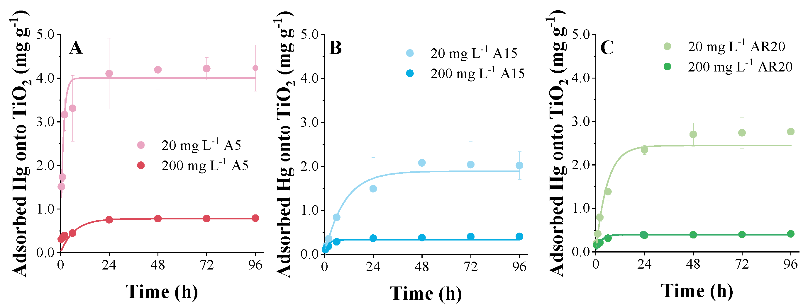
3.1. Adsorption Kinetics of Hg on nTiO2

All three tested nTiO2 materials adsorbed significant amounts of Hg (Figure 1). Hg adsorption on 20 and 200 mg L−1 nTiO2 increased rapidly in the first 24 h and tended to plateau at longer times. The adsorbed amount of Hg over time decreased with the increase in the primary particle size of the nTiO2 materials: A5 > AR20 > A15, despite more significant aggregation of the nTiO2 with the smaller primary size.
This finding agrees with previous studies showing that different nTiO2 materials adsorb more than 95% of the Hg present in the medium [31]. It was also suggested that the adsorption of Hg to nTiO2 takes place via the interaction of HgClOH and Hg(OH)2 with -TiOH and -TiO active sites [10,31]. A similar adsorption mechanism seems possible, given that chemical speciation calculations [10] showed that Hg is present mainly as -OH and -Cl complexes. The apparent adsorption and desorption rate constants for Hg adsorption on the nTiO2 materials (Table 1) depended on their primary size. The values of the apparent adsorption rate constants (kads,nTiO2) were in the ranges of (2.89−26) × 102, (1.8−3.2) × 102 and (1.3−1.4) × 102 kg L h−1 for A5, A15 and AR20, respectively. In addition to that, increasing kads,nTiO2 values were found with increasing nTiO2 concentrations. However, the initial concentration of nTiO2 that was used to obtain the apparent adsorption rate constants could not represent the quantification of the free binding sites at the surface, which led to the rate constants being dependent on the mass of the adsorbent given the significant aggregation and sedimentation of the nTiO2, in particular at high concentrations.

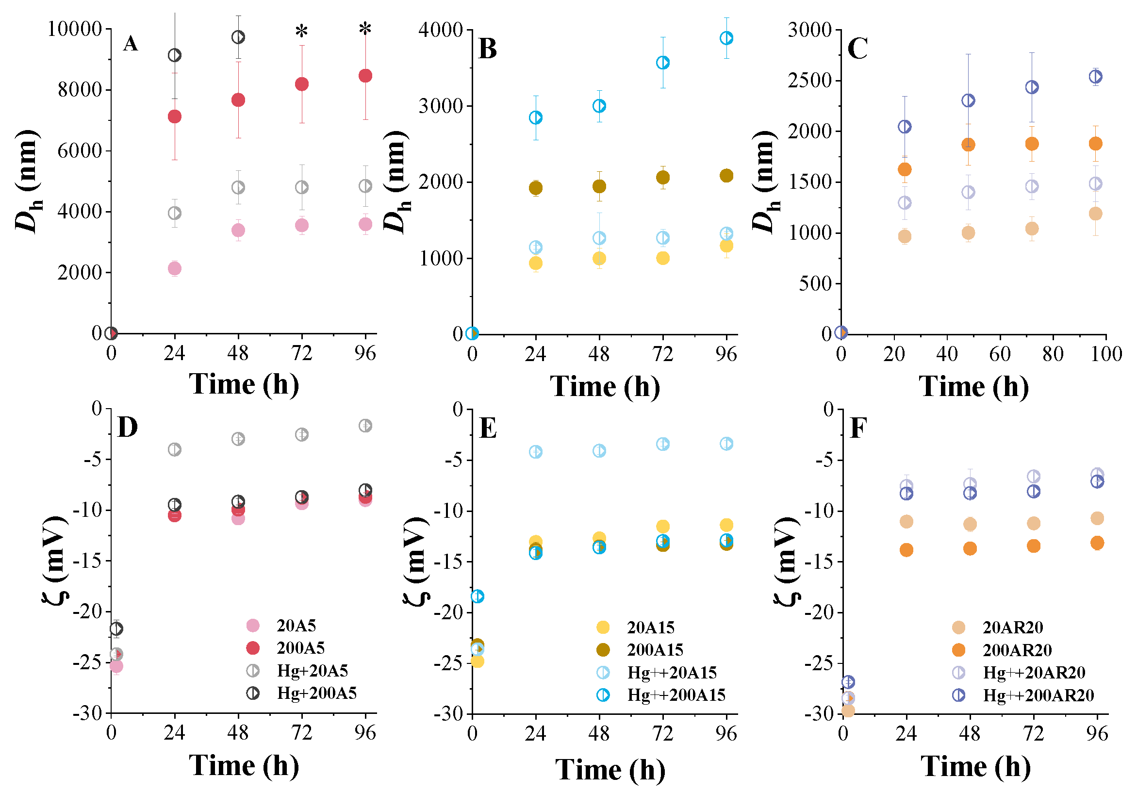
3.2. Time Course of Aggregation of nTiO2 and Hg-nTiO2 Complexes

Three nTiO2 materials strongly aggregated in the exposure medium and the size of the formed aggregates stayed stable over time (Figure 2A–C). A5 particles with the smallest primary size formed the largest aggregates with an average hydrodynamic size Dh that was more than two-fold higher than that of A15 and AR20 and increased gradually after 24 h. The Dh of the three materials increased significantly with the increase in nTiO2 concentration. This concentration effect was more pronounced for A5. For instance, the Dh of A5 at 24 h increased from 1940 to 7126 nm as the concentration increased from 20 to 200 mg L−1 (Figure 2A). These findings agree with the fact that the nTiO2 suspensions at higher concentrations were more prone to aggregate than for diluted suspensions, which may have been due to the increased collision probability between the particles [32]. The more pronounced aggregation of A5 than A15 and AR20 agreed with the measurements of the zeta potential (ζ). The absolute values of ζ increased in the order of 20 mV < ζ (A5) < ζ (A15) < ζ (AR20) < 30 mV (Figure 2D–F). After 24 h, the absolute values of ζ decreased to the range of 10–15 mV and stayed at comparable levels thereafter. The results are consistent with the existing literature reporting that nanoparticles tend to aggregate when the absolute value of ζ is small [33].
The aggregation of nTiO2 was more pronounced in mixtures due to the adsorption of Hg onto the surface of nTiO2 (see Section 3.1), which altered the nanoparticle surface charge and stability in the exposure medium. For instance, Hg led to 1.25- and 1.42-fold increases in Dh of 200 mg L−1 A5 at 24 and 96 h, 1.42 and 1.87 times for 200 mg L−1 A15 and 1.40 and 1.30 times for 200 mg L−1 AR20, respectively. nTiO2 is negatively charged, and can thus be aggregated through charge neutralization with cationic metallic coagulants. Values of ζ further revealed the interactions between nTiO2 particles and Hg, leading to a decrease in surface charge (Figure 2D–F). This finding agrees with the existing literature showing that Cd, Cu, Ni [34], Cd [35] and Hg [10] significantly enhanced the aggregation of different nTiO2 materials.

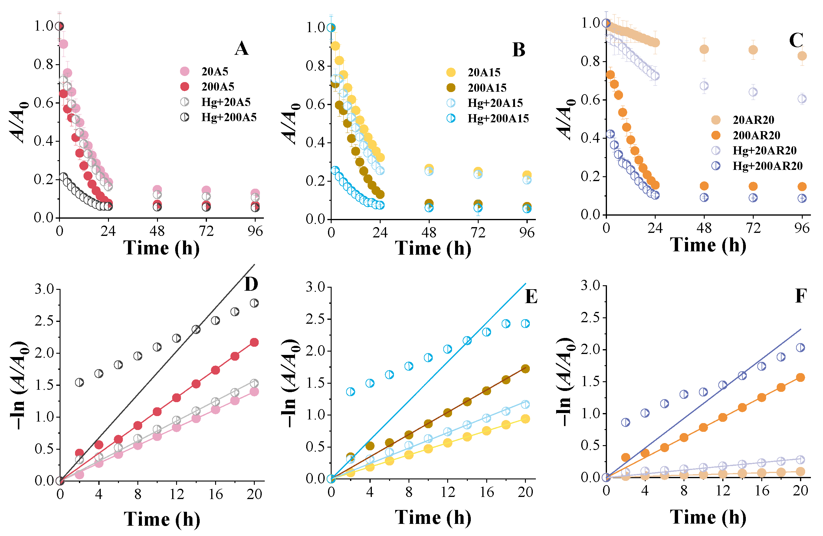
3.3. Kinetics of Sedimentation of nTiO2 and Hg-nTiO2 Complexes

In agreement with the strong aggregation, the nTiO2 aggregates settled down rapidly within the first 24 h and tended to stay constant from 24 to 96 h (Figure 3A–C). In general, the sedimentation of A5 aggregates (Figure 3A) was greater and much more rapid than A15 (Figure 3B) and AR20 aggregates (Figure 3C). Specifically, after 24 h with an initial concentration of 20 mg L−1, the suspended particles represented 18.64 wt%, 32.36 wt% and 89.85 wt% of the A5, A15 and AR20 suspensions, respectively. The sedimentation of the three nTiO2 materials at the initial concentration of 200 mg L−1 was more pronounced. Consequently, the percentages of particles suspended in the exposure medium were 13.07 wt%, 15.59 wt% and 17.64 wt% of the A5, A15 and AR20, respectively. The values of the settling rate constant (ksettle) decreased in the order of A5 > A15 > AR20 (Table 2).
As expected by the aggregation results, the sedimentation of all three nTiO2 was more pronounced in mixtures with Hg (Figure 3A). After 24 h, the A5 particles suspended in the solution decreased by 4.24 wt% in the presence of Hg compared to the treatment without Hg, while A15 particles decreased by 1.45 wt%, and AR20 particles decreased by 5.28 wt%. It is worth noting that AR20 was still the most sensitive material to the addition of Hg. The AR20 particles suspended in the solution with an initial concentration of 20 mg L−1 decreased by 17.39 wt% after adding Hg compared to the treatment without Hg after 24 h. The presence of Hg led to increases in ksettle values of 1.10-, 1.26- and 3.09-fold for 20 mg L−1 A5, A15 and AR20, respectively (Figure 3B).

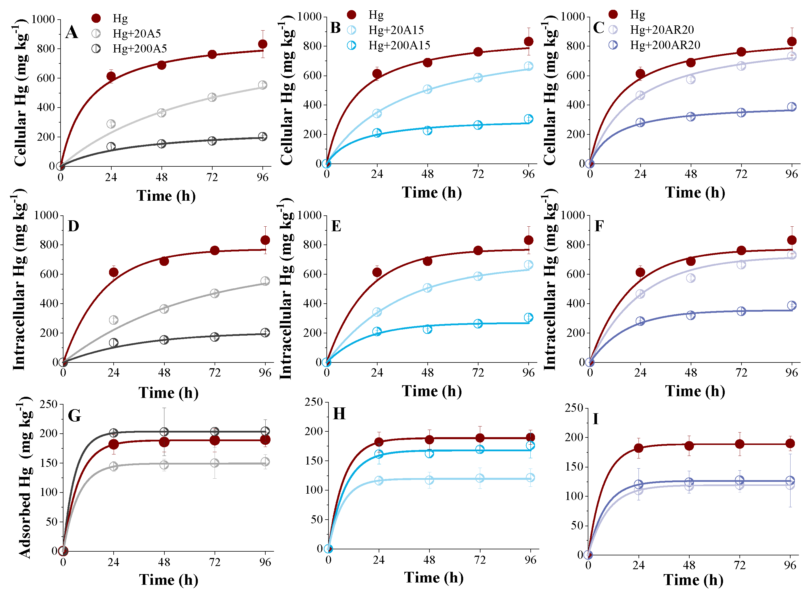
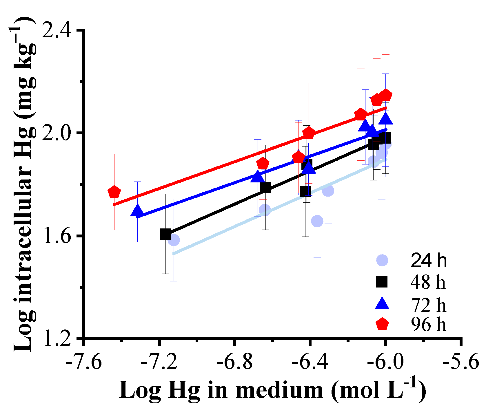
3.4. Kinetics of Hg Uptake by C. reinhardtii in Mixtures with nTiO2

Algal cells accumulated significant amounts of Hg, which increased over time (Figure 4). The contents of cellular (adsorbed + intracellular) Hg increased from 794 to 1021 mg Hg kg−1 algae with increased exposure time from 24 to 96 h in the absence of nTiO2 (Figure 4A–C). The intracellular Hg concentrations increased from 612 to 831 mg Hg kg−1 algae (Figure 4D–F) and adsorbed Hg (loosely bound) concentrations on the algal surface increased from 182 to 190 mg Hg kg−1 algae (Figure 4G–I). The ratios between intracellular and adsorbed Hg increased from 3.36 to 4.37 with an increased exposure time from 24 to 96 h. Similar ratios of intra- and extracellular Hg content in the diatom Thalassiosira pseudonana (8:2) and the flagellate Isochrysis galbana (9:1) were found, while the ratio was 1:1 in the green alga Chlorella autotrophica [36]. The different ratios indicated that cellular debris may play different roles in Hg detoxification in different species, which suggested that the surface-adsorbed mercury displayed no toxic effects [36]. In contrast, a ratio of about 2:8 (intracellular vs. adsorbed Hg content) was reported after 2 h exposure to 10−11 and 10−10 M Hg in C. reinhardtii [30]. The longer exposure duration and higher exposure concentration used in the present study may partly explain the dissimilarity. A rapid increase in the intracellular Hg content was observed in the diatom Thalassiosira weissflogii during the first day of exposure [37].
The addition of nTiO2 significantly reduced the total cellular, adsorbed and intracellular Hg contents as compared with individual Hg treatments (Figure 4). This decrease was more pronounced for A5 and higher concentrations. It diminished in the order A5 > A15 > AR20, showing the importance of the primary particle size. Specifically, the intracellular Hg decreased by 62.26%, 42.72% and 35.81% in the presence of 200 mg L−1 A5, A15 and AR20, and decreased by 29.26%, 14.06% and 4.48% in the presence of 20 mg L−1 A5, A15 and AR20, respectively, after 96 h. Correspondingly, the addition of 200 mg L−1 A5, A15 and AR20 decreased the concentrations of the dissolved mercury that was not adsorbed to nTiO2 in the exposure medium by 76.10%, 63.30% and 53.43% (see Section 3.1).
The presence of the nTiO2 decreased the Hg uptake (adsorbed and intracellular contents) by reducing the concentrations of the dissolved Hg as a result of the adsorption of Hg by aggregated nTiO2 and their sedimentation. This consideration is in line with the observed relationship between the intracellular Hg content and the concentration of Hg in the medium that was not adsorbed on the aggregates of nTiO2 (Figure 5). Similar results were obtained in our previous work with a short-term exposure time of 2 h for the same algae exposed to 10−9 and 5 × 10⁻⁷ M of Hg [10]. nTiO2 reduced adsorbed (loosely bound, EDTA-cysteine extractable) Hg content (Figure 4H–I). However, the reduction increased with the decreasing nTiO2 concentrations and decreased in the order A5 < A15 < AR20. This could be due to the “carry over” effect due to Hg adsorption on nTiO2 aggregates (Figure 1) that are trapping the cells in the case of A5 (Figure 6). This finding is consistent with a previous study indicating that cells of P. subcapitata adsorbed nTiO2 onto their surface and carried 2.3 times their weight in TiO2 particles [38]. A series of previous studies showed that nTiO2 cannot enter the cell wall of green alga [39,40,41].
In the absence of nTiO2, the kads and kdes characterizing Hg adsorption and desorption on the algal surface were (3.55 ± 0.29) × 10−2 and (1.39 ± 0.12) × 10−1 h−1 respectively. Co-exposure of C. reinhardtii to Hg and nTiO2 resulted in no significant changes in the kads and kdes, which were comparable with the values found for exposure to Hg alone. However, in the mixture containing A5, the kads were found to be approximately 3 to 15 times higher in the presence of 20 and 200 mg L−1 nTiO2. It is worth noting that the adsorption and desorption rate constants found for Hg adsorption on nTiO2 materials were higher as compared to the one determined for adsorption to algae (Table 1).
The apparent internalization kint and the efflux keff rate constant for Hg were almost comparable (6.04 ± 0.89) × 10−2 and (5.62 ± 1.06) × 10−2 h−1. The kint for Hg obtained in the present study was about 10 times lower than the value determined for the same algae using the same approach, but for a shorter exposure of 1 h (kint = 7.86 × 10⁻¹ h−1). The kint values were also lower than those determined for the internalization of Cd (13.68 h−1) and Ni (6.84 h−1) by C. reinhardtii [42,43] and Pb (1.54 h−1) by another green alga Chlorella vulgaris [44]. However, these apparent internalization rate constants were determined for short-term exposure corresponding to the linear rise of the intracellular metal content over time, where the efflux or depletion was negligible; therefore, a direct comparison was not straightforward.
nTiO2 also reduced the kint and kef and this reduction was more pronounced for A5 material with lower primary size. The addition of 20 and 200 mg L−1 nTiO2 decreased the kads, kdes and their ratios in all treatments with exception of 200 mg L−1 A5. Algal cells can be sequestered due to their co-agglomeration with ENPs; in this situation, the access of algae to nutrients present in the culture medium can be reduced or impaired, as well as mercury. The co-agglomeration of nTiO2 and algal cells was observed for A5, where this effect was particularly notorious at high NP concentrations (Figure 6D). In these algal-A5 agglomerates, it was possible to observe cells inside and at the periphery of the structures, as well as in the surrounding medium. For A15 and AR20, no such agglomerates were observed (Figure 6). In addition, the presence of nTiO2 could also favor the depletion of dissolved Hg in the exposure medium and increase the uncertainty of the apparent rate constants.
In addition, in the above considerations and modeling of Hg uptake kinetics by C. reinhardtii, Hg depletion in the exposure medium over the exposure duration was not considered. Indeed, the curvature of the uptake curves over time observed in the cellular, intracellular and adsorbed content could have also been due to the significant depletion of Hg in the exposure medium and not to the efflux processes. Therefore, the values of the apparent rate constants characterizing the above-mentioned processes have to be taken with caution.
The present study was performed under control conditions in the laboratory using a very high concentration of Hg that is representative of a highly contaminated environment, such as wastewater releases of chemical plants that historically used Hg in chlor-alkali electrolysis, acetaldehyde and vinyl chloride production. Given that both green algae and nanomaterials are used in the removal of hazardous heavy metals from contaminated waters, the present results present important implications for the development of efficient bioremediation technology for Hg.

4. Conclusions

The kinetics of the interactions in the ternary system containing mixtures of Hg, nTiO2 and the green alga C. reinhardtii were investigated under settings that were representative of a highly contaminated environment. The nTiO2 rapidly adsorbed significant amounts of Hg, leading to a 3–4-fold decrease in the dissolved Hg concentration after adding 200 mg L−1 A5, A15 and AR20 after 96 h. The tested nTiO2 materials and adsorbed Hg underwent fast aggregation and sedimentation in the algal exposure medium, which depended mostly on the initial concentration and the primary particle size of the nTiO2. The presence of nTiO2 significantly reduced the cellular, adsorbed and intracellular amounts of Hg in the algae. The obtained results significantly contribute to the enlargement of the understanding of the behavior of cocktails of engineered nanomaterials and hazardous pollutants, such as mercury, in a highly contaminated environment, such as wastewater effluent.

Author Contributions

M.L.: Methodology, Investigation, Visualization, Data Curation, Writing—Original Draft and Funding Acquisition; V.I.S.: Conceptualization, Resources, Methodology, Data Curation, Writing—Review and Editing, Supervision, and Funding Acquisition. All authors have read and agreed to the published version of the manuscript.

Funding

This research was funded by the China Scholarship Council grant [2016]3100 and the Swiss National Science Foundation, project N166089.

Institutional Review Board Statement

Not applicable.

Informed Consent Statement

Not applicable.

Data Availability Statement

The data presented in this study are available on request from the corresponding author.

Conflicts of Interest

The authors declare no conflict of interest.

References

  1. Li, M.T.; Liu, W.; Slaveykova, V.I. Effects of mixtures of engineered nanoparticles and metallic pollutants on aquatic organisms. Environments 2020, 7, 27. [Google Scholar] [CrossRef] [Green Version]
  2. Naasz, S.; Altenburger, R.; Kühnel, D. Environmental mixtures of nanomaterials and chemicals: The trojan-horse phenomenon and its relevance for ecotoxicity. Sci. Total Environ. 2018, 635, 1170–1181. [Google Scholar] [CrossRef] [PubMed]
  3. Auffan, M.; Rose, J.; Bottero, J.Y.; Lowry, G.V.; Jolivet, J.P.; Wiesner, M.R. Towards a definition of inorganic nanoparticles from an environmental, health and safety perspective. Nat. Nanotechnol. 2009, 4, 634–641. [Google Scholar] [CrossRef] [PubMed]
  4. Handy, R.D.; von der Kammer, F.; Lead, J.R.; Hassellov, M.; Owen, R.; Crane, M. The ecotoxicology and chemistry of manufactured nanoparticles. Ecotoxicology 2008, 17, 287–314. [Google Scholar] [CrossRef] [PubMed]
  5. Slaveykova, V.I.; Li, M.T.; Worms, I.A.; Liu, W. When environmental chemistry meets ecotoxicology: Bioavailability of inorganic nanoparticles to phytoplankton. Chimia 2020, 74, 115–121. [Google Scholar] [CrossRef] [PubMed]
  6. Chen, F.; Wu, L.; Xiao, X.; Rong, L.; Li, M.; Zou, X. Mixture toxicity of zinc oxide nanoparticle and chemicals with different mode of action upon Vibrio Fischeri. Environ. Sci. 2020, 32, 41. [Google Scholar] [CrossRef]
  7. Wang, D.; Hu, J.; Irons, D.R.; Wang, J. Synergistic toxic effect of nano-TiO2 and As (V) on Ceriodaphnia dubia. Sci. Total Environ. 2011, 409, 1351–1356. [Google Scholar] [CrossRef]
  8. Singh, J.; Lee, B.-K. Influence of nano-TiO2 particles on the bioaccumulation of cd in soybean plants (Glycine max): A possible mechanism for the removal of cd from the contaminated soil. J. Environ. Manag. 2016, 170, 88–96. [Google Scholar] [CrossRef]
  9. Li, M.; Slaveykova, V.I. Dual role of titanium dioxide nanoparticles in the accumulation of inorganic and methyl mercury by crustacean Daphnia magna through waterborne and dietary exposure. Environ. Pollut. 2022, 295, 118619. [Google Scholar] [CrossRef]
  10. Li, M.; Liu, W.; Slaveykova, V.I. NanoTiO2 materials mitigate mercury uptake and effects on green alga chlamydomonas reinhardtii in mixture exposure. Aquat. Toxicol. 2020, 224, 105502. [Google Scholar] [CrossRef]
  11. Yu, Z.; Hao, R.; Zhang, L.; Zhu, Y. Effects of TiO2, SiO2, Ag and CdTe/CdSquantum dots nanoparticles on toxicity of cadmium towards Chlamydomonas reinhardtii. Ecotoxicol. Environ. Saf. 2018, 156, 75–86. [Google Scholar] [CrossRef] [PubMed]
  12. Li, X.; Ma, Q.; Liu, T.; Dong, Z.; Fan, W. Effect of TiO2-nanoparticles on copper toxicity to bacteria: Role of bacterial surface. RSC Adv. 2020, 10, 5058–5065. [Google Scholar] [CrossRef] [Green Version]
  13. Fan, W.; Liang, D.; Wang, X.; Ren, J.; Xiao, S.; Zhou, T. Two-generational effects and recovery of arsenic and arsenate on Daphnia magna in the presence of nano-TiO2. Ecotoxicol. Environ. Saf. 2019, 172, 136–143. [Google Scholar] [CrossRef]
  14. Yi, X.; Chi, T.; Liu, B.; Liu, C.; Feng, G.; Dai, X.; Zhang, K.; Zhou, H. Effect of nano zinc oxide on the acute and reproductive toxicity of cadmium and lead to the marine copepod Tigriopus japonicus. Comp. Biochem. Physiol. Part C Toxicol. Pharmacol. 2019, 222, 118–124. [Google Scholar] [CrossRef] [PubMed]
  15. Sharma, V.K. Aggregation and toxicity of titanium dioxide nanoparticles in aquatic environment—A review. J. Environ. Sci. Health Part A 2009, 44, 1485–1495. [Google Scholar] [CrossRef] [PubMed]
  16. Keller, A.A.; Lazareva, A. Predicted releases of engineered nanomaterials: From global to regional to local. Environ. Sci. Technol. Lett. 2014, 1, 65–70. [Google Scholar] [CrossRef] [Green Version]
  17. Zhang, J.; Li, L.; Li, Y.; Yang, C. Microwave-assisted synthesis of hierarchical mesoporous nano-TiO2/cellulose composites for rapid adsorption of Pb2+. Chem. Eng. J. 2017, 313, 1132–1141. [Google Scholar] [CrossRef]
  18. Kuang, X.; Shao, J.; Peng, L.; Song, H.; Wei, X.; Luo, S.; Gu, J.-d. Nano-TiO2 enhances the adsorption of Cd(II) on biological soil crusts under mildly acidic conditions. J. Contam. Hydrol. 2020, 229, 103583. [Google Scholar] [CrossRef]
  19. Driscoll, C.T.; Mason, R.P.; Chan, H.M.; Jacob, D.J.; Pirrone, N. Mercury as a global pollutant: Sources, pathways, and effects. Environ. Sci. Technol. 2013, 47, 4967–4983. [Google Scholar] [CrossRef]
  20. Yang, L.; Zhang, Y.; Wang, F.; Luo, Z.; Guo, S.; Strähle, U. Toxicity of mercury: Molecular evidence. Chemosphere 2020, 245, 125586. [Google Scholar] [CrossRef]
  21. Le Faucheur, S.; Campbell, P.G.C.; Fortin, C.; Slaveykova, V.I. Interactions between mercury and phytoplankton: Speciation, bioavailability, and internal handling. Environ. Toxicol. Chem. 2014, 33, 1211–1224. [Google Scholar] [CrossRef] [PubMed]
  22. Dranguet, P.; Flück, R.; Regier, N.; Cosio, C.; Le Faucheur, S.; Slaveykova, V.I. Towards mechanistic understanding of mercury availability and toxicity to aquatic primary producers. Chimia 2014, 68, 799–805. [Google Scholar] [CrossRef] [PubMed] [Green Version]
  23. Cosio, C.; Flück, R.; Regier, N.; Slaveykova, V.I. Effects of macrophytes on the fate of mercury in aquatic systems. Environ. Toxicol. Chem. 2014, 33, 1225–1237. [Google Scholar] [CrossRef]
  24. Beauvais-Flück, R.; Slaveykova, V.I.; Cosio, C. Molecular effects of inorganic and methyl mercury in aquatic primary producers: Comparing impact to a macrophyte and a green microalga in controlled conditions. Geosciences 2018, 8, 393. [Google Scholar] [CrossRef] [Green Version]
  25. Harris, E.H. The Chlamydomonas Sourcebook: A Comprehensive Guide to Biology and Laboratory Use; Elsevier: Amsterdam, The Netherlands, 2013. [Google Scholar]
  26. Kahru, A.; Ivask, A. Mapping the dawn of nanoecotoxicological research. Acc. Chem. Res. 2013, 46, 823–833. [Google Scholar] [CrossRef] [PubMed]
  27. Brunelli, A.; Pojana, G.; Callegaro, S.; Marcomini, A. Agglomeration and sedimentation of titanium dioxide nanoparticles (n-TiO2) in synthetic and real waters. J. Nanoparticle Res. 2013, 15, 1684. [Google Scholar] [CrossRef]
  28. Li, M.; Slaveykova, V.I. A density gradient centrifugation method for rapid separation of nanoTiO2 and TiO2 aggregates from microalgal cells in complex mixtures with mercury. MethodsX 2020, 7, 101057. [Google Scholar] [CrossRef] [PubMed]
  29. Beauvais-Flück, R.; Slaveykova, V.I.; Cosio, C. Transcriptomic and physiological responses of the green microalga Chlamydomonas reinhardtii during short-term exposure to subnanomolar methylmercury concentrations. Environ. Sci. Technol. 2016, 50, 7126–7134. [Google Scholar] [CrossRef]
  30. Beauvais-Flück, R.; Slaveykova, V.I.; Cosio, C. Cellular toxicity pathways of inorganic and methyl mercury in the green microalga Chlamydomonas reinhardtii. Sci. Rep. 2017, 7, 8034. [Google Scholar] [CrossRef] [Green Version]
  31. Lopez-Munoz, M.J.; Arencibia, A.; Cerro, L.; Pascual, R.; Melgar, A. Adsorption of Hg(II) from aqueous solutions using TiO2 and titanate nanotube adsorbents. Appl. Surf. Sci. 2016, 367, 91–100. [Google Scholar] [CrossRef]
  32. Keller, A.A.; Wang, H.; Zhou, D.; Lenihan, H.S.; Cherr, G.; Cardinale, B.J.; Miller, R.; Ji, Z. Stability and aggregation of metal oxide nanoparticles in natural aqueous matrices. Environ. Sci. Technol. 2010, 44, 1962–1967. [Google Scholar] [CrossRef] [PubMed]
  33. Mandzy, N.; Grulke, E.; Druffel, T. Breakage of TiO2 agglomerates in electrostatically stabilized aqueous dispersions. Powder Technol. 2005, 160, 121–126. [Google Scholar] [CrossRef]
  34. Mahdavi, S. Nano-TiO2 modified with natural and chemical compounds as efficient adsorbents for the removal of Cd+ 2, Cu+ 2, and Ni+ 2 from water. Clean Technol. Environ. Policy 2016, 18, 81–94. [Google Scholar] [CrossRef]
  35. Han, Z.-X.; He, G.-D.; Wang, J.-H.; Lv, C.-x. Interaction influence of cd (ii) and nano-TiO2 on aggregation and adsorption kinetics toward marine algae. Int. J. Green Nanotechnol. 2011, 3, 229–237. [Google Scholar] [CrossRef]
  36. Wu, Y.; Wang, W.-X. Accumulation, subcellular distribution and toxicity of inorganic mercury and methylmercury in marine phytoplankton. Environ. Pollut. 2011, 159, 3097–3105. [Google Scholar] [CrossRef]
  37. Morelli, E.; Ferrara, R.; Bellini, B.; Dini, F.; Di Giuseppe, G.; Fantozzi, L. Changes in the non-protein thiol pool and production of dissolved gaseous mercury in the marine diatom Thalassiosira weissflogii under mercury exposure. Sci. Total Environ. 2009, 408, 286–293. [Google Scholar] [CrossRef] [Green Version]
  38. Huang, C.; Cha, D.; Ismat, S. Progress Report: Short-Term Chronic Toxicity of Photocatalytic Nanoparticles to Bacteria, Algae, and Zooplankton; University of Delaware: Newark, NJ, USA, 2005. [Google Scholar]
  39. Yang, W.-W.; Wang, Y.; Huang, B.; Wang, N.-X.; Wei, Z.-B.; Luo, J.; Miao, A.-J.; Yang, L.-Y. TiO2 nanoparticles act as a carrier of cd bioaccumulation in the ciliate Tetrahymena thermophila. Environ. Sci. Technol. 2014, 48, 7568–7575. [Google Scholar] [CrossRef]
  40. Tan, L.-Y.; Huang, B.; Xu, S.; Wei, Z.-B.; Yang, L.-Y.; Miao, A.-J. Aggregation reverses the carrier effects of TiO2 nanoparticles on cadmium accumulation in the waterflea Daphnia magna. Environ. Sci. Technol. 2017, 51, 932–939. [Google Scholar] [CrossRef]
  41. He, X.; Xie, C.; Ma, Y.; Wang, L.; He, X.; Shi, W.; Liu, X.; Liu, Y.; Zhang, Z. Size-dependent toxicity of tho2 nanoparticles to green algae Chlorella pyrenoidosa. Aquat. Toxicol. 2019, 209, 113–120. [Google Scholar] [CrossRef]
  42. Kola, H.; Wilkinson, K.J. Cadmium uptake by a green alga can be predicted by equilibrium modelling. Environ. Sci. Technol. 2005, 39, 3040–3047. [Google Scholar] [CrossRef]
  43. Worms, I.A.; Wilkinson, K.J. Ni uptake by a green alga. 2. Validation of equilibrium models for competition effects. Environ. Sci. Technol. 2007, 41, 4264–4270. [Google Scholar] [CrossRef] [PubMed]
  44. Slaveykova, V.I.; Wilkinson, K.J. Physicochemical Aspects of Lead Bioaccumulation by Chlorella vulgaris. Environ. Sci. Technol. 2002, 36, 969–975. [Google Scholar] [CrossRef] [PubMed] [Green Version]
Figure 1. Adsorption kinetics of Hg on three types of nTiO2: (A) anatase 5 nm (A5), (B) anatase 15 nm (A15) and (C) anatase/rutile 20 nm (AR20) at 20 and 200 mg L−1 suspended in an algal exposure medium. The measured initial Hg concentration in the medium was 6.3 × 10⁻⁷ M. Points are experimental data. Lines represent the fits with Equation (1).
Environments 09 00044 g001
Figure 2. Time-course changes of average hydrodynamic diameter (Dh, (AC)) and zeta potential (ζ, (DF)) of three different nTiO2 materials: anatase 5 nm (A5), anatase 15 nm (A15) and anatase/rutile 20 nm (AR20) at 20 and 200 mg L−1 suspended in an algal exposure medium in the absence and presence of Hg. * denotes the data out of the detection limit of the Zetasizer Nano ZS.
Environments 09 00044 g002
Figure 3. Time course of sedimentation of different nTiO2 materials: (A) anatase 5 nm (A5), (B) anatase 15 nm (A15) and (C) anatase/rutile 20 nm (AR20) at 20 and 200 mg L−1 in algal exposure medium in the absence and presence of Hg and (DF) first-order kinetics rate curves that were fit using Equation (2) for the experimental points until 24 h.
Environments 09 00044 g003
Figure 4. Time progression of Hg accumulation by C. reinhardtii. (AC) cellular, (DF) intracellular and (GI) adsorbed Hg contents in the absence and presence of three types of nTiO2: anatase 5 nm (A5), anatase 15 nm (A15) and anatase/rutile 20 nm (AR20) at 20 and 200 mg L−1. Initial Hg concentration: 6.3 × 10⁻⁷ M. Points are experimental data. Lines are model fittings with Equations (2) and (3). The apparent adsorption and desorption rate constants of Hg on algae, kads and kdes and the apparent internalization and efflux rate constants kint and kef obtained by using a non-linear pseudo-first-order one-compartment model are presented in Table 1.
Environments 09 00044 g004
Figure 5. Log–log plot of intracellular mercury content in C. reinhardtii and the concentration of Hg in exposure medium that was not adsorbed on nTiO2. Both results in the presence of 20 or 200 mg L−1 nTiO2 are presented.
Environments 09 00044 g005
Figure 6. Interactions between C. reinhardtii and three different types of nTiO2: anatase 5 nm (A5), anatase 15 nm (A15) and anatase/rutile 20 nm (AR20) after 24 h (AC) and 96 h of exposure (DF).
Environments 09 00044 g006
Table 1. Parameters of the non-linear pseudo-first-order one-compartment-model kinetic Hg adsorption on nTiO2 or algae and Hg internalization by algae. kads,nTiO2 and kdes,nTiO2 are the apparent adsorption and desorption rate constants of Hg on nTiO2, kads and kdes are the apparent adsorption and desorption rate constants of Hg on algae and kint and kef are the apparent internalization and efflux rate constants. The values are mean ± standard error. N = 3.
Parameter HgHg + 20 mg L−1 A5Hg + 200 mg L−1 A5Hg + 20 mg L−1 A15Hg + 200 mg L−1 A15Hg + 20 mg L−1 AR20Hg + 200 mg L−1 AR20
Hg adsorption on nTiO2
kads,nTiO2 * ×102 L kg−1 h−1-26.58 ± 4.252.89 ± 2.213.27 ± 0.631.85 ± 0.251.46 ± 0.251.31 ± 0.05
kdes,nTiO2×10−1 h−1-7.30 ± 2.704.10 ± 0.601.29 ± 0.015.10 ± 1.250.79 ± 0.193.56 ± 0.42
R2 -0.820.860.990.920.990.94
Hg adsorption on algae
kads * ×102 L kg−1 h−11.42 ± 0.151.10 ± 0.141.99 ± 0.090.96 ± 0.370.50 ± 0.260.71 ± 0.020.87 ± 0.07
kads ** ×10−2 h−13.55 ± 0.292.75 ± 0.344.98 ± 0.232.401.26 ± 0.541.76 ± 0.062.17 ± 0.15
kdes×10−1 h−11.39 ± 0.121.35 ± 0.181.80 ± 0.081.48 ± 0.250.44 ± 0.261.09 ± 0.051.26 ± 0.12
R2 0.990.991.001.000.881.001.00
Hg internalization
kint * ×102 L kg−1 h−12.42 ± 0.360.73 ± 0.110.45 ± 0.081.02 ± 0.050.71 ± 0.121.52 ± 0.171.10 ± 0.16
kint ** ×10−2 h−16.04 ± 0.891.83 ± 0.271.13 ± 0.222.55 ± 0.121.79 ± 0.403.81 ± 0.412.75 ± 0.41
kef×10−2 h−15.62 ± 1.062.23 ± 0.584.36 ± 1.132.66 ± 0.204.61 ± 0.143.88 ± 0.605.52 ± 1.05
R2 0.980.980.970.990.960.990.98
* The apparent rate constants were calculated using the measured initial concentration of the dissolved Hg without considering the adsorption on nTiO2 aggregates. ** The values of kads and kint expressed in h−1 were obtained from kads in L kg−1 h−1 by considering the initial concentration of Hg in the exposure medium of 6.3 × 10⁻⁷ mol L−1 and the cell density of 2.5 × 10⁻⁴ kg L−1.
Table 2. Parameters of the first-order decay kinetics of nTiO2 sedimentation of different materials in the absence and presence of Hg.
TreatmentSedimentation
ksettle (h−1) *R2Treatmentksettle (h−1) *R2
20A54.20.96Hg + 20A54.60.98
200A56.50.95Hg + 200A59.00.94
20A152.80.97Hg + 20A153.50.97
200A155.20.96Hg + 200A158.20.93
20AR200.30.98Hg + 20AR200.90.98
200AR204.70.98Hg + 200AR206.50.93
* The apparent rate constants were calculated using the nominal initial nTiO2 without considering the aggregation and sedimentation in the suspensions.
Publisher’s Note: MDPI stays neutral with regard to jurisdictional claims in published maps and institutional affiliations.

