

Atypical Carcinoid of the Thymus: Early Diagnosis in a Case Report


Abstract
1. Introduction
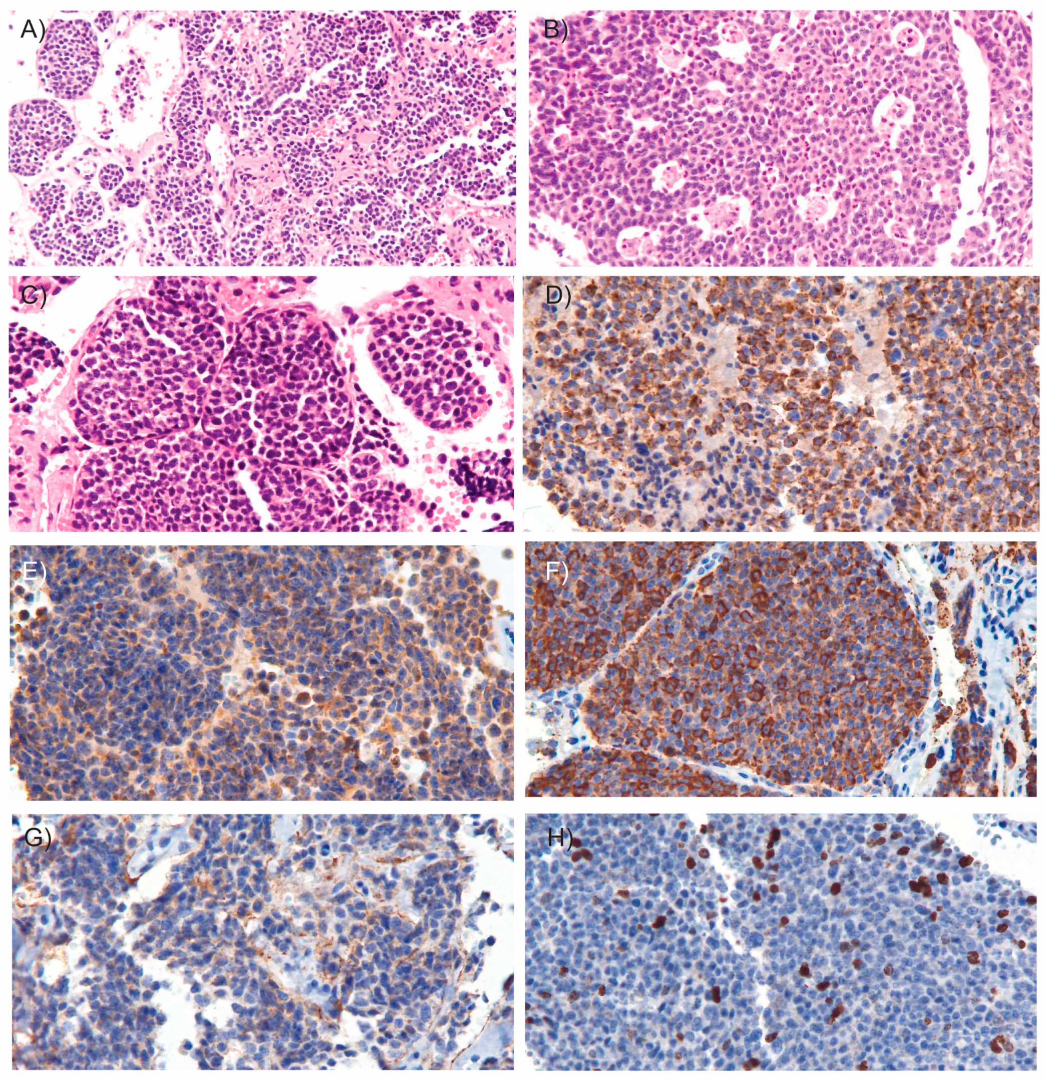
2. Case Report
3. Discussion
4. Conclusions
Author Contributions
Funding
Institutional Review Board Statement
Informed Consent Statement
Data Availability Statement
Conflicts of Interest
References
- Baloch, Z.W.; Asa, S.L.; Barletta, J.A.; Ghossein, R.A.; Juhlin, C.C.; Jung, C.K.; LiVolsi, V.A.; Papotti, M.G.; Sobrinho-Simões, M.; Tallini, G.; et al. Overview of the 2022 WHO Classification of Thyroid Neoplasms. Endocr. Pathol. 2022, 33, 27–63. [Google Scholar] [CrossRef]
- Han, B.; Sun, J.-M.; Ahn, J.S.; Park, K.; Ahn, M.-J. Clinical Outcomes of Atypical Carcinoid Tumors of the Lung and Thymus: 7-Year Experience of a Rare Malignancy at Single Institute. Med. Oncol. 2013, 30, 479. [Google Scholar] [CrossRef]
- Volante, M.; Mete, O.; Pelosi, G.; Roden, A.C.; Speel, E.J.M.; Uccella, S. Molecular Pathology of Well-Differentiated Pulmonary and Thymic Neuroendocrine Tumors: What Do Pathologists Need to Know? Endocr. Pathol. 2021, 32, 154–168. [Google Scholar] [CrossRef]
- Koehler, K.; Iams, W.T. Carcinoid Tumors Outside the Abdomen. Cancer Med. 2023, 12, 7893–7903. [Google Scholar] [CrossRef]
- Öberg, K.; Hellman, P.; Ferolla, P.; Papotti, M. Neuroendocrine Bronchial and Thymic Tumors: ESMO Clinical Practice Guidelines for Diagnosis, Treatment and Follow-Up. Ann. Oncol. 2012, 23, vii120–vii123. [Google Scholar] [CrossRef] [PubMed]
- Tiffet, O.; Nicholson, A.G.; Ladas, G.; Sheppard, M.N.; Goldstraw, P. A Clinicopathologic Study of 12 Neuroendocrine Tumors Arising in the Thymus. Chest 2003, 124, 141–146. [Google Scholar] [CrossRef] [PubMed]
- Zhu, S.; Wang, Z.-T.; Liu, W.-Z.; Zong, S.-X.; Li, B. Invasive Atypical Thymic Carcinoid: Three Case Reports And Literature Review. Onco. Targets. Ther. 2016, 9, 6171–6176. [Google Scholar] [CrossRef] [PubMed]
- Moran, C.A.; Suster, S. Neuroendocrine Carcinomas (Carcinoid Tumor) of the Thymus. Am. J. Clin. Pathol. 2000, 114, 100–110. [Google Scholar] [CrossRef]
- Rindi, G.; Mete, O.; Uccella, S.; Basturk, O.; La Rosa, S.; Brosens, L.A.A.; Ezzat, S.; de Herder, W.W.; Klimstra, D.S.; Papotti, M.; et al. Overview of the 2022 WHO Classification of Neuroendocrine Neoplasms. Endocr. Pathol. 2022, 33, 115–154. [Google Scholar] [CrossRef]
- Knipe, H. WHO Classification of Tumours Editorial Board Thymic Neuroendocrine Neoplasms. In WHO Classification of Tumours; IARC Publications: Lyon, France, 2021; pp. 389–397. [Google Scholar]
- Rao, X.; Chen, W.; Li, J.; Peng, G.; Wu, G.; Zhou, R.; Ding, Q. Primary Thymic Atypical Carcinoid with Rare Multiple Bone Metastasis: A Case Report and Literature Review. Mol. Clin. Oncol. 2021, 14, 78. [Google Scholar] [CrossRef]
- Li, L.-Y.; Zhao, H.-Y.; Tong, H.-C.; Li, Y.-C.; Xu, H.-T.; Ma, S.; Yang, L.-H.; Zhang, W.-L.; Wildes, T.; Wang, E. Atypical Thymic Carcinoid Tumor with Ectopic ACTH Syndrome in a 33-Year-Old Male Patient: A Rare Case Report and Literature Review. Medicine 2023, 102, e33847. [Google Scholar] [CrossRef]
- Fodil-Cherif, S.; Hadoux, J.; Mercier, O.; Benitez, J.C.; Durand, A.; Lasolle, H.; Perrier, M.; Jannin, A.; Laboureau, S.; Le Bras, M.; et al. Characterization, Prognosis, and Treatment of Patients With Locally Advanced or Metastatic Thymic Neuroendocrine Tumor: A Retrospective Study of the French GTE, ENDOCAN RENATEN, and RYTHMIC Networks. J. Thorac. Oncol. 2025. [Google Scholar] [CrossRef]
- Chaer, R.; Massad, M.G.; Evans, A.; Snow, N.J.; Geha, A.S. Primary Neuroendocrine Tumors of the Thymus. Ann. Thorac. Surg. 2002, 74, 1733–1740. [Google Scholar] [CrossRef] [PubMed]
- Hage, R.; de la Rivière, A.B.; Seldenrijk, C.A.; van den Bosch, J.M.M. Update in Pulmonary Carcinoid Tumors: A Review Article. Ann. Surg. Oncol. 2003, 10, 697–704. [Google Scholar] [CrossRef] [PubMed]
- Buchalska, B.; Solnik, M.; Maciejewski, K.; Fudalej, M.; Deptała, A.; Badowska-Kozakiewicz, A. Neuroendocrine Neoplasms of the Lungs, Thyroid, and Thymus. Biomedicines 2025, 13, 1028. [Google Scholar] [CrossRef] [PubMed]
- Bertero, L.; Metovic, J.; Vittone, F.; Cassoni, P.; Papotti, M. Overview of the Pathology of Thymic Neuroendocrine Tumors. Mediastinum 2017, 1, 10. [Google Scholar] [CrossRef]
- Brown, M.R.; Soto-Feliciano, Y.M. Menin: From Molecular Insights to Clinical Impact. Epigenomics 2025, 17, 489–505. [Google Scholar] [CrossRef]
- Vidal, A.; Lorenzo, M.J.; Isidro, M.L.; Cordido, F. Atypical Thymic Carcinoid in Multiple Endocrine Neoplasia Type 1 Syndrome. J. Endocrinol. Invest. 2007, 30, 601–602. [Google Scholar] [CrossRef]
- Londzin-Olesik, M.; Walter, A. Ectopic ACTH Syndrome Caused by Thymic Neuroendocrine Tumour—Stages of Treatment. Endokrynol. Pol. 2024, 75, 699–700. [Google Scholar] [CrossRef]
- Litvak, A.; Pietanza, M.C. Bronchial and Thymic Carcinoid Tumors. Hematol. Oncol. Clin. N. Am. 2016, 30, 83–102. [Google Scholar] [CrossRef]
- Shimamoto, A.; Ashizawa, K.; Kido, Y.; Hayashi, H.; Nagayasu, T.; Kawakami, A.; Mukae, H.; Hayashi, T.; Ohtsubo, M.; Shigematsu, K.; et al. CT and MRI Findings of Thymic Carcinoid. Br. J. Radiol. 2017, 90. [Google Scholar] [CrossRef]
- Girard, N. Neuroendocrine Tumors of the Thymus: The Oncologist Point of View. J. Thorac. Dis. 2017, 9, S1491–S1500. [Google Scholar] [CrossRef]
- Wang, X.; Li, Y.; Duan, J.; Chen, Y.; Yuan, B.; Qi, Z.; Tan, H. Capecitabine and Temozolomide as a Promising Therapy for Advanced Thymic Atypical Carcinoid. Oncologist 2019, 24, 798–802. [Google Scholar] [CrossRef]
- Ströbel, P.; Zettl, A.; Shilo, K.; Chuang, W.; Nicholson, A.G.; Matsuno, Y.; Gal, A.; Laeng, R.H.; Engel, P.; Capella, C.; et al. Tumor Genetics and Survival of Thymic Neuroendocrine Neoplasms: A Multi-institutional Clinicopathologic Study. Genes Chromosom. Cancer 2014, 53, 738–749. [Google Scholar] [CrossRef]


Disclaimer/Publisher’s Note: The statements, opinions and data contained in all publications are solely those of the individual author(s) and contributor(s) and not of MDPI and/or the editor(s). MDPI and/or the editor(s) disclaim responsibility for any injury to people or property resulting from any ideas, methods, instructions or products referred to in the content. |
© 2025 by the authors. Licensee MDPI, Basel, Switzerland. This article is an open access article distributed under the terms and conditions of the Creative Commons Attribution (CC BY) license (https://creativecommons.org/licenses/by/4.0/).
Share and Cite
Mier-Briseño, A.; Benavides-Huerto, M.A.; Padilla-Ponce, I.; Lagunas-Rangel, F.A. Atypical Carcinoid of the Thymus: Early Diagnosis in a Case Report. Med. Sci. 2025, 13, 96. https://doi.org/10.3390/medsci13030096
Mier-Briseño A, Benavides-Huerto MA, Padilla-Ponce I, Lagunas-Rangel FA. Atypical Carcinoid of the Thymus: Early Diagnosis in a Case Report. Medical Sciences. 2025; 13(3):96. https://doi.org/10.3390/medsci13030096
Chicago/Turabian StyleMier-Briseño, Antonio, Miguel Armando Benavides-Huerto, Ismael Padilla-Ponce, and Francisco Alejandro Lagunas-Rangel. 2025. "Atypical Carcinoid of the Thymus: Early Diagnosis in a Case Report" Medical Sciences 13, no. 3: 96. https://doi.org/10.3390/medsci13030096
APA StyleMier-Briseño, A., Benavides-Huerto, M. A., Padilla-Ponce, I., & Lagunas-Rangel, F. A. (2025). Atypical Carcinoid of the Thymus: Early Diagnosis in a Case Report. Medical Sciences, 13(3), 96. https://doi.org/10.3390/medsci13030096