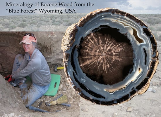
Mineralogy of Eocene Fossil Wood from the “Blue Forest” Locality, Southwestern Wyoming, United States


Abstract
1. Introduction
2. Geologic Setting
3. Paleontology
4. Methods
5. Previous Studies
6. Observations
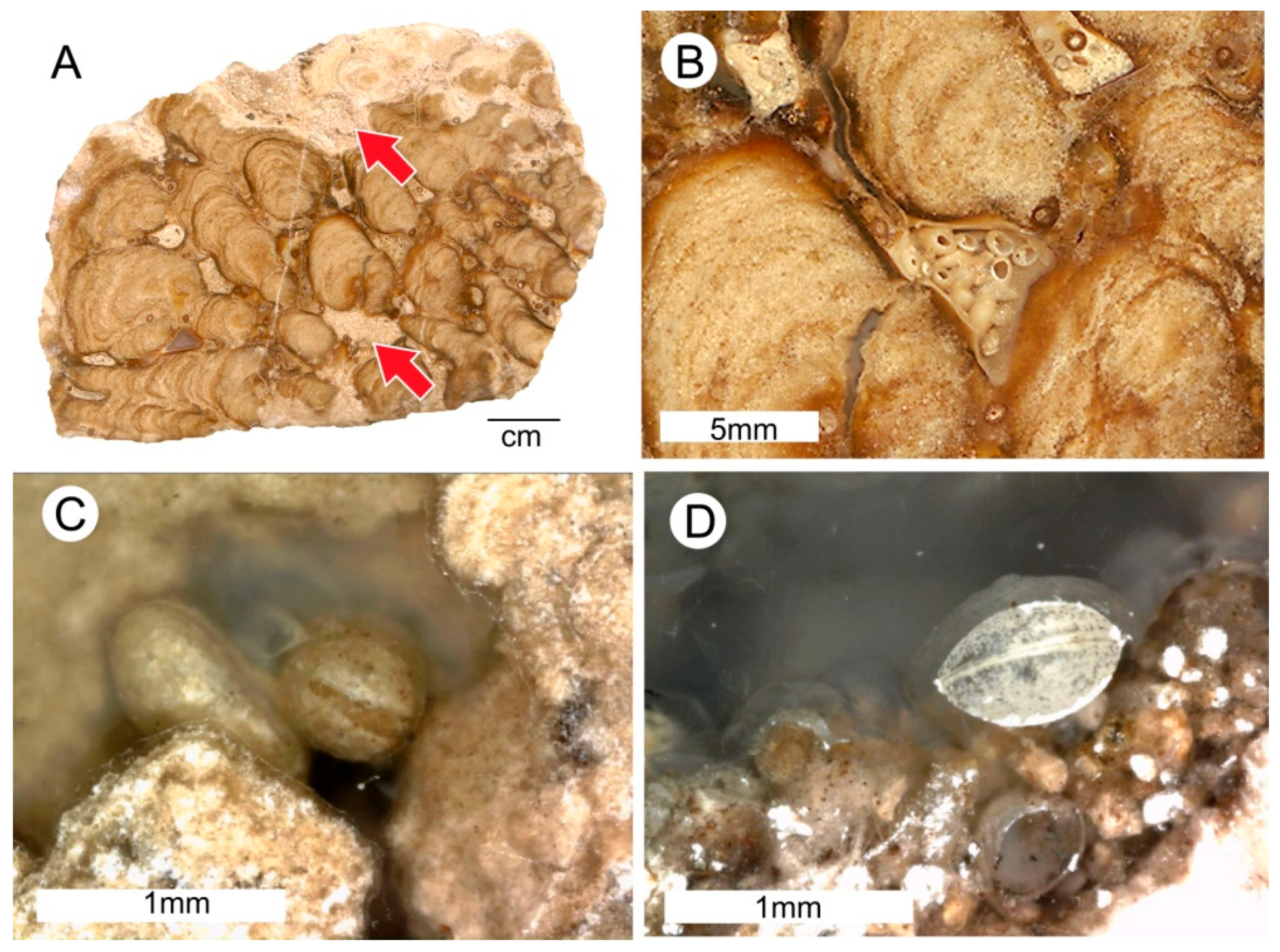
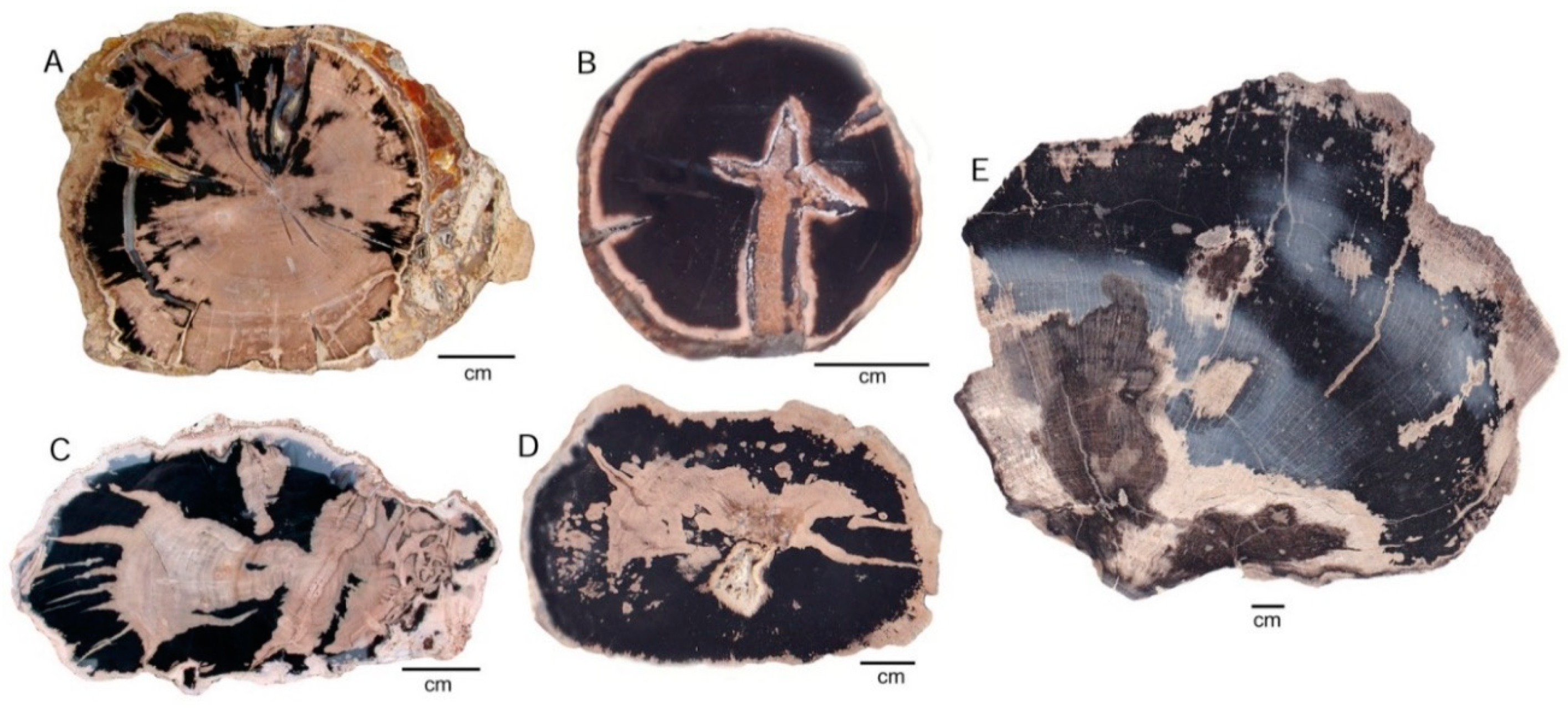
6.1. Taphonomy
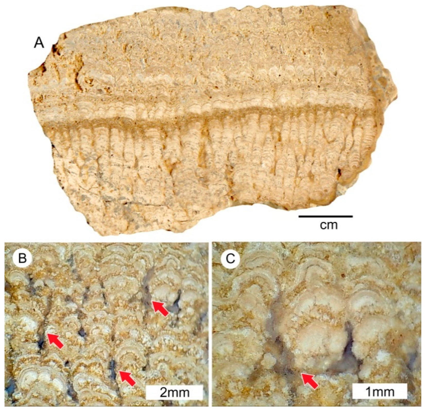
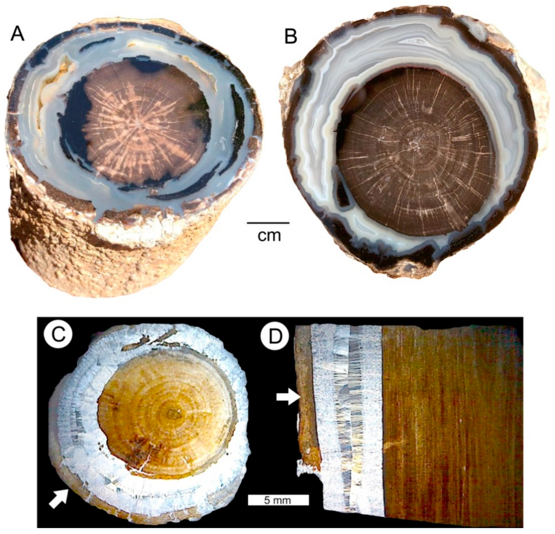
6.2. Stromatolitic Coatings
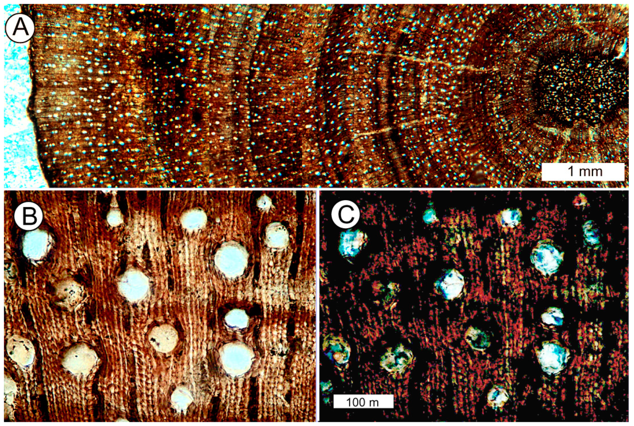
6.3. Wood Silicification
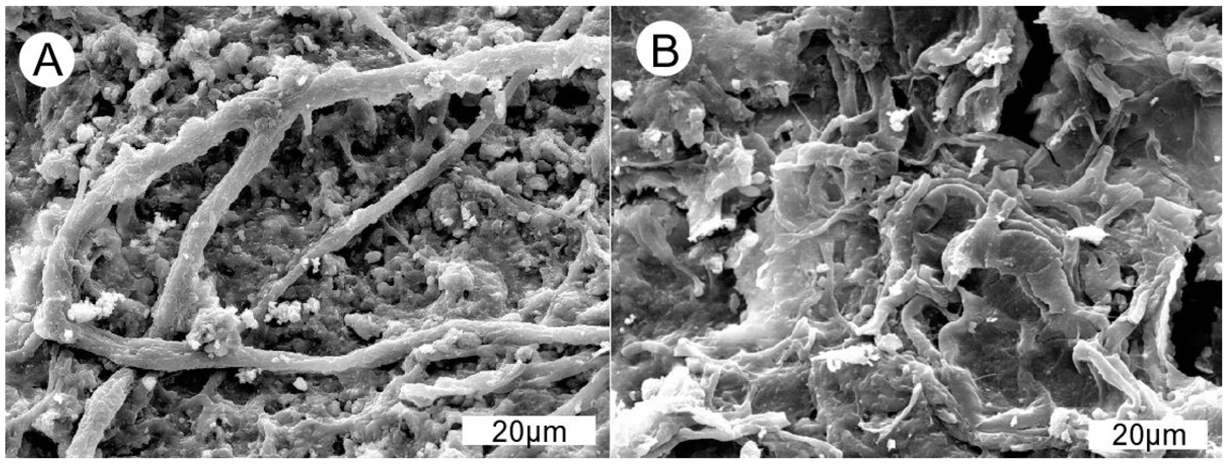
6.4. Relict Organic Matter
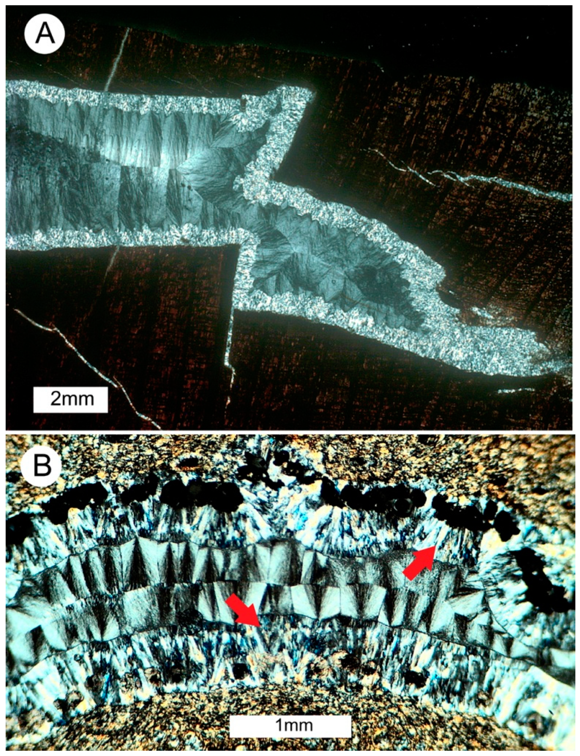
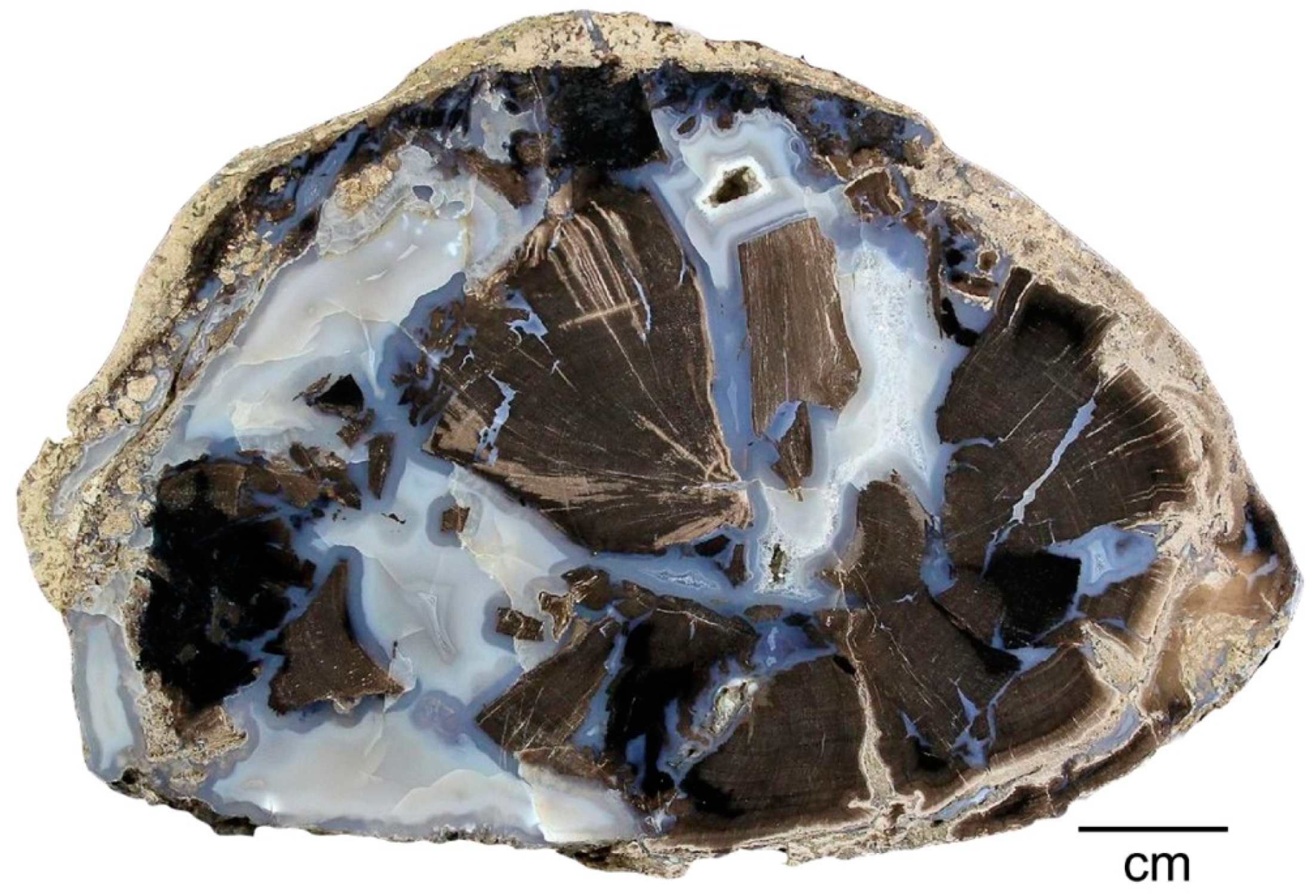
6.5. Secondary Chalcedony
6.6. Crystalline Quartz
6.7. Crystalline Calcite
6.8. Trace Element Geochemistry
7. Discussion
7.1. Fossilization Processes
7.2. Sequential Mineralization
7.2.1. Step 1: Wood Silicification
7.2.2. Step 2: Secondary Chalcedony
7.2.3. Step 3: Calcite Crystallization
7.2.4. Step 4: Quartz Crystallization
7.3. Evidence from Trace Elements
8. Conclusions
Author Contributions
Funding
Acknowledgments
Conflicts of Interest
References
- Grande, L. The Lost World of Fossil Lake—Snapshots from Deep Time; University of Chicago Press: Chicago, IL, USA, 2013; 425p. [Google Scholar]
- Murphy, P.C.; Evanoff, E. Paleontology and stratigraphy of the middle Eocene Bridger Formation, southern Green River basin, Wyoming. Brigh. Young Univ. Geol. Stud. 2011, 49, 83–109. [Google Scholar]
- Roehler, H.W. Eocene Climates, Depositional Environments, and Greography, Greater Green River Basin, Wyoming, Utah, and Colorado; U.S. Geological Survey Professional Paper 1056F; U.S. Government Printing Office: Washington, DC, USA, 1993; 74p.
- Sutherland, W.M.; Luhr, S.C. Preliminary Bedrock Geologic Map of the Farson 30’ × 60’ Quadrangle, Sweetwater, Sublette and Freemont Counties, Wyoming; Open File Report 11-6, scale 1:100,000; Wyoming State Geological Survey: Lander, WY, USA, 2011.
- Brand, L.R. Lacustrine deposition in the Bridger Formation: Lake Gosiute extended. Mt. Geol. 2007, 44, 69–77. [Google Scholar]
- Zeller, H.D.; Stephens, E.V. Geology of the Oregon Buttes Area, Sweetwater, Sublette and Freemont Counties, Southwest Wyoming; U.S. Geological Survey Bulletin 1256; U.S. Government Printing Office: Washington, DC, USA, 1969.
- Grande, L. Paleontology of the Green River Formation, with a Review of the Fish Fauna; Bulletin 63; Geological Survey of Wyoming: Laramie, WY, USA, 1984; 333p.
- Bradley, W.H. Limnology and the Eocene lakes of the Rocky Mountain region. Geol. Soc. Am. Bull. 1948, 59, 635–648. [Google Scholar] [CrossRef]
- Knowlton, F.H. Revision of the Flora of the Green River Formation, with Descriptions of New Species; U.S. Geological Survey Professional Paper 131-F; U.S. Government Printing Office: Washington, DC, USA, 1923; pp. 133–182.
- Berry, E.W. A Flora of Green River Age in the Wind River Basin of Wyoming; U.S. Geological Survey Professional Paper 165-B; U.S. Government Printing Office: Washington, DC, USA, 1930; pp. 55–81.
- Brown, R.W. Additions to the Flora of the Green River Formation; U.S. Geological Survey Professional Paper 1540-J; U.S. Government Printing Office: Washington, DC, USA, 1929; pp. 279–292.
- Cockerell, T.D.A. Plant and insect fossils from the Green River Eocene of Colorado. U.S. Natl. Mus. Proc. 1925, 66, 1–13. [Google Scholar] [CrossRef]
- Brown, R.W. The Recognizable Species of the Green River Flora; U.S. Geological Survey Professional Paper 185-C; U.S. Government Printing Office: Washington, DC, USA, 1934; pp. 45–77.
- Tschudy, R.H. Plant and Miscellaneous Microfossils from the Parachute Creek Member of the Green River Formation; U.S. Geological Survey Open-File Report 65; U.S. Government Printing Office: Washington, DC, USA, 1965; 2p.
- Newman, K.R. Palynomorph zones in early Tertiary formations of the Piceance Creel and Uinta Basins, Colorado and Utah. In Guidebook to the Energy Resources of the Piceance Creek Basin, Colorado; Rocky Mountain Association of Geologists Guidebook, 25th Annual Field Conference; Murray, D.K., Ed.; Rocky Mountain Association of Geologists: Denver, CO, USA, 1974; pp. 47–55. [Google Scholar]
- MacGinite, H.D. The Eocene Green River flora of northwestern Colorado and southeastern Utah. Calif. Univ. Publ. Geol. Sci. 1969, 83, 203. [Google Scholar]
- Johnson, K.R.; Plumb, C. Common plant fossils from the Green River Formation at Douglas Pass, Colorado. In The Green River Formation in Piceance Creek and Eastern Uinta Basins Field Trip; Grand Junction Geological Society: Grand Junction, CO, USA, 1975; pp. 121–130. [Google Scholar]
- Moussa, M.T. Fossil tracks from the Green River Formation (Eocene) near Soldier Summit, Utah. J. Paleontol. 1968, 42, 1433–1438. [Google Scholar]
- Eugster, H.P.; Surdam, R.C. Depositional environment of the Green River Formation of Wyoming: A preliminary report. Geol. Soc. Am. Bull. 1973, 84, 1115–1120. [Google Scholar] [CrossRef]
- Surdam, R.C.; Wolfbauer, A.A. The Green River Formation, Wyoming: A playa-lake complex. Geol. Soc. Am. Bull. 1973, 86, 335–345. [Google Scholar] [CrossRef]
- Bradley, W.H.; Eugster, H.P. Geochemistry and Paleolimnology of the Trona Deposits and Associated Authigenic Minerals of the Green River Formation of Wyoming; U.S. Geological Survey Professional Paper 496-B; U.S. Government Printing Office: Washington, DC, USA, 1969; pp. 1–71.
- Surdam, R.C.; Stanley, K.O. Lacustrine sedimentation during the culminating phase of Eocene Lake Gosiute, Wyoming (Green River Formation). Geol. Soc. Am. Bull. 1979, 90, 93–110. [Google Scholar] [CrossRef]
- Bucheim, H.P.; Brand, L.R.; Goodwin, H.T. Lacustrine to fluvial deposition in the Eocene Bridger Formation. Palaeogeogr. Palaeoclimatol. Palaeoecol. 2000, 162, 191–209. [Google Scholar] [CrossRef]
- Pietras, J.T.; Carroll, A.R.; Rhodes, M.K. Lake basin response to tectonic drainage diversion: Eocene Green River Formation, Wyoming. J. Paleontol. 2003, 30, 115–125. [Google Scholar]
- Hiza, M. Geologic history of the Absaroka volcanic province. Yellowstone Sci. 1998, 6, 2–7. [Google Scholar]
- Fritz, W.J. Reinterpretation of the depositional environment of the Yellowstone “fossil forests”. Geology 1980, 8, 309–313. [Google Scholar] [CrossRef]
- Leopold, E.B.; MacGinitie, H.D. Development and affinities of Tertiary floras in the Rocky Mountains. In Floristics and Paleofloristics of Asia and Eastern North America; Graham, A., Ed.; Elsevier: Amsterdam, The Netherlands, 1972; pp. 147–200. [Google Scholar]
- Leopold, E.B.; Denton, M.F. Comparative age of grassland and steppe east and west of the northern Rocky Mountains. Ann. Mo. Bot. Gard. 1987, 74, 841–867. [Google Scholar] [CrossRef]
- Mustoe, G.E. Density and loss on ignition as indicators of the fossilization of silicified wood. IAWA J. 2016, 37, 98–111. [Google Scholar] [CrossRef]
- ASTM. ASTM Method D-437396, Standard Test for Calcium Carbonate of Soils, Annual Book of ASTM Standards 2000, v. 408; ASTM: West Conshocker, PA, USA, 2000; pp. 573–575. [Google Scholar]
- Kruse, H.O. Some Eocene dicotyledonous woods from Eden Valley, Wyoming. Ohio J. Sci. 1954, 54, 243–268. [Google Scholar]
- Tidwell, W.D.; Simper, W.D.; Medlyn, D.A. A Palmoxylon from the Green River Formation (Eocene) of Eden Valley, Wyoming. Botanique 1971, 2, 93–102. [Google Scholar]
- Tidwell, W.D.; Medlyn, D.A.; Thayn, G.F. Three new species of Palmoxylon from the Eocene Green River Formation, Wyoming. Great Basin Nat. 1973, 33, 61–76. [Google Scholar]
- Boonchai, N.; Manchester, S.R. Systematic affinities of early Eocene petrified woods from Big Sandy Reservoir, southwestern Wyoming. Int. J. Plant Sci. 2012, 173, 209–227. [Google Scholar] [CrossRef]
- Boonchai, N. Systematic Affinities and Paleoenvironment of Eocene Petrified Woods from Southwestern Wyoming, USA. Ph.D. Thesis, Jilin University, Changchun, China, 2012; 224p. [Google Scholar]
- Nichols, D.J. Palynology of the Vermillion Creek coal bed and associated strata. U.S. Geol. Surv. Prof. Paper 1987, 1314D, 47–73. [Google Scholar]
- Boonchai, N.; Manchester, S.R.; Wheeler, E.A. Welkoetoxylon multiseriatum: Fossil moraceous wood from the Eocene Green River Formation, Wyoming, USA. IAWA J. 2015, 36, 158–166. [Google Scholar] [CrossRef]
- Bradley, W.H. Algae Reefs and Oolites of the Green River Formation; U.S. Geological Survey Professional Paper 154-G; U.S. Government Printing Office: Washington, DC, USA, 1929; pp. 203–223.
- Reis, M.O. Chlorellopsis coloniata Reis, Kalkgalen and Seesinterkalk aus dem rheinpalzischen Tertiar. Geognostische Jahresh. 1923, 36, 107–109. [Google Scholar]
- Saminpanya, S.; Sutherland, F.L. Silica phase-transformations during diagenesis within petrified woods found in fluvial deposits from Thailand-Myanmar. Sediment. Geol. 2013, 290, 15–26. [Google Scholar] [CrossRef]
- Mustoe, G.E. Late Tertiary petrified wood from Nevada, USA: Evidence of multiple silicification pathways. Geosciences 2015, 5, 286–309. [Google Scholar] [CrossRef]
- Viney, M.; Deitrich, D.; Mustoe, G.; Link, P.; Lampke, T.; Götze, J.; Röβer, R. Multi-stage silicification of Pliocene wood: Re-examination of an 1895 discovery from Idaho, USA. Geosciences 2016, 6, 21. [Google Scholar] [CrossRef]
- Mustoe, G.E.; Viney, M. Mineralogy of Paleocene petrified wood from Cherokee Ranch Fossil Forest, central Colorado, USA. Geosciences 2017, 239, 23. [Google Scholar] [CrossRef]
- Mustoe, G.E. Wood petrifaction: A new view of permineralization and replacement. Geosciences 2017, 7, 119. [Google Scholar] [CrossRef]
- Bradley, W.H. Fresh water algae from the Green River Formation of Colorado. Bull. Torrey Bot. Club 1929, 37, 232–233. [Google Scholar] [CrossRef]
- Brunskill, G.J. Fayetteville Green Lake, New York, II. Precipitation and sedimentation of calcite in a meromictic lake with laminated sediments. Limnol. Oceanogr. 1969, 14, 830–847. [Google Scholar] [CrossRef]
- Eggleston, J.R.; Dean, W.E. Freshwater stromatolitic bioherms in Green Lake, New York. In Developments in Sedimentology, v. 20; Walter, M.R., Ed.; Elsevier: New York, NY, USA, 1976; pp. 479–488. [Google Scholar]
- Koban, C.G.; Schweigert, G. Microbial origin of travertine fabrics—Two examples from southern Germany (Pleistocene Stuttgart travertines and Miocene Reidöschingen travertine). Facies 1993, 29, 251–264. [Google Scholar] [CrossRef]
- Francis, J.E. The Fossil Forests of the Basal Purebeck Formation (Upper Jurassic) of Dorset, Southern England. Ph.D. Thesis, University of Southampton, Southhampton, UK, October 1992; 295p. [Google Scholar]
- Ford, T.D.; Pedley, H.M. A review of tufa and travertine deposits of the world. Earth Sci. Rev. 1996, 41, 117–175. [Google Scholar] [CrossRef]
- Surdam, R.C.; Wray, J.L. Lacustrine stromatolites, Eocene Green River Formation, Wyoming. In Developments in Sedimentology, v. 20; Walter, M.R., Ed.; Elsevier: New York, NY, USA, 1976; pp. 535–541. [Google Scholar]
- Utami, W.S.; Herdianita, N.R.; Atmaja, R.W. The effect of temperature and pH on the formation of silica scaling of Dieng Geothermal Field, Central Java, Indonesia. In Proceedings of the Thirty-Ninth Workshop on Geothermal Reservoir Engineering Stanford University, Stanford, CA, USA, 24–26 February 2014. [Google Scholar]
- Nicholson, K. Geothermal Fluids; Chemistry and Exploration Techniques; Springer-Verlag: Berlin, Germany, 1968; 263p. [Google Scholar]
- Mustoe, G.E. Mineralogy of non-silicified fossil wood. Geosciences 2018, 8, 85. [Google Scholar] [CrossRef]
- Polgári, M.; Bajnóczi, B.; Gótzi, J.; Vigh, T. Cathodoluminescence behavior of Mn-rich carbonates. Goldschmidt 2007 conference, August 20–24, Cologne, Germany. Geochim. Cosmochim. Acta 2007, 71, A80. [Google Scholar]
- Mustoe, G.E.; Acosta, M. Origin of petrified wood color. Geosciences 2016, 6, 25. [Google Scholar] [CrossRef]


| LOCATION | FAMILY | REFERENCE |
|---|---|---|
| HAY’S RANCH: SE of Big Sandy Reservoir | ||
| Myrica scalariforme | Myricaceae | [31] |
| Talauma multiperforata | Lauracea | [31] |
| Forchammerioxylon scleroticum | Capparidaceae | [31] |
| Amridoxylon ordinatum | Rutaceae | [31] |
| Fagara monophlloides | Rutaceae | [31] |
| Fagara biseriata | Rutaceae | [31] |
| Suriana inordinata | Simaroubaceae | [31] |
| Heveoxylon microsporosum | Euphoribiaceae | [31] |
| Schinoxylon actinoporosum | Anacardiaceae | [31] |
| Edenoxylon paviareolatum | Anacardiaceae | [31] |
| Aspidospermoxylon uniseriatum | Apocynaceae | [31] |
| BIG SANDY RESERVOIR UF327: NE of Big Sandy Reservoir | ||
| Palmoxylon macginitiei | Arecaceae (Palmae) | [32] |
| Palmoxylon contortum | Arecaceae (Palmae) | [33] |
| Palmoxylon colei | Arecaceae (Palmae) | [33] |
| Palmoxylon edenense | Arecaceae (Palmae) | [33] |
| Edenoxylon paviareolatum | Anacardiaceae | [34,35] |
| Laurinoxylon stickai | Lauraceae | [34,35] |
| Wilsonoxylon edenense | Cancellaceae | [34,35] |
| PARNELL DRAW: 42 km east of Farson, WY | ||
| Cupressinoxylon sp. | Cupressaceae | [35] |
| Pinus sp. | Pinaceae | [35] |
| Palmoxylon sp. | Arecaceae (Palmae) | [35] |
| Edenoxylon | Anacardiaceae | [35] |
| Cf. Laurinoxylon stickai | Lauraceae | [35] |
| Cf. Mastixia sp. | Cornaceae | [35] |
| Platanoxylon sp. | Platacaneae | [35] |
| Dicotyloxylon spp. (7 unknown taxa) | Unknown | [35] |
| Welkotopoxylon multiseriata | Moraceae | [37] |
| Sample | Type | Calculated Density | LOI 450 °C | Assumed Original Wood Density | % of Original Organic Matter |
|---|---|---|---|---|---|
| BF1 | Dicot wood | 2.42 | 2.94% | 0.60 | 7.1% |
| BF3 | Dicot wood | 2.52 | 4.40% | 0.60 | 11.1% |
| BFTS | Dicot wood | 2.39 | 6.19% | 0.60 | 14.8% |
| BF2018 | Dicot Wood | 2.31 | 5.96% | 0.60 | 13.8% |
| H3W2 | Dicot wood | 2.52 | 0.97% | 0.60 | 2.4% |
| WP | Palmoxylon | 2.50 | 1.39% | 0.56 | 6.2% |
| Sample | Ti | V | Cr | Mn | Fe | Co | Ni | Cu | U |
|---|---|---|---|---|---|---|---|---|---|
| calcite | 4 | 2 | 1 | 7545 | 458 | 3 | 2 | 4 | 0 |
| chalcedony | 47 | 28 | 34 | 186 | 1087 | 3 | 6 | 54 | 1 |
| wood #1 | 17 | 768 | 22 | 105 | 461 | 1 | 1 | 12 | 3 |
| wood #2 | 133 | 699 | 22 | 171 | 675 | 2 | 1 | 25 | 4 |
© 2019 by the authors. Licensee MDPI, Basel, Switzerland. This article is an open access article distributed under the terms and conditions of the Creative Commons Attribution (CC BY) license (http://creativecommons.org/licenses/by/4.0/).
Share and Cite
Mustoe, G.E.; Viney, M.; Mills, J. Mineralogy of Eocene Fossil Wood from the “Blue Forest” Locality, Southwestern Wyoming, United States. Geosciences 2019, 9, 35. https://doi.org/10.3390/geosciences9010035
Mustoe GE, Viney M, Mills J. Mineralogy of Eocene Fossil Wood from the “Blue Forest” Locality, Southwestern Wyoming, United States. Geosciences. 2019; 9(1):35. https://doi.org/10.3390/geosciences9010035
Chicago/Turabian StyleMustoe, George E., Mike Viney, and Jim Mills. 2019. "Mineralogy of Eocene Fossil Wood from the “Blue Forest” Locality, Southwestern Wyoming, United States" Geosciences 9, no. 1: 35. https://doi.org/10.3390/geosciences9010035
APA StyleMustoe, G. E., Viney, M., & Mills, J. (2019). Mineralogy of Eocene Fossil Wood from the “Blue Forest” Locality, Southwestern Wyoming, United States. Geosciences, 9(1), 35. https://doi.org/10.3390/geosciences9010035