Article

Grape Seed Proanthocyanidin Extract Improves Growth Performance and Protects Against Hydrogen Peroxide-Induced Oxidative Stress to the Liver and Intestine in Weaned Hyla Rabbits

Key Laboratory of Efficient Utilization of Non-Grain Feed Resources (Co-Construction by Ministry and Province), Ministry of Agriculture and Rural Affairs, Shandong Provincial Key Laboratory of Animal Nutrition and Efficient Feeding, Department of Animal Science, Shandong Agricultural University, Tai’an 271017, China
*
Authors to whom correspondence should be addressed.
Animals 2025, 15(3), 327; https://doi.org/10.3390/ani15030327
Submission received: 22 December 2024 / Revised: 20 January 2025 / Accepted: 22 January 2025 / Published: 24 January 2025
(This article belongs to the Special Issue Plant Extracts as Feed Additives in Animal Nutrition and Health)

Simple Summary

Weaning is one of the most stressful events in a rabbit’s life, which could induce oxidative stress, leading to growth retardation. The liver and intestine are the body’s most important metabolic and digestive organs, respectively. However, these organs are the most sensitive to oxidative stress and highly susceptible to injury when oxidative stress occurs. Because using antibiotics as growth promoters is prohibited, it is essential to identify new antioxidants that can promote growth performance and protect rabbits against the damage caused by weaning-induced oxidative stress. Proanthocyanidins are naturally polyphenolic compounds with strong free radical scavenging ability and antioxidant properties, and they are especially abundant in grape seeds. However, there is limited research on the effects of grape seed proanthocyanidin extract (GSPE) on the growth performance of weaned meat rabbits and their role in alleviating oxidative stress in the liver and intestine. Our findings showed that supplementation with 400 mg/kg GSPE could improve growth performance and alleviate hydrogen peroxide-induced adverse effects on the liver and small intestine through enhancing the antioxidative capacity in weaned Hyla rabbits, providing support for the application of GSPE in rabbit farming.

Abstract

Three experiments were conducted to investigate the effects of grape seed proanthocyanidin extract (GSPE) on the growth performance of weaned Hyla rabbits and explore its protective effects against oxidative stress in the liver and intestine by establishing a hydrogen peroxide (H2O2)-induced oxidative stress model. In Exp.1, ninety-six weaned rabbits were used to evaluate the effects of dietary GSPE level on growth performance, and the results showed that a 400 mg/kg GSPE addition increased the feed conversion ratio and liver coefficient and promoted cholesterol metabolism. Exp.2 was conducted to explore the H2O2 concentration required to establish an oxidative stress model, indicating that the model could be established by an intraperitoneal injection of 10% H2O2 at a dosage of 1 mL/kg body weight. In Exp.3, seventy-two weaned rabbits were used to investigate the protective effects against H2O2-induced oxidative stress in the liver and intestine. Our findings showed that 400 mg/kg GSPE supplementation could alleviate the adverse effects of H2O2 injection on the antioxidant capacity in the liver and intestine as well as liver morphology. Therefore, an addition of 400 mg/kg GSPE could improve growth performance and alleviate H2O2-induced adverse effects on the liver and small intestine by enhancing the antioxidative capacity in weaned Hyla rabbits.

1. Introduction

Reactive oxygen species (ROS), including hydrogen peroxide (H2O2) and superoxide, are continuously produced from oxygen (O2) during aerobic metabolism and various pathogenic processes [1]. Oxidative stress arises when the balance between ROS production and the body’s antioxidant defense mechanisms is disrupted [2]. This imbalance can trigger a cascade of reactions that damage lipids, proteins, and/or DNA, potentially leading to the development of numerous diseases [3]. The liver and intestine are the body’s most important metabolic and digestive organs, respectively. Meanwhile, they are especially vulnerable to oxidative stress due to the high number of mitochondria in their cells, which are the primary sites of ROS production [4,5,6]. Weaned rabbits become extremely vulnerable to oxidative stress due to the alteration of feed and environment [7]. Numerous studies have shown that oxidative stress can impair liver and intestinal morphology and function, leading to increased hepatic cell apoptosis and intestinal barrier permeability and ultimately resulting in growth retardation in rabbits [8,9]. Because using antibiotics as growth promoters is prohibited, it is essential to identify new antioxidants that protect rabbits against the damage caused by ROS.
Proanthocyanidins are naturally polyphenolic compounds with strong free radical scavenging ability and antioxidant properties. They have gained increasing interest due to their non-toxicity, bioavailability, and ability to provide multi-organ protection against toxicity induced by drugs and chemicals [10]. Grape seeds are the most abundant source of proanthocyanidins. Fawzia et al. [11] indicated that dietary 300 mg/kg grape seed extract containing 41.6% proanthocyanidins increased body weight (BW) and feed conversion ratio (FCR), enhanced plasma antioxidant enzyme activities, and reduced plasma malonaldehyde (MDA) concentration in rabbits. Grape seed proanthocyanidin extract (GSPE) was also found to play a beneficial role in the enhancement of liver function of rabbits in a heat stress-induced oxidative stress model [12]. Similarly, 250 and 500 mg/kg GSPE relieved Aflatoxin B1 (AFB1) exposure-induced adverse effects on growth performance and liver histopathological lesions via enhancing serum and hepatic antioxidant capacity [13]. A previous study in weaned piglets showed that a dietary addition of 50–150 mg/kg GSPE consisting of 79.28% oligomeric procyanidins enhanced antioxidant enzyme capacities and antioxidant-related gene expressions and decreased MDA concentration in the intestine [14]. Zheng et al. [15] found that 30–120 mg/kg GSPE (containing 86.81% proanthocyanidin oligomers, 2.41% epicatechin, 1.52% catechin, and 0.98% proanthocyanidin B2) decreased the feed-to-gain ratio (F/G) and elevated serum total superoxide dismutase (T-SOD) and glutathione peroxidase (GSH-Px) activities in growing pigs. However, there is limited research on the effects of GSPE on the growth performance of weaned meat rabbits and their role in alleviating oxidative stress in the liver and intestine.
H2O2 injection is a widely used procedure to establish the oxidative stress model in animals [16,17]. Hence, three experiments were conducted in this study to investigate the effects of GSPE on the growth performance of weaned meat rabbits and explore its role in alleviating oxidative stress in the liver and intestine by establishing an H2O2-induced oxidative stress model, providing support for the application of GSPE in rabbit farming.

2. Materials and Methods

2.1. Animals and Treatments

In Exp.1, a total of 96 weaned Hyla rabbits (35 d of age) were randomly assigned to four treatment groups with 6 replicates per treatment and 4 rabbits per replicate. The rabbits in the four groups were fed a basal diet supplemented with 0, 200, 400, or 800 mg/kg GSPE, denoted as G0, G200, G400, and G800, respectively. The individual fasting BWs at the start and end days of the trial, the daily feed intake per cage, and the number of deaths were recorded. Finally, the average daily gain (ADG), average daily feed intake (ADFI), F/G, and survival rate were calculated. The trial lasted for 35 days.
In Exp.2, to explore the concentration of H2O2 required to establish the oxidative stress model, sixty-four Hyla rabbits of 56 d age were randomly allocated to one of four treatment groups (8 replicates each group and 2 rabbits per group): (1) Blank, rabbits not injected with sterile saline (SS) or H2O2; (2) SS, rabbits injected with 0.9% SS intraperitoneally; (3) 5% H2O2, rabbits injected with 5% H2O2 intraperitoneally; (4) 10% H2O2, rabbits injected with 10% H2O2 intraperitoneally. The trial lasted for 13 days, consisting of a 6-day adaptive period and a 7-day formal experiment period. The SS or H2O2 was administered once on the first day of the formal experiment. The intraperitoneal injection dosage of SS or H2O2 in this study was 1 mL/kg BW. The injection dosage, frequency, and duration of the trial referred to the results in Yin et al. [17]. All the rabbits were fed the same basal diet during the formal experiment period, and the ADF, ADFI, F/G, and survival rate were calculated according to the method in Exp.1.
In Exp.3, seventy-two weaned Hyla rabbits of 35 d age were randomly divided into three treatment groups (6 replicates with 4 rabbits each) as follows: control group, H2O2 group, and H2O2+GSPE group. The rabbits in the control and H2O2 groups were fed the basal diet, whereas the rabbits in the H2O2+GSPE group were fed the basal diet supplemented with 400 mg/kg GSPE according to the results of Exp1. The trial lasted for 35 days. On day 29 of the trial, the rabbits in the H2O2 group and H2O2+GSPE group were injected with 10% H2O2 (1 mL/kg BW) intraperitoneally according to the results of Exp.2, while the rabbits in the control group were administered with an equivalent volume of 0.9% SS.
All the rabbits used in this study were purchased from Tai’an Xinglong Rabbit Farming (Tai’an, China). The basal diet (Table 1) used in the trials was formulated in line with the nutrient requirements recommended by the Nutrition Requirements of Meat Rabbits (NY/T 4049-2021, Ministry of Agriculture of China), and the GSPE (CAS 4852-22-6, purity ≥ 95%) was purchased from Solarbio Science & Technology Co., Ltd. (Beijing, China). All rabbits were housed in cages and were provided with ad libitum access to fresh water and feed during the trials. The lighting was controlled under a 12 h light and 12 h dark condition with room temperature between 20 and 23 °C.

2.2. Sampling Procedure

In Exp.1, at the end of the trial, eight rabbits from each group, with BWs closest to the average, were selected for collecting blood by heart punctures [8]. The serum was obtained after centrifugation for 15 min at 3000× g and immediately stored at −20 °C for further analysis. Subsequently, the rabbits were slaughtered after being euthanized. The full eviscerated weight and half eviscerated weight were recorded to calculate the full eviscerated weight ratio and half eviscerated weight ratio, respectively, as described by Kong et al. [18]. Additionally, the thymus, spleen, liver, and kidney were weighed to calculate the ratios of individual organs over BW accordingly.
In Exp.2, seven days after H2O2 injection (day 7 of the formal experiment period), one rabbit from each replicate with similar weight to the replicate average was selected for blood sampling to obtain the serum. The procedure was the same as that in Exp.1.
In Exp.3, on day 35 of the trial (seven days after H2O2 injection), eight rabbits per group with a similar BW to the average group weight were chosen and slaughtered immediately after deep anesthesia. After evisceration, the small intestine was dissected according to previous study [19]. The segments (2~3 cm) of duodenum, jejunum, and ileum were immediately cut and rinsed by sterile saline. One part of the intestinal tissues was fixed in 4% paraformaldehyde for morphological examination, and another part was stored at −80 °C after being snap-frozen in liquid nitrogen. Additionally, liver samples weighing about 5 g were promptly obtained from the central region of the hepatic lobule. A portion of these samples was rapidly frozen using liquid nitrogen and subsequently stored at −80 °C for future analysis, while the remaining portion was fixed in a 4% paraformaldehyde solution at room temperature.

2.3. Determination of Serum Biochemical Indicators and MDA

Serum total protein (TP), urea, albumin (ALB), triglycerides (TG), total cholesterol (TCHO), calcium (Ca), phosphorus (P), alkaline phosphatase (ALP), and MDA were determined using the commercial kits purchased from Nanjing Jiancheng Bioengineering Institute (Nanjing, China).

2.4. Measurements of Intestinal and Liver Morphology

After 24 h fixation in 4% paraformaldehyde solution, intestinal and liver tissue samples were dehydrated through graded levels of ethyl alcohol and embedded in liquid paraffin according to conventional paraffin-embedding protocol [20]. Then, paraffin wax embedded tissues were cut into 5 µm sections using a semi-automatic microtome (Leica Co., Wetzlar, Germany) and stained with hematoxylin and eosin (H&E). Photomicrographs were obtained using an Olympus BX51 microscope (Tokyo, Japan). Intestinal villus height (VH) and crypt depth (CD) were measured with JD801 morphologic image analysis software (Nanjing, China) as previously described [21], and the ratio of VH to CD (V/C) was also calculated.

2.5. Determination of Antioxidant Indexes in the Small Intestine and Liver

The intestinal and liver tissue samples were homogenized in ice-cold saline solution, and the supernatants were obtained after being centrifugated at 2500× g at 4 °C for 10 min. The levels of MDA, SOD, catalase (CAT), and glutathione (GSH) were determined using the commercial kits (Nanjing Jiancheng Bioengineering Institute, Nanjing, China) as described in detail in Chen et al. [19].

2.6. Statistical Analysis

The average cage data were used to assess the impacts on growth performance, and the individual rabbit was considered the experimental unit for the other variables using one-way ANOVA of SAS 9.4 (Institute Inc., Cary, NC, USA). Variations among the group means were compared using Duncan’s multiple comparisons. Results are expressed as means with the root mean square error (RMSE). A level of p < 0.05 was considered statistically significant.

3. Results

3.1. Effects of Dietary Supplementation with GSPE on the Growth Performance of Weaned Rabbits (Exp.1)

As shown in Table 2, the rabbits in the G400 group had a significantly lower F/G compared to the rabbits in the G0 group (p < 0.05). There were no significant differences in final BW (FBW), ADFI, and ADG among the four groups (p > 0.05). The survival rates were 79.8%, 79.8%, 91.7%, and 83.4%, respectively, for the G0, G200, G400, and G800 groups.

3.2. Effects of Dietary Supplementation with GSPE on the Slaughter Performance of Weaned Rabbits (Exp.1)

The data of the slaughter performance of meat rabbits are shown in Table 3. No significant differences were observed in the full eviscerated weight, half eviscerated weight, full eviscerated weight ratio, and half eviscerated weight ratio of rabbits among all the groups (p > 0.05).

3.3. Effects of Dietary Supplementation with GSPE on the Organ Coefficients of Weaned Rabbits (Exp.1)

The organ coefficients of meat rabbits are shown in Table 4. The organ coefficient of the liver was significantly higher in the G400 group than in the G0 group (p < 0.05). There were no significant differences among the four treatments concerning the ratios of thymus, spleen, and kidney weights over BW (p > 0.05).

3.4. Effects of Dietary Supplementation with GSPE on the Serum Biochemical Indicators of Weaned Rabbits (Exp.1)

The serum biochemical indicators of meat rabbits are shown in Table 5. The serum TCHO concentration in the G400 group was significantly lower than that in the G0 group (p < 0.05). There were no significant differences in the serum concentrations of TP, UREA, ALB, TG, Ca, P, and ALP among the four groups (p > 0.05).

3.5. Effects of H2O2 Injection on the Growth Performance of Hyla Rabbits (Exp.2)

As shown in Table 6, SS injection had no significant effects on the growth performance of rabbits (p < 0.05), but 10% H2O2 injection significantly decreased the FBW, ADFI, and ADG and increased F/G ratio compared to the other three groups (p < 0.05). Additionally, the 5% H2O2 group had significantly lower ADG and a higher F/G ratio than the Blank and SS groups (p < 0.05). No rabbits died in the four treatments during the trial.

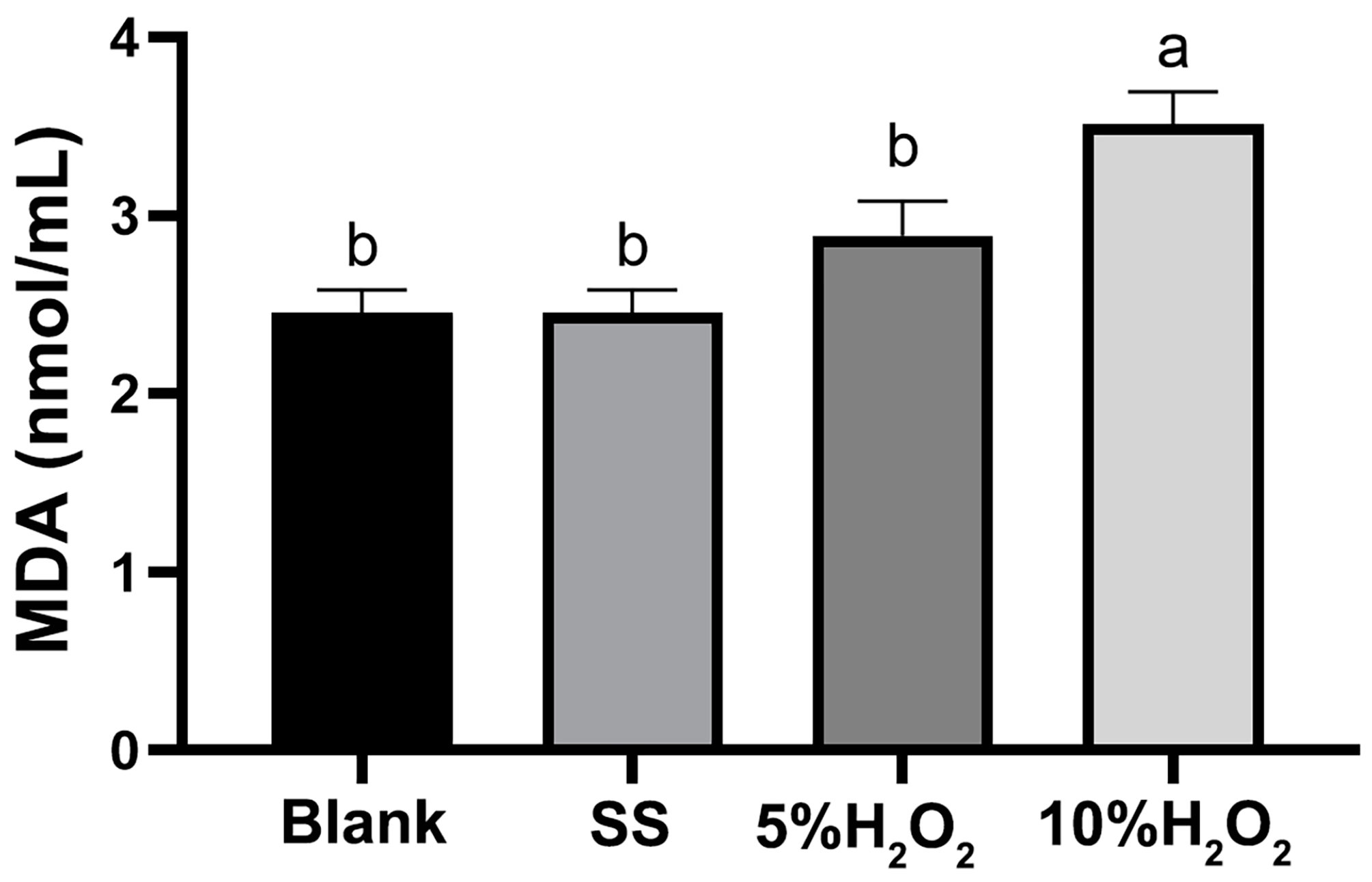
3.6. Effects of H2O2 Injection on the Serum MDA Concentration of Hyla Rabbits (Exp.2)

Figure 1 showed that 10% H2O2 injection had the highest serum MDA concentration, which was significantly higher than the other three groups (p < 0.05).

3.7. Effects of Dietary Supplementation with GSPE on Liver Morphology of Weaned Rabbits Challenged with H2O2 (Exp.3)

As shown in Figure 2, the liver cells in the control group were structurally intact and arranged in an orderly manner to form liver cords, with a clear nucleus located centrally in the cell. Meanwhile, H2O2 injection resulted in liver cell swelling, vacuolization, and unclear cell spaces and liver cords, with the cell nucleus shifting or even disappearing. However, GSPE supplementation alleviated the adverse changes in liver morphology caused by the intraperitoneal injection of H2O2.

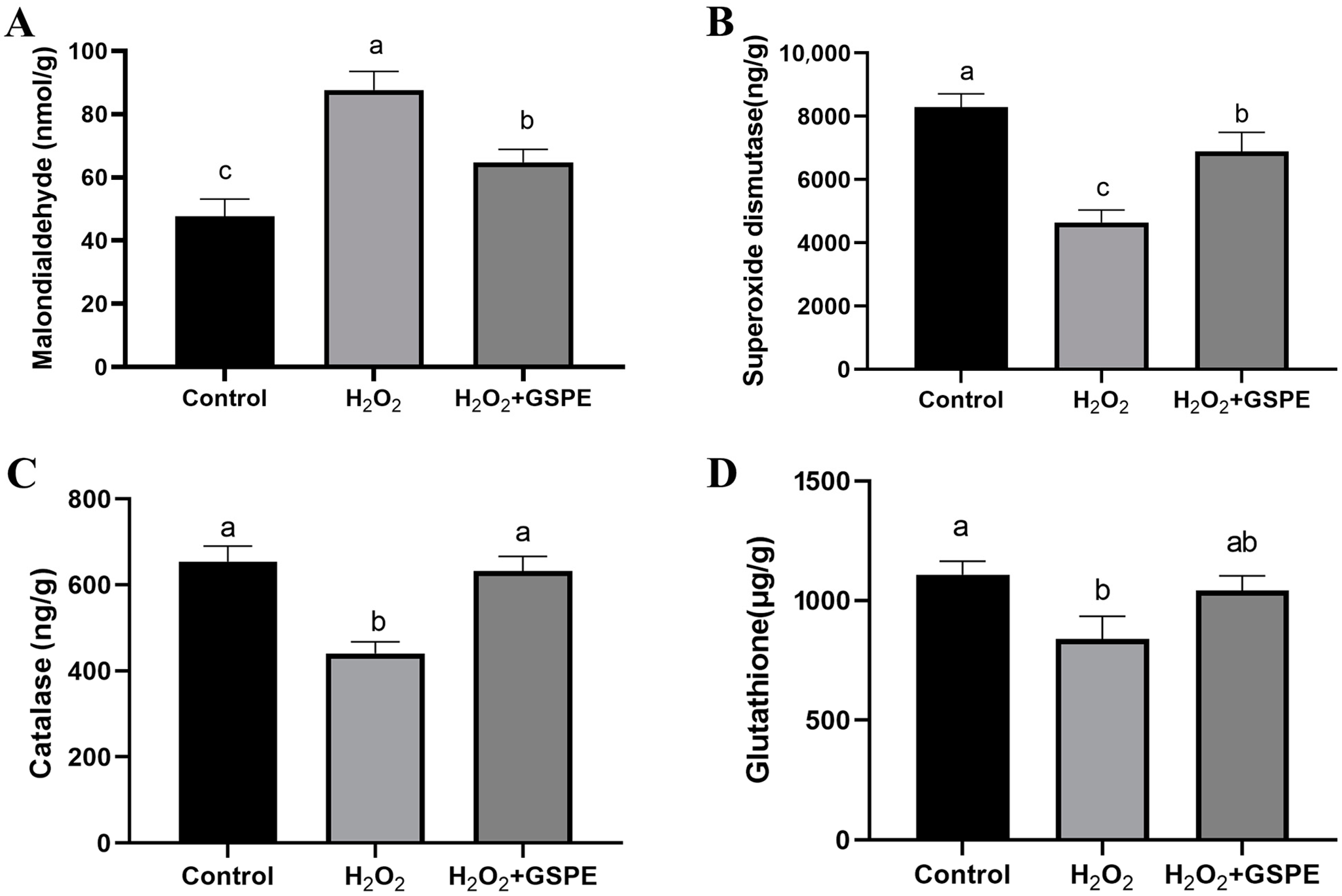
3.8. Effects of Dietary Supplementation with GSPE on Liver Oxidative Capacity of Weaned Rabbits Challenged with H2O2 (Exp.3)

Relative to the control group, H2O2 injection significantly elevated the MDA concentration (p < 0.05) and significantly reduced the levels of SOD, CAT, and GSH in the liver (p < 0.05) (Figure 3). In H2O2-challenged rabbits, dietary GSPE supplementation significantly (p < 0.05) decreased MDA concentration and increased SOD and CAT activities in the liver. The rabbits in the H2O2+GSPE group had significantly higher MDA concentration and lower SOD activity than the rabbits in the control group (p < 0.05), and no significant differences were observed in the CAT and GSH activities between the two groups (p > 0.05).

3.9. Effects of Dietary Supplementation with GSPE on Small Intestinal Morphometric Measurements of Weaned Rabbits Challenged with H2O2 (Exp.3)

Compared to the control group, H2O2 administration had no significant effects on the VH, CD, and V/C of duodenum, jejunum, and ileum (p > 0.05) (Figure 4). The rabbits in the H2O2+GSPE group had significantly higher duodenal VH and lower ileal CD than the rabbits in the H2O2 group (p < 0.05) and showed significantly lower duodenal CD than the rabbits in the control group (p < 0.05). Moreover, the H2O2+GSPE group had significantly lower jejunal CD (p < 0.05) and significantly higher V/C in the duodenum, jejunum, and ileum than the other two groups (p < 0.05).

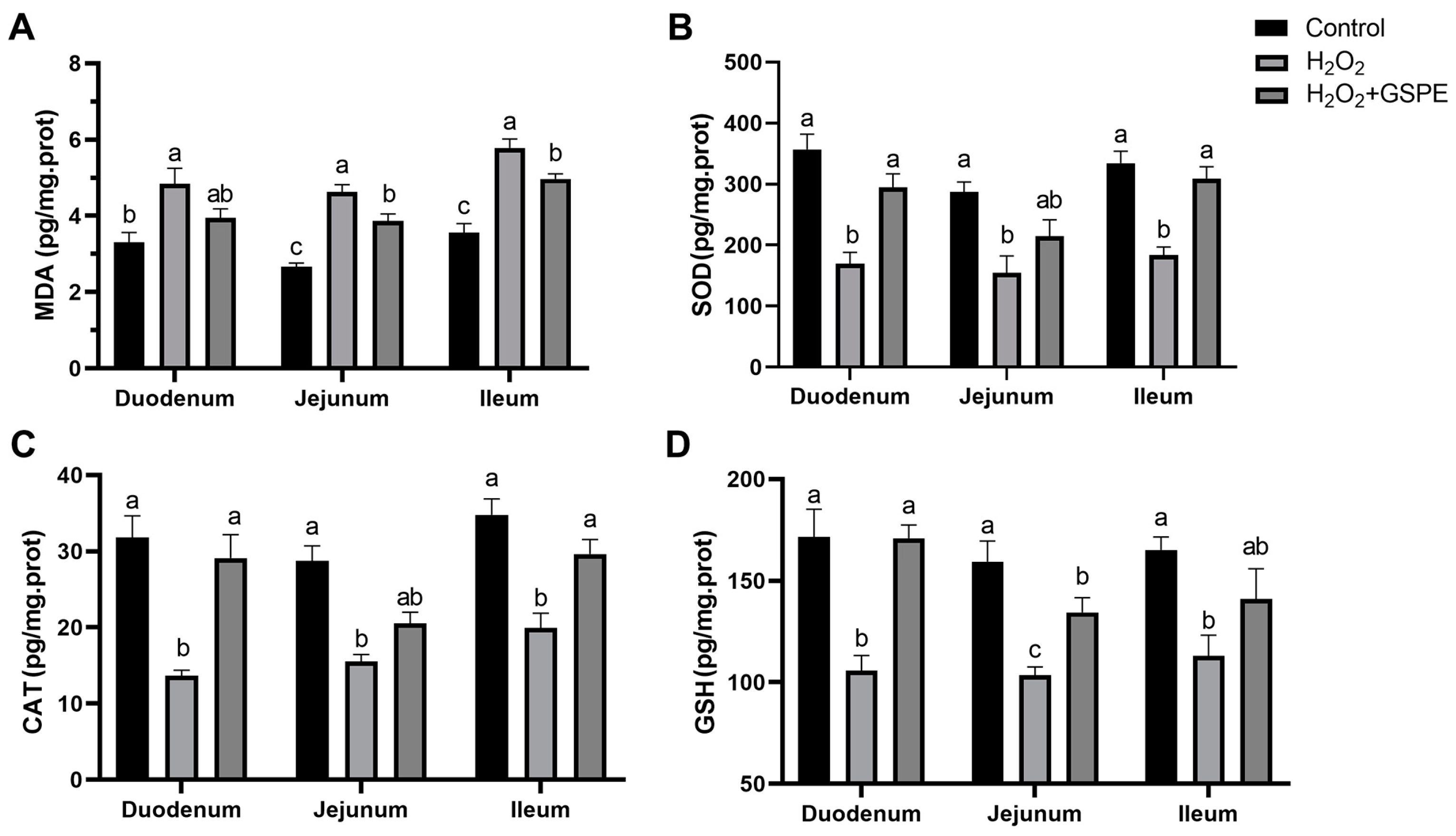
3.10. Effects of Dietary Supplementation with GSPE on Small Intestinal Oxidative Capacity of Weaned Rabbits Challenged with H2O2 (Exp.3)

Compared to the control group, H2O2 injection significantly increased the MDA concentration and inhibited the activities of SOD, CAT, and GSH in the duodenum, jejunum, and ileum of rabbits (p < 0.05) (Figure 5). In H2O2-challenged rabbits, dietary GSPE supplementation significantly decreased the MDA concentrations in the jejunum and ileum (p < 0.05) and significantly increased the SOD and CAT activities in the duodenum and ileum, as well as GSH activity in the jejunum of meat rabbits (p < 0.05). Additionally, the rabbits in the H2O2+GSPE group showed significantly higher MDA concentrations in the jejunum and ileum (p < 0.05) and significantly lower GSH activity in the jejunum relative to the control group (p < 0.05).

4. Discussion

In the present study, dietary supplementation with 400 mg/kg GSPE decreased the F/G ratio, but GSPE addition had no significant effects on the slaughter performance in meat rabbits. A previous study in broilers also showed that 7.5 and 15 mg/kg GSPE decreased the F/G ratio without affecting the eviscerated yield [22]. Additionally, 200 and 400 mg/kg GSPE supplementation was found to increase the FCR in goldfish [23]. Many studies have attributed the growth-promoting effects of GSPE to its antioxidant activity [24,25]. It is well known that weaning of rabbits offers nutritional and animal health benefits in rabbit production. But undeniably, weaning rabbits for does is one of the most stressful events, as rabbits often suffer from physiological, nutritional, and social stresses during the weaning progress. It was reported that the weaning process could induce abnormal behavioral traits and physiological responses, such as increased cortisol, CAT, and SOD levels [7]. An elevated blood cortisol level was usually associated with oxidative stress, and increasing antioxidative enzyme activities was a way to overcome the produced ROS [7,26]. Oxidative stress caused by weaning is a significant factor leading to growth retardation in livestock. Previous studies have shown that oxidative stress can damage the intestinal mucosal barrier and inhibit gut enzyme activity, reducing nutrient digestion and absorption [27,28]. Moreover, oxidative stress also could induce liver injury and loss of liver function and thus suppress the secretion of growth and development-related hormones [29,30]. GSPE has been shown to possess a wide range of pharmacological and medicinal properties in mitigating oxidative stress. It has been proven that GSPE can alleviate exogenous (such as cisplatin, lipopolysaccharides, zearalenone, and H2O2) and endogenous (such as diabetes) oxidative stress [31,32,33,34]. Mu et al. [35] found that the supplementation of GSPE (0–40 mg/kg BW/day) linearly increased the liver weight of ram lambs, suggesting that GSPE potentially plays a crucial role in the metabolism of organisms. Similarly, 400 mg/kg GSPE supplementation significantly increased the liver coefficient of rabbits. These results suggest that the optimal level of GSPE supplementation in the diet promoted the development of liver in the rabbits. The liver plays a key role in the synthesis, transport, conversion, and excretion of cholesterol. In the present study, the serum TCHO of weaned rabbits was significantly decreased by dietary 400 mg/kg GSPE supplementation. Previous studies in rats have indicated that GSPE can promote lipid metabolism and inhibit fat deposition [36]. Feng et al. [37] also showed that adding 100 and 200 mg/kg GSPE to the diet reduced serum TCHO levels but increased serum high-density lipoprotein cholesterol (HDL-C) levels in pigs. HDL-C, produced by the liver, helps to transport excess cholesterol from peripheral tissues back to the liver, preventing cholesterol accumulation in the blood vessels [38]. Therefore, the decreased serum TCHO of rabbits in the G400 group might be related to the regulation of lipid metabolism by the GSPE, which needs to be studied further. However, exposure to higher concentrations of GSPE could result in ROS generation and cell death [39], which might be the reason for the decreased liver coefficient and increased serum TCHO level in the G800 group. The above results also indicated the importance of optimizing the concentration of GSPE for application to avoid potential harmful pro-oxidant effects in animal feed.
H2O2 is one of the major and the most stable ROS in the redox regulation of biological activities. Excessive H2O2 is harmful to almost all cell components, so it is often used to establish the oxidative stress model [16,17]. In this study, 10% H2O2 injection showed an adverse effect on the growth performance of rabbits. Similar results have also been reported in a 10% H2O2-induced oxidative stress model [17,40]. Additionally, increased MDA concentrations in the serum, liver, and small intestine were found in the rabbits injected with 10% H2O2 in the current study. MDA is usually considered a reliable biomarker of oxidative stress [41]. Therefore, a rabbit oxidative stress model was well established by an intraperitoneal injection of 10% H2O2 in the present study.
The liver and intestine are the organs that are most sensitive to oxidative stress and are highly susceptible to injury when the oxidative stress occurs [42]. In this study, the liver morphological changes of rabbits were induced by 10% H2O2 injection. A previous study in vitro showed that H2O2 treatment could result in the accumulation of the ROS through disrupting the intracellular oxidative homeostasis via inhibiting antioxidant enzyme activities, inducing the oxidative damage in HepG2 cells [43]. Reduced SOD, CAT, and GSH activities and elevated MDA level by H2O2 injection were also found in the current study, but GSPE supplementation could alleviate these changes to the levels observed in the control group. SOD and CAT play crucial roles in protecting the body from oxidative damage. SOD converts ROS into H2O2, while CAT is responsible for detoxifying H2O2, breaking it down into H2O and O2 [44]. GSH is a low molecular weight thiol-tripeptide capable of directly reacting with free radicals and peroxides to reduce oxidative stress and protect cells from damage [6]. It has been proven that PPARα, Nrf2/ARE, and PI3K/AKT signaling pathways are all involved in the protective effect of GSPE against liver oxidative damage [33,45]. Rajput et al. [13] found that 250 and 400 mg/kg GSPE decreased MDA concentration and increased T-SOD, CAT, GSH, and GSH-Px activities in the liver, alleviating the liver injury caused by the ABF1 of broilers. A study in Kunming mice also showed that 100 mg/kg GSPE could activate the Nrf2/ARE signaling pathways in the liver and protect against zearalenone-induced liver oxidative damage [34]. Therefore, GSPE supplementation could mitigate H2O2-induced liver injury by enhancing liver antioxidant capacity in meat rabbits. However, the mechanism by which GSPE alleviates H2O2-induced hepatic oxidative damage in rabbits still requires further investigation.
The crypt–villus architecture of the small intestine is essential for maintaining the structural integrity of the intestinal epithelium and preservation of gut homeostasis [46]. The V/C ratio is regarded as a valuable criterion for assessing the nutrient absorption capacity of the small intestine [18]. In the present study, 10% H2O2 injection was not found to significantly damage the crypt–villus architecture of the small intestine compared with the control group, although it disrupted the small intestinal redox homeostasis of rabbits. This might be attributed to the intestinal property of self-repair to maintain mucosal homeostasis [46], and 10% H2O2 injection once was not enough to induce extensive damage to intestinal morphology. However, GSPE supplementation showed beneficial effects to improve the small intestinal morphology of meat rabbits, especially under H2O2-induced oxidative stress conditions. Previous studies in pigs [14], broiler hens [47], and mice [48] have manifested that GSPE supplementation could improve intestinal health through enhancing antioxidant capacity and modulating gut microbiota. Consistently, enhanced SOD, CAT, and GSH activities and reduced MDA concentration in the small intestine were found in the H2O2-challenged rabbits fed the diet supplemented with GSPE. Therefore, GSPE supplementation could mitigate the H2O2-induced intestinal oxidative stress of the meat rabbits in this study.

5. Conclusions

Collectively, supplementation with 400 mg/kg GSPE could increase the FCR and promote the development of liver and cholesterol metabolism in weaned Hyla rabbits. The intraperitoneal injection of 10% H2O2 at dosage of 1 mL/kg BW decreased growth performance, induced oxidative stress of the liver and small intestine, and damaged the liver morphology in the rabbits, showing that this dosage could be used to establish a rabbit oxidative stress model. However, dietary 400 mg/kg GSPE supplementation alleviated H2O2-induced adverse effects on the liver and small intestine by enhancing the antioxidative capacity in weaned Hyla rabbits.

Author Contributions

Conceptualization, L.L. and F.L.; Data curation, M.G.; Formal analysis, M.G. and J.C.; Funding acquisition, L.L. and F.L.; Investigation, L.L.; Methodology, F.L. and J.C.; Project administration, M.G.; Software, M.G. and J.C.; Supervision, F.L. and J.C.; Validation, L.L. and F.L.; Writing—original draft, M.G. and J.C.; Writing—review and editing, L.L., F.L. and J.C. All authors have read and agreed to the published version of the manuscript.

Funding

This research was funded by Project of Introducing Urgently Needed Talents in Key Supporting Regions of Shandong Province (2024); Key R&D Program of Shandong Province (2023TZXD044); the Earmarked Fund for CARS (CARS-43-B-1); Special Economic Animal Industry Technology System of Shandong Province (SDAIT-21-16).

Institutional Review Board Statement

The animal study protocol was approved by the Care and Use Committee of Shandong Agricultural University (protocol code SDAUA-2020-201).

Informed Consent Statement

The authors obtained permission to publish this detailed information, along with evidence that Tai’an Xinglong Rabbit Farming was aware and allowed the data to be published.

Data Availability Statement

The original contributions presented in this study are included in the article. Further inquiries can be directed to the corresponding authors.

Conflicts of Interest

The authors declare no conflicts of interest.

References

  1. Averill-Bates, D. Reactive oxygen species and cell signaling. Review. Biochim. Biophys. Acta (BBA)-Mol. Cell Res. 2024, 1871, 119573. [Google Scholar] [CrossRef]
  2. Machado, I.F.; Miranda, R.G.; Dorta, D.J.; Rolo, A.P.; Palmeira, C.M. Targeting oxidative stress with polyphenols to fight liver diseases. Antioxidants 2023, 12, 1212. [Google Scholar] [CrossRef] [PubMed]
  3. An, X.; Yu, W.; Liu, J.; Tang, D.; Yang, L.; Chen, X. Oxidative cell death in cancer: Mechanisms and therapeutic opportunities. Cell Death Dis. 2024, 15, 556. [Google Scholar] [CrossRef] [PubMed]
  4. Figueira, T.R.; Barros, M.H.; Camargo, A.A.; Castilho, R.F.; Ferreira, J.C.; Kowaltowski, A.J.; Sluse, F.E.; Souza-Pinto, N.C.; Vercesi, A.E. Mitochondria as a source of reactive oxygen and nitrogen species: From molecular mechanisms to human health. Antioxid. Redox Signal. 2013, 18, 2029–2074. [Google Scholar] [CrossRef] [PubMed]
  5. Chen, J.; Luo, Y.; Li, Y.; Chen, D.; Yu, B.; He, J. Chlorogenic acid attenuates oxidative stress-induced intestinal epithelium injury by co-regulating the PI3K/Akt and IκBα/NF-κB signaling. Antioxidants 2021, 10, 1915. [Google Scholar] [CrossRef]
  6. Zhu, R.; Wang, Y.; Zhang, L.; Guo, Q. Oxidative stress and liver disease. Hepatol. Res. 2012, 42, 741–749. [Google Scholar] [CrossRef]
  7. Gharib, H.S.; Abdel-Fattah, A.F.; Mohammed, H.A.; Abdel-Fattah, D.M. Weaning induces changes in behavior and stress indicators in young New Zealand rabbits. J. Adv. Vet. Anim. Res. 2018, 5, 166–172. [Google Scholar] [CrossRef]
  8. Ji, R.; Chen, J.; Xu, J.; Zhang, L.; Liu, L.; Li, F. Protective effect of chlorogenic acid on liver injury in heat-stressed meat rabbits. J. Anim. Physiol. Anim. Nutr. 2024, 108, 1203–1213. [Google Scholar] [CrossRef]
  9. Somi, M.H.; Hajipour, B.; Abad, G.D.A.; Hemmati, M.R.; Ghabili, K.; Khodadadi, A.; Vatankhah, A.M. Protective role of lipoic acid on methotrexate induced intestinal damage in rabbit model. Indian J. Gastroenterol. 2011, 30, 38–40. [Google Scholar] [CrossRef]
  10. Rauf, A.; Imran, M.; Abu-Izneid, T.; Patel, S.; Pan, X.; Naz, S.; Silva, A.S.; Saeed, F.; Suleria, H.A.R. Proanthocyanidins: A comprehensive review. Biomed. Pharmacother. 2019, 116, 108999. [Google Scholar] [CrossRef]
  11. Hassan, F.A.; Mahrose, K.M.; Basyony, M.M. Effects of grape seed extract as a natural antioxidant on growth performance, carcass characteristics and antioxidant status of rabbits during heat stress. Arch. Anim. Nutr. 2016, 70, 141–154. [Google Scholar] [CrossRef] [PubMed]
  12. Abd Allah, E.; Abdel Hady, D.E.; Ghodaia, A.E.; Ameen, H.; Eliraqy, E.Z.; Farouk, Z.; Shedeed, S. Impact of Grape Seed Extract (Proanthocyanidins) on Pathophysiological Changes and Antioxidant Capacity in Rabbit Does Expose to Heat Stress. Egypt. J. Vet. Sci. 2024, 55, 2139–2146. [Google Scholar] [CrossRef]
  13. Rajput, S.A.; Sun, L.; Zhang, N.; Khalil, M.M.; Gao, X.; Ling, Z.; Zhu, L.; Khan, F.A.; Zhang, J.; Qi, D. Ameliorative effects of grape seed proanthocyanidin extract on growth performance, immune function, antioxidant capacity, biochemical constituents, liver histopathology and aflatoxin residues in broilers exposed to aflatoxin B1. Toxins 2017, 9, 371. [Google Scholar] [CrossRef]
  14. Wei, X.; Li, L.; Yan, H.; Li, Q.; Gao, J.; Hao, R. Grape seed procyanidins improve intestinal health by modulating gut microbiota and enhancing intestinal antioxidant capacity in weaned piglets. Livest. Sci. 2022, 264, 105066. [Google Scholar] [CrossRef]
  15. Zheng, Y.; Li, Y.; Yu, B.; Huang, Z.; Luo, Y.; Zheng, P.; Mao, X.; Yu, J.; Tan, H.; Luo, J. Grape seed proanthocyanidins improves growth performance, antioxidative capacity, and intestinal microbiota in growing pigs. Front. Microbiol. 2024, 15, 1501211. [Google Scholar] [CrossRef]
  16. Feng, W.; Xu, Y.; Su, S.; Yu, F.; Li, J.; Jia, R.; Song, C.; Li, H.; Xu, P.; Tang, Y. Transcriptomic analysis of hydrogen peroxide-induced liver dysfunction in Cyprinus carpio: Insights into protein synthesis and metabolism. Sci. Total Environ. 2024, 917, 170393. [Google Scholar] [CrossRef]
  17. Yin, J.; Duan, J.; Cui, Z.; Ren, W.; Li, T.; Yin, Y. Hydrogen peroxide-induced oxidative stress activates NF-κB and Nrf2/Keap1 signals and triggers autophagy in piglets. Rsc Adv. 2015, 5, 15479–15486. [Google Scholar] [CrossRef]
  18. Kong, F.; Wu, F.; Liu, Y.; Lai, N.; Wang, G.; Shen, S.; Han, S.; Li, B.; Zhi, Y.; Chen, S. Effects of enzymolytic soybean meal on the growth performance, digestive enzyme activity, some serum indexes, carcase performance and meat quality of Rex rabbits. Ital. J. Anim. Sci. 2022, 21, 1307–1314. [Google Scholar] [CrossRef]
  19. Chen, J.; Li, F.; Yang, W.; Jiang, S.; Li, Y. Supplementation with exogenous catalase from Penicillium notatum in the diet ameliorates lipopolysaccharide-induced intestinal oxidative damage through affecting intestinal antioxidant capacity and microbiota in weaned pigs. Microbiol. Spectr. 2021, 9, e00654-21. [Google Scholar] [CrossRef]
  20. Zhang, P.; Jing, C.; Liang, M.; Jiang, S.; Huang, L.; Jiao, N.; Li, Y.; Yang, W. Zearalenone exposure triggered cecal physical barrier injury through the TGF-β1/Smads signaling pathway in weaned piglets. Toxins 2021, 13, 902. [Google Scholar] [CrossRef] [PubMed]
  21. Li, Y.; Zhao, X.; Zhang, L.; Zhan, X.; Liu, Z.; Zhuo, Y.; Lin, Y.; Fang, Z.; Che, L.; Feng, B. Effects of a diet supplemented with exogenous catalase from penicillium notatum on intestinal development and microbiota in weaned piglets. Microorganisms 2020, 8, 391. [Google Scholar] [CrossRef] [PubMed]
  22. Yang, J.Y.; Zhang, H.J.; Wang, J.; Wu, S.G.; Yue, H.Y.; Jiang, X.R.; Qi, G.H. Effects of dietary grape proanthocyanidins on the growth performance, jejunum morphology and plasma biochemical indices of broiler chicks. Animal 2017, 11, 762–770. [Google Scholar] [CrossRef] [PubMed]
  23. Jahanbakhshi, A.; Pourmozaffar, S.; Mozanzadeh, M.T.; Adeshina, I.; Zehra, S.; Vega-Heredia, S. Dietary Effect of Grape Seed Proanthocyanidin Extract on Growth Performance, Serum Biochemical Parameters, Skin Mucosal Immune Response, and Antioxidant Capacity in Goldfish (Carassius auratus). Ann. Anim. Sci. 2023, 23, 215–223. [Google Scholar] [CrossRef]
  24. Mohammadi, Y.; Kamangar, B.B.; Zarei, M.A. Effects of diets containing grape seed proanthocyanidin extract on the growth and oxidative capacity of common carp (Cyprinus carpio). Aquaculture 2021, 540, 736689. [Google Scholar] [CrossRef]
  25. Zheng, Y.; Li, Y.; Yu, B.; Luo, Y.; Huang, Z.; Zheng, P.; Mao, X.; Dai, Z.; Yu, J.; Yan, H. Dietary supplementation of grape seed proanthocyanidins improves growth performance, carcass traits, and meat quality in growing-finishing pigs. Anim. Nutr. 2024; in press. [Google Scholar] [CrossRef]
  26. Kim, J.; Yun, K.; Cho, A.; Kim, D.H.; Lee, Y.K.; Choi, M.J.; Kim, S.-h.; Kim, H.; Yoon, J.W.; Park, H.C. High cortisol levels are associated with oxidative stress and mortality in maintenance hemodialysis patients. BMC Nephrol. 2022, 23, 98. [Google Scholar] [CrossRef]
  27. Lykkesfeldt, J.; Svendsen, O. Oxidants and antioxidants in disease: Oxidative stress in farm animals. Vet. J. 2007, 173, 502–511. [Google Scholar] [CrossRef]
  28. Yuan, S.; Chen, D.; Zhang, K.; Yu, B. Effects of oxidative stress on growth performance, nutrient digestibilities and activities of antioxidative enzymes of weanling pigs. Asian-Australas. J. Anim. Sci. 2007, 20, 1600–1605. [Google Scholar] [CrossRef]
  29. Sánchez-Valle, V.; Chavez-Tapia, N.C.; Uribe, M.; Méndez-Sánchez, N. Role of oxidative stress and molecular changes in liver fibrosis: A review. Curr. Med. Chem. 2012, 19, 4850–4860. [Google Scholar] [CrossRef]
  30. Takahashi, Y. Nonalcoholic fatty liver disease and adult growth hormone deficiency: An under-recognized association? Best Pract. Res. Clin. Endocrinol. Metab. 2023, 37, 101816. [Google Scholar] [CrossRef]
  31. Yousef, M.; Saad, A.; El-Shennawy, L. Protective effect of grape seed proanthocyanidin extract against oxidative stress induced by cisplatin in rats. Food Chem. Toxicol. 2009, 47, 1176–1183. [Google Scholar] [CrossRef] [PubMed]
  32. Mansouri, E.; Panahi, M.; Ghaffari, M.A.; Ghorbani, A. Effects of grape seed proanthocyanidin extract on oxidative stress induced by diabetes in rat kidney. Iran. Biomed. J. 2011, 15, 100. [Google Scholar]
  33. Liu, X.; Lin, X.; Mi, Y.; Li, J.; Zhang, C. Grape seed proanthocyanidin extract prevents ovarian aging by inhibiting oxidative stress in the hens. Oxidative Med. Cell. Longev. 2018, 2018, 9390810. [Google Scholar] [CrossRef] [PubMed]
  34. Long, M.; Yang, S.-H.; Han, J.-X.; Li, P.; Zhang, Y.; Dong, S.; Chen, X.; Guo, J.; Wang, J.; He, J.-B. The protective effect of grape-seed proanthocyanidin extract on oxidative damage induced by zearalenone in Kunming mice liver. Int. J. Mol. Sci. 2016, 17, 808. [Google Scholar] [CrossRef]
  35. Mu, C.; Yang, W.; Wang, P.; Zhao, J.; Hao, X.; Zhang, J. Effects of high-concentrate diet supplemented with grape seed proanthocyanidins on growth performance, liver function, meat quality, and antioxidant activity in finishing lambs. Anim. Feed Sci. Technol. 2020, 266, 114518. [Google Scholar] [CrossRef]
  36. Gu, A.; Zhang, T.; Wang, T.; Wu, Z.; Shan, A.; Li, J. Effects of grape seed proanthocyanidins extract on fat deposition and lipid metabolism in rats. Chin. J. Anim. Nutr. 2021, 33, 4133–4144. [Google Scholar]
  37. Feng, Y.; Chen, X.; Chen, D.; He, J.; Zheng, P.; Luo, Y.; Yu, B.; Huang, Z. Dietary grape seed proanthocyanidin extract supplementation improves antioxidant capacity and lipid metabolism in finishing pigs. Anim. Biotechnol. 2023, 34, 4021–4031. [Google Scholar] [CrossRef]
  38. Habib, A.; Mihas, A.A.; Abou-Assi, S.G.; Williams, L.M.; Gavis, E.; Pandak, W.M.; Heuman, D.M. High-density lipoprotein cholesterol as an indicator of liver function and prognosis in noncholestatic cirrhotics. Clin. Gastroenterol. Hepatol. 2005, 3, 286–291. [Google Scholar] [CrossRef]
  39. Shao, Z.-H.; Vanden Hoek, T.L.; Xie, J.; Wojcik, K.; Chan, K.C.; Li, C.-Q.; Hamann, K.; Qin, Y.; Schumacker, P.T.; Becker, L.B. Grape seed proanthocyanidins induce pro-oxidant toxicity in cardiomyocytes. Cardiovasc. Toxicol. 2003, 3, 331–339. [Google Scholar] [CrossRef]
  40. Xing, T.; Chen, X.; Li, J.; Zhang, L.; Gao, F. Dietary taurine attenuates hydrogen peroxide-impaired growth performance and meat quality of broilers via modulating redox status and cell death signaling. J. Anim. Sci. 2021, 99, skab089. [Google Scholar] [CrossRef]
  41. Del Rio, D.; Stewart, A.J.; Pellegrini, N. A review of recent studies on malondialdehyde as toxic molecule and biological marker of oxidative stress. Nutr. Metab. Cardiovasc. Dis. 2005, 15, 316–328. [Google Scholar] [CrossRef] [PubMed]
  42. Aw, T.Y. Molecular and cellular responses to oxidative stress and changes in oxidation-reduction imbalance in the intestine. Am. J. Clin. Nutr. 1999, 70, 557–565. [Google Scholar] [CrossRef] [PubMed]
  43. Li, X.; Hua, Y.; Yang, C.; Liu, S.; Tan, L.; Guo, J.; Li, Y. Polysaccharides extracted from mulberry fruits (Morus nigra L.): Antioxidant effect of ameliorating H2O2-induced liver injury in HepG2 cells. BMC Complement. Med. Ther. 2023, 23, 112. [Google Scholar] [CrossRef] [PubMed]
  44. Finkel, T.; Holbrook, N.J. Oxidants, oxidative stress and the biology of ageing. Nature 2000, 408, 239–247. [Google Scholar] [CrossRef]
  45. Yang, D.; Jiang, H.; Lu, J.; Lv, Y.; Baiyun, R.; Li, S.; Liu, B.; Lv, Z.; Zhang, Z. Dietary grape seed proanthocyanidin extract regulates metabolic disturbance in rat liver exposed to lead associated with PPARα signaling pathway. Environ. Pollut. 2018, 237, 377–387. [Google Scholar] [CrossRef]
  46. Beumer, J.; Clevers, H. Cell fate specification and differentiation in the adult mammalian intestine. Nat. Rev. Mol. Cell Biol. 2021, 22, 39–53. [Google Scholar] [CrossRef]
  47. Grandhaye, J.; Douard, V.; Rodriguez-Mateos, A.; Xu, Y.; Cheok, A.; Riva, A.; Guabiraba, R.; Zemb, O.; Philippe, C.; Monnoye, M. Microbiota changes due to grape seed extract diet improved intestinal homeostasis and decreased fatness in parental broiler hens. Microorganisms 2020, 8, 1141. [Google Scholar] [CrossRef]
  48. Sheng, K.; Zhang, G.; Sun, M.; He, S.; Kong, X.; Wang, J.; Zhu, F.; Zha, X.; Wang, Y. Grape seed proanthocyanidin extract ameliorates dextran sulfate sodium-induced colitis through intestinal barrier improvement, oxidative stress reduction, and inflammatory cytokines and gut microbiota modulation. Food Funct. 2020, 11, 7817–7829. [Google Scholar] [CrossRef]
Figure 1. Effects of hydrogen peroxide (H2O2) injection on serum malondialdehyde (MDA) content of Hyla rabbits. Blank, rabbits not injected with sterile saline (SS) or H2O2 intraperitoneally; SS, rabbits injected with SS intraperitoneally; 5% H2O2, rabbits injected with 5% H2O2 intraperitoneally; 10% H2O2, rabbits injected with 10% H2O2 intraperitoneally. a,b Means with different lowercase letters differ significantly among treatments (p < 0.05). n = 8.
Animals 15 00327 g001
Figure 2. Effects of dietary supplementation with grape seed proanthocyanidin extract (GSPE) on liver morphology of weaned rabbits challenged with hydrogen peroxide (H2O2). Control, rabbits fed the basal diet; H2O2, rabbits fed the basal diet and given intraperitoneal injection of H2O2; H2O2+GSPE, rabbits fed the basal diet supplemented with 400 mg/kg and given intraperitoneal injection of H2O2.
Animals 15 00327 g002
Figure 3. Effects of dietary supplementation with grape seed proanthocyanidin extract (GSPE) on liver oxidative capacity of weaned rabbits challenged with hydrogen peroxide (H2O2). (A) malondialdehyde; (B) superoxide dismutase; (C) catalase; (D) glutathione. Control, rabbits fed the basal diet; H2O2, rabbits fed the basal diet and given intraperitoneal injection of H2O2; H2O2+GSPE, rabbits fed the basal diet supplemented with 400 mg/kg and given intraperitoneal injection of H2O2. a,b,c Means with different lowercase letters differ significantly among treatments (p < 0.05). n = 8.
Animals 15 00327 g003
Figure 4. Effects of dietary supplementation with grape seed proanthocyanidin extract (GSPE) on small intestinal morphometric measurements of weaned rabbits challenged with hydrogen peroxide (H2O2). (A) Villus height; (B) crypt depth; (C) villus height/crypt depth. Control, rabbits fed the basal diet; H2O2, rabbits fed the basal diet and given intraperitoneal injection of H2O2; H2O2+GSPE, rabbits fed the basal diet supplemented with 400 mg/kg and given intraperitoneal injection of H2O2. a,b Means with different lowercase letters differ significantly among treatments (p < 0.05). n = 8.
Animals 15 00327 g004
Figure 5. Effects of dietary supplementation with grape seed proanthocyanidin extract (GSPE) on small intestinal oxidative capacity of weaned rabbits challenged with hydrogen peroxide (H2O2). (A) MDA, malondialdehyde; (B) SOD, superoxide dismutase;(C) CAT, catalase; (D) GSH, glutathione. Control, rabbits fed the basal diet; H2O2, rabbits fed the basal diet and given intraperitoneal injection of H2O2; H2O2+GSPE, rabbits fed the basal diet supplemented with 400 mg/kg and given intraperitoneal injection of H2O2. a,b,c Means with different lowercase letters differ significantly among treatments (p < 0.05). n = 8.
Animals 15 00327 g005
Table 1. Composition and nutrient levels of the experimental diet.
Ingredients (%)ContentNutrient Levels 2 (%)Content
Corn17Digestible energy (MJ/kg)10.25
Rice husk5Crude protein15.86
Wheat bran20Crude fiber18.28
Soybean meal14Acid detergent fiber19.98
Alfalfa meal24Neutral detergent fiber36.25
Bean straw powder16Ether extract3.71
Premix 14Calcium0.78
Total100Phosphorus0.43
1 The premix provided the following per kg of diets: Vitamin D3 2000 IU, Vitamin B5 375 mg, Vitamin B7 1.5 mg, Vitamin E 22.5 mg, Vitamin A 6000 IU, Vitamin K 15 mg, Vitamin B2 45 mg, Vitamin B3 375 mg, Vitamin B1 15 mg, Vitamin B6 15 mg, Vitamin B12 0.15 mg, choline chloride 625 mg, Se 0.05 mg, Zn 50 mg, ferrous sulfate 100 mg, iodine 0.6 mg, methionine 1500 mg, lysine 1500 mg, diclazuril 5mg. 2 Nutrient levels were measured values.
Table 2. Effects of dietary supplementation with grape seed proanthocyanidin extract (GSPE) on the growth performance of rabbits.
Items 1Treatment 2RMSEp Value
G0G200G400G800
IBW (g)80079780180921.100.756
FBW (g)185318631832183576.660.881
ADFI (g/d)125.18124.66118.13120.398.120.391
ADG (g/d)30.1030.4729.4529.311.980.715
F/G4.16 a4.09 ab4.01 b4.11 ab0.080.043
Survival rate (%)79.879.891.783.4--
1 IBW, initial body weight; FBW, final body weight; ADFI, average daily feed intake; ADG, average daily gain; F/G, ratio of ADFI to ADG. 2 G0, G200, G400, and G800 were rabbits fed the basal diet supplemented with 0, 200, 400, and 800 mg/kg GSPE, respectively. a,b Means with different letters within a row differ (p < 0.05). n = 8.
Table 3. Effects of dietary supplementation with grape seed proanthocyanidin extract (GSPE) on slaughter performance of meat rabbits.
ItemsTreatment 1RMSEp Value
G0G200G400G800
Full eviscerated weight (g)7857907998167.340.894
Full eviscerated weight ratio (%)41.2941.3141.5842.622.150.681
Half eviscerated weight (g)8728718948978.270.918
Half eviscerated weight ratio (%)45.8545.5846.5046.842.280.764
1 G0, G200, G400, and G800 were rabbits fed the basal diet supplemented with 0, 200, 400, and 800 mg/kg GSPE, respectively. n = 8.
Table 4. Effects of dietary supplementation with grape seed proanthocyanidin extract (GSPE) on organ coefficients in meat rabbits.
ItemsTreatment 1RMSEp Value
G0G200G400G800
Thymus (g/kg)2.562.312.482.260.240.117
Spleen (g/kg)0.8640.7850.7610.7640.170.683
Liver (g/kg)26.69 b28.41 ab33.65 a29.11 ab3.390.013
Kidney (g/kg)7.226.527.136.860.880.519
1 G0, G200, G400, and G800 were rabbits fed the basal diet supplemented with 0, 200, 400, and 800 mg/kg GSPE, respectively. a,b Means with different letters within a row differ (p < 0.05). n = 8.
Table 5. Effects of dietary supplementation with grape seed proanthocyanidin extract (GSPE) on serum biochemical indicators in meat rabbits.
Items 2Treatment 1RMSEp Value
G0G200G400G800
TP (g/L)57.1559.2855.7556.423.820.429
UREA (g/L)8.519.007.847.571.510.363
ALB (g/L)30.5532.1731.5730.821.970.492
TG (mmol/L)0.661.200.700.820.580.393
TCHO (mmol/L)1.93 a1.74 ab1.49 b1.68 ab0.200.012
Ca (mmol/L)4.604.624.694.610.200.869
P (mmol/L)3.063.283.113.130.500.738
ALP (U/L)116.17125.00132.00111.5024.370.486
1 G0, G200, G400, and G800 were rabbits fed the basal diet supplemented with 0, 200, 400, and 800 mg/kg GSPE, respectively. 2 TP, total protein; UREA, urea; ALB, albumin; TG, triglycerides; TCHO, total cholesterol; Ca, calcium; P, phosphorus; ALP, alkaline phosphatase. a,b Means with different letters within a row differ (p < 0.05). n = 8.
Table 6. Effects of hydrogen peroxide (H2O2) injection on the growth performance of Hyla rabbits.
Items 2Treatment 1RMSEp Value
BlankSS5% H2O210% H2O2
IBW (g)146814741469146630.770.961
FBW (g)1681 a1688 a1656 a1612 b37.820.002
ADFI (g/d)126.10 a125.72 a118.64 a105.28 b13.510.015
ADG (g/d)30.38 a30.54 a26.73 b20.74 c3.41<0.001
F/G4.15 c4.12 c4.45 b5.10 a0.17<0.001
Survival rate (%)100100100100--
1 Blank, rabbits not injected with sterile saline (SS) or H2O2 intraperitoneally; SS, rabbits injected with SS intraperitoneally; 5% H2O2, rabbits injected with 5% H2O2 intraperitoneally; 10% H2O2, rabbits injected with 10% H2O2 intraperitoneally. 2 IBW, initial BW; FBW, final BW; ADFI, average daily feed intake; ADG, average daily gain; F/G, ratio of ADFI to ADG. a,b,c Means with different letters within a row differ (p < 0.05). n = 8.
Disclaimer/Publisher’s Note: The statements, opinions and data contained in all publications are solely those of the individual author(s) and contributor(s) and not of MDPI and/or the editor(s). MDPI and/or the editor(s) disclaim responsibility for any injury to people or property resulting from any ideas, methods, instructions or products referred to in the content.

