Article

Antimicrobial Resistance and Virulence Determinants of Escherichia coli Isolates from Raw Milk from Dairy Cows with Subclinical Mastitis

by
Ntelekwane George Khasapane
1,*,
Olga de Smidt
1,
Kgaugelo Edward Lekota
2,
Jane Nkhebenyane
1,
Oriel Thekisoe
2 and
Tsepo Ramatla
1
1
Centre for Applied Food Safety and Biotechnology, Department of Life Sciences, Central University of Technology, 1 Park Road, Bloemfontein 9300, South Africa
2
Unit for Environmental Sciences and Management, North-West University, Potchefstroom 2531, South Africa
*
Author to whom correspondence should be addressed.
Animals 2025, 15(13), 1980; https://doi.org/10.3390/ani15131980
Submission received: 27 May 2025 / Revised: 2 July 2025 / Accepted: 4 July 2025 / Published: 5 July 2025

Simple Summary

Subclinical mastitis (SCM) is a major but often undetected issue in dairy farming, causing economic losses and affecting milk production. This study analyzed 174 milk samples and found SCM in 39.08% of them. Of these SCM-positive cases, 88.23% were caused by Escherichia coli. The most common serogroups detected were O113 and O111, and virulence genes Stx1 and Stx2 were found in 15% and 1.7% of isolates, respectively. Alarmingly, E. coli showed high resistance to several antibiotics, particularly penicillin (71.6%), ciprofloxacin (70%), and gentamicin (30%). All isolates carried the blaVIM resistance gene, and many also harbored blaKPC, blaNDM, sul1, and msrA. Half of the E. coli isolates were multidrug-resistant (MDR). These results highlight the critical need for better monitoring and responsible antibiotic use in dairy farming to prevent the spread of resistant bacteria to humans and improve mastitis treatment outcomes.

Abstract

Subclinical mastitis (SCM) is a stealthy but devastating challenge in the dairy industry, leading to economic losses and hindering efforts to achieve milk self-sufficiency. This study investigated the prevalence of SCM, antimicrobial resistance, and virulence profiles of Escherichia coli. A total of 174 milk samples were analyzed using the California mastitis test (CMT), somatic cell counts (SCCs), bacteriological culture, MALDI-TOF MS, and polymerase chain reaction (PCR). The findings revealed that the SCM prevalence was 68/174 (39.08%) based on CMT and SCC. Among SCM-positive samples, 60/68 (88.23%) were identified as E. coli, confirmed by MALDI-TOF MS and PCR assay. The most frequently detected serogroups were 0113 (11.6%) and 0113 (3.3%). Additionally, the genes for Stx1 and Stx2 were also detected in nine (15%) and one (1.7%), respectively. Antimicrobial susceptibility tests showed widespread resistance, with E. coli isolates demonstrating resistance to penicillin in 43 (71.6%), followed by ciprofloxacin in 42 (70%) and gentamicin in 18 (30%). A larger proportion of the E. coli strains (100%) harbored the blaVIM gene, while 23 (38.3%), 20%, 20%, and 1.47% contained blaKPC, blaNMD, suli1, and msrA. Thirty (50%) isolates were considered multidrug-resistant (MDR). These findings underscore the urgent need for enhanced surveillance and antibiotic stewardship in dairy farming. The presence of MDR E. coli in SCM poses a dual threat of potential transmission to humans and treatment failures in mastitis management. This study highlights the importance of proactive control strategies to mitigate the spread of antimicrobial resistance in livestock and beyond.

1. Introduction

Mastitis in cattle is one of the most significant illnesses that causes substantial financial losses in animal farms in developing nations and most of the world’s nations [1]. The expulsion of sick animals from the herd, significant milk production deficiencies, and high veterinary medication expenses are all consequences of mastitis [2]. Furthermore, a careful zoonotic viewpoint links mastitis to separating different bacteria and their harmful compounds in milk [3]. The ability of disease-causing microbes and their toxins to permeate the food chain and cause major foodborne illnesses has made mastitis a clear health risk [4], as noted by Oliver et al. [5] and Hennekinne et al. [6]. A significant cause of symptomatic and asymptomatic mastitis in dairy cows is the bacterium E. coli, although many other microbes can also lead to this condition [7].
Most E. coli strains are harmless, but some can cause foodborne diseases, such as those that generate Shiga toxins [8]. Humans are mostly exposed to this pathogen by consuming contaminated food, such as unpasteurized milk and dairy products [9]. While Shiga toxins (Stx1 and Stx2) and intimin (eae) are among the numerous extremely pathogenic genes produced by E. coli, they are thought to be the most frequently found genes in cows with mastitis symptoms, which poses a clear risk to human health. Through direct contact with diseased animals or the consumption of food, water, or vegetables contaminated with animal feces, humans can occasionally be infected with STEC, which first appeared as a foodborne pathogen. STEC infections can thus be spread from person to person with ease. STEC isolates, particularly serotype O157:H7, can cause a variety of illnesses, from mild diarrhea to hemolytic uremic syndrome (HUS) and hemorrhagic colitis (HC). Children, the elderly, and patients with impaired immune systems are usually affected by these problems [9]. Naturally occurring after gastrointestinal infections with STEC, the systemic sickness of HC usually starts with diarrhea and cramping in the abdomen, followed by bloody diarrhea. Furthermore, in nearly 80% of cases in both children and adults, HUS, which is defined by acute renal failure, changes from bloody diarrhea at lunchtime to bloody diarrhea after one to five days [10]. Worldwide, the emergence of multidrug-resistant E. coli strains from clinical samples and mastitis milk is considered a public health concern [11]. The remarkable evolution of multidrug-resistant E. coli strains indicates that the strains from various animals and those from human clinical samples are closely correlated, according to earlier scientific research [12].
Antibiotic resistance to different pathogens is a vital community health issue that has been linked to increasing infection rates in some parts of the world [13]. According to Frieri et al. [14], multidrug-resistant bacteria are difficult to treat and may not even be curable with traditional antimicrobial medications. According to the World Health Organization, one of the greatest threats to public health in the twenty-first century is the resistance of different microorganisms to numerous antibiotics. This worldwide issue has compelled the hunt for new drugs with lower resistance.
The susceptibility of E. coli to various antibiotics has been found to decline recently. E. coli, which produces extended-spectrum β-lactamase (ESBL), is expanding globally in humans and farm animals. ESBL-encoding genes on mobile elements, the transfer of ESBL gene-carrying plasmids, and high-virulence gene content are some of the mechanisms linked to its successful dissemination [15]. According to Yu et al. [16], food animals and animal products contain a range of tetracycline resistance genes (TRGs) and aminoglycoside-modifying enzyme (AME) genes. Currently, there is limited public health surveillance data on the prevalence of E. coli in the research area. In such a context, it is challenging to define events and directions of transmission and evaluate the risk of disease transmission between humans, cattle, and the environment [17]. The aim of this study was therefore to characterize the molecular and phenotypic characteristics of E. coli strains isolated from the milk of dairy cows with subclinical mastitis from the Free State in South Africa.

2. Materials and Methods

2.1. Sampling and Mastitis Screening

In total, 174 milk samples were collected from dairy cows with subclinical mastitis on six farms distributed in the Thabo Mofutsanyana District Municipality of Free State Province in March 2024 (Supplementary Figure S1). To avoid cross-contamination, each cow’s udder was cleaned with distilled water and dried with a disposable paper towel before sample collection. A 10 mL sample of milk was collected from each cow after the teats were cleaned using towels and disinfected with 70% ethanol. A cow was defined as having subclinical mastitis after being subjected to screening for SCM using a somatic cell count (SCC) assay from Mérieux NutriSciences(South Africa, Cape Town). The SCC results were scored and interpreted as a healthy quarter if the SCCs were ≤100,000 cells/mL milk; weakly positive quarter if the SCCs were between >100,000 <500,000 cells/mL milk; distinctly positive if the SCCs were >500,000 <1,000,000 cells/mL milk; and strongly positive if the SCCs were ≥1,000,000 cells/mL milk, as recommended by Karzis et al. [18] and Khasapane et al. [19].

2.2. Bacterial Isolation and Identification

Milk samples (10 μL) were streaked onto Harlequin® E. coli/Coliform Agar (Neogen, Scotland, UK) and incubated for 24 to 48 h at 37 °C. The sample culture was deemed negative if no bacterium could be isolated following the direct plating process. On the other hand, samples that isolated more than two bacterial species were deemed tainted. Colonies with the same chromogenic characteristics (blue-green colonies) were reisolated on nutrient agar (Neogen, Scotland, UK), and single colonies were sequentially transferred to create a pure culture. Then, as previously mentioned by Cameron et al. [20], the isolates were identified using matrix-assisted laser desorption ionization–time-of-flight (MALDI-TOF) mass spectrometry using MALDI-Biotyper 3.0 software (Bruker Daltonics, Bremen, Germany). Each identification of an isolate was performed twice. If isolates were not resolved after two rounds of MALDI-TOF MS analysis, they were deemed unidentified. A cut-off score of ≥1.7 was employed as a threshold for quality control. All pure E. coli isolate colonies were stored at −80 °C in Brain Heart Infusion (BHI) broth containing 15% glycerol for subsequent investigations.

2.3. DNA Extraction from E. coli Isolates

DNA was extracted from a single colony of verified E. coli cultured overnight on nutrient agar plates at 37 °C. Each colony was inoculated into 200 μL of sterile distilled water and vortexed for two minutes. Cells were centrifuged (Thermo Fisher Scientific, Waltham, MA, USA) at 13,000 rpm for 10 min. After pipetting 500 µL of distilled water into the Eppendorf tubes and vortexing, the cells were lysed for 15 min at 100 °C in a heat block [21]. A Mini Spin centrifuge (Thermo Fisher Scientific, Waltham, MA, USA) was used to remove the cell debris by centrifuging it for 5 min at 10.000 rpm. Immediately after extraction, the supernatant was used as a PCR template. The E. coli ATCC 35218 (Thermo Fisher Scientific, Waltham, MA, USA) was used as a positive control, while the negative control was nuclease-free water.

2.4. Molecular Identification of E. coli Using uidA PCR Assay

Singleplex PCR was used to amplify the uidA gene using the reaction conditions outlined in Supplementary Table S1. A total of 30 μL of the PCR reaction mixture consisted of the E. coli DNA template (4 μL), 15 μL of Taq 2X Master Mix from New England Biolabs distributed by Inqaba Biotech (Pretoria, South Africa) 1 μM each primer (1 μL), and 9 μL of double-distilled water [22].

2.5. Detection of Virulence Factors, O Serogroups, and Antibiotic-Resistant Genes

For the purpose of this study, Supplementary Table S1 shows the list of primers and the reaction conditions which were used to detect four E. coli O serogroups, namely O157 (rfbE), O145 (wzx), O113 (wzy), and O45 (wzx1); virulence genes; H7 flagellar protein (flicH7); STEC; Shiga toxin 1 (stx1) and Shiga toxin 2 (Stx2); and antimicrobial-resistant genes carbapenemases (KPC, VIM, and NDM), lincosamide (mefA/E), sulphonamide (sulI), streptomycin (strA), tetracycline (tetB), and macrolides (msrA and ermA). A programmable DNA thermo-cycler (Eppendorf FlexCycler 2, Hamburg, Germany) was used in all PCR reactions. The PCR master mix DNA template (4 μL), 15 μL of Taq 2X Master Mix from New England Biolabs distributed by Inqaba Biotech (Pretoria, South Africa), 1 μM of each primer (1 μL), and 9 μL of double-distilled water were mixed. A DNA sample (S4DEC) from Shiga toxin (Stx)-producing E. coli (isolated from our previous study) was used as a positive control [23].

2.6. Phenotypic Antimicrobial Resistance

Antimicrobial susceptibility was assessed using the disc diffusion method on Mueller–Hinton agar (Merck, Darmstadt, Germany). After vaccination, the intervertebral discs were placed on a plate and incubated aerobically at 37 °C for 18–24 h. The results were interpreted according to the M100-ED 31:2021 Performance Standards for Antimicrobial Susceptibility Testing by the Clinical and Laboratory Standards Institute [24]. Eight antibiotics were used in this study, including ampicillin (10 μg), ciprofloxacin (5 μg), erythromycin (15 μg), gentamicin (10 μg), penicillin (10 μg), tetracycline (30 μg), imipenem (10 μg) and meropenem (10 μg), purchased from Thermo Fischer Scientific™ (Thermo Fisher Scientific, Waltham, MA, USA). E. coli ATCC 25922 was used as the control for antimicrobial resistance determination. A positive Mueller–Hinton agar plate was interpreted if there was an indentation in the E. coli inhibition zone or clove-shaped growth of E. coli around the disks [25].

3. Results

3.1. California Mastitis Test (CMT) and Somatic Cell Counts (SCCs)

Out of the 174 cows sampled, the CMT results showed that 84 (54.3%) cows had an indication of intramammary infection. In addition, only 68 (39.1%) of those cows were positive for subclinical mastitis at a cow level based on the SCC assay.

3.2. Identification of E. coli

A total of 68 presumptive E. coli strains were isolated from the 174 milk samples collected from dairy cows with subclinical mastitis on six farms. MALDI-TOF-MS analysis and uidA gene PCR assay confirmed the identification of all 60 presumptive E. coli strains. However, eight isolates were not identified by MALDI-TOF-MS and did not possess the uidA gene.

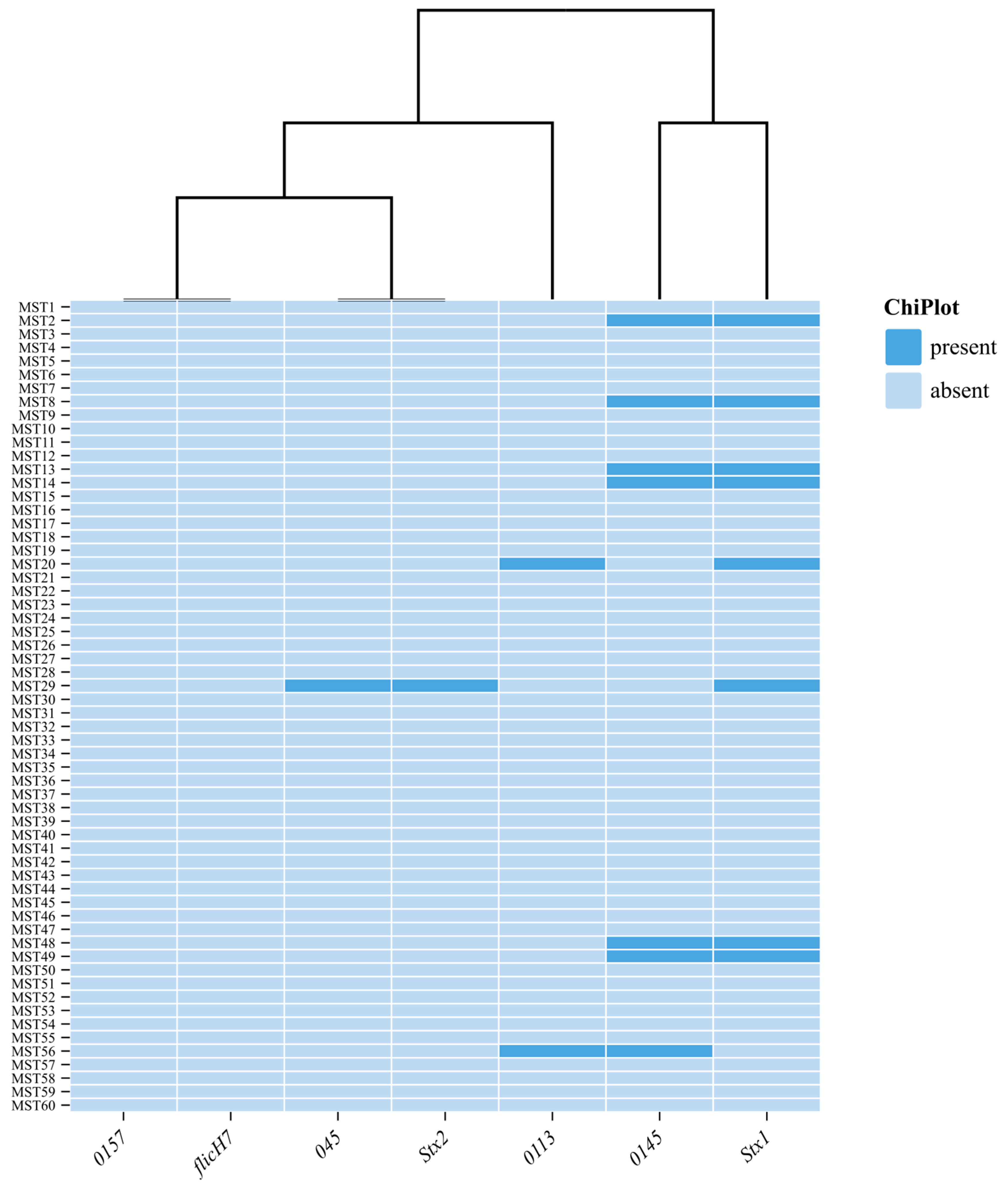
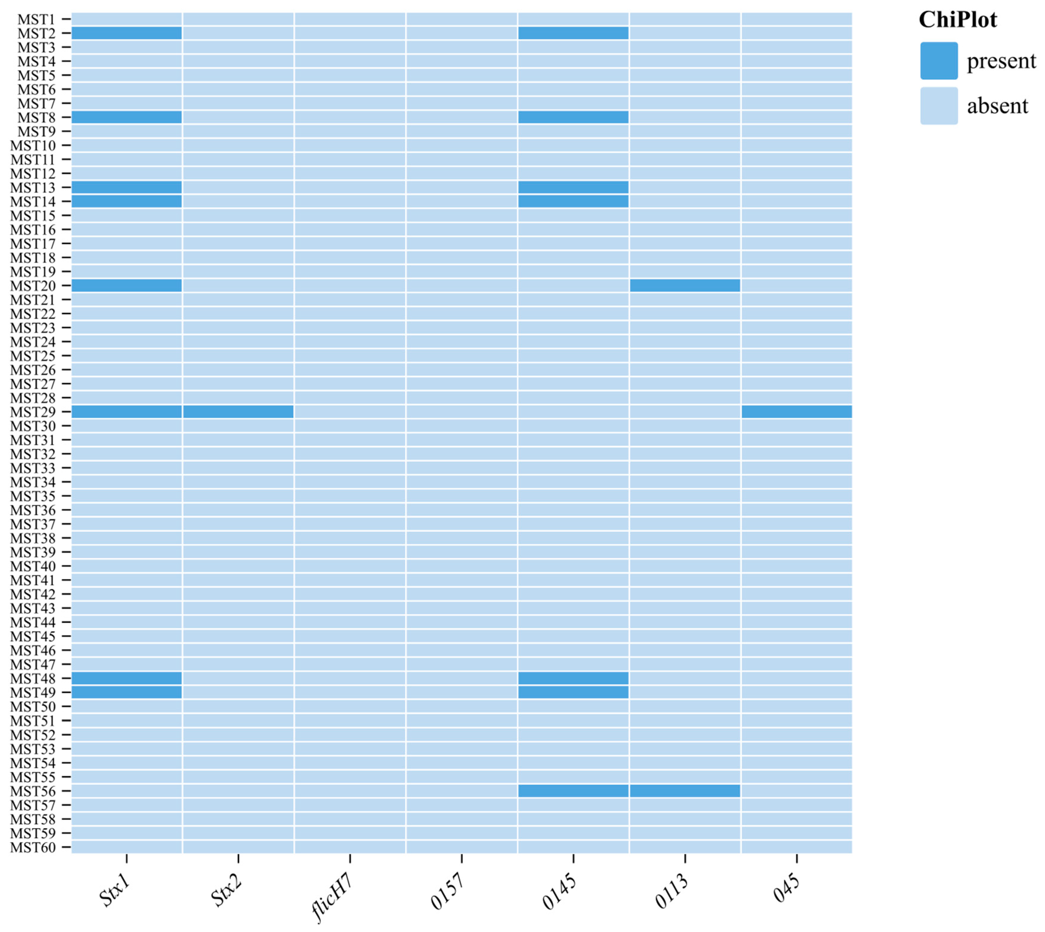
3.3. Detection of Virulent Genes and O Serogroups

The major virulence/toxin genes, including Enterohemorrhagic Flagella H7 E. coli (flicH7) and rfbO157, a virulence gene that codes for the O-antigen specific to the E. coli O157:H7 strain and serogroup 0157, were not detected in any of the isolates; nine (15%) isolates harbored stx1, and only one isolate harbored stx2. One (1.7%) harbored a combination of stx1 and stx2 genes. Shiga-like toxin-producing E. coli 045 and 0113 were detected in 7/60 (11.7%) and 2/60 (3.3%), respectively (Figure 1).

3.4. Antibiotic Sensitivity and Resistance Genes Detected in E. coli Isolates

According to the results of the disc diffusion test, the majority of the isolates were resistant to penicillin, followed by ciprofloxacin and gentamicin at 43/60 (71.6%), 42/60 (70%), and 18/60 (30%), respectively. Furthermore, 8/60 (13.3%) and 1/60 (1.6%) of the isolates were resistant to meropenem and imipenem, respectively (Figure 2). Genomic antimicrobial resistance profiling indicated that 100% of all isolates carried the ß-lactamase gene blaVIM, followed by blaKPC in 23/68 (33.82%) and blaNMD in 12/68 (17.64%). Moreover, in 12/68 (12.64%) of the isolates, the suli1 gene encoding for sulphonamide was also detected, while 1/68 (11.47%) of the isolates were found to have the msrA gene encoding for macrolides. Seven (11.6%) isolates were found to harbor both phenotypic and genotypic resistance to carbapenem.

3.5. Multidrug-Resistant E. coli Isolates

This study found that 30 out of 60 isolates (50%) resisted three or more antibiotics. Among these isolates, 15 (25%) were resistant to three antibiotics, 12 (20%) were resistant to four, 2 (3.3%) were resistant to six, and 1 (1.7%) was resistant to five antibiotics (Table 1). Of the nine strains of E. coli that produce Shiga toxin, only 77.7% were MDR.

4. Discussion

Small-scale dairy farming is an important economic sector for improving agriculture and livelihoods in developing nations [26,27]. According to the Food and Agriculture Organization’s 2014 assessment, mastitis is a multifactorial production illness that poses a concern to small-scale producers because of its high prevalence of over 50% [28]. Subclinical mastitis is one of the main issues facing the dairy sector worldwide. The most common cause of subclinical mastitis worldwide is E. coli [29].
The current study’s findings about the subclinical mastitis status of cows were consistent with reports of 32.5% and 33% subclinical mastitis, respectively [30]. In contrast to the results of the current study conducted in the same area, subclinical mastitis was found to be 61.60% in the district of Faisalabad’s conventional dairy systems [31]. Likewise, a recent study found that the same district had a 45.9% prevalence of subclinical mastitis [32]. Somatic cell count is a beneficial technique for tracking SCM at individual quarters [33]. The present investigation showed that SCCs increased with higher CMT scores and that the mean SCC was relatively high, ranging from 1.48 to 8.66 × 10⁵ cells/mL of milk. Dos Reis and colleagues concurred that SCCs are the cause of changes in milk supply and composition and increase with CMT scores [34]. Geographical location, breed, age, lactation stages, udder condition, number of parties, immunity, management, hygiene, and milking procedures on dairy farms all affect the incidence of mastitis in lactating cows [35,36].
The present study used PCR targeting the uidA gene and MALDI-TOF MS to confirm 60/68 (88.2%) of E. coli strains. Our findings were nearly identical to those of Islam et al. [36] in Bangladesh (75%) and Shakya et al. [37] in India (81.1%), who isolated the bacteria from goats, chicken meat, chevon meat, raw milk, and human urine and stool samples. However, studies conducted in South Africa by Ntuli et al. [38] (36%) and China by Liu et al. [39] (34.4%) revealed considerably contrasting outcomes, where their results showed a low prevalence of the isolate in bulk milk and individual milk from dairy herds. The high occurrence of E. coli in raw milk and milk products is of concern, as it has been associated with fecal contamination and the consequent risk of enteric pathogens in foodstuffs [40].
STEC is considered to be responsible for causing life-threatening diseases like hemorrhagic colitis. STEC is also considered a significant contributor to kidney failure in children. Hence, consumable milk and milk products contaminated with STEC are serious concerns [41]. The results of the current investigation show that 15% of the isolates harbored sxt1, while only one harbored the stx2 gene encoding for STEC. This is in agreement with the findings of Momtaz et al. [42], Tavakoli and Pourtaghi [43], and Mashak [44], who identified STEC in 13.88%, 22.2%, and 7.7% of the isolates reported from Shahrekord, Tehran, and Alborz Province, respectively, in Iran; the simultaneous presence of stx1 and stx2 genes was found in 20.51% of the STEC isolates, in contrast with the current study [42,43,44]. According to Momtaz et al. [42] and Zafarane et al. [45], raw milk from Iranian cows with subclinical mastitis showed a distribution of stx1 and stx2, with 15.06% and 11.11%, respectively. Their findings were supported by those of Brenjchi et al. [46], who found that raw bulk tank milk samples sent to Mashhad’s Pegah Pasteurization Factory exhibited a 6.15% frequency of STEC, with O157:H7 found in just one isolate (12.5%) of these samples.
In contrast, the current study found the prevalence of pathogenic STEC non-0157, which indicates milk contamination and potential public health risks in the dairy value chain system. The prevalence of Shiga-like toxin-producing E. coli 045 and 0113 was detected in 11.7% and 3.3%, respectively. This was in agreement with the results of Ullah et al. [47], who found STEC in 5% of bovine milk samples in Pakistan. By comparison, this subgroup was present in Germany at a rate of 24.7%, Egypt at 18.0%, California at 58.1%, and Spain at 35.9%. In this study, serogroup O157 was found to be non-existent, whereas other studies reported low prevalences of 3.8% in Spain and 0% in Egypt. The non-O157 STEC subgroup’s superiority over O157 is therefore consistent with the study conducted by Amézquita-López et al. [48], who reported the prevalence of O157 and non-O157 STEC ranging from 0.42 to 74% and 0.2 to 48.8%, respectively, in Mexico. The variations in prevalence values might be due to the differences in epidemiological determinants like stocking density, age, season, spatial distribution, sampling time, strategy, handling, and laboratory practices.
In the current investigation, the isolates tested positive for the blaKPC, blaNDM, and blaVIM carbapenemase genes, regardless of whether they were imipenem- or meropenem-resistant. Our study’s findings were consistent with those of another study conducted in Uganda by Okoche et al. [49], which also identified blaVIM, blaKPC, and blaNDM genes. However, the most common gene was blaVIM (10.7%), followed by blaKPC (5.1%) and blaNDM-1 (2.6%). A 2019 study by Hoelle et al. [50] in the USA revealed that roughly 55% of the E. coli isolates tested positive for the blaVIM gene and 1% for the blaIMP gene. These discrepancies may be the result of differences in the samples gathered, the location of the collection, and their large sample sizes. A previous study suggested that beta-lactamases by Gram-negative organisms are usually secreted, especially when antibiotics are present in the environment [51], which probably explains the observation of unexpressed genes in our strain collection.
This study’s results further revealed that most isolates were resistant to penicillin, followed by ciprofloxacin and gentamicin. Our study was similar to those of Zafarane et al. [45] and Tavakoli and Pourtaghi [43], who found that most E. coli isolates were resistant to penicillin in Iran. Moreover, a study by Tahar et al. [51] found that ciprofloxacin was also among the antibiotics that E. coli was greatly resistant to in Algeria, at 13.5%, even though it was lower than our prevalence. The rate of 18% for gentamicin was comparable to that found in Egypt (13.3%) [52], Tunisia (19.5%) [17], and China (12%) [16]. Furthermore, compared to published rates of 67.9%, 77%, and 49.2% in Brazil [53], Lebanon [54], and Iran [55], the current study’s results were in contrast. In this study, the prevalence of MDR E. coli was 30%. This prevalence was higher than the 14.8% recorded by Ngaywa et al. [56] in comparable investigations carried out in Egypt and Northern Kenya, respectively. Nevertheless, it was comparable to the 34.7% noted by Dowidar and Khalifa [57].

5. Conclusions

In conclusion, there is a serious risk to animal health, dairy productivity, and public safety due to the high frequency of subclinical mastitis (39.08%) and the predominance of Escherichia coli, especially multidrug-resistant strains that carry crucial resistance genes. The zoonotic potential of these pathogens and their function as antimicrobial resistance (AMR) reservoirs in the food production chain were highlighted by the identification of virulence genes (Stx1 and Stx2) and resistance determinants, including blaVIM, blaKPC, and blaNDM. In order to reduce productivity losses and stop the spread of infection, the results call for improved screening programs for the early detection and management of subclinical mastitis in dairy herds. To support evidence-based policy and One Health initiatives, ongoing AMR surveillance in cattle and the environment is crucial, as is genetic tracking of resistance genes.

Supplementary Materials

The following supporting information can be downloaded at: https://www.mdpi.com/article/10.3390/ani15131980/s1, Figure S1: The study site where samples were collected; Table S1: The primers used in this study.

Author Contributions

N.G.K.: writing—original draft, visualization, software, resources, methodology, investigation, formal analysis, data curation, funding acquisition, conceptualization, and project administration. O.d.S.: writing—review and editing. J.N.: writing—review and editing, supervision. K.E.L.: writing—review and editing, data curation. T.R.: writing—review and editing, methodology, data curation, supervision. O.T.: writing—review and editing, validation, supervision, and project administration. All authors have read and agreed to the published version of the manuscript.

Funding

This study was supported financially by the National Research Foundation (NRF) grant number: NFSG23042496860.

Institutional Review Board Statement

This study was approved by the Department of Agriculture, Land Reform, and Rural Development under Section 20 of the Animal Diseases Act, 1984 (Ref Number: 12/11/1/4/6 (6335 ON)) on 10 October 2024 and the Animal Research Ethics Committee (UFS-AED2024/0015) from the University of the Free State on 22 October 2024.

Informed Consent Statement

The farmers were verbally consulted and asked for consent to publish the results.

Data Availability Statement

All data generated in this study are contained within the manuscript.

Acknowledgments

The authors would like to acknowledge the dairy farmers and their staff for their support with sample collection.

Conflicts of Interest

The authors declare no conflicts of interest.

References

  1. Abebe, R.; Hatiya, H.; Abera, M.; Megersa, B.; Asmare, K. Bovine mastitis: Prevalence, risk factors and isolation of Staphylococcus aureus in dairy herds at Hawassa milk shed, South Ethiopia. BMC Veter. Res. 2016, 12, 270. [Google Scholar] [CrossRef] [PubMed]
  2. Seegers, H.; Fourichon, C.; Beaudeau, F. Production effects related to mastitis and mastitis economics in dairy cattle herds. Vet. Res. 2003, 34, 475–491. [Google Scholar] [CrossRef] [PubMed]
  3. González, R.N.; Wilson, D.J. Mycoplasmal mastitis in dairy herds. Veter. Clin. Food Anim. Pract. 2003, 19, 199–221. [Google Scholar] [CrossRef] [PubMed]
  4. Jeffrey, G.; Nehring, R.; Hallahan, C.; Sandretto, C. Pasture-based dairy systems: Who are the producers and are their operations more profitable than conventional dairies? J. Agric. Resour. Econ. 2009, 34, 412–427. [Google Scholar]
  5. Oliver, S.P.; Jayarao, B.M.; Almeida, R.A. Foodborne pathogens, mastitis, milk quality, and dairy food safety. In NMC Annual Meeting Proceedings; National Mastitis Council: New Prague, MN, USA, 2005; Volume 1, pp. 3–27. [Google Scholar]
  6. Hennekinne, J.-A.; De Buyser, M.-L.; Dragacci, S. Staphylococcus aureus and its food poisoning toxins: Characterization and outbreak investigation. FEMS Microbiol. Rev. 2012, 36, 815–836. [Google Scholar] [CrossRef]
  7. Burvenich, C.; Van Merris, V.; Mehrzad, J.; Diez-Fraile, A.; Duchateau, L. Severity of E. coli mastitis is mainly determined by cow factors. Vet. Res. 2003, 34, 521–564. [Google Scholar] [CrossRef]
  8. Abebe, M.; Hailelule, A.; Abrha, B.; Nigus, A.; Birhanu, M.; Adane, H.; Genene, T.; Daniel, H.; Getachew, G.; Merga, G.; et al. Antibiogram of Escherichia coli strains isolated from food of bovine origin in selected Woredas of Tigray, Ethiopia. J. Bacteriol. Res. 2014, 6, 17–22. [Google Scholar]
  9. CfD Control. Prevention. Outbreaks of Escherichia coli O157: H7 infections among children associated with farm visits–Pennsylvania and Washington, 2000. MMWR Morb. Mortal. Wkly. Rep. 2001, 50, 293–297. [Google Scholar]
  10. Boerlin, P.; McEwen, S.A.; Boerlin-Petzold, F.; Wilson, J.B.; Johnson, R.P.; Gyles, C.L. Associations between virulence factors of Shiga toxin-producing Escherichia coli and disease in humans. J. Clin. Microbiol. 1999, 37, 497–503. [Google Scholar] [CrossRef]
  11. Bag, M.S.; Khan, M.R.; Sami, M.H.; Begum, F.; Islam, M.; Rahman, M.; Rahman, M.; Hassan, J. Virulence determinants and antimicrobial resistance of E. coli isolated from bovine clinical mastitis in some selected dairy farms of Bangladesh. Saudi J. Biol. Sci. 2021, 28, 6317–6323. [Google Scholar] [CrossRef]
  12. Pereira, E.d.S.; Crippa, B.L.; Morasi, R.M.; de Almeida, J.M.; Gebara, C.; Langoni, H.; Neto, A.T.; Gonçalves, M.C.; Silva, N.C.C. Identification of enteropathogenic Escherichia coli as the cause of mastitis in cows from Brazil. Pesqui. Veter. Bras. 2024, 44, e07430. [Google Scholar] [CrossRef]
  13. Goulart, D.B.; Mellata, M. Escherichia coli mastitis in dairy cattle: Etiology, diagnosis, and treatment challenges. Front. Microbiol. 2022, 13, 928346. [Google Scholar] [CrossRef] [PubMed]
  14. Frieri, M.; Kumar, K.; Boutin, A. Antibiotic resistance. J. Infect. Public Health 2017, 10, 369–378. [Google Scholar] [CrossRef] [PubMed]
  15. Eisenberger, D.; Carl, A.; Balsliemke, J.; Kämpf, P.; Nickel, S.; Schulze, G.; Valenza, G. Molecular characterization of extended-spectrum β-lactamase-producing Escherichia coli isolates from milk samples of dairy cows with mastitis in Bavaria, Germany. Microb. Drug Resist. 2018, 24, 505–510. [Google Scholar] [CrossRef]
  16. Yu, Z.; Wang, J.; Ho, H.; Wang, Y.; Huang, S.; Han, R. Prevalence and antimicrobial-resistance phenotypes and genotypes of Escherichia coli isolated from raw milk samples from mastitis cases in four regions of China. J. Glob. Antimicrob. Resist. 2020, 22, 94–101. [Google Scholar] [CrossRef]
  17. Saidani, M.; Messadi, L.; Soudani, A.; Daaloul-Jedidi, M.; Châtre, P.; Ben Chehida, F.; Mamlouk, A.; Mahjoub, W.; Madec, J.-Y.; Haenni, M. Epidemiology, antimicrobial resistance, and extended-spectrum beta-lactamase-producing enterobacteriaceae in clinical bovine mastitis in Tunisia. Microb. Drug Resist. 2018, 24, 1242–1248. [Google Scholar] [CrossRef]
  18. Karzis, J.; Donkin, E.F.; Webb, E.C.; Etter, E.M.; Petzer, I.M. Somatic cell count thresholds in composite and quarter milk samples as indicator of bovine intramammary infection status. Onderstepoort J. Veter. Res. 2017, 84, 1–10. [Google Scholar] [CrossRef]
  19. Khasapane, N.G.; Koos, M.; Nkhebenyane, S.J.; Khumalo, Z.T.H.; Ramatla, T.; Thekisoe, O. Detection of Staphylococcus isolates and their antimicrobial resistance profiles and virulence genes from subclinical mastitis cattle milk using MALDI-TOF MS, PCR and sequencing in free state province, South Africa. Animals 2024, 14, 154. [Google Scholar] [CrossRef]
  20. Cameron, M.; Perry, J.; Middleton, J.; Chaffer, M.; Lewis, J.; Keefe, G. Evaluation of MALDI-TOF mass spectrometry and a custom reference spectrum expanded database for the identification of bovine-associated coagulase-negative staphylococci. J. Dairy Sci. 2018, 101, 590–595. [Google Scholar] [CrossRef]
  21. Nessa, K.; Ahmed, D.; Islam, J.; Kabir, F.L.; Hossain, M.A. Usefulness of a multiplex PCR for detection of diarrheagenic Escherichia coli in a diagnostic microbiology laboratory setting. Bangladesh J. Med. Microbiol. 2007, 1, 38–42. [Google Scholar] [CrossRef]
  22. Ramatla, T.; Tutubala, M.; Motlhaping, T.; de Wet, L.; Mokgokong, P.; Thekisoe, O.; Lekota, K. Molecular detection of Shiga toxin and extended-spectrum beta-lactamase (ESBL)-producing Escherichia coli isolates from sheep and goats. Mol. Biol. Rep. 2024, 51, 57. [Google Scholar] [CrossRef] [PubMed]
  23. Abdlla, Y.; Sheet, O.H.; Alsanjary, R.A.; Plötz, M.; Abdulmawjood, A.A. Isolation and Identification of Escherichia coli from Buffalo’s Milk using PCR Technique in Nineveh Governorate. Egypt. J. Veter. Sci. 2024, 55, 1881–1887. [Google Scholar] [CrossRef]
  24. CLSI (Clinical and Laboratory Standards Institute). Performance Standards for Antimicrobial Disk and Dilution Susceptibility Tests for Bacteria Isolated from Animals, 6th ed.; CLSI Document VET01-S; CLSI: Pittsburgh, PA, USA, 2023. [Google Scholar]
  25. Dehkordi, F.S.; Yazdani, F.; Mozafari, J.; Valizadeh, Y. Virulence factors, serogroups and antimicrobial resistance properties of Escherichia coli strains in fermented dairy products. BMC Res. Notes 2014, 7, 217. [Google Scholar] [CrossRef]
  26. Ranjbar, R.; Dehkordi, F.S.; Shahreza, M.H.S.; Rahimi, E. Prevalence, identification of virulence factors, O-serogroups and antibiotic resistance properties of Shiga-toxin producing Escherichia coli strains isolated from raw milk and traditional dairy products. Antimicrob. Resist. Infect. Control. 2018, 7, 53. [Google Scholar] [CrossRef]
  27. Thrusfield, M. Veterinary Epidemiology; John Wiley & Sons: Hoboken, NJ, USA, 2018. [Google Scholar]
  28. FAO; GDP; IFCN. Dairy Development’s Impact on Poverty Reduction; FAO: Chicago, IL, USA, 2018. [Google Scholar]
  29. Mekonnen, S.; Koop, G.; Melkie, S.; Getahun, C.; Hogeveen, H.; Lam, T. Prevalence of subclinical mastitis and associated risk factors at cow and herd level in dairy farms in North-West Ethiopia. Prev. Veter. Med. 2017, 145, 23–31. [Google Scholar] [CrossRef]
  30. Fahim, K.M.; Ismael, E.; Khalefa, H.S.; Farag, H.S.; Hamza, D.A. Isolation and characterization of E. coli strains causing intramammary infections from dairy animals and wild birds. Int. J. Veter. Sci. Med. 2019, 7, 61–70. [Google Scholar] [CrossRef]
  31. Zuo, J.; Lv, Z.; Lian, L.; Wu, Z.; Fu, S.; Zhang, H.; Wu, J.; Pan, Z.; Yu, Y.; Chen, W.; et al. Difference Analysis on Virulence Genes, Biofilms and Antimicrobial Susceptibility of Escherichia coli from Clinical and Subclinical Bovine Mastitis. Veter. Sci. 2025, 12, 132. [Google Scholar] [CrossRef]
  32. Naseer, M.A.; Aqib, A.I.; Ashar, A.; Saleem, M.I.; Shoaib, M.; Kulyar, M.F.-E.; Ashfaq, K.; Bhutta, Z.A.; Nighat, S. Detection of Altered Pattern of Antibiogram and Biofilm Character in Staphylococcus aureus Isolated from Dairy Milk. Pak. J. Zool. 2021, 53, 191–199. [Google Scholar] [CrossRef]
  33. Javed; Umar, M.; Ijaz, M.; Fatima, Z.; Anjum, A.A.; Aqib, A.I.; Ali, M.M.; Rehman, A.; Ahmed, A.; Ghaffar, A. Frequency and antimicrobial susceptibility of methicillin and vancomycin-resistant Staphylococcus aureus from bovine milk. Pak. Veter. J. 2021, 41, 463–468. [Google Scholar] [CrossRef]
  34. Abed, A.H.; Menshawy, A.M.S.; Zeinhom, M.M.A.; Hossain, D.; Khalifa, E.; Wareth, G.; Awad, M.F. Subclinical mastitis in selected bovine dairy herds in North Upper Egypt: Assessment of prevalence, causative bacterial pathogens, antimicrobial resistance and virulence-associated genes. Microorganisms 2021, 9, 1175. [Google Scholar] [CrossRef]
  35. Hope, A. Laboratory Handbook on Bovine Mastitis. Aust. Veter- J. 2000, 78, 488. [Google Scholar] [CrossRef]
  36. Islam, K.; Ahad, A.; Barua, M.; Islam, A.; Chakma, S.; Dorji, C.; Uddin, M.A.; Islam, S.; Ahasan, A.S.M.L. Isolation and epidemiology of multidrug resistant Escherichia coli from goats in Cox’s Bazar, Bangladesh. J. Adv. Veter. Anim. Res. 2016, 3, 166–172. [Google Scholar] [CrossRef]
  37. Bhoomika; Shakya, S.; Patyal, A.; Gade, N.E. Occurrence and characteristics of extended-spectrum β-lactamases producing Escherichia coli in foods of animal origin and human clinical samples in Chhattisgarh, India. Veter. World 2016, 9, 996–1000. [Google Scholar] [CrossRef] [PubMed]
  38. Ntuli, V.; Njage, P.; Buys, E. Characterization of Escherichia coli and other Enterobacteriaceae in producer-distributor bulk milk. J. Dairy Sci. 2016, 99, 9534–9549. [Google Scholar] [CrossRef]
  39. Liu, H.; Meng, L.; Dong, L.; Zhang, Y.; Wang, J.; Zheng, N. Prevalence, antimicrobial susceptibility, and molecular characterization of Escherichia coli isolated from raw milk in dairy herds in Northern China. Front. Microbiol. 2021, 12, 730656. [Google Scholar] [CrossRef]
  40. Ramírez-Castillo, F.Y.; Moreno-Flores, A.C.; Avelar-González, F.J.; Márquez-Díaz, F.; Harel, J.; Guerrero-Barrera, A.L. An evaluation of multidrug-resistant Escherichia coli isolates in urinary tract infections from Aguascalientes, Mexico: Cross-sectional study. Ann. Clin. Microbiol. Antimicrob. 2018, 17, 34. [Google Scholar] [CrossRef]
  41. Ashraf, A.; Imran, M.; Chang, Y.-F. Antimicrobial resistance of Escherichia coli isolates from mastitic milk and its possible relationship with resistance and virulence genes. Pak. J. Zool. 2018, 50, 1435–1441. [Google Scholar] [CrossRef]
  42. Momtaz, H.; Rahimi, E.; Moshkelani, S. Molecular detection of antimicrobial resistance genes in E. coli isolated from slaughtered commercial chickens in Iran. Veter. Med. 2012, 57, 193–197. [Google Scholar] [CrossRef]
  43. Tavakoli, M.; Pourtaghi, H. Molecular detection of virulence genes and multi-drug resistance patterns in Escherichia coli (STEC) in clinical bovine mastitis: Alborz province, Iran. Iran. J. Vet. Res. 2017, 18, 208–211. [Google Scholar]
  44. Mashak, Z. Prevalence and antibiotic resistance of Escherichia coli O157:H7 isolated from raw meat samples of ruminants and poultry. J. Food Nutr. Res. 2018, 6, 96–102. [Google Scholar] [CrossRef]
  45. Zafarane, S.; Houri, H.; Kazemian, H.; Heidari, H.; Amiri, P.; Tabarraei, B. Characterization of virulence genes, serogroups and antimicrobial susceptibility of Shiga toxin producing Escherichia coli isolated from bovine mastitic milk in Tehran, Iran. Trop Biomed. 2017, 34, 295–304. [Google Scholar] [PubMed]
  46. Brenjchi, M.; Jamshidi, A.; Farzaneh, N.; Bassami, M.R. Short paper: Identification of shiga toxin producing Escherichia coli o157: H7 in raw cow milk samples from dairy farms in mashhad using multiplex pcr assay. Iran. J. Vet. Res. 2011, 12, 145–149. [Google Scholar]
  47. Ullah, S.; Khan, S.U.H.; Ali, T.; Zeb, M.T.; Riaz, M.H.; Khan, S.; Goyal, S.M.; Rouby, S.R. Molecular characterization and antibiotic susceptibility of Shiga toxin- producing Escherichia coli (STEC) isolated from raw milk of dairy bovines in Khyber Pakhtunkhwa, Pakistan. PLoS ONE 2024, 19, e0307830. [Google Scholar] [CrossRef] [PubMed]
  48. Amézquita-López, B.A.; Quiñones, B.; Cooley, M.B.; León-Félix, J.; Campo, N.C.-D.; Mandrell, R.E.; Jiménez, M.; Chaidez, C.; Ibekwe, A.M. Genotypic analyses of shiga toxin-producing Escherichia coli O157 and Non-O157 recovered from feces of domestic animals on rural farms in Mexico. PLoS ONE 2012, 7, e51565. [Google Scholar] [CrossRef]
  49. Okoche, D.; Asiimwe, B.B.; Katabazi, F.A.; Kato, L.; Najjuka, C.F.; Zhang, Q. Prevalence and characterization of carbapenem-resistant Enterobacteriaceae isolated from Mulago National Referral Hospital, Uganda. PLoS ONE 2015, 10, e0135745. [Google Scholar] [CrossRef]
  50. Hoelle, J.; Johnson, J.R.; Johnston, B.D.; Kinkle, B.; Boczek, L.; Ryu, H.; Hayes, S. Survey of US wastewater for carbapenem-resistant Enterobacteriaceae. J. Water Health 2019, 17, 219–226. [Google Scholar] [CrossRef]
  51. Tahar, S.; Nabil, M.M.; Safia, T.; Ngaiganam, E.P.; Omar, A.; Hafidha, C.; Hanane, Z.; Rolain, J.-M.; Diene, S.M. Molecular characterization of multidrug-resistant Escherichia coli isolated from milk of dairy cows with clinical mastitis in Algeria. J. Food Prot. 2020, 83, 2173–2178. [Google Scholar] [CrossRef]
  52. Ameen, F.; Reda, S.A.; El-Shatoury, S.A.; Riad, E.M.; Enany, M.E.; Alarfaj, A.A. Prevalence of antibiotic resistant mastitis pathogens in dairy cows in Egypt and potential biological control agents produced from plant endophytic actinobacteria. Saudi J. Biol. Sci. 2019, 26, 1492–1498. [Google Scholar] [CrossRef]
  53. Rangel, P.M.; Marin, J.M. Antimicrobial resistance in brazilian isolates of Shiga toxin-encoding Escherichia coli from cows with mastitis. Ars Vet. Jaboticabal SP 2009, 25, 18–23. [Google Scholar]
  54. Barbour, E.K.; Kassabian, T.J.; Shaib, H.; Kassaify, Z.; Iyer, A.; Azhar, E.; Harakeh, S.; Kumosani, T. The significance of Escherichia coli-induced mastitis in cows associated with the presence of virulence genes and wide range-resistance to twenty antimicrobials. Int. J. Appl. Res. Vet. Med. 2015, 13, 51–63. [Google Scholar]
  55. Fazel, F.; Jamshidi, A.; Khoramian, B. Phenotypic and genotypic study on antimicrobial resistance patterns of E. coli isolates from bovine mastitis. Microb. Pathog. 2019, 132, 355–361. [Google Scholar] [CrossRef] [PubMed]
  56. Ngaywa, C.; Aboge, G.O.; Obiero, G.; Omwenga, I.; Ngwili, N.; Wamwere, G.; Wainaina, M.; Bett, B. Antimicrobial resistant Escherichia coli isolates detected in raw milk of livestock in pastoral areas of northern Kenya. Food Control. 2019, 102, 173–178. [Google Scholar] [CrossRef]
  57. Dowidar, H.A.; Khalifa, M.I. Molecular Isolation and Identification of Multidrug-resistant Escherichia coli from Milk, Meat, and Product Samples. J. Adv. Vet. Res. 2023, 13, 643–646. Available online: https://advetresearch.com/index.php/AVR/article/view/1267 (accessed on 15 March 2025).
Figure 1. The number of detected virulence genes and serotypes from E. coli strains.
Animals 15 01980 g001
Figure 2. Phenotypic AMR results and their respective AMR genes detected in E. coli strains.
Animals 15 01980 g002
Table 1. Multi-drug resistance Escherichia coli isolates.
Antibiotic DisksNumber of IsolatesPercentage
P, CIP, MEM11.6
P, TET, IMP11.6
P, CIP, CN813.3
P, CIP, AMP 35
P, CIP, AMP, CN 11.6
P, CIP, AMP, E 23.3
P, CIP, AMP, TET46.6
P, CIP, E, TET 11.6
P, CIP, CN, MEM11.6
P, AMP, MEM, CN23.3
P, AMP, MEM, IMP11.6
E, AMP, CIP11.6
E, CN, CIP, MEM11.6
AMP, CN, CIP11.6
P, CIP, CN, IMP, MEM11.6
P, E, TET, CIP, IMP, MEM11.6
P = Penicillin, CIP = Ciprofloxacin, CN = Gentamicin, MEM = Meropenem, E = Erythromycin, TET = Tetracycline, AMP = Ampicillin.
Disclaimer/Publisher’s Note: The statements, opinions and data contained in all publications are solely those of the individual author(s) and contributor(s) and not of MDPI and/or the editor(s). MDPI and/or the editor(s) disclaim responsibility for any injury to people or property resulting from any ideas, methods, instructions or products referred to in the content.

