
Surgical Correction of a Sinus Venosus Atrial Septal Defect with Partial Anomalous Pulmonary Venous Connections Using Cardiac Computed Tomography Imaging and a 3D-Printed Model
 , , , , , , and
Abstract
Simple Summary
Abstract
1. Introduction
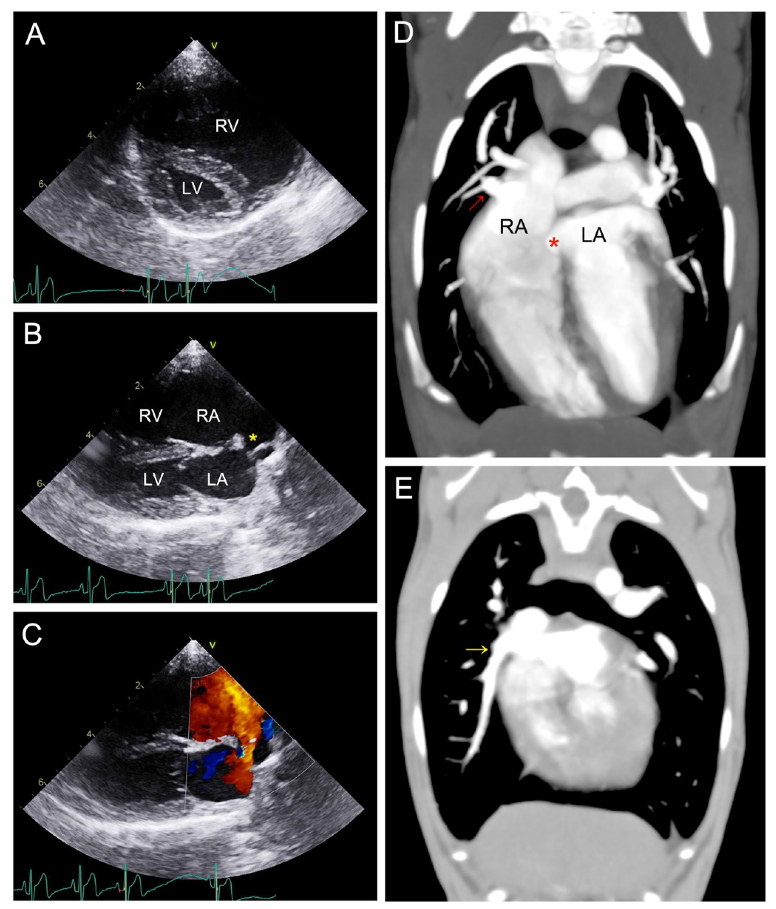
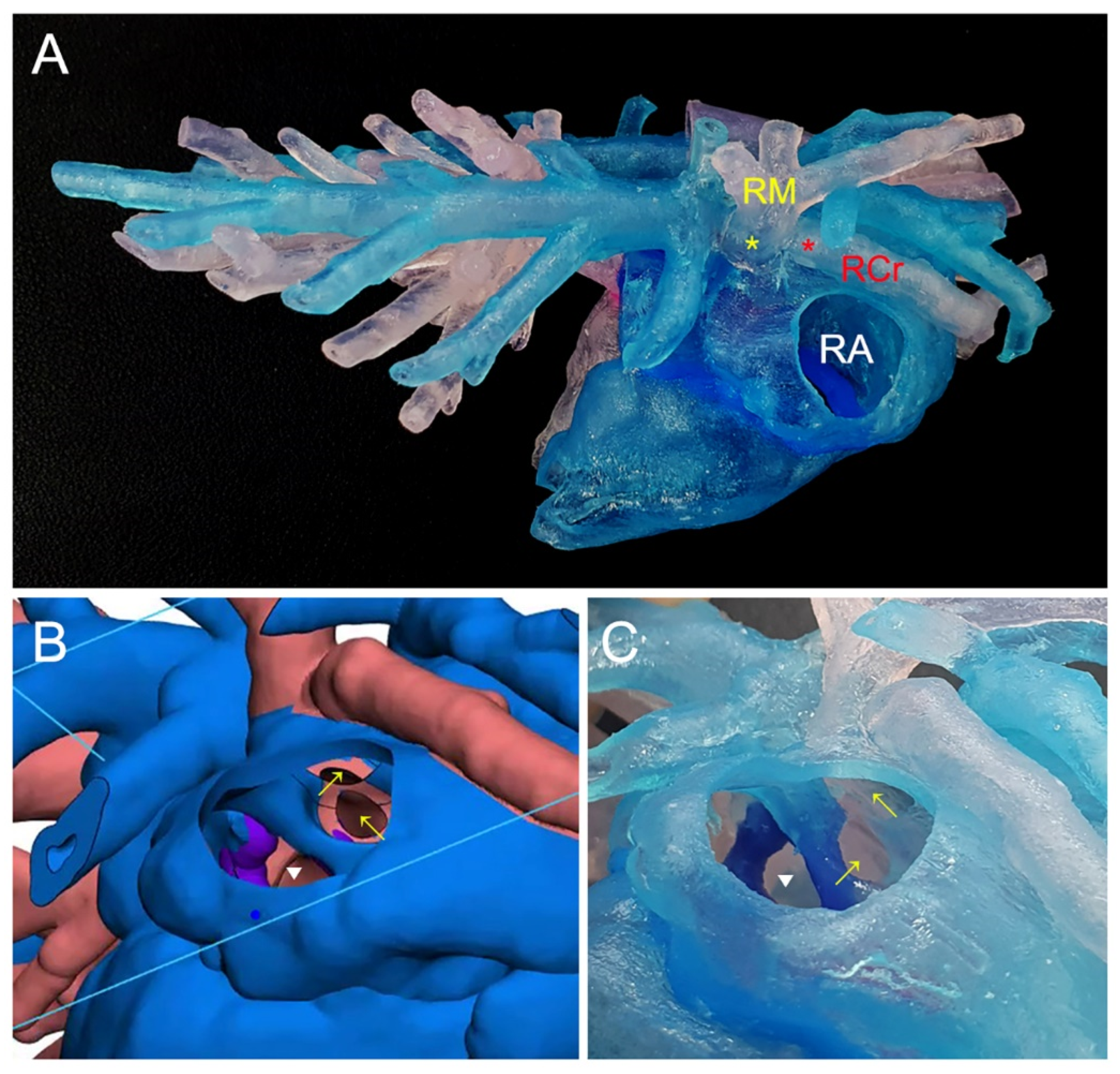
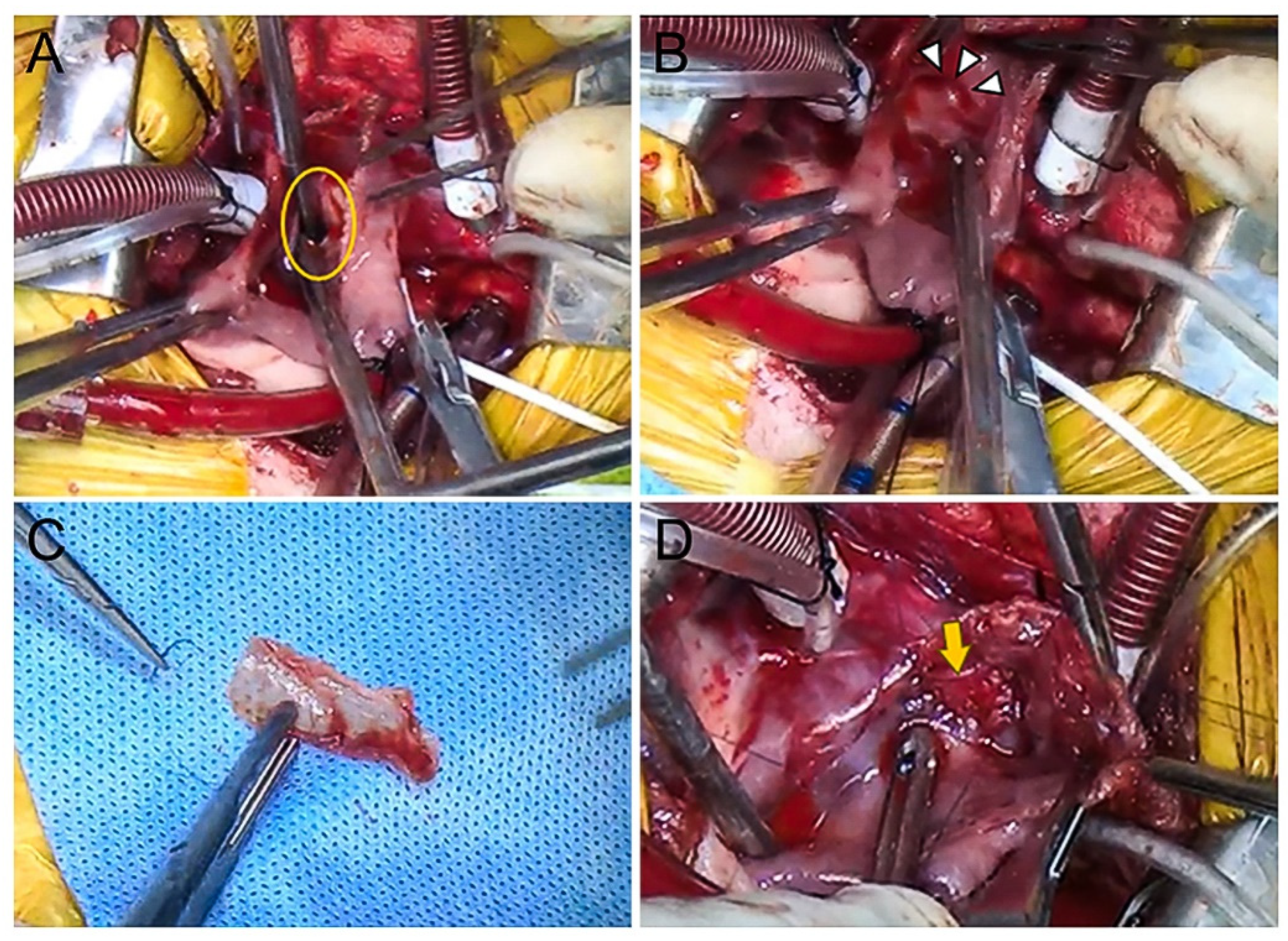
2. Case
3. Discussion
4. Conclusions
Author Contributions
Funding
Institutional Review Board Statement
Informed Consent Statement
Data Availability Statement
Acknowledgments
Conflicts of Interest
References
- Oliveira, P.; Domenech, O.; Silva, J.; Vannini, S.; Bussadori, R.; Bussadori, C. Retrospective review of congenital heart disease in 976 dogs. J. Vet. Intern. Med. 2011, 25, 477–483. [Google Scholar] [CrossRef] [PubMed]
- Chetboul, V.; Charles, V.; Nicolle, A.; Sampedrano, C.C.; Gouni, V.; Pouchelon, J.L.; Tissier, R. Retrospective study of 156 atrial septal defects in dogs and cats (2001–2005). J. Vet. Med. A Physiol. Pathol. Clin. Med. 2006, 53, 179–184. [Google Scholar] [CrossRef] [PubMed]
- Paslawska, U.; Noszczyk-Nowak, A.; Cepiel, A.; Staszczyk, M.; Janiszewski, A. Atrial septal defect ostium primum type in a dog—A case report. Electron. J. Pol. Agric. Univ. 2013, 16, 6. [Google Scholar]
- Torres, A.J. Hemodynamic assessment of atrial septal defects. J. Thorac. Dis. 2018, 10, S2882–S2889. [Google Scholar] [CrossRef] [PubMed]
- Fraisse, A.; Latchman, M.; Sharma, S.R.; Bayburt, S.; Amedro, P.; di Salvo, G.; Baruteau, A.E. Atrial septal defect closure: Indications and contra-indications. J. Thorac. Dis. 2018, 10, S2874–S2881. [Google Scholar] [CrossRef] [PubMed]
- Stout, K.K.; Daniels, C.J.; Aboulhosn, J.A.; Bozkurt, B.; Broberg, C.S.; Colman, J.M.; Crumb, S.R.; Dearani, J.A.; Fuller, S.; Gurvitz, M.; et al. 2018 AHA/ACC Guideline for the Management of Adults with Congenital Heart Disease: Executive Summary: A Report of the American College of Cardiology/American Heart Association Task Force on Clinical Practice Guidelines. J. Am. Coll. Cardiol. 2019, 73, 1494–1563, Correction in J. Am. Coll. Cardiol. 2019, 73, 2361. [Google Scholar] [CrossRef] [PubMed]
- Mizuno, T.; Mizuno, M.; Harada, K.; Takano, H.; Shinoda, A.; Takahashi, A.; Mamada, K.; Takamura, K.; Chen, A.; Iwanaga, K.; et al. Surgical correction for sinus venosus atrial septal defect with partial anomalous pulmonary venous connection in a dog. J. Vet. Cardiol. 2020, 28, 23–30. [Google Scholar] [CrossRef] [PubMed]
- Han, C.; Liu, C.; Sun, X.; Pan, S. Surgical treatment of partial anomalous pulmonary venous connection misdiagnosed as atrial septal defect underwent transcatheter occlusion: A case report. Clin. Case Rep. 2021, 9, 2345–2349. [Google Scholar] [CrossRef] [PubMed]
- Dillman, J.R.; Hernandez, R.J. Role of CT in the evaluation of congenital cardiovascular disease in children. Am. J. Roentgenol. 2009, 192, 1219–1231. [Google Scholar] [CrossRef]
- Desjardins, B.; Kazerooni, E.A. ECG-gated cardiac CT. Am. J. Roentgenol. 2004, 182, 993–1010. [Google Scholar] [CrossRef]
- Kappanayil, M.; Koneti, N.R.; Kannan, R.R.; Kottayil, B.P.; Kumar, K. Three-dimensional-printed cardiac prototypes aid surgical decision-making and preoperative planning in selected cases of complex congenital heart diseases: Early experience and proof of concept in a resource-limited environment. Ann. Pediatr. Cardiol. 2017, 10, 117–125. [Google Scholar] [CrossRef]
- Kim, M.K.; Kim, S.M.; Kim, E.K.; Chang, S.A.; Jun, T.G.; Choe, Y.H. Three-Dimensional Printed Model of Partial Anomalous Pulmonary Venous Return with Biatrial Connection. Taehan Yongsang Uihakhoe Chi. 2020, 81, 1523–1528. [Google Scholar] [CrossRef]
- Serres, F.; Chetboul, V.; Gouni, V.; Tissier, R.; Sampedrano, C.C.; Pouchelon, J.L. Diagnostic value of echo-Doppler and tissue Doppler imaging in dogs with pulmonary arterial hypertension. J. Vet. Intern. Med. 2007, 21, 1280–1289. [Google Scholar] [CrossRef] [PubMed]
- Serres, F.; Chetboul, V.; Tissier, R.; Gouni, V.; Desmyter, A.; Sampedrano, C.C.; Pouchelon, J.L. Quantification of pulmonary to systemic flow ratio by a Doppler echocardiographic method in the normal dog: Repeatability, reproducibility, and reference ranges. J. Vet. Cardiol. 2009, 11, 23–29. [Google Scholar] [CrossRef]
- Malik, P. Grossman’s Cardiac Catheterization, Angiography, and Intervention, 7th edn (2005). Can. J. Cardiol. 2007, 23, 602. [Google Scholar] [CrossRef]
- Gordon, S.G.; Miller, M.W.; Roland, R.M.; Saunders, A.B.; Achen, S.E.; Drourr, L.T.; Nelson, D.A. Transcatheter atrial septal defect closure with the Amplatzer atrial septal occluder in 13 dogs: Short- and mid-term outcome. J. Vet. Intern. Med. 2009, 23, 995–1002. [Google Scholar] [CrossRef]
- Jost, C.H.A.; Connolly, H.M.; Danielson, G.K.; Bailey, K.R.; Schaff, H.V.; Shen, W.K.; Warnes, C.A.; Seward, J.B.; Puga, F.J.; Tajik, A.J. Sinus venosus atrial septal defect: Long-term postoperative outcome for 115 patients. Circulation 2005, 112, 1953–1958. [Google Scholar] [CrossRef] [PubMed]
- Shimajiri, H.; Harada, Y.; Kinoshita, M.; Mikami, S. Sinus venosus atrial septal defect and partial anomalous pulmonary venous connection in a patient with dextrocardia. BMJ Case Rep. 2022, 15, e245523. [Google Scholar] [CrossRef]
- Sahay, S.; Krasuski, R.A.; Tonelli, A.R. Partial anomalous pulmonary venous connection and pulmonary arterial hypertension. Respirology 2012, 17, 957–963. [Google Scholar] [CrossRef]
- Yang, J.C.; Lin, M.T.; Jaw, F.S.; Chen, S.J.; Wang, J.K.; Shih, T.T.; Wu, M.H.; Li, Y.W. Trends in the utilization of computed tomography and cardiac catheterization among children with congenital heart disease. J. Formos. Med. Assoc. 2015, 114, 1061–1068. [Google Scholar] [CrossRef]
- Schmauss, D.; Haeberle, S.; Hagl, C.; Sodian, R. Three-dimensional printing in cardiac surgery and interventional cardiology: A single-centre experience. Eur. J. Cardiothorac. Surg. 2015, 47, 1044–1052. [Google Scholar] [CrossRef] [PubMed]
- Lee, S.; Squelch, A.; Sun, Z. Quantitative Assessment of 3D Printed Model Accuracy in Delineating Congenital Heart Disease. Biomolecules 2021, 11, 270. [Google Scholar] [CrossRef] [PubMed]
- Sun, Z.; Lau, I.; Wong, Y.H.; Yeong, C.H. Personalized Three-Dimensional Printed Models in Congenital Heart Disease. J. Clin. Med. 2019, 8, 522. [Google Scholar] [CrossRef] [PubMed]
- Sun, Z.; Wee, C. 3D Printed Models in Cardiovascular Disease: An Exciting Future to Deliver Personalized Medicine. Micromachines 2022, 13, 1575. [Google Scholar] [CrossRef] [PubMed]
- Seo, J.W.; Kim, J.S.; Cha, M.J.; Yoon, J.K.; Kim, M.J.; Tsao, H.M.; Lee, C.H.; Oh, S. Surgical and Electrical Anatomy of the Inter-Nodal and Intra-Atrial Conduction System in the Heart. J. Chest Surg. 2022, 55, 364–377. [Google Scholar] [CrossRef]
- Batra, A.S.; Balaji, S. Post operative temporary epicardial pacing: When, how and why? Ann. Pediatr. Cardiol. 2008, 1, 120–125. [Google Scholar] [CrossRef]
- Gregoratos, G.; Cheitlin, M.D.; Conill, A.; Epstein, A.E.; Fellows, C.; Ferguson, T.B., Jr.; Freedman, R.A.; Hlatky, M.A.; Naccarelli, G.V.; Saksena, S.; et al. ACC/AHA Guidelines for Implantation of Cardiac Pacemakers and Antiarrhythmia Devices: Executive Summary—A report of the American College of Cardiology/American Heart Association Task Force on Practice Guidelines (Committee on Pacemaker Implantation). Circulation 1998, 97, 1325–1335. [Google Scholar] [CrossRef]

Disclaimer/Publisher’s Note: The statements, opinions and data contained in all publications are solely those of the individual author(s) and contributor(s) and not of MDPI and/or the editor(s). MDPI and/or the editor(s) disclaim responsibility for any injury to people or property resulting from any ideas, methods, instructions or products referred to in the content. |
© 2024 by the authors. Licensee MDPI, Basel, Switzerland. This article is an open access article distributed under the terms and conditions of the Creative Commons Attribution (CC BY) license (https://creativecommons.org/licenses/by/4.0/).
Share and Cite
Kim, K.-M.; Moon, C.-H.; Lee, W.-J.; Kim, W.-J.; Kim, M.; Jeong, J.; Lee, H.-B.; Jeong, S.-M.; Choi, H.-J.; Hwang, T.S.; et al. Surgical Correction of a Sinus Venosus Atrial Septal Defect with Partial Anomalous Pulmonary Venous Connections Using Cardiac Computed Tomography Imaging and a 3D-Printed Model. Animals 2024, 14, 1094. https://doi.org/10.3390/ani14071094
Kim K-M, Moon C-H, Lee W-J, Kim W-J, Kim M, Jeong J, Lee H-B, Jeong S-M, Choi H-J, Hwang TS, et al. Surgical Correction of a Sinus Venosus Atrial Septal Defect with Partial Anomalous Pulmonary Venous Connections Using Cardiac Computed Tomography Imaging and a 3D-Printed Model. Animals. 2024; 14(7):1094. https://doi.org/10.3390/ani14071094
Chicago/Turabian StyleKim, Kyung-Min, Chang-Hwan Moon, Won-Jong Lee, Woo-Jin Kim, Mihyung Kim, Jaemin Jeong, Hae-Beom Lee, Seong-Mok Jeong, Ho-Jung Choi, Tae Sung Hwang, and et al. 2024. "Surgical Correction of a Sinus Venosus Atrial Septal Defect with Partial Anomalous Pulmonary Venous Connections Using Cardiac Computed Tomography Imaging and a 3D-Printed Model" Animals 14, no. 7: 1094. https://doi.org/10.3390/ani14071094
APA StyleKim, K.-M., Moon, C.-H., Lee, W.-J., Kim, W.-J., Kim, M., Jeong, J., Lee, H.-B., Jeong, S.-M., Choi, H.-J., Hwang, T. S., Lee, H. C., Yu, J. H., Nam, A., & Kim, D.-H. (2024). Surgical Correction of a Sinus Venosus Atrial Septal Defect with Partial Anomalous Pulmonary Venous Connections Using Cardiac Computed Tomography Imaging and a 3D-Printed Model. Animals, 14(7), 1094. https://doi.org/10.3390/ani14071094