

Computed Tomographic Findings in Dogs with Hepatic Bacterial Parenchymal Infection and Abscessation

 , , ,
, , ,
Simple Summary
Abstract
1. Introduction
2. Materials and Methods
2.1. Selection and Description of Subjects
2.2. Data Recording and Analysis
2.3. CT Acquisition Parameters
2.4. Statistical Analysis
3. Results
3.1. Signalment
3.2. History and Clinical Findings
3.3. Laboratory Findings
3.4. Cytological, Histological and Bacteriological Findings
3.5. CT Studies
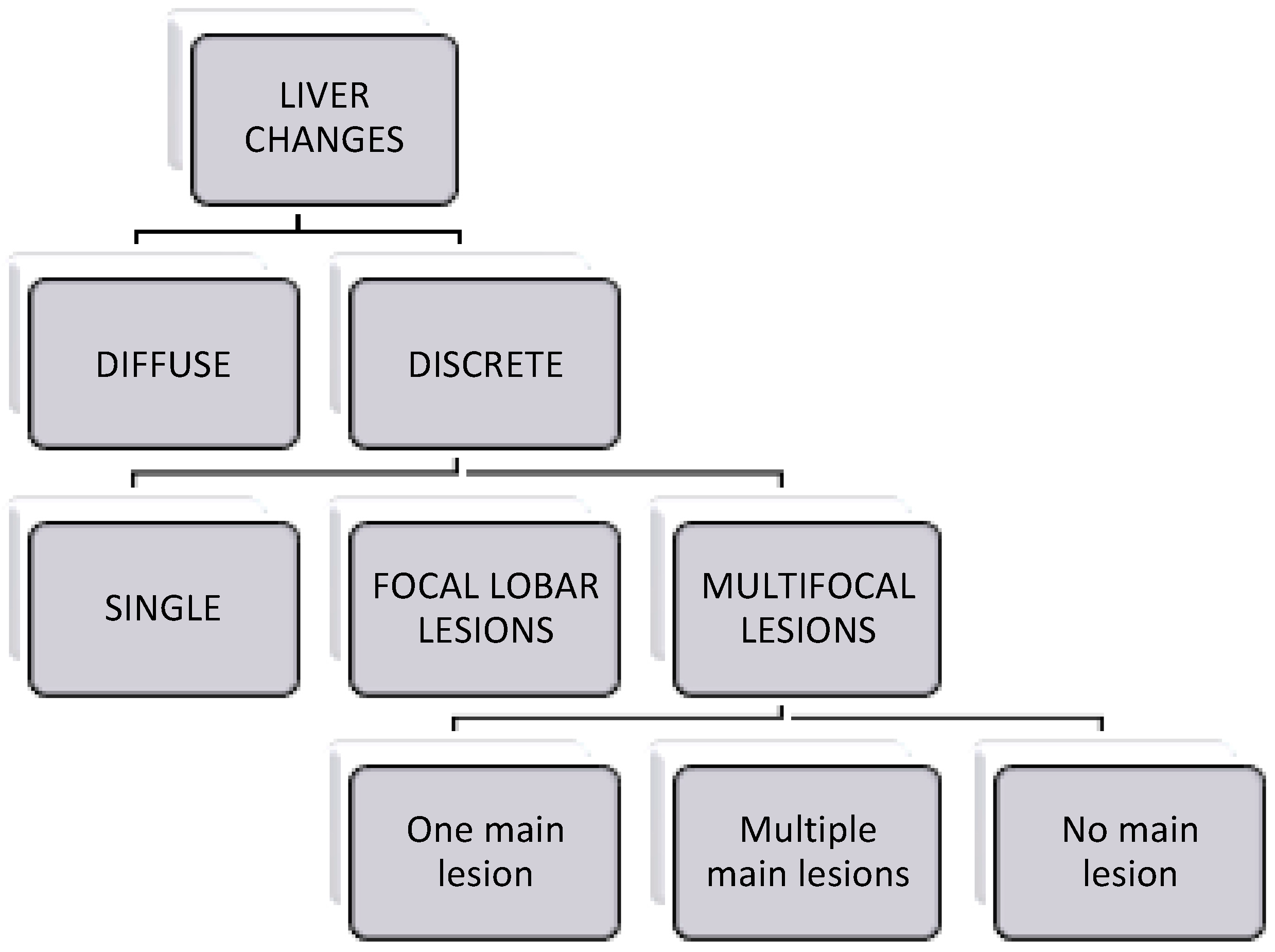
3.6. CT Findings
3.6.1. “Diffuse” Changes
3.6.2. “Discrete” Changes
3.7. Additional Changes Accompanying the Presence of Liver Infection
3.8. Association of CT Findings with Cytology/Histopathology/Culture
4. Discussion
5. Conclusions
Supplementary Materials
Author Contributions
Funding
Institutional Review Board Statement
Informed Consent Statement
Data Availability Statement
Acknowledgments
Conflicts of Interest
References
- Im, J.; Burney, D.P.; McDonough, S.P.; Nicholson, B.; Eatroff, A.; Simpson, K.W. Canine Hepatitis Associated with Intrahepatic Bacteria in Three Dogs. J. Am. Anim. Hosp. Assoc. 2018, 54, 65–70. [Google Scholar] [CrossRef] [PubMed]
- Twedt, D.; Cullen, J.; Rothuizen, J.; Desmet, V.; Bunch, S.; Van Winkle, T.; Charles, J.; Absent, R.W.; Szatmári, V. WSAVA Liver Standardization Group; Elsevier: Amsterdam, The Netherlands, 2006. [Google Scholar]
- Bächler, P.; Baladron, M.J.; Menias, C.; Beddings, I.; Loch, R.; Zalaquett, E.; Vargas, M.; Connolly, S.; Bhalla, S.; Huete, Á. Multimodality Imaging of Liver Infections: Differential Diagnosis and Potential Pitfalls. Radiographics 2016, 36, 1001–1023. [Google Scholar] [CrossRef] [PubMed]
- Wisplinghoff, H.; Appleton, D.L. Bacterial Infection of the Liver. In Comparative Hepatitis (Birkhäuser Advances in Infectious Diseases); Weber, O., Protzer, U., Eds.; Birkhäuser: Basel, Switzerland, 2008; pp. 143–161. [Google Scholar]
- Burke, J.E.; Hess, R.S.; McEntee, E.P.; Griffin, M.A.; Harmon, S.M.; Silverstein, D.C. Hepatic Abscessation in Dogs: A Multicenter Study of 56 Cases (2010–2019). J. Vet. Emerg. Crit. Care 2023, 33, 665–675. [Google Scholar] [CrossRef] [PubMed]
- Lord, P.F.; Carb, A.; Halliwell, W.H.; Prueter, J.C. Emphysematous Hepatic Abscess Associated with Trauma, Necrotic Hepatic Nodular Hyperplasia and Adenoma in a Dog: A Case History Report. Vet. Radiol. 1982, 23, 46–49. [Google Scholar] [CrossRef]
- Ettinger, S.J.; Feldman, E.C.; Côté, E. Textbook of Veterinary Internal Medicine: Diseases of the Dog and the Cat, 8th ed.; Ettinger, S.J., Feldman, E.C., Côté, E., Eds.; Elsevier: St. Louis, MI, USA, 2017; Volume 2. [Google Scholar]
- Halvorsen, R.A.; Korobkin, M.; Foster, W.L.; Silverman, P.M.; Thompson, W.M. The Variable CT Appearance of Hepatic Abscesses. Am. J. Roentgenol. 1984, 142, 941–946. [Google Scholar] [CrossRef]
- Mathieu, D.; Vasile, N.; Fagniez, P.-L.; Segui, S.; Grably, D.; Lard, D. Dynamic CT Features of Hepatic Abscesses. Radiology 1985, 154, 749–752. [Google Scholar] [CrossRef]
- Choi, B.I. (Ed.) Focal Hepatic Infections. In Radiology Illustrated: Hepatobiliary and Pancreatic Radiology; Springer: Seoul, Republic of Korea, 2014; pp. 234–262. [Google Scholar]
- Khim, G.; Em, S.; Mo, S.; Townell, N. Liver Abscess: Diagnostic and Management Issues Found in the Low Resource Setting. Br. Med. Bull. 2019, 132, 45–52. [Google Scholar] [CrossRef]
- Mazza, O.; Santibanes, M.; Santibanes, E. Pyogenic Liver Abscess. In Blumgart’s Surgery of the Liver, Billiary Tract and Pancreas; Jarnagin, W.R., Blungart, L.H., Eds.; Elsevier: St. Louis, MI, USA, 2016; Volume II, pp. 1073–1082. [Google Scholar]
- Jeffrey, R.B.; Tolentino, C.S.; Chang, F.C.; Federle, M.P. CT of Small Pyogenic Hepatic Abscesses: The Cluster Sign. Am. J. Roentgenol. 1988, 151, 487–489. [Google Scholar] [CrossRef]
- Toshifumi, G.; Masumi, K.; Osamu, M.; Takeshi, K.; Yasuhiro, K.; Junichiro, S.; Noboru, T.; Satoshi, K. Dynamic CT of Hepatic Abscesses: Significance of Transient Segmental Enhancement. Am. J. Roentgenol. 2001, 176, 675–679. [Google Scholar]
- Giambelluca, D.; Panzuto, F.; Giambelluca, E.; Midiri, M. The “Double Target Sign” in Liver Abscess. In Abdominal Radiology; Springer: New York, NY, USA, 2018; pp. 2885–2886. [Google Scholar] [CrossRef]
- Grooters, A.M.; Sherding, R.G.; Biller, D.S.; Johnson, S.E. Hepatic Abscesses Associated with Diabetes Mellitus in Two Dogs. J. Veter Intern. Med. 1994, 8, 203–206. [Google Scholar] [CrossRef]
- Schwarz, L.A.; Penninck, D.G.; Leveille-Webster, C. Hepatic Abscesses in 13 Dogs: A Review of the Ultrasonographic Findings, Clinical Data and Therapeutic Options. Veter Radiol. Ultrasound 1998, 39, 357–365. [Google Scholar] [CrossRef] [PubMed]
- Carrillo, J.; Escobar, M.T.; Porlan, S.; Rodenas, C.; Agut, A. Emphysematous Hepatic Abscess and Pyometra, Both Caused by Burkholderia cepacia, in a Bitch. Vet. Rec. Case Rep. 2021, 9, e53. [Google Scholar] [CrossRef]
- Konde, L.J.; Lebel, J.L.; Park, R.D.; Wrigley, R.H. Sonographic Application in the Diagnosis of Intraabdominal Abscess in the Dog. Vet. Radiol. 1986, 27, 151–154. [Google Scholar] [CrossRef]
- Dickerson, V.; Poses, B.; Hyndman, P.; McPhetridge, J.; Scharf, V.; Matz, B.; Singh, A.; Grimes, J.A. Outcome in 38 Dogs Surgically Treated for Hepatic Abscessation. Vet. Surg. 2023, 52, 127–133. [Google Scholar] [CrossRef]
- Cordella, A.; Bertolini, G. Multiphase Multidetector-Row Computed Tomographic and Ultrasonographic Findings in Dogs with Spontaneous Liver Lobe Torsion. Res. Vet. Sci. 2021, 135, 192–199. [Google Scholar] [CrossRef] [PubMed]
- Köhler, C.; Jopp, I.; Bosch, B.; Pfeifer, S.; Alef, M.; Oechtering, G.; Kiefer, I. Liver abscess in a dog. A case report. I. Leberabszess Bei Einem Hund. Tierärztliche Prax. Ausg. K Kleintiere Heimtiere 2012, 40, 211–218. [Google Scholar] [CrossRef]
- Milodowski, E.J.; Lamb, C.R.; Lee, K.C.L. Salmonella Hepatic Abscess as a Sequel to Liver Lobectomy and Biopsy in a Dog with Hepatocellular Adenoma. Vet. Rec. Case Rep. 2017, 5, e000511. [Google Scholar] [CrossRef]
- Kameda, S.; Okamura, Y.; Miyazaki, A.; Katayama, M.; Suzuki, H.; Uzuka, Y. A Case of Liver Abscess in a Dog with Surgical Removal Performed and CT Imaging Findings. J. Jpn. Vet. Med. Assoc. 2015, 68, 128–133. [Google Scholar] [CrossRef]
- Percival, A.; Lopez, D.J.; Miller, A.; Scrivani, P.V. Computed Tomography of Suppurative and Neoplastic Diseases Involving the Canine Omenta and Omental Bursa. Vet. Radiol. Ultrasound 2023, 64, 851–863. [Google Scholar] [CrossRef]
- Sonet, J.; Barthélemy, A.; Goy-Thollot, I.; Pouzot-Nevoret, C. Prospective Evaluation of Abdominal Ultrasonographic Findings in 35 Dogs with Leptospirosis. Vet. Radiol. Ultrasound 2018, 59, 98–106. [Google Scholar] [CrossRef]
- Mattoon, J.S.; Nyland, T.G. Small Animal Diagnostic Ultrasound, 3rd ed.; Elsevier: St. Louis, MI, USA, 2015. [Google Scholar]
- Engelmann, N.; Ondreka, N.; Michalik, J.; Neiger, R. Intra-Abdominal Mycobacterium Tuberculosis Infection in a Dog. J. Vet. Intern. Med. 2014, 28, 934–938. [Google Scholar] [CrossRef] [PubMed]
- R Core Team. R: A Language and Environment for Statistical Computing; R Foundation for Statistical Computing: Vienna, Austria, 2023; Available online: https://www.r-project.org/ (accessed on 20 May 2024).
- Schwarz, T.; Rossi, F.; Morandi, F. Liver, Gallbladder and Spleen. In Veterinary Computed Tomography; Schwarz, T., Ed.; Jong Wiley & Sons Ltd.: Oxford, UK, 2011; pp. 297–314. [Google Scholar]
- Alvarez, J.A.; Gonzá Lez, J.J.; Baldonedo, R.F.; Sanz, L.; Carreño, G.; Jorge, J.I.; Antonio, J.; Pérez, A. Single and Multiple Pyogenic Liver Abscesses: Etiology, Clinical Course, and Outcome. Dig. Surg. 2001, 18, 283–288. [Google Scholar] [CrossRef] [PubMed]
- Sharma, V.; Kalantri, A.; Gupta, P.; Mandavdhare, H.; Samanta, J.; Sinha, S.K. Hepatic Infections: A Comprehensive Imaging Review. J. Gastrointest. Infect. 2019, 9, 38–49. [Google Scholar] [CrossRef]
- Uršic, M.; Ravnik, D.; Hribernik, M.; Pečar, J.; Butinar, J.; Fazarinc, G. Gross Anatomy of the Portal Vein and Hepatic Artery Ramifications in Dogs: Corrosion Cast Study. J. Vet. Med. Ser. C Anat. Histol. Embryol. 2007, 36, 83–87. [Google Scholar] [CrossRef]
- Hermanson, J.W.; de Lahunta, A.; Evans, H.E. Miller and Evans’ Anatomy of the Dog, 5th ed.; Elsevier: St. Louis, MI, USA, 2020. [Google Scholar]
- Hennessey, E.; Cassel, N.; Nuth, E.; Biller, D. CT Can Identify Characteristic Features of Hypaxial Muscle Abscesses in Dogs Due to Presumed Migrating Vegetal Foreign Material as Well as Additional Changes along the Migratory Tract in Other Anatomic Regions. Vet. Radiol. Ultrasound 2022, 63, 691–698. [Google Scholar] [CrossRef]
- Leela Arporn, R.; Ohta, H.; Shimbo, G.; Hanazono, K.; Osuga, T.; Morishita, K.; Sasaki, N.; Takiguchi, M. Computed Tomographic Features for Differentiating Benign from Malignant Liver Lesions in Dogs. J. Vet. Med. Sci. 2019, 81, 1697–1704. [Google Scholar] [CrossRef]
- Fukushima, K.; Kanemoto, H.; Ohno, K.; Takahashi, M.; Nakashima, K.; Fujino, Y.; Uchida, K.; Fujiwara, R.; Nishimura, R.; Tsujimoto, H. Ct Characteristics of Primary Hepatic Mass Lesions in Dogs. Vet. Radiol. Ultrasound 2012, 53, 252–257. [Google Scholar] [CrossRef] [PubMed]
- Matteini, F.; Cannella, R.; Garzelli, L.; Dioguardi Burgio, M.; Sartoris, R.; Brancatelli, G.; Vilgrain, V.; Ronot, M.; Vernuccio, F. Benign and Malignant Focal Liver Lesions Displaying Rim Arterial Phase Hyperenhancement on CT and MRI. Insights Into Imaging 2024, 15, 178. [Google Scholar] [CrossRef] [PubMed]
- Malekzadeh, S.; Widmer, L.; Salahshour, F.; Egger, B.; Ronot, M.; Thoeny, H.C. Typical Imaging Finding of Hepatic Infections: A Pictorial Essay. Abdom. Imaging 2021, 46, 544–561. [Google Scholar] [CrossRef]
- Leotta, D.F.; Bruce, M.; Wang, Y.N.; Kucewicz, J.; Khokhlova, T.; Chan, K.; Monsky, W.; Matula, T.J. Sonographic Features of Abscess Maturation in a Porcine Model. Ultrasound Med. Biol. 2021, 47, 1920–1930. [Google Scholar] [CrossRef]
- Teixeira, G.; Faria, R. Inflammatory Mediators Leading to Edema Formation through Plasma Membrane Receptors. In Infectious Diseases and Sepsis Development; IntechOpen: London, UK, 2021. [Google Scholar] [CrossRef]
- Baqueiro Canto, R.; Álvarez Ibarra, S.; Torres Vera, M.; Padrón Arredondo, G.; Canto, B.; Padrón Arredondo Hepatic, G. Hepatic Abscess with Gas by Streptococcus Pyogenes (Group A). Gastroenterology 2019, 2, 121. [Google Scholar]
- Mizandari, M.; Azrumelashvili, T.; Toria, N.; Nanava, N.; Pantsulaia, I.; Kikodze, N.; Janikashvili, N.; Chikovani, T. Cured Giant Hepatocellular Carcinoma after Transarterial Embolization Complicated with Liver Abscess Formation. Radiol. Case Rep. 2020, 15, 1485–1492. [Google Scholar] [CrossRef] [PubMed]
- Rosen, S.; Lumbrezer-Johnson, S.; Hostnik, E.; Salyer, S.; Selmic, L.E. Recurrent Liver Abscessation in a Dog with an Incompletely Resected Hepatocellular Adenoma. Vet. Rec. Case Rep. 2023, 11, e536. [Google Scholar] [CrossRef]
- Manfredi, S.; Fabbi, M.; Bonazzi, M.; Leonardi, F.; Miduri, F.; Parroccini, I.; Daga, E.; Gnudi, G.; Volta, A. Ultrasonographic Differentiation between Portal Venous and Parenchymal Gas May Be Important for the Prognosis of Canine and Feline Hepatic Emphysema: 37 Cases. Vet. Radiol. Ultrasound 2019, 60, 734–744. [Google Scholar] [CrossRef]
- Ghosn, Y.; Abdallah, A.; Hussein Kamareddine, M.; Geahchan, A.; Baghdadi, A.; El-Rassi, Z.; Chamseddine, A.; Ashou, R. Gas-Forming Liver Abscess versus Emphysematous Hepatitis: A Radiologic Diagnostic Dilemma—A Case Report and Review of the Literature. Case Rep. Hepatol. 2019, 2019, 5274525. [Google Scholar] [CrossRef]
- Porez, D.; Kallel, H.; Dobian, S.; Gerbert-Ferrendier, T.; Nacher, M.; Djossou, F.; Demar, M.; Amroun, H.; Zappa, M.; Drak Alsibai, K. Diagnostic and Management of Emphysematous Hepatitis with Emphasis on Biopathology. Microorganisms 2023, 11, 2137. [Google Scholar] [CrossRef] [PubMed]
- Jang, S.; Lee, S.; Choi, J. CT Imaging Features of Fat Stranding in Cats and Dogs with Abdominal Disorder. J. Vet. Sci. 2022, 23, e70. [Google Scholar] [CrossRef]
- Kanemoto, H.; Fukushima, K.; Tsujimoto, H.; Ohno, K. Brief Communication Communication Brève Intrahepatic Cholelithiasis in Dogs and Cats: A Case Series. Can. Vet. J. 2017, 58, 971–973. [Google Scholar]
- Park, H.Y.; Cho, Y.G.; Lee, Y.W.; Choi, H.J. Evaluation of Gallbladder and Common Bile Duct Size and Appearance by Computed Tomography in Dogs. J. Vet. Sci. 2018, 19, 653–659. [Google Scholar] [CrossRef]
- Cordella, A.; Bertolini, G. Multiphase Multidetector-Row CT Reveals Different Patterns of Hepatic Portal Venous Gas and Pneumobilia. Vet. Radiol. Ultrasound 2021, 62, 68–75. [Google Scholar] [CrossRef]
- Policelli Smith, R.; Gookin, J.L.; Smolski, W.; Di Cicco, M.F.; Correa, M.; Seiler, G.S. Association between Gallbladder Ultrasound Findings and Bacterial Culture of Bile in 70 Cats and 202 Dogs. J. Vet. Intern. Med. 2017, 31, 1451–1458. [Google Scholar] [CrossRef] [PubMed]
- Ghielmetti, G.; Giger, U. Mycobacterium Avium: An Emerging Pathogen for Dog Breeds with Hereditary Immunodeficiencies. Curr. Clin. Microbiol. Rep. 2020, 7, 67–80. [Google Scholar] [CrossRef] [PubMed]
- Eggers, J.S.; Parker, G.A.; Braaf, H.A.; Mense, M.G. Disseminated Mycobacterium Avium Infection in Three Miniature Schnauzer Litter Mates. J. Vet. Diagn. Investig. 1997, 9, 424–427. [Google Scholar] [CrossRef] [PubMed]
- Webster, C.R.L.; Center, S.A.; Cullen, J.M.; Penninck, D.G.; Richter, K.P.; Twedt, D.C.; Watson, P.J. ACVIM Consensus Statement on the Diagnosis and Treatment of Chronic Hepatitis in Dogs. J. Vet. Intern. Med. 2019, 33, 1173–1200. [Google Scholar] [CrossRef] [PubMed]
- McCallum, K.E.; Constantino-Casas, F.; Cullen, J.M.; Warland, J.H.; Swales, H.; Linghley, N.; Kortum, A.J.; Sterritt, A.J.; Cogan, T.; Watson, P.J. Hepatic Leptospiral Infections in Dogs without Obvious Renal Involvement. J. Vet. Intern. Med. 2019, 33, 141–150. [Google Scholar] [CrossRef]
- Sykes, J.E.; Hartmann, K.; Lunn, K.F.; Moore, G.E.; Stoddard, R.A.; Goldstein, R.E. 2010 ACVIM Small Animal Consensus Statement on Leptospirosis: Diagnosis, Epidemiology, Treatment, and Prevention. J. Vet. Intern. Med. 2011, 25, 1–13. [Google Scholar] [CrossRef]
- Raskin, R.E.; Meyer, D.J.; Boes, K.M. Canine and Feline Cytopathology: A Color Atlas and Interpretation Guide, 4th ed.; Elsevier: St Louis, MI, USA, 2023. [Google Scholar]
- Sergeeff, J.S.; Armstrong, P.J.; Bunch, S.E. Hepatic Abscesses in Cats: 14 Cases (1985–2002). J. Vet. Intern. Med. 2004, 18, 295–300. [Google Scholar] [CrossRef]
- Carloni, A.; Terragni, R.; Morselli-Labate, A.M.; Paninarova, M.; Graham, J.; Valenti, P.; Alberti, M.; Albarello, G.; Millanta, F.; Vignoli, M. Prevalence, Distribution, and Clinical Characteristics of Hemangiosarcoma-Associated Skeletal Muscle Metastases in 61 Dogs: A Whole Body Computed Tomographic Study. J. Vet. Intern. Med. 2019, 33, 812–819. [Google Scholar] [CrossRef]
- Lamb, C.R.; Whitlock, J.; Foster-Yeow, A.T.L. Prevalence of Pulmonary Nodules in Dogs with Malignant Neoplasia as Determined by CT. Vet. Radiol. Ultrasound 2019, 60, 300–305. [Google Scholar] [CrossRef]
- López, M.C.; Vila, A.; Rodón, J.; Roura, X. Leptospira Seroprevalence in Owned Dogs from Spain. Heliyon 2019, 5, e02373. [Google Scholar] [CrossRef]
- Taylor, C.; O’Neill, D.G.; Catchpole, B.; Brodbelt, D.C. Incidence and Demographic Risk Factors for Leptospirosis in Dogs in the UK. Vet. Rec. 2022, 190, e512. [Google Scholar] [CrossRef] [PubMed]
- Andre-Fontaine, G. Diagnosis Algorithm for Leptospirosis in Dogs: Disease and Vaccination Effects on the Serological Results. Vet. Rec. 2013, 172, 502. [Google Scholar] [CrossRef] [PubMed]
- Soudah, B.; Schirakowski, A.; Gebel, M.; Potthoff, A.; Braubach, P.; Schlue, J.; Krech, T.; Dämmrich, M.E.; Kreipe, H.H.; Abbas, M. Overview and Evaluation of the Value of Fine Needle Aspiration Cytology in Determining the Histogenesis of Liver Nodules: 14 Years of Experience at Hannover Medical School. Oncol. Rep. 2015, 33, 81–87. [Google Scholar] [CrossRef]
- Nasit, J.; Patel, V.; Parikh, B.; Shah, M.; Davara, K. Fine-Needle Aspiration Cytology and Biopsy in Hepatic Masses: A Minimally Invasive Diagnostic Approach. Clin. Cancer Investig. J. 2013, 2, 132. [Google Scholar] [CrossRef]


| Bacterial Shape | Fisher’s Exact Test p-Value | |||
|---|---|---|---|---|
| Bacilli | Cocci | Cocci and Bacilli | ||
| Gas visible | 0.591 | |||
| Yes | 2 (10.0%) | 2 (10.0%) | 0 (0.0%) | |
| No | 4 (20.0%) | 9 (45.0%) | 3 (15.0%) | |
| Cluster sign | 1.00 | |||
| Yes | 2 (10.5%) | 5 (26.3%) | 1 (5.3%) | |
| No | 3 (15.8%) | 6 (31.6%) | 2 (10.5%) | |
| Cystic appearance | 0.787 | |||
| Yes | 1 (5.3%) | 4 (21.1%) | 0 (0.0%) | |
| No | 4 (21.1%) | 7 (36.8%) | 3 (15.8%) | |
| Peritoneal free fluid | 0.574 | |||
| Yes | 4 (20.0%) | 8 (40.0%) | 1 (5.5%) | |
| No | 2 (10.0%) | 3 (15.0%) | 2 (10.0%) | |
| Fat stranding | 0.357 | |||
| Yes | 5 (25.0%) | 8 (40.0%) | 1 (5.0%) | |
| No | 1 (5.0%) | 3 (15.0%) | 2 (10.0%) | |
| Pneumoperitoneum | 1.00 | |||
| Yes | 1 (5.0%) | 3 (15.0%) | 0 (0.0%) | |
| No | 5 (25.0%) | 8 (40.0%) | 3 (15.0%) | |
| Lymphadenomegaly | 0.363 | |||
| Yes | 6 (30.0%) | 10 (50.0%) | 5 (25.0%) | |
| No | 0 (0.0%) | 1 (5.0%) | 1 (5.0%) | |
| Lesion Type | Fisher’s Exact Test p-Value | ||
|---|---|---|---|
| Multifocal | Single | ||
| Body weight category | 1.00 | ||
| <10 kg | 2 (11.1%) | 0 (0.0%) | |
| 10–25 kg | 5 (27.8%) | 2 (11.1%) | |
| >25 kg | 7 (38.9%) | 2 (11.1%) | |
| Sex | 1.00 | ||
| Female | 6 (31.6%) | 2 (10.5%) | |
| Male | 8 (42.1%) | 3 (15.8%) | |
| Neuter status | 1.00 | ||
| Neutered | 11 (57.9%) | 4 (21.1%) | |
| Entire | 3 (15.8%) | 1 (5.3%) | |
| Anorexia/inappetence | 0.338 | ||
| Yes | 7 (36.8%) | 4 (21.1%) | |
| No | 7 (36.8%) | 1 (5.3%) | |
| Pyrexia | 0.305 | ||
| Yes | 10 (52.6%) | 2 (10.5%) | |
| No | 4 (21.1%) | 3 (15.8%) | |
| Vomiting | 0.272 | ||
| Yes | 2 (10.5%) | 2 (10.5%) | |
| No | 12 (63.2%) | 3 (15.8%) | |
| Weight loss | 0.272 | ||
| Yes | 2 (10.5%) | 2 (10.5%) | |
| No | 12 (63.2%) | 3 (15.8%) | |
| Clinical presentation | 0.405 | ||
| Acute (<2 weeks) | 13 (72.2%) | 3 (16.7%) | |
| Chronic (>2 weeks) | 1 (5.6%) | 1 (5.6%) | |
| Previous antibiotic treatment | 1.00 | ||
| Yes | 5 (27.8%) | 1 (5.6%) | |
| No | 9 (50.0%) | 3 (16.7%) | |
| Demeanour | 0.237 | ||
| Alert | 7 (41.2%) | 4 (23.5%) | |
| Depressed | 6 (35.3%) | 0 (0.0%) | |
| Abdominal pain | 0.023 | ||
| Yes | 10 (55.6%) | 0 (0.0%) | |
| No | 4 (22.2%) | 4 (22.2%) | |
| Tachycardia | 1.00 | ||
| Yes | 5 (27.8%) | 2 (11.1%) | |
| No | 9 (50.0%) | 2 (11.1%) | |
| Tachypnoea | 0.405 | ||
| Yes | 1 (5.6%) | 1 (5.6%) | |
| No | 13 (72.2%) | 3 (16.7%) | |
| Anaemia | 1.00 | ||
| Yes | 6 (33.3%) | 1 (5.6%) | |
| No | 8 (44.4%) | 3 (16.7%) | |
| Neutrophilia | 0.533 | ||
| Yes | 11 (61.1%) | 2 (11.1%) | |
| No | 3 (16.7%) | 2 (11.1%) | |
| Raised alanine aminotransferase (ALT) | 1.00 | ||
| Yes | 12 (66.7%) | 4 (22.2%) | |
| No | 2 (11.1%) | 0 (0.0%) | |
| Raised alkaline phosphatase (ALKP) | 0.533 | ||
| Yes | 11 (61.1%) | 2 (11.1%) | |
| No | 3 (16.7%) | 2 (11.1%) | |
| Bacteria shape | 0.276 | ||
| Bacilli | 4 (21.1%) | 1 (5.3%) | |
| Cocci | 9 (47.4%) | 2 (10.5%) | |
| Bacilli and Cocci | 1 (5.3%) | 2 (10.5%) | |
| Liver size | 0.081 | ||
| Decreased | 0 (0.0%) | 1 (5.6%) | |
| Normal | 8 (42.1%) | 4 (21.1%) | |
| Increased | 6 (31.6%) | 0 (0.0%) | |
| Peritoneal free fluid | 1.00 | ||
| Yes | 9 (47.4%) | 3 (15.8%) | |
| No | 5 (26.3%) | 2 (10.5%) | |
| Fat stranding | 1.00 | ||
| Yes | 10 (52.6%) | 3 (15.8%) | |
| No | 4 (21.1%) | 2 (10.5%) | |
| Pneumoperitoneum | 1.00 | ||
| Yes | 3 (15.8%) | 1 (5.6%) | |
| No | 11 (61.1%) | 4 (21.1%) | |
| Lymphadenomegaly | 0.468 | ||
| Yes | 13 (68.4%) | 4 (21.1%) | |
| No | 1 (5.6%) | 1 (5.6%) | |
Disclaimer/Publisher’s Note: The statements, opinions and data contained in all publications are solely those of the individual author(s) and contributor(s) and not of MDPI and/or the editor(s). MDPI and/or the editor(s) disclaim responsibility for any injury to people or property resulting from any ideas, methods, instructions or products referred to in the content. |
© 2024 by the authors. Licensee MDPI, Basel, Switzerland. This article is an open access article distributed under the terms and conditions of the Creative Commons Attribution (CC BY) license (https://creativecommons.org/licenses/by/4.0/).
Share and Cite
Maté de Haro, L.; Vila, A.; Di Bella, A.; Mallol, C.; Anselmi, C.; Barreiro-Vazquez, J.-D.; Pollard, D.; Salgüero, R.; Fitzgerald, E.; Moreno-Aguado, B. Computed Tomographic Findings in Dogs with Hepatic Bacterial Parenchymal Infection and Abscessation. Animals 2024, 14, 3399. https://doi.org/10.3390/ani14233399
Maté de Haro L, Vila A, Di Bella A, Mallol C, Anselmi C, Barreiro-Vazquez J-D, Pollard D, Salgüero R, Fitzgerald E, Moreno-Aguado B. Computed Tomographic Findings in Dogs with Hepatic Bacterial Parenchymal Infection and Abscessation. Animals. 2024; 14(23):3399. https://doi.org/10.3390/ani14233399
Chicago/Turabian StyleMaté de Haro, Luis, Andrea Vila, Andrea Di Bella, Claudia Mallol, Carlo Anselmi, Jose-Daniel Barreiro-Vazquez, Danica Pollard, Raquel Salgüero, Ella Fitzgerald, and Beatriz Moreno-Aguado. 2024. "Computed Tomographic Findings in Dogs with Hepatic Bacterial Parenchymal Infection and Abscessation" Animals 14, no. 23: 3399. https://doi.org/10.3390/ani14233399
APA StyleMaté de Haro, L., Vila, A., Di Bella, A., Mallol, C., Anselmi, C., Barreiro-Vazquez, J.-D., Pollard, D., Salgüero, R., Fitzgerald, E., & Moreno-Aguado, B. (2024). Computed Tomographic Findings in Dogs with Hepatic Bacterial Parenchymal Infection and Abscessation. Animals, 14(23), 3399. https://doi.org/10.3390/ani14233399