Article

Exploring the Antioxidant and Genoprotective Potential of Salicornia ramosissima Incorporation in the Diet of the European Seabass (Dicentrarchus labrax)

1
CESAM—Centre for Environmental and Marine Studies and Department of Biology, University of Aveiro, 3810-193 Aveiro, Portugal
2
Riasearch, Lda., 3870-168 Murtosa, Portugal
3
Centro Interdisciplinar de Investigação Marinha e Ambiental (CIIMAR), Universidade do Porto, 4450-208 Matosinhos, Portugal
4
School of Medicine and Biomedical Sciences (ICBAS-UP), University of Porto, 4050-313 Porto, Portugal
*
Author to whom correspondence should be addressed.
Animals 2024, 14(1), 93; https://doi.org/10.3390/ani14010093
Submission received: 14 November 2023 / Revised: 14 December 2023 / Accepted: 16 December 2023 / Published: 27 December 2023
(This article belongs to the Special Issue Functional Feeds to Improve Shrimp and Fish Aquaculture)

Abstract

:

Simple Summary

Salicornia ramosissima was studied as a potential feed material for juvenile European seabass in aquaculture. This halophyte was incorporated in three different levels (2.5, 5 and 10%) into the fish diet for two months. The results showed no negative impact on the fish’s growth, and there was an improvement in antioxidant activity in specific tissues, particularly through the GSH-related defense subsystem. A genotoxic trigger was also identified in the first month, which was considered a protective mechanism, and in the second month, a clear protection of DNA integrity was observed, especially at higher supplementation levels. Overall, the findings suggest that supplemented diets with S. ramosissima have functional benefits, offering a potential way to enhance aquaculture practices and utilize a novel, low-value raw material.

Abstract

The identification of novel feed materials as a source of functional ingredients is a topical priority in the finfish aquaculture sector. Due to the agrotechnical practices associated and phytochemical profiling, halophytes emerge as a new source of feedstuff for aquafeeds, with the potential to boost productivity and environmental sustainability. Therefore, the present study aimed to assess the potential of Salicornia ramosissima incorporation (2.5, 5, and 10%), for 2 months, in the diet of juvenile European seabass, seeking antioxidant (in the liver, gills, and blood) and genoprotective (DNA and chromosomal integrity in blood) benefits. Halophyte inclusion showed no impairments on growth performance. Moreover, a tissue-specific antioxidant improvement was apparent, namely through the GSH-related defense subsystem, but revealing multiple and complex mechanisms. A genotoxic trigger (regarded as a pro-genoprotective mechanism) was identified in the first month of supplementation. A clear protection of DNA integrity was detected in the second month, for all the supplementation levels (and the most prominent melioration at 10%). Overall, these results pointed out a functionality of S. ramosissima-supplemented diets and a promising way to improve aquaculture practices, also unraveling a complementary novel, low-value raw material, and a path to its valorization.

1. Introduction

Arable lands are diminishing globally because of soil salinization as well as freshwater scarcity due to the lack of precipitation and improper water resource management practices [1]. Thus, the use of salt-tolerant plants (halophytes) to produce food and feed emerges as a sound and timely alternative to conventional crop production, overcoming constraints such as the limited adoption of sustainable and responsible agricultural practices [2], coupled with competition with direct human consumption. In this direction, the cultivation of halophytes is gaining recognition and, recently, its valorization as a food product and novel feedstock for nutraceuticals [3] and pharmaceutical [4] applications has been investigated (revised by [5]).
These premises also make halophytes good candidates as a new source of feedstuff for aquafeeds, with potential economic viability and gains in terms of environmental sustainability. However, investigations in this direction are scarce and completely absent in carnivorous fish species. To our knowledge, only a couple of studies have been carried out addressing the replacement of fish meal with Salicornia bigelovii in Nile tilapia (Oreochromis niloticus) feed [6,7], showing no compromise to growth parameters or body composition. Therefore, research and development efforts focused on this topic appear perfectly justifiable, innovative, and opportune, namely concerning high-value aquaculture species, mainly represented in the European context by marine teleosts (e.g., salmon, seabream, and seabass).
The halophyte Salicornia ramosissima is an annual green tip plant that can be found in saltmarshes, with a broad geographical distribution (from Artic to Mediterranean regions), characterized by a pleasant texture, a juicy and salty taste [8], as well as phytochemical profiling that supports its edibility and food relevance [9]. Beyond the traditional culinary applications, Salicornia spp. are rich in fibers, minerals, polyphenols, flavonoids, and proteins, and poor in fat, emerging thus as valuable nutritional and healthy food for humans [10,11,12,13]. These halophytes also showed medicinal attributes, namely that they are immunomodulatory, lipid- and sugar-lowering, antiproliferative, and osteoprotective, as well as reinforcing antioxidant and DNA repair systems [9,12,13,14]. Bearing in mind these properties, Salicornia spp. should be cogitated as a promising supplement ingredient for inclusion in feeds for marine fish farming.
The major challenge of seeking novel feed supplements/materials is the combined achievement of environmental sustainability, on the one hand, and maintenance of fish growth, overall health/survival, while meeting fillet quality and consumer expectations, on the other [15,16,17]. Furthermore, “the icing on the cake” would be the development of aquafeeds that, besides satisfying the previous features, present the added value of exhibiting functional properties. The development of functional feeds, characterized as having physiological effects in fish beyond those of nutrient effects (e.g., improvement of stress tolerance, disease resistance, and health performance) [18], has, in past years, increasingly attracted the attention of research in food sciences applied to aquaculture [19]. Functional ingredients/nutrients in aquafeeds can be designed to achieve a plethora of expected benefits, but the strengthening of antioxidant defenses and DNA integrity, due to their pivotal roles determining plentiful upstream functions, stands among those that could result in greater physiological improvements and therefore have higher potential to generate outcomes in this productive activity.
The antioxidant defense system is essential to maintain cell homeostasis. When it fails or is overwhelmed, oxidative stress products (such as reactive oxygen species; ROS) can damage cell constituents such as lipids, proteins, and DNA. Fish can adapt to ROS overgeneration by increasing antioxidant defenses; however, the amount of ROS that can be neutralized is limited, which may disturb the redox state [20,21,22], impairing critical functions at higher levels. It has been demonstrated that antioxidant shielding in fish can be strengthened by dietary approaches, through supplementation with natural or artificial ingredients such as selenium [23], oligosaccharides of plant origin and phytogenic feed additives [24], carotenoids [25], and diludine [26].
Genome integrity and stability have central importance to organisms’ health, fitness, and ultimately survival. Intensive aquaculture practices (e.g., health management through the use of anesthetics, antiparasitics, and antibiotics) and the manipulation of rearing conditions (e.g., stressing temperature and high stocking density) pose recurring challenges to the DNA integrity of farmed fish [27]. Thus, the improvement of basal levels of DNA integrity (translating an efficacy to neutralize endogenous sources of damage), or the enhanced capacity to cope with the exogenous threats previously mentioned, play a key role in the balance between health and disease, in the proper functioning of metabolic pathways, and thereby on the general status and growth performance of fish. As stated above for oxidative status, genome integrity can be modulated, including in fish, by dietary factors, which was clearly demonstrated, for instance, with supplementation with vitamin C [28] and marine macroalgae [29]. Nevertheless, to the authors’ knowledge, the incorporation of halophytes in fish feeds as a factor of genoprotection was never investigated.
The European seabass (Dicentrarchus labrax), from the Moronidae family, was the first species of non-salmonid marine fish to be produced in Europe, being currently one of the most important species commercially exploited in the Mediterranean area (e.g., Greece, Turkey, Italy, Spain, Croatia, and Egypt) [30]. In 2020, global production of farmed D. labrax reached 243.9 thousand tons, its highest value ever [1]. Its predatory nature and inherent dietary requirements make the inclusion of plant-based ingredients more challenging. Nevertheless, the partial replacement of fish meal by plant protein sources in the diet of D. labrax has shown to be viable [30], though requiring a careful evaluation of the adequate percentage [31]. Concerning the development of functional feeds, in the last decades, different ingredients have been tested on D. labrax. For instance, the introduction of yeast as a complementary ingredient showed no negative effects on growth and improved feed efficiency [32]. Medicinal plants such as Yucca schidigera improved hematological parameters and immunological responses [33], while the introduction of prebiotics acted as immunostimulants through the increase of phagocytic activity [34]. It was also suggested that dietary supplementation with a macro- and microalgae blend may improve the intestinal health of D. labrax [35]. Recently, a study investigated the use of alternative feed resources from insects, crustacea, or microalgae biomass as functional supplements in a diet for juvenile D. labrax totally deprived of fish meal [36]. The results pointed out that supplementation with insect and crayfish meals improve fish growth and gut health, while the use of microalgae still requires some refinement.
Therefore, having as a backdrop the importance of D. labrax production in the European aquaculture sector and the promising attributes of halophytes enunciated above, the present work was designed to assess the functional properties of S. ramosissima when incorporated in D. labrax feed, coupled with an evaluation of the impact on growth performance. Subsequently, the specific goals considered were: (i) to evaluate the protection promoted by S. ramossima specifically towards the improvement of DNA and chromosomal integrity (in blood) and antioxidant system strengthening (in the liver, gills, and blood); (ii) to ensure that S. ramossima supplementation does not present adverse effects to seabass, translated into health impairments that may affect fish growth and welfare.
Ultimately, the pursuit of the previous goals will contribute to shedding light on the potential of halophytes as an emerging feed ingredient, towards the improvement of aquaculture practices and the valorization of low-value plant-based material.

2. Materials and Methods

2.1. Experimental Diets

Salicornia ramosissima biomass (21.5 kg) was collected in Praia da Areia Branca, Torreira (40°46′22.1″ N 8°39′29.5″ W; Portugal), in May 2019. Plant portions not used for human consumption (i.e., the whole plant, except the green tips and the roots) were isolated, and dried in Riasearch Lda. facilities (Murtosa, Portugal), in mesh mats that allowed air circulation, to prevent any degradation promoted by humidity. The resulting 6 kg were sent to Sparos Lda. (Olhão, Portugal) for feed formulation and manufacture, by extrusion (pellet size: 1.2 and 2.0 mm), after proper grinding (below 400 µm, as all the other ingredients). A control diet (C) was formulated to meet the nutritional requirements of juvenile D. labrax. Three isoproteic (wheat gluten levels were slightly adjusted to ensure protein levels remained similar between diets), isolipidic, and isoenergetic diets were produced, differing in the ingredient formulation from diet C only by replacing wheat meal with S. ramosissima whole-plant biomass, incorporated at 2.5%, 5%, and 10% of feed weight (S2.5, S5, and S10, respectively).
The main ingredients and analyzed proximate composition of the experimental diets are detailed in Table 1 and Table 2, respectively.

2.2. Rearing System, Feeding Trial, and Fish Sampling

Juveniles of European seabass (D. labrax), with a mean initial weight of 7.3 ± 0.1 g, were obtained from Sonríonansa, S.L. hatchery (Cantabria, Spain) and allocated in Riasearch Lda. facilities (Murtosa, Portugal). The trial, started in July 2020, consisted of assessing the growth performance and health condition (cytogenetic and biochemical evaluations) of D. labrax after the inclusion of different percentages of S. ramosissima in the feed. For that, fish were divided into 12 tanks (cylindrical fiberglass tanks of 350 L, each containing 200 L of water and 80 fish), corresponding to three tanks per condition/diet and a closed saltwater recirculation system (RAS) with 40 m3 (with a water renewal of 200 L per hour). During the experimental period, water parameters were measured once a day using commercial probes and maintained as follows: temperature at 21.6 ± 0.2 °C, dissolved oxygen at 6.4 ± 0.6 mg L−1, salinity at 18.2 ± 0.2, pH at 7.5 ± 0.2, and nitrogen compounds below 0.1 mg L−1.
A feeding trial with a total duration of 2 months, and an intermediate sampling after 1 month were performed, involving the diets described above (see Section 2.1). Fish were kept under a 14 h light:10 h dark photoperiod and fed by hand until visual satiety in three daily meals.
For the assessment of growth performance, body weight was determined at the end of the experimental period for all fish. The occurrence of mortality was monitored daily, and survival was expressed as a percentage and calculated as: S = (Sf/Si) × 100, where Si and Sf correspond to the initial and final number of individuals in the tanks, respectively.
At each sampling moment, for cytogenetic and biochemical evaluations, five fish were randomly collected from each thank (15 fish per condition/diet). Fish were anesthetized with 200 mg L−1 tricaine methanesulfonate (MS-222; buffered with NaHCO3) for approximately 10 min, and blood was drawn from the posterior cardinal vein using heparinized (27 mg mL−1 heparin) glass Pasteur pipettes and collected in 2 mL microtubes. Thus, one microtube, containing 0.002 mL of blood diluted in 1 mL of chilled PBS (pH = 7.4; 0.01 M), constituted the cell suspension for comet assay, and the other, with the remaining blood volume, was assigned for antioxidant analysis. Aliquots for comet assay were kept cold up to further procedures, while the aliquots for antioxidants determination were immediately frozen in liquid nitrogen. Blood smears were immediately prepared for erythrocytic nuclear abnormalities (ENA) assay and erythrocyte maturity index (EMI). Immediately after blood sampling, the fish were sacrificed by cervical transection. The gills and liver were removed and frozen in liquid nitrogen for later determination of oxidative stress endpoints.

2.3. Biochemical and Cytogenetic Evaluations

2.3.1. Antioxidant System Status and Peroxidative Damage

All measurements were carried out in a SpectraMax 190 microplate reader, at 25 °C. Tissue samples were homogenized using a Potter–Elvehjem homogenizer, in chilled phosphate buffer (0.1 M, pH 7.4) in a 1:11 (blood and liver) and 1:6 (gills) ratio [tissue mass (mg): buffer volume (mL)]. The resulting homogenate of each tissue was then divided into two aliquots, for lipid peroxidation (LPO) measurement and post-mitochondrial supernatant (PMS) preparation. The PMS preparation was obtained by centrifugation in a refrigerated centrifuge (Eppendorf 5415R) at 13,400× g for 20 min at 4 °C. Aliquots of PMS were then divided into microtubes and stored at −80 °C until further antioxidant analyses. The activities of superoxide dismutase (SOD), catalase (CAT), glutathione peroxidase (GPx), and glutathione transferase (GST), as well as total glutathione (GSHt) content were measure in all the tissues addressed, while glutathione reductase (GR) activity was assessed only in the gills and liver (blood showed no measurable levels of this enzymatic activity).
SOD activity was assayed in PMS (at 25 °C) with a Ransod kit (Randox Laboratories Ltd., Crumlin, UK). The method employs xanthine and xanthine oxidase to generate superoxide radicals, which react with 2-(4-iodo-phenyl)-3-(4-nitrophenol)-5-phenyltetrazolium chloride (INT) to form a red formazan dye determined at 505 nm. SOD activity was then measured by the degree of inhibition of this reaction, considering that one unit of SOD causes a 50% inhibition of the rate of reduction of INT, under the conditions of the assay. The results were expressed as the SOD unit’s mg protein−1.
CAT activity was assayed in PMS (at 25 °C) by the method of [37], with slight modifications. Briefly, the assay mixture consisted of 0.190 mL phosphate buffer (0.05 M, pH 7.0) with hydrogen peroxide (H2O2; 0.010 M) and 0.010 mL of PMS, in a final volume of 0.2 mL. Change in absorbance was measured in appropriated UV transparent microplates (UV-Star® flat-bottom microplates, Greiner Bio-One GmbH, Germany), recorded at 240 nm, and CAT activity was calculated in terms of μmol H2O2 consumed min−1 mg−1 protein, using a molar extinction coefficient (ε) of 43.5 M−1 cm−1.
GPx activity was determined in PMS (at 25 °C) according to the method described by [38] and modified by [39]. The assay mixture consisted of 0.09 mL phosphate buffer (0.05 M, pH 7.0), 0.03 mL ethylenediaminetetraacetic acid (EDTA; 0.010 M), 0.03 mL sodium azide (0.010 M), 0.03 mL glutathione reductase (2.4 U mL−1), 0.03 mL reduced glutathione (GSH; 0.010 M), 0.03 mL nicotinamide adenine dinucleotide phosphate-oxidase (NADPH; 0.0015 M), 0.03 mL H2O2 (0.0025 M), and 0.03 mL of PMS in a total volume of 0.3 mL. Oxidation of NADPH to NADP+ was recorded at 340 nm, and GPx activity was calculated in terms of nmol NADPH oxidized min−1 mg protein−1 using a ε of 6.22 × 10³ M−1 cm−1.
GST activity was determined in PMS (at 25 °C) with CDNB (1-chloro-2,4-dinitrobenzene) as a substrate, according to the method of [40]. The assay mixture consisted of 0.1 mL of PMS and 0.17 mL of phosphate buffer (0.2 M, pH 7.9) and GSH (0.0018 M). The reaction was initiated by addition of 0.03 mL of CDNB (0.01 M), and the increase in absorbance was recorded at 340 nm. The enzyme activity was calculated as nmol CDNB conjugate formed min−1 mg−1 protein (ε = 9.6 mM−1 cm−1).
GR activity was measured according to [41] in PMS (at 25 °C), with minor modifications. Briefly, the assay mixture contained 0.050 mL of PMS fraction and 0.250 mL of reaction medium consisting of phosphate buffer (0.05 M, pH 7.0), NADPH (0.0002 M), glutathione disulfide (GSSG; 0.001 M) and diethylenetriaminepentaacetic acid (DTPA; 0.0005 M). The activity was determined by measuring the oxidation of NADPH at 340 nm and calculated as nmol NADPH oxidized min−1 mg protein−1, using a ε of 6.22 × 10³ M−1 cm−1.
For GSHt content determination, PMS was precipitated with trichloroacetic acid (TCA 12%) for 1 h and then centrifuged at 12,000× g for 5 min at 4 °C. GSHt was determined (in deproteinated PMS) by adopting the enzymatic recycling method with GR excess, whereby the sulfhydryl group of GSH reacts with 5,5-dithiobis-(2-nitrobenzoic acid) (DTNB; Ellman’s reagent) and produces a yellow-colored 5-thio-2-ni-trobenzoic acid (TNB) [42,43]. The rate of TNB production is directly proportional to this recycling reaction, which is in turn directly proportional to the GSH concentration in the sample. The assay mixture consisted of 0.2 mL sodium phosphate buffer (0.143 M, pH 8), EDTA (0.0063 M), DTNB (0.001 M), and NADPH (0.00034 M), added to 0.04 mL of deproteinated PMS. The reaction was initiated with 0.04 mL of GR (8.5 UmL−1). The formation of TNB was measured at 415 nm. It should be noted that GSSG is converted to GSH by GR in this system, which consequently measures total GSH. The results were expressed as nmol TNB formed min−1 mg protein−1 (ε = 14.1 × 10³ M−1 cm−1).
As an estimation of LPO, the quantification of thiobarbituric acid reactive substances (TBARS) was carried out in the previously prepared lysate according to the procedure of [44,45], adapted by [46,47]. Briefly, 0.005 mL of butylatedhydroxytoluene (BHT; 4% in methanol) and 0.045 mL of potassium phosphate buffer (0.05 M, pH 7.4) were added to 0.075 mL of lysate and mixed well to prevent oxidation. To 0.05 mL of this mixture, 0.25 mL of TCA (12%) were added and vortexed, and then, 0.225 mL of Tris–HCl (0.06 M) and DTPA (0.0001 M) (pH 7.4) and 0.25 mL of thiobarbituric acid (TBA; 0.73%) were added. This mixture was heated for 1 h in a water bath set at 100 °C and then cooled to room temperature and centrifuged (Eppendorf 5415R) at 15,700× g for 5 min. The absorbance of each sample supernatant was measured at 535 nm. LPO was expressed in nmol of TBARS-formed mg tissue−1 using a ε of 1.56 × 10⁵ M−1 cm−1.

2.3.2. DNA and Chromosomal Integrity

The alkaline comet assay was performed based on the method described by [48], with slight modifications according to [49,50]. All slides were freshly prepared. Briefly, 20 µL of cell suspension (using the whole blood previously diluted in PBS) was resuspended in 70 µL of low melting point agarose (1%; dissolved in PBS). Twelve drops (gels) with 6 µL of cell suspension were placed on a pre-coated glass slide with 1% of normal melting-point agarose (1%; dissolved in distilled water), as two rows of six gels (six groups of two replicates), without coverslips, containing approximately 1.5 × 10³ cells/gel. Gels were kept for 5 min at 4 °C to let agarose polymerise and then immersed in a lysis solution (2.5 M NaCl, 0.1 M EDTA, 10 mM Tris, 1% Triton X-100, pH 10) for 1 h. Thereafter, slides were gently placed in a horizontal electrophoresis tank filled with fresh electrophoresis solution (0.3 M NaOH, 1 mM EDTA, pH 13), for alkaline treatment. DNA was then allowed to unwind for 20 min. Electrophoresis was performed under 1.04 V cm−1 for 15 min. DNA unwinding and electrophoresis (as well as the preceding lysis) were carried out in the dark, at 4 °C. Once finished, the electrophoresis slides were washed in PBS (10 min), distilled water (10 min), and then gels were fixed for 10 min in absolute ethanol. Slides were stained with ethidium bromide (20 g L−1) and analyzed on a Leica DM2000 fluorescence microscope (×100 magnification). Images were captured with a Leica DFC7000 T camera. Tail DNA % was determined with OpenComet software [51] with a minimum of 100 nucleoids per sample. This metric for the comet assay is the most accepted in use for determining DNA breaks [52,53,54]. Automatically generated scores corresponding to multiple nuclei, non-cellular debris, or obstructions were manually removed from the analysis.
The erythrocytic nuclear abnormalities (ENA) assay was carried out in mature peripheral erythrocytes, according to the procedure of [55]. Briefly, one blood smear per animal was fixed with methanol for 10 min and stained with Giemsa (5%) for 30 min. Slides were coded and scored blind. From each smear (one slide per fish), 1000 erythrocytes were scored, under 1000× magnification (microscope Olympus BX50), to evaluate the relative frequency of the following nuclear lesions: kidney shaped nuclei (K), lobed nuclei (L), segmented nuclei (S), vacuolated nuclei (V), and micronuclei (MN). The results were expressed as the sum of frequencies for all the categories observed (K + L + S + V + MN).

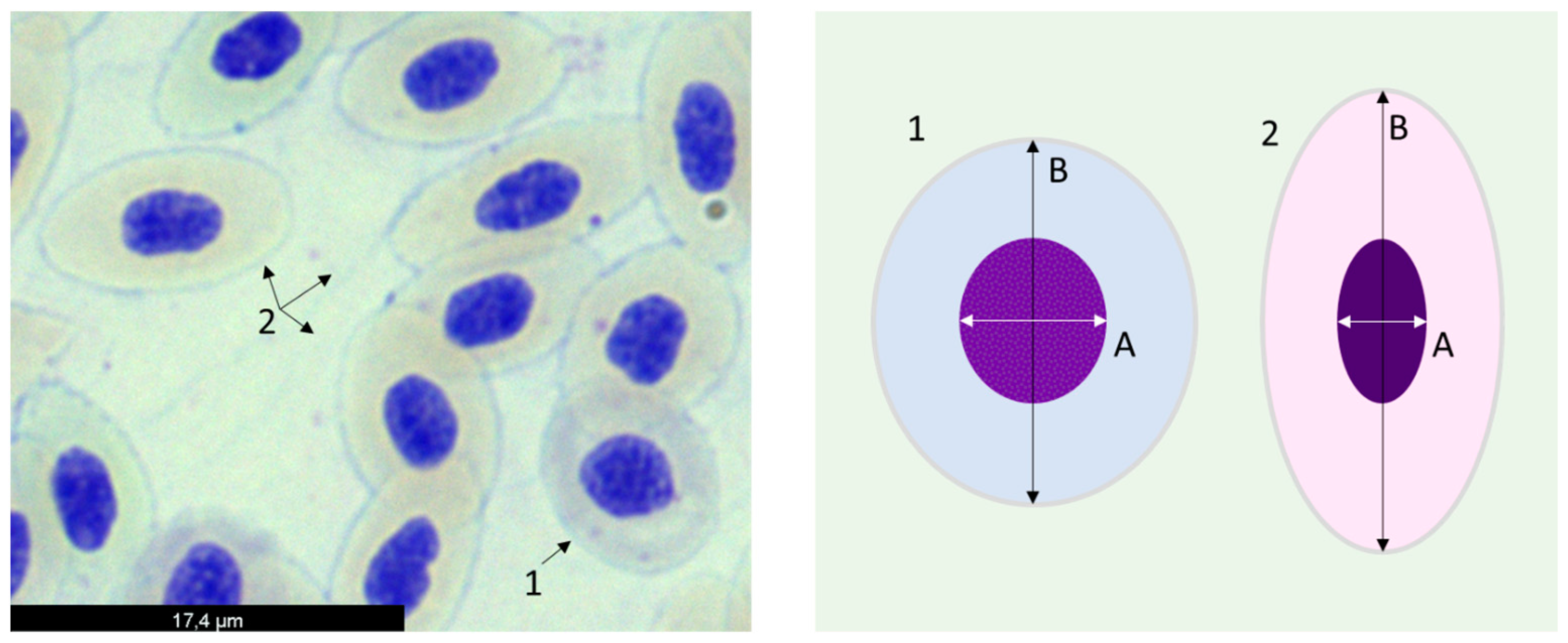
2.3.3. Erythrocyte Population Dynamics

The erythrocyte population dynamics were assessed by the calculation of the erythrocyte maturity index (EMI), determined according to [56], with the modifications proposed by [57]. Briefly, 10 microscopic fields were randomly selected per slide (one slide per fish; the same slides used for the ENA assay) and photographed under 400× magnification (microscope Olympus BXB50). Then, in each microscopic field, 25 random cells were analyzed with ImageJ software, measuring the minor axis of the nucleus and the major axis of the cell (A and B, respectively; see Figure 1).
The EMI was calculated for each cell by dividing A by B values, to a total of 250 cells. From the values of the ratio, cells were then categorized into one of the 10 maturity classes: [0.0 ≤ class 1 < 0.1]; [0.1 ≤ class 2 < 0.2]; [0.2 ≤ class 3 < 0.3]; [0.3 ≤ class 4 < 0.4]; [0.4 ≤ class 5 < 0.5]; [0.5 ≤ class 6 < 0.6]; [0.6 ≤ class 7 < 0.7]; [0.7 ≤ class 8 < 0.8]; [0.8 ≤ class 9 < 0.9]; and [0.9 ≤ class 10 ≤ 1], where class 1 represented erythrocytes with the higher maturity level and class 10 corresponded to cells with lower maturity status. Finally, average values for the frequency (%) of cells observed in each maturity class were represented for each experimental group.

2.4. Statistical Analyses

The statistical analysis was conducted on the free trial of Statistica 10.0 software (StatSoft, Inc., Street Tulsa, OK, USA), considering each sampling moment individually. The normality and homogeneity of variances were confirmed by Shapiro–Wilk’s W and Brown–Forsythe (HOV) tests, respectively, in order to meet the required statistical demands.
Differences in growth performance and survival between dietary treatments were evaluated using one-way ANOVA, followed by Tukey HSD multiple comparison tests. Kruskal–Wallis one-way analysis of variance tests, followed by Wilcoxon pairwise comparison tests were used when data did not comply with the one-way ANOVA’s assumptions.
For biochemical and cytogenetic data, one-way ANOVA followed by the post hoc Dunnett test were used to compare test-diet groups with the corresponding control. When the parametric demands failed, the statistical nonparametric Kruskal–Wallis test was performed. When the data did not fulfil the assumptions for normality, a non-parametric test was executed (Kruskal–Wallis ANOVA) and, when detected differences, a post-hoc Dunn’s test was applied.
In results expressed as percentages, an arcsine transformation was performed prior to any statistical test: T = ASIN (SQRT (value/100)), and then tested for normality (Shapiro–Wilk test and graphical analysis) and homogeneity of variances (Levene’s test).
In all the analyses, differences between means were considered significant when p < 0.05 [58].

3. Results

3.1. Growth Performance

Fish grew properly, and no significant differences between dietary treatments were found at the end of the feeding trial (month 2), in body weight as well as in survival values. Thus, final body weights (g) were 43.70 ± 0.32, 43.30 ± 1.28, 43.60 ± 0.98 and 43.50 ± 0.95, respectively for C, S2.5, S5, and S10. Survival values (%) were 94.60 ± 4.10, 97.10 ± 1.60, 97.90 ± 1.20, and 95.80 ± 1.60, respectively for C, S2.5, S5, and S10.

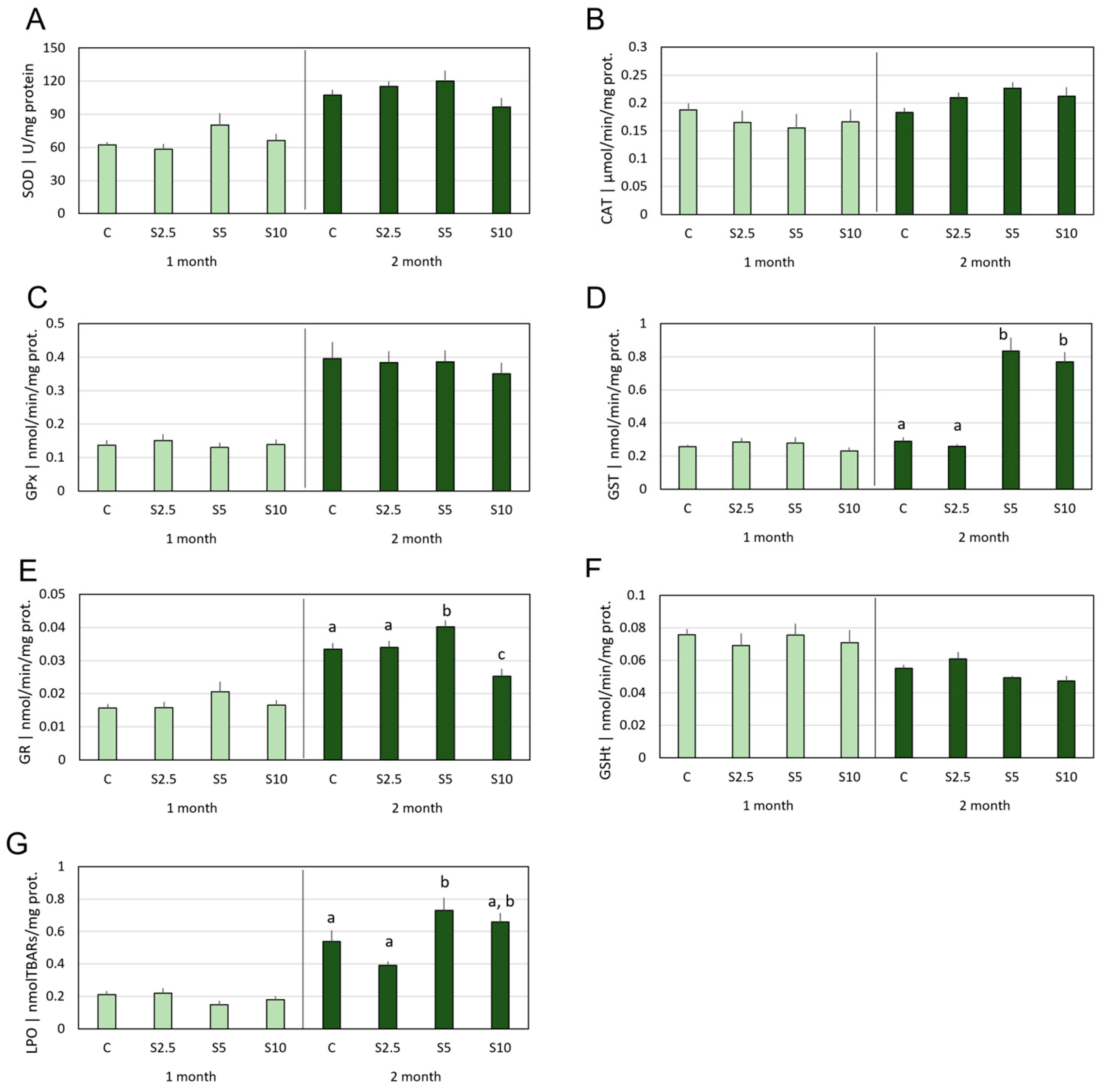
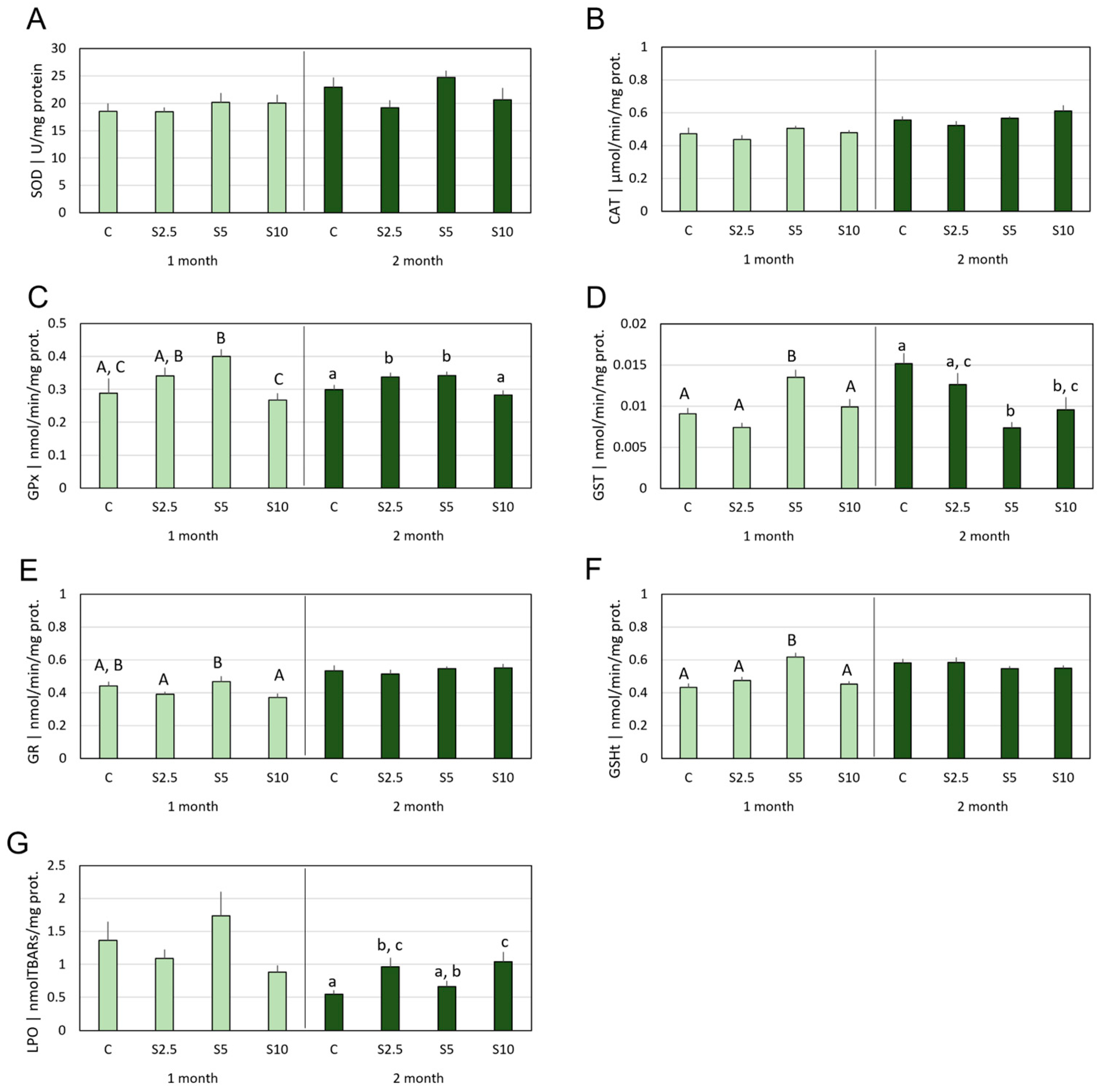
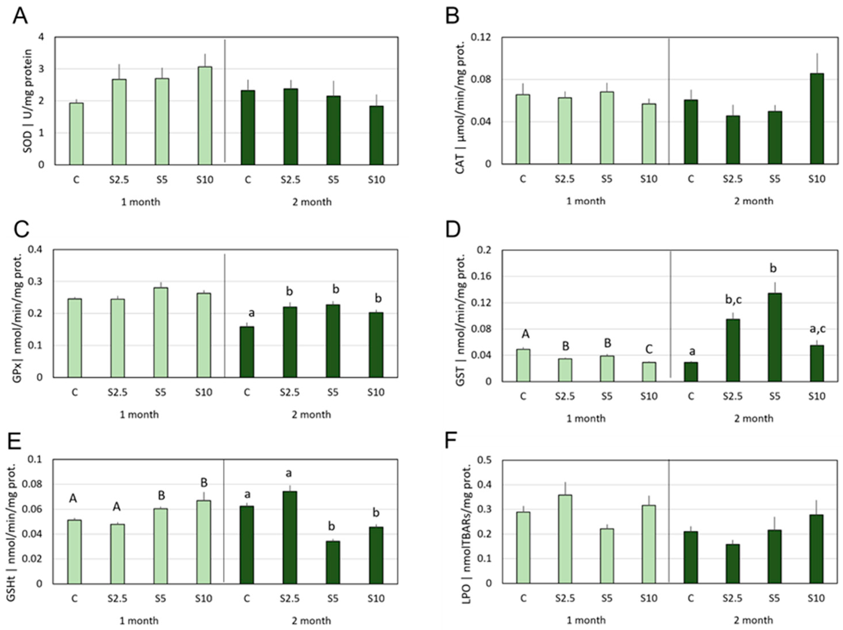
3.2. Antioxidant Defenses vs. Oxidative Damage

In the first month of the feeding trial, no variations in antioxidants nor lipid peroxidation were observed in the livers of the fish. Nevertheless, following 2 months of dietary supplementation, the S5 group showed significantly increased activities of GST and GR (Figure 2D,E), as well as an elevation of LPO levels (Figure 2G), when compared with the C group. At the same point in time, the S10 group also showed a GST activity increase, but occurring concomitantly with a GR activity decrease.
Concerning gills, in the first month, significant alterations in relation to group C were restricted to the S5 dietary group. Hence, this group depicted an increase of GPx and GST activities (Figure 2C and Figure 3D), as well as GSHt content (Figure 3F). Simultaneously, for the same group, no alterations were detected in SOD and CAT activities (Figure 3A,B), nor LPO (Figure 3G). A different response pattern was perceived in month 2, when a significant increase of GPx activity was measured in the S2.5 and S5 groups (Figure 3C), and a GST activity decrease in S5 and S10 (Figure 3D), while SOD and CAT activities remained unaltered (Figure 3A,B). Lipid peroxidation showed significant increases in the S2.5 and S10 groups (Figure 3G).
In blood, as reported for the other tissues, no significant differences were detected between the experimental groups (in both sampling moments) for SOD and CAT activities (Figure 4A,B).
The other antioxidants measured displayed distinct response patterns in the two trial lengths. Hence, regarding GPx activity, significant differences were only detected in the second month, with all the supplemented groups (S2.5, S5, and S10), showing higher levels in comparison with the control group, but no differences among them (Figure 4C).
Regarding GST activity (Figure 4D), after 1 month, all the supplemented groups displayed lower levels when compared to the respective control, with the highest supplementation group (S10) also showing significantly lower activity in comparison with the other supplemented groups (S2.5 and S5). Differently, after 2 months, groups S2.5 and S5 showed significantly elevated GST activity in comparison to the control, with the highest level reported for S5 (also significantly higher than S10). Concerning GSHt, time-related profiles were also found (Figure 4E), since, in the first month, groups S5 and S10 revealed significantly higher contents in comparison with the control group (as well as with S2.5), while, after 2 months, the same groups displayed significantly lower content in comparison with the control group (as well as with S2.5). In terms of LPO, no significant alterations were detected either in the first or in the second month (Figure 4F).

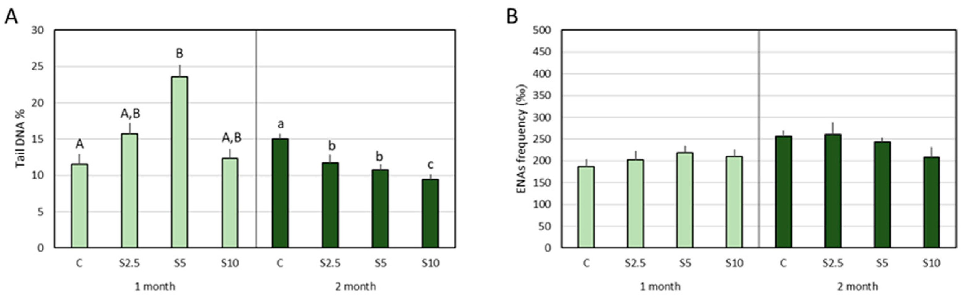
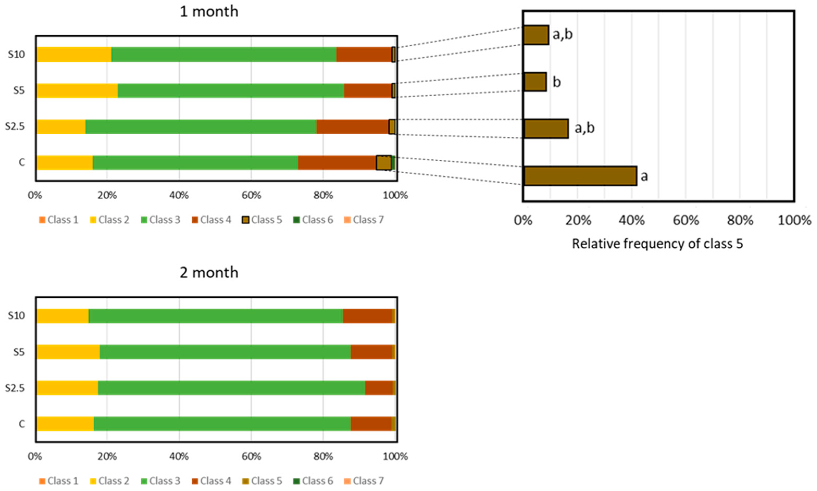
3.3. DNA and Chromosomal Integrity

Significant differences in terms of tail DNA% were detected in both months but translating contrasting variation profiles. In the first month, the S5 group revealed significantly higher DNA damage when compared to the control group. In the second month, all the supplemented groups (S2.5, S5, and S10) showed significantly lower values of DNA damage when compared to the control group, with the lowest percentage (highest DNA integrity) measured for S10 (Figure 5A).
During the two months of the trial, no significant differences were detected for the ENA frequency (Figure 5B).

3.4. Erythrocyte Population Dynamics (EMI Assay)

The cell frequency for each maturity class is depicted in Figure 6 (classes 1 to 7; no cells in classes 8 to 10 were observed). The most representative class in both sampling moments, regardless of the experimental group, was class 3.
Significant differences between the groups were detected only for class 5 and in the first month of the experimental trial. The group fed with 5% S. ramosissima (S5) revealed a lower frequency of these cells when compared to the control group (Figure 6).

4. Discussion

In past decades, developments in fish nutrition towards the identification of functional feeds and a thorough understanding of underlying mechanisms have been made; however, research in this area has been mainly focused on animal growth, feed intake, fillets’ physicochemical and sensory properties, and histological analysis of the digestive apparatus and gut microbiome [59]. In more recent years, studies of DNA and chromosomal integrity, as well antioxidant system modulation, have been performed in fish [60,61], highlighting the potential advances for the aquaculture sector resulting from the scientific knowledge to be developed in this context.
About 2000 plants are known worldwide to possess some salinity tolerance, but only a few species have been investigated for their antioxidant capacities [3], thus adding a greater scientific interest to the current study.
In the light of the statement “a feed is only as good as its ingredients” [62], and considering the phytochemical profiling of Salicornia spp. described in the literature [10,11,12] and the beneficial health properties already demonstrated in other contexts, the potential of these halophytes as functional ingredients of fish feeds bursts forth as a plausible hypothesis. Nevertheless, this has never been addressed, including in the context of carnivorous species like the European seabass (Dicentrarchus labrax), which points out the novelty of the present investigation.
Though this research was focused on the functionality of dietary supplementation with S. ramosissima biomass, some considerations on aquafeed environmental sustainability can still be made. Functional additives are supposed to be incorporated at low concentrations, but it must be highlighted that the highest inclusion level currently tested (10%), replacing wheat meal, represents a saving in cereal use of 63%, compared with a regular feed, which is likely to have impacts beyond functionality (such as in terms of economic and environmental sustainability). This should be just one aspect of a larger goal of improving the environmental performance of global aquaculture [63] that also includes the reduction the Fish In: Fish Out (FIFO) ratio [64], and has been increasingly placed on the agenda of researchers, feed and fish meal/oil producers, and regulation and advisory agencies (e.g., the European Aquaculture Advisory Council).

4.1. Growth Performance of European Seabass following S. ramosissima Dietary Supplementation

As previously stated, from the point of view of farmers and feed manufacturers, the primary condition for a novel plant-based raw material to be pondered as a functional ingredient for fish feeds (in addition to costs) is the maintenance of zootechnical performance (e.g., survival, growth, feed palatability, and feeding efficiency) [15,16,17]. In this direction, current data indicate that S. ramosissima biomass can be included in diets for juvenile D. labrax, at up to 10% of their composition, with no detrimental effects to growth performance and survival. This agrees with previous works on D. labrax nutrition, which demonstrated that macroalgae- [65] and plant-based [66] meals do not negatively affect growth.
In the present work, a percentage of wheat meal was replaced by halophyte biomass in D. labrax feed. Wheat meal has been introduced in fish feed formulations since it is a good source of energy due to its high carbohydrate content and dietary fiber, and can also complement protein sources [67]. Hence, we also demonstrated the correctness of the decision regarding the partial replacement of wheat, instead of another ingredient, in regular D. labrax feeds.
Hence, once the key factor regarding growth performance and survival was guaranteed, the conditions were met to move on to the next step, exploring the potential functional properties of S. ramosissima.

4.2. Incorporation of S. ramosissima in European Seabass Feed and Modulation of the Antioxidant System and Oxidative Status

Salicornia spp. are known to be a good source of antioxidants [9,10,11,68]. For instance, it has been demonstrated that S. herbacea could be a useful antioxidant due to its phenolic (salycilic, p-coumaric, ferulic, caffeic acids) and flavonoid (quercetin, acacetin) compounds [68]. Phenolic compounds are known to have direct antioxidant actions due to the presence of hydroxyl groups, which function as hydrogen donors [69]. Concerning specifically S. ramosissima, it was reported that its antioxidant capacity may be related to compounds such as vitamins (e.g., β-carotene and α-tocopherol) [10], flavonoids, phenolics, and hydroxycinnamic acid derivatives [11,70]. Therefore, the assumptions are met to put forward the hypothesis that dietary supplementation with S. ramosissima can improve the oxidative status of D. labrax.
The antioxidant system is essential for maintaining cellular and tissue integrity, as fish, like all aerobic organisms, are exposed to pro-oxidant pressures due to normal (endogenous) metabolic processes and exogenous factors [71,72]. It has been clearly established for fish, including for D. labrax [73], that antioxidant responses are tissue specific, showing differential expression of defense mechanisms in various organs/tissues due to their distinct functions, metabolic rates, and exposure to environmental stressors resulting from their anatomical position. Hence, though an integrated picture of the different tissues addressed is envisioned, the current results will first be discussed, respecting this specificity.
The liver is the central metabolic organ in fish [74], responsible for an array of functions, and the principal site of xenobiotic metabolism and detoxification of harmful substances immediately after their absorption from the gastrointestinal tract [74]. Therefore, this organ developed a robust antioxidant defense system to protect its own cellular structures and maintain overall health. This robustness of the hepatic antioxidant system (which also includes a variety of non-enzymatic antioxidants not measured in the present study, like vitamin C, vitamin E, and various metal-binding proteins) may justify the “immobility” (stability) of the antioxidants addressed in the liver after the first month of supplementation with S. ramosissima. Unlike gills and blood, the liver took 2 months for the GSH-associated enzymes (GST and GR) to respond, which, in coherence, only occurred in the higher levels of supplementation (5% and 10%), with the lower level (2.5%) remaining unresponsive. Moreover, it seems that the incorporation of 5% of S. ramosissima acted as a challenge to the hepatic antioxidant system, which, in addition to increasing GST and GR activities, allowed the rise of LPO levels. A preliminary interpretation of these results would point to the existence of a certain level of toxicity associated with the halophyte ingestion. However, a more comprehensive discussion will be developed below, pointing out another interpretation.
Antioxidant defenses in organs that interface with the external medium and are involved in gas exchange, such as fish gills, are reinforced as they are highly exposed to environmental factors that can lead to the production of ROS. Thus, the branchial GSH-related defense subsystem was particularly responsive in fish fed with 5% of S. ramosissima for 1 month, displaying increased GPx, GST, and GR activities, as well as GSHt content. Interestingly, the gills of fish fed with the diets incorporating 2.5% and 10% remained unresponsive. The GSH-related defense subsystem plays a crucial role in protecting cells and organisms from oxidative stress and various toxic substances [75], and refers to a set of biological mechanisms and molecules centered around the tripeptide molecule glutathione (GSH), which, in addition to the elements analyzed here, further includes GSH biosynthesis and GSH transporters. It seems clear that, within relatively narrow limits, one month of S. ramosissima supplementation can upregulate GSH-related defenses in juvenile seabass. After 2 months of dietary supplementation, the modulation of these antioxidants followed a distinct profile, with a less distinguishable pattern (GPx induction in S2.5 and S5; GST decrease in S5 and S10), coupled with the emergence of lipid peroxidation in the S2.5 and S10 groups.
A solid discussion on these results is necessary here, considering the complexity of the underlying processes. There is not a universally agreed-upon specific level of lipid peroxidation increase that is considered toxicologically worrying to fish, because it can depend on several factors, including baseline levels, duration of exposure, tissue/organ specificities, and concurrent factors. Considering the interference of all these factors, there may be a dual interpretation for an increase of LPO; on the one hand, it can be a consequence of a failure/overwhelm of the antioxidant system, but on the other, it can signal a trigger for the activation of the antioxidant system. In some cases, exposure to moderate levels of ROS and lipid peroxidation can stimulate an adaptive response, making the cells more resilient to future oxidative pressures [61]. This interplay between lipid peroxidation and the antioxidant system is a vital part of an integrated response to oxidative stress and the efforts to maintain/restore cellular homeostasis and tissue integrity. Therefore, the most plausible explanation for the punctual elevations of LPO, observed following 2 months of dietary supplementation with S. ramosissima, is that some plant constituents generate an external stimulus for the liver (at 5%) and gill (at 2.5% and 10%) cells of seabass, with the goal of strengthening the antioxidant defense system.
It has been demonstrated that polyphenols, also known for their antioxidant effects, can also have a pro-oxidant action associated with their instability and easy oxidation, generating H2O2, quinones, and semiquinones, among other products [69]. These compounds can thus exert a mild stressful challenge that triggers a rapid response, leading to increased levels of endogenous antioxidants, such as GSH [69,76]. Hence, flavonoids, a subgroup of polyphenols occurring in Salicornia spp. [11,68,70], are plausible candidates to play that dual role.
Corroborating the previous interpretation, it should be highlighted that certain foods or isolated compounds that are usually described as antioxidants also display pro-oxidant actions in humans [77,78]. In addition, several studies reported pro-oxidant properties of certain phytocompounds, namely carotenoids [79], vitamin C [80], and polyphenols [81].
It is worth noting that the cogitated mild pro-oxidant action of S. ramosissima displayed a dose-dependence specificity for the organs in equation, i.e., it occurs at 5% supplementation for the liver and at 2.5% and 10% for the gills. Moreover, the relevance of the determinants mentioned above emerges as obvious, namely organ specificities, duration of exposure (response evident only for 2 months of supplementation), and concurrent factors, which can explain the non-linear relationship to the dose (likely following hormetic or J-shaped dose-response curves).
Concerning blood cell responses, a distinct pattern was observed in comparison to the liver and gills. The only similarities that could be invoked between the three tissues addressed are the unresponsiveness of SOD and CAT activities to the S. ramosissima supplementation and the occurrence of clearly different response patterns in the two trial lengths (1 vs. 2 months). Therefore, S. ramosissima was shown to modulate the GSH-related subsystem, translated into a GST activity decrease (all the dietary groups) and increased GSH content (S5 and S10) in the first month of supplementation, while in the second month, a GPx (all the dietary groups) and GST (S2.5 and S5) induction was observed.
It is noteworthy from these results that the GSH-related subsystem in blood cells can be up- or down-regulated by S. ramosissima supplementation. Both directions of this modulation seem to represent improvements of antioxidant status due to supplementation, though the decrease of GST activity (month 1) and GSHt content (month 2) can be particularly interesting considering the potential to favorably impact fish growth. This assertion relies on the contribution of an exogenous source (dietary supplementation) of nonenzymatic antioxidants, saving endogenous resources via a lower expression/synthesis of antioxidant/free radical scavenger molecules, as suggested by [60,82] for the mechanism underlying the benefits of a macroalgae-enriched fish feed. A fish study carried out by [83] involving dietary vitamins also lends support to this interpretation, configuring what was called the theory of sparing.
Specifically regarding blood GSHt in the first month, the increased contents displayed by groups S5 and S10 can rely on the concomitant decrease of GST activity. GSTs represent a multigene family of primarily soluble enzymes involved in the detoxification of electrophilic and genotoxic xenobiotics, as well as endogenous reactive intermediates produced during cellular oxidative stress like peroxidative products [84,85]. Hence, a decrease in GST activity means that less GSH is being used for detoxification processes, allowing more GSH to accumulate within the cells.
Overall, the present results are in conformity with [82,86], who stated that LPO increase cannot be predicted only on the basis of antioxidant depletion. Predicting peroxidative damage as a function of antioxidant responses can be challenging due to the complex, intricate, and multifaceted nature of the processes involved, depending on factors such as the involvement of multiple antioxidant sub-systems, interactions between antioxidants, time-dependent dynamics, and feedback mechanisms.
During the whole trial, and in all tissues addressed, there were no variations in the activity of two key enzymatic antioxidants—SOD and CAT—categorized as a first line of defense. Moreover, this unresponsiveness was registered frequently in concomitance with the modulation of the GSH-related antioxidant subsystem. Both lines of defense represent different and complementary mechanisms for combating oxidative stress. SOD and CAT primarily deal with the breakdown of superoxide radicals (O2•-) and hydrogen peroxide (H2O2), while the GSH-related subsystem focuses on neutralizing other ROS, such as lipid peroxides and organic hydroperoxides [72,87]. Thus, it may be hypothesized that S. ramosissima supplementation entails a specific form of (mild) oxidative challenge, shared by the three tissues studied, pointing out the ability of seabass to fine-tune its defenses to match the specific nature of the oxidative stress it encounters.

4.3. Incorporation of S. ramosissima in European Seabass Feed and DNA/Chromosomal Integrity

The antigenotoxic properties of natural edible products (e.g., fruit, green tea) have been frequently investigated, using human experimental approaches, aiming at the identification of functional foods [88,89,90]. Differently, in the context of aquafeed formulation, though the concept has been also valued, research is much less profuse. Promising results were obtained, for instance, with the introduction of a macroalgae mixture in feeds for S. aurata, demonstrating a genoprotection effect [60], including against aqua-medicines frequently adopted in aquaculture (the antibiotic oxytetracycline and the antiparasitic formalin) [27]. However, the genoprotective potential of S. ramosissima incorporated in fish feeds, to the authors’ knowledge, is a completely unexplored topic.
Antigenotoxic protection was investigated in the present study through two complementary endpoints (both focused on blood cells): (i) DNA breaks measured by the comet assay (tail DNA %), a type of damage that can be repaired, and (ii) ENA frequency, translating to a more severe and less transient type of genetic damage (clastogenic and/or aneugenic lesions).
The results showed that chromosomal integrity (measured by ENA frequency) was not affected by the dietary treatments, indicating that more subtle modulation processes are at play. In fact, comet assay data pointed out measurable alterations, reflecting (apparently) contrasting results for 1-month and 2-month supplementation. In the first month, a punctual elevation of DNA damage was registered for the group fed with 5% S. ramosissima. As discussed above for oxidative stress data, this effect should be interpreted cautiously, taking into consideration the fact that it resulted from a delicate balance between genotoxic and antigenotoxic pressures. In fact, it is well established that substances described as DNA integrity promoters depict a mild genotoxic action, which may be a path to trigger the antigenotoxic defenses (e.g., [91,92,93,94]). These complex mechanisms, representing “two sides of the same redox coin” [88], were partially decoded using in vitro approaches (human lymphocytes) and addressing the protective effects of green tea (Camellia sinensis) [88]. Thus, it was suggested that genoprotection by low-dose green tea could be due to the direct antioxidant protection of polyphenols, while genotoxicity is induced by H2O2 (generated by autoxidation of catechins).
The integrated analysis of the different results presently obtained provide support to the interpretation of the DNA integrity alteration registered for the S5 group (month 1) in light of the mechanisms enunciated above, thus translating as mild and temporary damage that works as a trigger to activate the DNA protective pathways. In this direction, it should be considered that: (i) no increased DNA breaks were observed, for the same trial duration, in fish fed with 10% of S. ramosissima (which would be expected if it were a real toxic action); (ii) no chromosomal damage nor lipid peroxidation were concomitantly observed (S5 in month 1); (iii) the effect was no longer noticeable after 2 months. Hence, what could be described as a sign of genotoxicity is better described as a pro-genoprotective action.
As a further reinforcement of this explanation, following 2 months, comet assay results demonstrated that Salicornia-enriched diets offer solid genoprotection to all the groups with Salicornia supplementation, with emphasis on the S10 group that showed the most significant improvement to DNA integrity.
Similar to what was described by [88], it can be hypothesized that the genoprotective and genotoxic (pro-genoprotective) actions may be mediated by different S. ramosissima constituents, with distinct toxicokinetics and toxidynamics in the fish body. Nevertheless, no support is currently found for H2O2 to play the role of genotoxicant, since the results regarding antioxidant modulation do not confirm that specific mechanism, since CAT activity was regularly unresponsive. Elucidation of these complex mechanisms requires further investigation.
Looking closely, the results obtained after 1 and 2 months of supplementation will be complementary rather than contrasting, as the protective potential expressed over the longer period will have an underlying and intermediate state of preparation.
DNA integrity is vital for any organism, in any context, but it assumes paramount importance in fish under intensive culture (involving, for instance, high-density populations, close confinement, controlled environments, variations in water quality, and exposure to aqua-medicines) for several reasons: (i) is essential for preserving genetic diversity within fish populations, also associated with the ability to cope with stressors, reducing the risk of stress-related illnesses; (ii) it is linked to the immune system’s ability to recognize and combat pathogens, thereby promoting a robust immune response; (iii) any genetic defects or mutations can compromise growth; (iv) it allows saving the energy that would be allocated to defense mechanisms related, for example, to DNA damage repair or replacement of damaged cells.
In addition to the previous aspects, centered on fish health, it is also relevant to point out that maintaining DNA integrity helps ensure that the final products meet safety and quality standards, as well countering some criticism related to animal welfare and ethical concerns.
In future works, it will be important to understand the effect of a diet enriched with Salicornia when fish are facing genotoxic and/or oxidative challenges arising from routine aquaculture practices.
To shed some light on the dynamics of the circulating erythrocyte population and possible interferences of the dietary backgrounds tested, the erythrocyte maturity index (EMI) was assessed (based on the analysis of nucleo-cytoplasmatic ratios). This index also complements the information provided by the ENA assay, elucidating the balance between erythropoiesis and cell removal, and thus about recovery pathways [27] (the fate of DNA/chromosomal lesions will depend upon replacement of damaged cells) [95]. The dynamics can be altered, for instance, through interference with hemoglobin production, assembly, or stability, as well as with the cell cycle and maturation of precursor cells [96].
Current EMI results demonstrated that, in general, the dietary backgrounds tested did not affect D. labrax erythrocyte dynamics, which also means that the genomic integrity assessment was not affected by fluctuations in the erythrocyte dynamics/lifespan. A single alteration was pointed out, which concerned a frequency reduction of cell in class 5 (a stage tending to be immature) for the S5 group at 1 month. The sporadic and transient nature of this fluctuation reduces its significance. However, it can be an indication that erythrocytes from seabass fed with 5% Salicornia had a slightly higher lifespan, which could reflect an improvement of blood cells’ condition due to halophyte ingestion. Moreover, the benefit of avoiding energy expenditure in cell rejuvenation is implicit.

5. Conclusions

Overall, the outputs of the present study, addressing the potential of the incorporation of the halophyte S. ramosissima in the diet of the European seabass (D. labrax), allow the following conclusions:
(i)
The inclusion of whole-plant biomass up to 10%, for 2 months, replacing wheat meal, showed no impairments on growth performance, pointing out that the use of S. ramosissima routinely as a feed ingredient can be considered, though its scalability and economic viability need to be evaluated.
(ii)
S. ramosissima was shown to affect the oxidative status of D. labrax, namely modulating the GSH-related defense subsystem in a tissue-specific manner and with higher acuity in the second month of feeding. Though an antioxidant improvement was apparent, it followed multiple and complex mechanisms, which also included a mild pro-oxidant that, expectantly, triggered beneficial adaptations of the endogenous antioxidant systems.
(iii)
A clear protection of DNA integrity (potential to reduce DNA damage or to increase the resistance of DNA to routine challenges) was detected in the second month for all the inclusion levels, though the most prominent benefit was achieved for the highest level (10%). Again, complex genoprotective pathways were disclosed, involving an intermediary step (expressed in the first month) where a genotoxic trigger (regarded as a pro-genoprotective mechanism) was identified.
(iv)
The use of Salicornia-enriched formulations as complementary feeds with functional properties showed potential, but the choice of dose and supplementation time tended to be critical.
In short, these results highlight a promising solution to improve aquaculture practices, also unraveling a complementary novel, low-value raw material, and a valorization pathway.

Author Contributions

Conceptualization, B.C., R.J.M.R. and M.P.; Investigation, R.M. and M.P.; Methodology, R.M., P.S., A.M., V.P. and A.B.; Supervision, M.P.; Writing—original draft, R.M., P.S. and S.G.; Writing—review & editing, B.C., R.J.M.R. and M.P. All authors have read and agreed to the published version of the manuscript.

Funding

This study has received funding from the European Union’s Horizon 2020 research and innovation programme under Grant Agreement No. 862834. Any results of this project reflect only this consortium’s view; the European Commission is not responsible for any use that may be made of the information it contains.

Institutional Review Board Statement

Fish rearing and sampling were carried out employing established routine and animal husbandry practices commonly employed in commercial farming operations. The procedures were carried out by technicians certified by FELASA. The fish trial and experimental protocols were approved by the Ethical Committee of Riasearch Lda. (Murtosa, Portugal) overseen by the National Veterinary Authority (DGAV, Portugal), authorization Nº 0421/0000/0000/2021, in compliance with the guidelines of the European Union (Directive 2010/63/EU) and Portuguese law (Artigo 44, Decreto-Lei no. 113/2013, de 7 de Agosto) on the protection of animals used for scientific purposes.

Data Availability Statement

Data are contained within the article.

Acknowledgments

Thanks are due to CESAM by FCT/MCTES (UIDP/50017/2020 + UIDB/50017/2020 + LA/P/004/2020), through National funds, as well as to FCT—Foundation for Science and Technology within the scope of UIDB/0443/2020 and UIDP/04423/2020. BC was supported by FCT (2020.00290.CEECIND).

Conflicts of Interest

The authors declare no conflicts of interest.

References

  1. Turcios, A.E.; Cayenne, A.; Uellendahl, H.; Papenbrock, J. Halophyte Plants and Their Residues as Feedstock for Biogas Production—Chances and Challenges. Appl. Sci. 2021, 11, 2746. [Google Scholar] [CrossRef]
  2. Fry, J.P.; Love, D.C.; MacDonald, G.K.; West, P.C.; Engstrom, P.M.; Nachman, K.E.; Lawrence, R.S. Environmental Health Impacts of Feeding Crops to Farmed Fish. Environ. Int. 2016, 91, 201–214. [Google Scholar] [CrossRef] [PubMed]
  3. Ksouri, R.; Ksouri, W.M.; Jallali, I.; Debez, A.; Magné, C.; Hiroko, I.; Abdelly, C. Medicinal Halophytes: Potent Source of Health Promoting Biomolecules with Medical, Nutraceutical and Food Applications. Crit. Rev. Biotechnol. 2012, 32, 289–326. [Google Scholar] [CrossRef] [PubMed]
  4. Giordano, R.; Saii, Z.; Fredsgaard, M.; Hulkko, L.S.S.; Poulsen, T.B.G.; Thomsen, M.E.; Henneberg, N.; Zucolotto, S.M.; Arendt-Nielsen, L.; Papenbrock, J.; et al. Pharmacological Insights into Halophyte Bioactive Extract Action on Anti-Inflammatory, Pain Relief and Antibiotics-Type Mechanisms. Molecules 2021, 26, 3140. [Google Scholar] [CrossRef] [PubMed]
  5. Hulkko, L.S.S.; Chaturvedi, T.; Thomsen, M.H. Extraction and Quantification of Chlorophylls, Carotenoids, Phenolic Compounds, and Vitamins from Halophyte Biomasses. Appl. Sci. 2022, 12, 840. [Google Scholar] [CrossRef]
  6. Belal, I.E.H.; Al-Dosari, M. Replacement of Fish Meal with Salicornia Meal in Feeds for Nile Tilapia Oreochromis niloticus. J. World Aquac. Soc. 1999, 30, 285–289. [Google Scholar] [CrossRef]
  7. Ríos-Durán, M.G.; Valencia, I.R.; Ross, L.G.; Martínez-Palacios, C.A. Nutritional Evaluation of Autoclaved Salicornia bigelovii Torr. Seed Meal Supplemented with Varying Levels of Cholesterol on Growth, Nutrient Utilization and Survival of the Nile Tilapia (Oreochromis niloticus). Aquac. Int. 2013, 21, 1355–1371. [Google Scholar] [CrossRef]
  8. Ventura, Y.; Eshel, A.; Pasternak, D.; Sagi, M. The Development of Halophyte-Based Agriculture: Past and Present. Ann. Bot. 2015, 115, 529–540. [Google Scholar] [CrossRef]
  9. Patel, S. Salicornia: Evaluating the Halophytic Extremophile as a Food and a Pharmaceutical Candidate. 3 Biotech 2016, 6, 104. [Google Scholar] [CrossRef]
  10. Lima, A.R.; Castañeda-Loaiza, V.; Salazar, M.; Nunes, C.; Quintas, C.; Gama, F.; Pestana, M.; Correia, P.J.; Santos, T.; Varela, J.; et al. Influence of Cultivation Salinity in the Nutritional Composition, Antioxidant Capacity and Microbial Quality of Salicornia Ramosissima Commercially Produced in Soilless Systems. Food Chem. 2020, 333, 127525. [Google Scholar] [CrossRef] [PubMed]
  11. Cárdenas-Pérez, S.; Piernik, A.; Chanona-Pérez, J.J.; Grigore, M.N.; Perea-Flores, M.J. An Overview of the Emerging Trends of the Salicornia L. Genus as a Sustainable Crop. Environ. Exp. Bot. 2021, 191, 104606. [Google Scholar] [CrossRef]
  12. Barreira, L.; Resek, E.; Rodrigues, M.J.; Rocha, M.I.; Pereira, H.; Bandarra, N.; da Silva, M.M.; Varela, J.; Custódio, L. Halophytes: Gourmet Food with Nutritional Health Benefits? J. Food Compos. Anal. 2017, 59, 35–42. [Google Scholar] [CrossRef]
  13. Kim, S.; Lee, E.Y.; Hillman, P.F.; Ko, J.; Yang, I.; Nam, S.J. Chemical Structure and Biological Activities of Secondary Metabolites from Salicornia Europaea l. Molecules 2021, 26, 2252. [Google Scholar] [CrossRef] [PubMed]
  14. Custódio, M.; Lillebø, A.I.; Calado, R.; Villasante, S. Halophytes as Novel Marine Products—A Consumers’ Perspective in Portugal and Policy Implications. Mar. Policy 2021, 133, 104731. [Google Scholar] [CrossRef]
  15. Herman, E.M.; Schmidt, M.A. The Potential for Engineering Enhanced Functional-Feed Soybeans for Sustainable Aquaculture Feed. Front. Plant Sci. 2016, 7, 440. [Google Scholar] [CrossRef]
  16. Soto, J.O.; Paniagua-Michel, J.D.J.; Lopez, L.; Ochoa, L. Functional feeds in aquaculture. In Springer Handbook of Marine Biotechnology; Springer: Berlin/Heidelberg, Germany, 2015. [Google Scholar]
  17. Wan, A.H.L.; Davies, S.J.; Soler-Vila, A.; Fitzgerald, R.; Johnson, M.P. Macroalgae as a Sustainable Aquafeed Ingredient. Rev. Aquac. 2019, 11, 458–492. [Google Scholar] [CrossRef]
  18. Guerreiro, I.; Magalhães, R.; Coutinho, F.; Couto, A.; Sousa, S.; Delerue-Matos, C.; Domingues, V.F.; Oliva-Teles, A.; Peres, H. Evaluation of the Seaweeds Chondrus Crispus and Ulva Lactuca as Functional Ingredients in Gilthead Seabream (Sparus aurata). J. Appl. Phycol. 2019, 31, 2115–2124. [Google Scholar] [CrossRef]
  19. Hoseinifar, S.H.; Yousefi, S.; Van Doan, H.; Ashouri, G.; Gioacchini, G.; Maradonna, F.; Carnevali, O. Oxidative Stress and Antioxidant Defense in Fish: The Implications of Probiotic, Prebiotic, and Synbiotics. Rev. Fish. Sci. Aquac. 2020, 29, 198–217. [Google Scholar] [CrossRef]
  20. Barata, C.; Varo, I.; Navarro, J.C.; Arun, S.; Porte, C. Antioxidant Enzyme Activities and Lipid Peroxidation in the Freshwater Cladoceran Daphnia Magna Exposed to Redox Cycling Compounds. Comp. Biochem. Physiol. C Toxicol. Pharmacol. 2005, 140, 175–186. [Google Scholar] [CrossRef]
  21. Kovacic, P. Free Radicals in Biology and Medicine. J. Pharm. Sci. 1986, 75, 105–106. [Google Scholar] [CrossRef]
  22. Lushchak, V.I. Environmentally Induced Oxidative Stress in Aquatic Animals. Aquat. Toxicol. 2011, 101, 13–30. [Google Scholar] [CrossRef] [PubMed]
  23. Wischhusen, P.; Larroquet, L.; Durand, T.; Oger, C.; Galano, J.M.; Rocher, A.; Vigor, C.; Antony Jesu Prabhu, P.; Véron, V.; Briens, M.; et al. Oxidative Stress and Antioxidant Response in Rainbow Trout Fry Exposed to Acute Hypoxia Is Affected by Selenium Nutrition of Parents and during First Exogenous Feeding. Free Radic. Biol. Med. 2020, 155, 99–113. [Google Scholar] [CrossRef] [PubMed]
  24. Torrecillas, S.; Terova, G.; Makol, A.; Serradell, A.; Valdenegro-Vega, V.; Izquierdo, M.; Acosta, F.; Montero, D. Dietary Phytogenics and Galactomannan Oligosaccharides in Low Fish Meal and Fish Oil-Based Diets for European Sea Bass (Dicentrarchus labrax) Juveniles: Effects on Gill Structure and Health and Implications on Oxidative Stress Status. Front. Immunol. 2021, 12, 663106. [Google Scholar] [CrossRef] [PubMed]
  25. Sahin, K.; Yazlak, H.; Orhan, C.; Tuzcu, M.; Akdemir, F.; Sahin, N. The Effect of Lycopene on Antioxidant Status in Rainbow Trout (Oncorhynchus mykiss) Reared under High Stocking Density. Aquaculture 2014, 418–419, 132–138. [Google Scholar] [CrossRef]
  26. Hisar, O.; Yanik, T.; Kocaman, E.M.; Arslan, M.; Slukvin, A.; Goncharova, R. Effects of Diludine Supplementation on Growth Performance, Liver Antioxidant Enzyme Activities and Muscular Trace Elements of Rainbow Trout (Oncorhynchus mykiss) Juveniles at a Low Water Temperature. Aquac. Nutr. 2012, 18, 211–219. [Google Scholar] [CrossRef]
  27. Marques, A.; Marçal, R.; Pereira, V.; Pereira, P.; Mieiro, C.; Guilherme, S.; Marques, C.; Santos, M.A.; Pereira, R.; Abreu, H.; et al. Macroalgae-Enriched Diet Protects Gilthead Seabream (Sparus aurata) against Erythrocyte Population Instability and Chromosomal Damage Induced by Aqua-Medicines. J. Appl. Phycol. 2020, 32, 1477–1493. [Google Scholar] [CrossRef]
  28. Cheng, C.H.; Liang, H.Y.; Luo, S.W.; Wang, A.L.; Ye, C.X. The Protective Effects of Vitamin C on Apoptosis, DNA Damage and Proteome of Pufferfish (Takifugu obscurus) under Low Temperature Stress. J. Therm. Biol. 2018, 71. [Google Scholar] [CrossRef]
  29. Directorate-General for Maritime Affairs and Fisheries. Case Study in the EU Seabass Maritime Affairs and Fisheries Price Structure in the Supply Chain for Seabass focus on Greece, Croatia and Spain. 2021. Available online: https://op.europa.eu/en/publication-detail/-/publication/4577254b-8251-11eb-9ac9-01aa75ed71a1/language-en (accessed on 21 February 2023).
  30. Messina, M.; Piccolo, G.; Tulli, F.; Messina, C.M.; Cardinaletti, G.; Tibaldi, E. Lipid Composition and Metabolism of European Sea Bass (Dicentrarchus labrax L.) Fed Diets Containing Wheat Gluten and Legume Meals as Substitutes for Fish Meal. Aquaculture 2013, 376–379, 6–14. [Google Scholar] [CrossRef]
  31. Kaushik, S.J.; Covès, D.; Dutto, G.; Blanc, D. Almost Total Replacement of Fish Meal by Plant Protein Sources in the Diet of a Marine Teleost, the European Seabass, Dicentrarchus labrax. Aquaculture 2004, 230, 391–404. [Google Scholar] [CrossRef]
  32. Oliva-Teles, A.; Gonçalves, P. Partial Replacement of Fishmeal by Brewers Yeast (Saccaromyces cerevisae) in Diets for Sea Bass (Dicentrarchus labrax) Juveniles. Aquaculture 2001, 202, 269–278. [Google Scholar] [CrossRef]
  33. Tadese, D.A.; Song, C.; Sun, C.; Liu, B.; Liu, B.; Zhou, Q.; Xu, P.; Ge, X.; Liu, M.; Xu, X.; et al. The Role of Currently Used Medicinal Plants in Aquaculture and Their Action Mechanisms: A Review. Rev. Aquac. 2022, 14, 816–847. [Google Scholar] [CrossRef]
  34. Carbone, D.; Faggio, C. Importance of Prebiotics in Aquaculture as Immunostimulants. Effects on Immune System of Sparus aurata and Dicentrarchus labrax. Fish. Shellfish. Immunol. 2016, 54, 172–178. [Google Scholar] [CrossRef] [PubMed]
  35. Ferreira, M.; Sousa, V.; Oliveira, B.; Canadas-Sousa, A.; Abreu, H.; Dias, J.; Kiron, V.; Valente, L.M.P. An In-Depth Characterisation of European Seabass Intestinal Segments for Assessing the Impact of an Algae-Based Functional Diet on Intestinal Health. Sci. Rep. 2023, 13, 11686. [Google Scholar] [CrossRef] [PubMed]
  36. Randazzo, B.; Di Marco, P.; Zarantoniello, M.; Daniso, E.; Cerri, R.; Finoia, M.G.; Capoccioni, F.; Tibaldi, E.; Olivotto, I.; Cardinaletti, G. Effects of Supplementing a Plant Protein-Rich Diet with Insect, Crayfish or Microalgae Meals on Gilthead Sea Bream (Sparus aurata) and European Seabass (Dicentrarchus labrax) Growth, Physiological Status and Gut Health. Aquaculture 2023, 575, 739811. [Google Scholar] [CrossRef]
  37. Claiborne, A. Catalase activity. In Hand Book of Methods for Oxygen Radical Research; Greenwald, R.A., Ed.; CRC Press: Boca Raton, FL, USA, 1985. [Google Scholar]
  38. Mohandas, J.; Marshall, J.J.; Duggin, G.G.; Horvath, J.S.; Tiller, D.J. Differential Distribution of Glutathione and Glutathione-Related Enzymes in Rabbit Kidney. Possible Implications in Analgesic Nephropathy. Biochem. Pharmacol. 1984, 33, 1801–1807. [Google Scholar] [CrossRef]
  39. Athar, M.; Iqbal, M. Ferric Nitrilotriacetate Promotes N-Diethylnitrosamine-Induced Renal Tumorigenesis in the Rat: Implications for the Involvement of Oxidative Stress. Carcinogenesis 1998, 19, 1133–1139. [Google Scholar] [CrossRef]
  40. Habig, W.H.; Pabst, M.J.; Jakoby, W.B. Glutathione S Transferases. The First Enzymatic Step in Mercapturic Acid Formation. J. Biol. Chem. 1974, 249, 7130–7139. [Google Scholar] [CrossRef]
  41. Cribb, A.E.; Leeder, J.; Spielberg, S.P. Use of a Microplate Reader in an Assay of Glutathione Reductase Using 5,5′-Dithiobis(2-Nitrobenzoic Acid). Anal. Biochem. 1989, 183, 195–196. [Google Scholar] [CrossRef]
  42. Baker, M.A.; Cerniglia, G.J.; Zaman, A. Microtiter Plate Assay for the Measurement of Glutathione and Glutathione Disulfide in Large Numbers of Biological Samples. Anal. Biochem. 1990, 190, 360–365. [Google Scholar] [CrossRef]
  43. Tietze, F. Enzymic method for quantitative determination of nanogram amounts of total and oxidized glutathione: Applications to mammalian blood and other tissues. Anal. Biochem. 1969, 27, 502–522. [Google Scholar] [CrossRef]
  44. Ohkawa, H.; Ohishi, N.; Yagi, K. Assay for lipid peroxides in animal tissues by thiobarbituric acid reaction. Anal. Biochem. 1979, 95, 351–358. [Google Scholar] [CrossRef] [PubMed]
  45. Bird, R.P.; Draper, H.H. Comparative Studies on Different Methods of Malonaldehyde Determination. Methods Enzymol. 1984, 105, 299–305. [Google Scholar] [CrossRef] [PubMed]
  46. Filho, D.W.; Torres, M.; Tribess, T.; Pedrosa, R.; Soares, C. Influence of season and pollution on the antioxidant defenses of the cichlid fish acará (Geophagus brasiliensis). Braz. J. Med Biol. Res. 2001, 34, 719–726. [Google Scholar] [CrossRef] [PubMed]
  47. Filho, D.W.; Tribess, T.; Gáspari, C.; Claudio, F.; Torres, M.; Magalhães, A. Seasonal changes in antioxidant defenses of the digestive gland of the brown mussel (Perna perna). Aquaculture 2001, 203, 149–158. [Google Scholar] [CrossRef]
  48. Collins, A.R. The Comet Assay for DNA Damage and Repair: Principles, Applications, and Limitations. Mol. Biotechnol. 2004, 26, 249–261. [Google Scholar] [CrossRef] [PubMed]
  49. Guilherme, S.; Gaivao, I.; Santos, M.A.; Pacheco, M. European eel (Anguilla anguilla) genotoxic and pro-oxidant responses following short-term exposure to Roundup(R)—A glyphosate-based herbicide. Mutagenesis 2010, 25, 523–530. [Google Scholar] [CrossRef]
  50. Shaposhnikov, S.; Azqueta, A.; Henriksson, S.; Meier, S.; Gaivão, I.; Huskisson, N.H.; Smart, A.; Brunborg, G.; Nilsson, M.; Collins, A.R. Twelve-gel slide format optimised for comet assay and fluorescent in situ hybridisation. Toxicol. Lett. 2010, 195, 31–34. [Google Scholar] [CrossRef]
  51. Gyori, B.M.; Venkatachalam, G.; Thiagarajan, P.S.; Hsu, D.; Clement, M.-V. OpenComet: An automated tool for comet assay image analysis. Redox Biol. 2014, 2, 457–465. [Google Scholar] [CrossRef]
  52. Kumaravel, T.S.; Vilhar, B.; Faux, S.P.; Jha, A.N. Comet Assay measurements: A perspective. Cell Biol. Toxicol. 2009, 25, 53–64. [Google Scholar] [CrossRef]
  53. Møller, P. The comet assay: Ready for 30 more years. Mutagenesis 2018, 33, 1–7. [Google Scholar] [CrossRef]
  54. Møller, P.; Azqueta, A.; Boutet-Robinet, E.; Koppen, G.; Bonassi, S.; Milić, M.; Gajski, G.; Costa, S.; Teixeira, J.P.; Pereira, C.C.; et al. Minimum Information for Reporting on the Comet Assay (MIRCA): Recommendations for describing comet assay procedures and results. Nat. Protoc. 2020, 15, 3817–3826. [Google Scholar] [CrossRef] [PubMed]
  55. Pacheco, M.; Santos, M.A. Biotransformation, genotoxic, and histopathological effects of environmental contaminants in European eel (Anguilla anguilla L.). Ecotoxicol. Environ. Saf. 2002, 53, 331–347. [Google Scholar] [CrossRef] [PubMed]
  56. Maceda-Veiga, A.; Monroy, M.; Viscor, G.; De Sostoa, A. Changes in non-specific biomarkers in the Mediterranean barbel (Barbus meridionalis) exposed to sewage effluents in a Mediterranean stream (Catalonia, NE Spain). Aquat. Toxicol. 2010, 100, 229–237. [Google Scholar] [CrossRef] [PubMed]
  57. Castro, D.; Mieiro, C.; Coelho, J.; Guilherme, S.; Marques, A.; Santos, M.; Duarte, A.; Pereira, E.; Pacheco, M. Addressing the impact of mercury estuarine contamination in the European eel (Anguilla anguilla L., 1758)—An early diagnosis in glass eel stage based on erythrocytic nuclear morphology. Mar. Pollut. Bull. 2018, 127, 733–742. [Google Scholar] [CrossRef] [PubMed]
  58. Zar, J. Multiple comparisons. In Biostatistical Analysis; Prentice Hall International Inc.: Upper Saddle River, NJ, USA, 2010; pp. 226–248. [Google Scholar]
  59. Zarantoniello, M.; Randazzo, B.; Secci, G.; Notarstefano, V.; Giorgini, E.; Lock, E.; Parisi, G.; Olivotto, I. Application of laboratory methods for understanding fish responses to black soldier fly (Hermetia illucens) based diets. J. Insects Food Feed. 2022, 8, 1173–1195. [Google Scholar] [CrossRef]
  60. Pereira, V.; Marques, A.; Gaivão, I.; Rego, A.; Abreu, H.; Pereira, R.; Santos, M.A.; Guilherme, S.; Pacheco, M. Marine macroalgae as a dietary source of genoprotection in gilthead seabream (Sparus aurata) against endogenous and exogenous challenges. Comp. Biochem. Physiol. Part C Toxicol. Pharmacol. 2019, 219, 12–24. [Google Scholar] [CrossRef]
  61. Marques, A.; Ferreira, J.; Abreu, H.; Pereira, R.; Pinto, D.; Silva, A.; Gaivão, I.; Pacheco, M. Comparative genoprotection ability of wild-harvested vs. aqua-cultured Ulva rigida coupled with phytochemical profiling. Eur. J. Phycol. 2021, 56, 105–118. [Google Scholar] [CrossRef]
  62. Glencross, B.D.; Booth, M.; Allan, G.L. A feed is only as good as its ingredients? A review of ingredient evaluation strategies for aquaculture feeds. Aquac. Nutr. 2007, 13, 17–34. [Google Scholar] [CrossRef]
  63. Henriksson, P.J.G.; Troell, M.; Banks, L.K.; Belton, B.; Beveridge, M.C.M.; Klinger, D.H.; Pelletier, N.; Phillips, M.J.; Tran, N. Interventions for improving the productivity and environmental performance of global aquaculture for future food security. One Earth 2021, 4, 1220–1232. [Google Scholar] [CrossRef]
  64. Kok, B.; Malcorps, W.; Tlusty, M.F.; Eltholth, M.M.; Auchterlonie, N.A.; Little, D.C.; Harmsen, R.; Newton, R.W.; Davies, S.J. Fish as feed: Using economic allocation to quantify the Fish In: Fish Out ratio of major fed aquaculture species. Aquaculture 2020, 528, 735474. [Google Scholar] [CrossRef]
  65. Peixoto, M.J.; Salas-Leitón, E.; Pereira, L.F.; Queiroz, A.; Magalhães, F.; Pereira, R.; Abreu, H.; Reis, P.A.; Gonçalves, J.F.M.; Ozório, R.O.d.A. Role of dietary seaweed supplementation on growth performance, digestive capacity and immune and stress responsiveness in European seabass (Dicentrarchus labrax). Aquac. Rep. 2016, 3, 189–197. [Google Scholar] [CrossRef]
  66. Torrecillas, S.; Mompel, D.; Caballero, M.; Montero, D.; Merrifield, D.; Rodiles, A.; Robaina, L.; Zamorano, M.; Karalazos, V.; Kaushik, S.; et al. Effect of fishmeal and fish oil replacement by vegetable meals and oils on gut health of European sea bass (Dicentrarchus labrax). Aquaculture 2017, 468, 386–398. [Google Scholar] [CrossRef]
  67. Apper-Bossard, E.; Feneuil, A.; Wagner, A.; Respondek, F. Use of vital wheat gluten in aquaculture feeds. Aquat. Biosyst. 2013, 9, 21. [Google Scholar] [CrossRef] [PubMed]
  68. Essaidi, I.; Brahmi, Z.; Snoussi, A.; Koubaier, H.B.H.; Casabianca, H.; Abe, N.; El Omri, A.; Chaabouni, M.M.; Bouzouita, N. Phytochemical investigation of Tunisian Salicornia herbacea L., antioxidant, antimicrobial and cytochrome P450 (CYPs) inhibitory activities of its methanol extract. Food Control. 2013, 32, 125–133. [Google Scholar] [CrossRef]
  69. Halliwell, B. The antioxidant paradox: Less paradoxical now? Br. J. Clin. Pharmacol. 2012, 75, 637–644. [Google Scholar] [CrossRef] [PubMed]
  70. Antunes, M.D.; Gago, C.; Guerreiro, A.; Sousa, A.R.; Julião, M.; Miguel, M.G.; Faleiro, M.L.; Panagopoulos, T. Nutritional Characterization and Storage Ability of Salicornia ramosissima and Sarcocornia perennis for Fresh Vegetable Salads. Horticulturae 2021, 7, 6. [Google Scholar] [CrossRef]
  71. Velisek, J.; Stara, A.; Li, Z.-H.; Silovska, S.; Turek, J. Comparison of the effects of four anaesthetics on blood biochemical profiles and oxidative stress biomarkers in rainbow trout. Aquaculture 2011, 310, 369–375. [Google Scholar] [CrossRef]
  72. Tkachenko, H.; Kurhaluk, N.; Grudniewska, J.; Andriichuk, A. Tissue-specific responses of oxidative stress biomarkers and antioxidant defenses in rainbow trout Oncorhynchus mykiss during a vaccination against furunculosis. Fish Physiol. Biochem. 2014, 40, 1289–1300. [Google Scholar] [CrossRef]
  73. Mieiro, C.L.; Dolbeth, M.; Marques, T.A.; Duarte, A.C.; Pereira, M.E.; Pacheco, M. Mercury accumulation and tissue-specific antioxidant efficiency in the wild European sea bass (Dicentrarchus labrax) with emphasis on seasonality. Environ. Sci. Pollut. Res. 2014, 21, 10638–10651. [Google Scholar] [CrossRef]
  74. Wu, P.; Liu, Y.; Jiang, W.-D.; Jiang, J.; Zhao, J.; Zhang, Y.-A.; Zhou, X.-Q.; Feng, L. A Comparative Study on Antioxidant System in Fish Hepatopancreas and Intestine Affected by Choline Deficiency: Different Change Patterns of Varied Antioxidant Enzyme Genes and Nrf2 Signaling Factors. PLoS ONE 2017, 12, e0169888. [Google Scholar] [CrossRef]
  75. Hayes, J.D.; McLellan, L.I. Glutathione and glutathione-dependent enzymes represent a co-ordinately regulated defence against oxidative stress. Free. Radic. Res. 1999, 31, 273–300. [Google Scholar] [CrossRef] [PubMed]
  76. Mattson, M.P.; Cheng, A. Neurohormetic phytochemicals: Low-dose toxins that induce adaptive neuronal stress responses. Trends Neurosci. 2006, 29, 632–639. [Google Scholar] [CrossRef] [PubMed]
  77. Yen, G.-C.; Duh, P.-D.; Tsai, H.-L. Antioxidant and pro-oxidant properties of ascorbic acid and gallic acid. Food Chem. 2002, 79, 307–313. [Google Scholar] [CrossRef]
  78. Lambert, J.D.; Elias, R.J. The antioxidant and pro-oxidant activities of green tea polyphenols: A role in cancer prevention. Arch. Biochem. Biophys. 2010, 501, 65–72. [Google Scholar] [CrossRef] [PubMed]
  79. El-Agamey, A.; Lowe, G.M.; McGarvey, D.J.; Mortensen, A.; Phillip, D.M.; Truscott, T.; Young, A.J. Carotenoid radical chemistry and antioxidant/pro-oxidant properties. Arch. Biochem. Biophys. 2004, 430, 37–48. [Google Scholar] [CrossRef]
  80. Podmore, I.D.; Griffiths, H.R.; Herbert, K.E.; Mistry, N.; Mistry, P.; Lunec, J. Vitamin C exhibits pro-oxidant properties. Nature 1998, 392, 559. [Google Scholar] [CrossRef]
  81. Halliwell, B. Are Polyphenols Antioxidants or Pro-Oxidants? What Do We Learn from Cell Culture and in Vivo Studies? Arch. Biochem. Biophys. 2008, 476, 107–112. [Google Scholar] [CrossRef]
  82. Ahmad, I.; Pacheco, M.; Santos, M.A. Anguilla anguilla L. oxidative stress biomarkers: An in situ study of freshwater wetland ecosystem (Pateira de Fermentelos, Portugal). Chemosphere 2006, 65, 952–962. [Google Scholar] [CrossRef]
  83. Sealey, W.M.; Gatlin, D.M. Dietary Vitamin C and Vitamin E Interact to Influence Growth and Tissue Composition of Juvenile Hybrid Striped Bass (Morone chrysops ♀ × M. saxatilis ♂) but Have Limited Effects on Immune Responses. J. Nutr. 2002, 132, 748–755. [Google Scholar] [CrossRef]
  84. Reiser, S.; Wuertz, S.; Schroeder, J.; Kloas, W.; Hanel, R. Risks of seawater ozonation in recirculation aquaculture—Effects of oxidative stress on animal welfare of juvenile turbot (Psetta maxima, L.). Aquat. Toxicol. 2011, 105, 508–517. [Google Scholar] [CrossRef]
  85. Van der Oost, R.; Beyer, J.; Vermeulen, N.P.E. Fish bioaccumulation and biomarkers in environmental risk assessment: A review. Environ. Toxicol. Pharmacol. 2003, 13, 57–149. [Google Scholar] [CrossRef] [PubMed]
  86. Mieiro, C.L.; Ahmad, I.; Pereira, M.E.; Duarte, A.C.; Pacheco, M. Antioxidant system breakdown in brain of feral golden grey mullet (Liza aurata) as an effect of mercury exposure. Ecotoxicology 2010, 19, 1034–1045. [Google Scholar] [CrossRef] [PubMed]
  87. Ighodaro, O.M.; Akinloye, O.A. First line defence antioxidants-superoxide dismutase (SOD), catalase (CAT) and glutathione peroxidase (GPX): Their fundamental role in the entire antioxidant defence grid. Alex. J. Med. 2018, 54, 287–293. [Google Scholar] [CrossRef]
  88. Ho, C.K.; Siu-Wai, C.; Siu, P.M.; Benzie, I.F. Genoprotection and genotoxicity of green tea (Camellia sinensis): Are they two sides of the same redox coin? Redox Rep. 2013, 18, 150–154. [Google Scholar] [CrossRef] [PubMed]
  89. Izquierdo-Vega, J.A.; Morales-González, J.A.; Sánchez-Gutiérrez, M.; Betanzos-Cabrera, G.; Sosa-Delgado, S.M.; Sumaya-Martínez, M.T.; Morales-González, Á.; Paniagua-Pérez, R.; Madrigal-Bujaidar, E.; Madrigal-Santillán, E. Evidence of Some Natural Products with Antigenotoxic Effects. Part 1: Fruits and Polysaccharides. Nutrients 2017, 9, 102. [Google Scholar] [CrossRef] [PubMed]
  90. Madrigal-Santillán, E.; Fragoso-Antonio, S.; Valadez-Vega, C.; Solano-Solano, G.; Pérez, C.Z.; Sánchez-Gutiérrez, M.; Izquierdo-Vega, J.A.; Gutiérrez-Salinas, J.; Esquivel-Soto, J.; Esquivel-Chirino, C.; et al. Investigation on the Protective Effects of Cranberry Against the DNA Damage Induced by Benzo[a]pyrene. Molecules 2012, 17, 4435–4451. [Google Scholar] [CrossRef] [PubMed]
  91. Alves, A.B.C.R.; dos Santos, R.S.; Calil, S.d.S.; Niero, R.; Lopes, J.d.S.; Perazzo, F.F.; Rosa, P.C.P.; Andrade, S.F.; Cechinel-Filho, V.; Maistro, E.L. Genotoxic assessment of Rubus imperialis (Rosaceae) extract in vivo and its potential chemoprevention against cyclophosphamide-induced DNA damage. J. Ethnopharmacol. 2014, 153, 694–700. [Google Scholar] [CrossRef] [PubMed]
  92. Azqueta, A.; Collins, A. Polyphenols and DNA Damage: A Mixed Blessing. Nutrients 2016, 8, 785. [Google Scholar] [CrossRef]
  93. Leandro, L.F.; Munari, C.C.; Sato, V.L.F.L.; Alves, J.M.; de Oliveira, P.F.; Mastrocola, D.F.P.; Martins, S.D.P.L.; da Silva Moraes, T.; de Oliveira, A.I.; Tozatti, M.G. Assessment of the genotoxicity and antigenotoxicity of (+)-usnic acid in V79 cells and Swiss mice by the micronucleus and comet assays. Mutat. Res. Toxicol. Environ. Mutagen. 2013, 753, 101–106. [Google Scholar] [CrossRef]
  94. Marques, A.; Ferreira, J.; Cerqueda-Pacheco, A.; Pereira, V.; Abreu, H.; Pereira, R.; Pires, M.J.; Seixas, F.; Oliveira, P.; Gaivão, I.; et al. Genoprotection and metabolic benefits of marine macroalgae—Insights into the concept of functional foods through direct and indirect consumption. Food Biosci. 2022, 47. [Google Scholar] [CrossRef]
  95. El-Bibany, A.H.; Bodnar, A.G.; Reinardy, H.C. Comparative DNA Damage and Repair in Echinoderm Coelomocytes Exposed to Genotoxicants. PLoS ONE 2014, 9, e107815. [Google Scholar] [CrossRef] [PubMed]
  96. Ramaiah, L.; Bounous, D.I.; Elmore, S.A. Chapter 50—Hematopoietic system. In Haschek and Rousseaux’s Handbook of Toxicologic Pathology, 3rd ed.; Academic Press: Cambridge, MA, USA, 2013. [Google Scholar]
Figure 1. Peripheral erythrocytes of Dicentrarchus labrax. (Left): blood smear (Giemsa stain) showing erythrocytes in different stages of maturation. (Right): representation of erythrocytes (with nuclear normal shape) elucidating measurements performed for the calculation of the erythrocyte maturity index (EMI), viz. minor axis of the nucleus (A) and major axis of the cell (B). In both images, erythrocytes in earlier (1) and later (2) maturity stages are represented.
Animals 14 00093 g001
Figure 2. Mean values of oxidative stress parameters in the liver, namely (A) superoxide dismutase (SOD), (B) catalase (CAT), (C) glutathione peroxidase (GPx), (D) glutathione-S-transferase (GST), (E) glutathione reductase (GR) activities, and (F) total glutathione content (GSHt), as well as (G) lipid peroxidation (LPO), following 1 (light green) and 2 months (dark green) of dietary supplementation. Experimental groups concern: control (C), fed with standard feed, and Salicornia-supplemented diets (S2.5, S5, and S10, corresponding to 2.5%, 5%, and 10% supplementation, respectively). Bars represent standard errors. Different letters correspond to statistically significant differences (p < 0.05), within the same trial duration (lowercase letters for 2 months).
Animals 14 00093 g002
Figure 3. Mean values of oxidative stress parameters in the gills, namely (A) superoxide dismutase (SOD), (B) catalase (CAT), (C) glutathione peroxidase (GPx), (D) glutathione-S-transferase (GST) activities, (E) glutathione reductase, and (F) total glutathione content (GSHt), as well as (G) lipid peroxidation (LPO), following 1 (light green) and 2 months (dark green) of dietary supplementation. Experimental groups concern: control (C), fed with standard feed, and Salicornia-supplemented diets (S2.5, S5, and S10, corresponding to 2.5%, 5%, and 10% supplementation, respectively). Bars represent standard errors. Different letters correspond to statistically significant differences (p < 0.05), within the same trial duration (capital letters for 1 month; lowercase letters for 2 months).
Animals 14 00093 g003
Figure 4. Mean values of oxidative stress parameters in the blood, namely (A) superoxide dismutase (SOD), (B) catalase (CAT), (C) glutathione peroxidase (GPx), (D) glutathione-S-transferase (GST) activities, and (E) total glutathione content (GSHt), as well as (F) lipid peroxidation (LPO), following 1 (light green) and 2 months (dark green) of dietary supplementation. Experimental groups concern: control (C), fed with standard feed, and Salicornia-supplemented diets (S2.5, S5, and S10, corresponding to 2.5%, 5%, and 10% supplementation, respectively). Bars represent standard errors. Different letters correspond to statistically significant differences (p < 0.05), within the same trial duration (capital letters for 1 month; lowercase letters for 2 months).
Animals 14 00093 g004
Figure 5. DNA and chromosomal damage in blood cells of the European seabass. DNA strand breaks measured by the comet assay (A), and expressed as percentages of DNA in the tail, and erythrocytic nuclear abnormalities frequency (‰) (B), following 1 (light green) and 2 months (dark green) of dietary supplementation. Experimental groups concern: control (C), fed with standard feed, and Salicornia-supplemented diets (S2.5, S5, and S10, corresponding to 2.5%, 5%, and 10% supplementation, respectively). Bars represent standard errors. Different letters correspond to statistically significant differences (p < 0.05), within the same trial duration (capital letters for 1 month; lowercase letters for 2 months).
Animals 14 00093 g005
Figure 6. Representation of frequency (%) of classes 1 to 7 of the erythrocyte maturity index (EMI) evaluated in peripheral erythrocytes, following 1 (top-left) and 2 months (bottom) of dietary supplementation, and class 5 (top-right) in the first month. Experimental groups concern: control (C), fed with standard feed, and Salicornia-supplemented diets (S2.5, S5, and S10, corresponding to 2.5%, 5%, and 10% supplementation, respectively). Bars represent standard errors. Different lowercase letters correspond to statistically significant differences (p < 0.05).
Animals 14 00093 g006
Table 1. Main ingredients of the experimental diets: standard feed or control (C) and feeds with S. ramosissima incorporation at 2.5% (S2.5), 5% (S5), and 10% (S10).
Ingredients (%)Diets
CS2.5S5S10
Fish meal LT70 135.035.035.035.0
Kill meal 25.05.05.05.0
Soy protein concentrate 313.013.013.013.0
Wheat gluten 410.010.110.110.3
Corn gluten meal 58.08.08.08.0
Wheat meal 616.313.711.26.0
Vitamin and mineral premix 71.01.01.01.0
Monocalcium phosphate 80.80.80.80.8
Fish oil 95.25.25.25.2
Rapeseed oil 105.75.75.75.7
Salicomia-2.55.010.0
1 LT70 steam dried. 70.7% crude protein (CP). 8.1% crude fat (CF). Pesquera Diamante. Peru. 2 Krill meal: 52% CP, 22% CF, Aker Biomarine, Norway. 3 Soycomil P: 63% CP. 0.8% CF. ADM. The Netherlands. 4 VITAL: 83.7% CP. 1.6% CF. ROQUETTE Frères. France. 5 Corn gluten meal: 61% CP. 6% CF. COPAM. Portugal. 6 Wheat meal: 10.2% CP; 1.2% CF. Casa Lanchinha. Portugal. 7 20 PREMIX Lda. Portugal. Vitamins (IU or mg/kg diet): DL-alpha tocopheryl acetate 100 mg; sodium menadione bisulphate 25 mg; retinyl acetate 20.000 IU; DL-cholecalciferol 2.000 IU; thiamine 30 mg; riboflavin 30 mg; pyridoxine 20 mg; cyanocobalamin 0.1 mg; nicotinic acid 200 mg; folic acid 15 mg; ascorbic acid 500 mg; inositol 500 mg; biotin 3 mg; calcium panthotenate 100 mg; choline chloride 1.000 mg; betaine 500 mg. Minerals (g or mg/kg diet): copper sulfate 9 mg; ferric sulfate 6 mg; potassium iodide 0.5 mg; manganese oxide 9.6 mg; sodium selenite 0.01 mg; zinc sulfate 7.5 mg; sodium chloride 400 mg; excipient wheat middlings.8 21.8% phosphorus, 18.4% calcium, Fosfitalia, Italy. 9 98.1% CF (16% EPA; 12% DHA), Sopropêche, France. 10 Henry Lamotte Oils GmbH. Germany.
Table 2. Analyzed proximate composition of the experimental diets: standard feed or control (C) and feeds with S. ramosissima incorporation at 2.5% (S2.5), 5% (S5), and 10% (S10).
% of Dry MatterDiets
CS2.5S5S10
Dry matter94.196.595.594.5
Crude protein52.451.453.752.1
Crude lipids16.816.817.317.3
Ash9.09.810.511.3
Energy (KJ g−1 DM)23.423.123.322.7
Disclaimer/Publisher’s Note: The statements, opinions and data contained in all publications are solely those of the individual author(s) and contributor(s) and not of MDPI and/or the editor(s). MDPI and/or the editor(s) disclaim responsibility for any injury to people or property resulting from any ideas, methods, instructions or products referred to in the content.

