Next Article in Journal
The Postural and Body Surface Temperature Response of Leisure Horses to Lunging with Selected Lunging Aids
Previous Article in Journal
Profitability Optimization of Dairy Farms: The Effect of Pregnancy Rate and Culling Decision
Previous Article in Special Issue
Airsacculitis Caused by Enterobacteria and Occurrence of Eggs of the Superfamily Diplotriaenoidea in Feces of Tropical Screech Owl (Megascops choliba) in the Amazon Biome
 
 
animals-logo
Article Menu

Article Menu

Article

Low Occurrence of Salmonella spp. in Wild Animals in Bahia, Brazil—Population Assessment and Characterization in the Caatinga and Atlantic Forest Biomes

by
Eliege Jullia Eudoxia dos Santos
1,
Amanda Teixeira Sampaio Lopes
1,
Hllytchaikra Ferraz Fehlberg
1,
Josiane Moreira Rocha
1,
Pedro de Alcântara Brito Júnior
1,
Fernanda Coelho Simas Bernardes
1,
Thaise da Silva Oliveira Costa
1,
Elisa Arcanjo Guilherme
2,
Kristel Myriam De Vleeschouwer
3,
Leonardo de Carvalho Oliveira
4,
Beatris Felipe Rosa
5,
Beatricy Silva de Amorim
5,
Leildo Machado Carilo Filho
5,
Elson Oliveira Rios
6,
Suelen Sanches Ferreira
7,
Dália dos Prazeres Rodrigues
8,
George Rêgo Albuquerque
9,
Flávia Regina Miranda
9,
Martin Roberto Del Valle Alvarez
10,
Victor Goyannes Dill Orrico
10,
Rachel Passos Rezende
10,
Selene Siqueira da Cunha Nogueira
10,
Lucas Jose Luduverio Pizauro
9 and
Bianca Mendes Maciel
10,*
add Show full author list remove Hide full author list
1
Graduate Program in Animal Science, Santa Cruz State University, Ilhéus 45662-900, BA, Brazil
2
Undergraduate Program in Veterinary Medicine, Santa Cruz State University, Ilhéus 45662-900, BA, Brazil
3
Centre for Research and Conservation, Royal Zoological Society of Antwerp, B-2018 Antwerpen, Belgium
4
Bicho do Mato Research Institute, Belo Horizonte 30360-082, MG, Brazil
5
Graduate Program in Zoology, Santa Cruz State University, Ilhéus 45662-900, BA, Brazil
6
Mammals Collection Alexandre Rodrigues Ferreira (CMARF-UESC), Santa Cruz State University, Ilhéus 45662-900, BA, Brazil
7
North Fluminense Foundation for Regional Development, Campos dos Goytacazes 28053-100, RJ, Brazil
8
Enterobacteria Laboratory, Oswaldo Cruz Institute, Rio de Janeiro 21041-250, RJ, Brazil
9
Department of Agricultural and Environmental Sciences, Santa Cruz State University, Ilhéus 45662-900, BA, Brazil
10
Department of Biological Sciences, Santa Cruz State University, Ilhéus 45662-900, BA, Brazil
*
Author to whom correspondence should be addressed.
Animals 2024, 14(1), 21; https://doi.org/10.3390/ani14010021
Submission received: 13 June 2023 / Revised: 6 September 2023 / Accepted: 9 September 2023 / Published: 20 December 2023
(This article belongs to the Special Issue Bacteria and Parasites in Wildlife)

Abstract

:

Simple Summary

This study evaluated the possible role of wildlife in the Atlantic Forest and Caatinga biomes of Bahia, Brazil, as reservoirs of Salmonella. Very low frequencies (4/674 = 0.59%) of Salmonella infections and antibiotic resistance were observed. Thus, the findings of this study indicated that a wide variety of wildlife species do not carry Salmonella. This may be attributed to minimal human interference. Bacteria of potential public health concern were only detected in areas with high human interaction; therefore, we propose that Salmonella may be a good indicator of degradation in wildlife environments.

Abstract

Salmonella spp. are known to persist in the environment. Wild animals are believed to act as important reservoirs, with antimicrobial resistance frequently occurring in the environment. However, little is known about the role of the wildlife in Bahia as a reservoir for Salmonella in Brazil. This study aimed to isolate and characterize Salmonella spp. from wildlife in the Atlantic Forest and Caatinga biomes considering indicators such as the animal species, degree of anthropization, sampling area, and feeding habits. Convenience wildlife sampling and characterization were conducted, followed by microbiological and molecular identification of Salmonella isolates, serotyping, and antimicrobial susceptibility testing. A total of 674 fecal samples were collected from 12 municipalities during 2015–2021, and 4 were positive for the following Salmonella species: Salmonella enterica subspecies enterica serovar Agona (n = 1), Salmonella enterica subsp. enterica serogroup O:16 (n = 2), and Salmonella enterica subsp. enterica serovar Muenchen (n = 1). Antimicrobial susceptibility analysis revealed that one isolate was resistant to six antibiotics, including extended-spectrum penicillins and beta-lactamase inhibitors. These results indicated a low frequency of Salmonella spp. in the sampled forest fragments. The presence of Salmonella in wild animals increases the risk to public health and biodiversity and indicates that they can act as sentinels of environmental contamination or indicators of preservation.

1. Introduction

The current concept of health is extended to the idea of “One World, One Health” as it considers the tripartite relationship between humans, animals, and the environment [1]. In this regard, wildlife is considered to play an important role as sentinels for the detection of infectious diseases related to the impact of anthropogenic actions and by taking an active role in transmitting pathogens to other animals and humans [2]. Anthropogenic influences in forest areas occur due to demographic expansion related to economic activities, which generates waste that contaminates ecosystems and consequently alters the ecological relationships between species in different habitats [3]. This combination of factors directly and indirectly promotes environmental and health changes in biomes and the agent/host/disease relationship [4].
Contact with wild fauna can increase the occurrence and/or incidence of pathogens both unidirectionally and bidirectionally [5], allowing organisms limited to certain areas and species to be transported to others [6]. This relationship intensifies if commercial animal breeding sites are located close to forested areas, as this promotes the sharing of food, water, and shelter [7]. Wild animals play an important role in the epidemiology of Salmonella spp. because they act as asymptomatic latent carriers of various serovars that can be isolated from a wide variety of wild animals [8] and survive in aquatic environments for long periods [2]. Infectious pathogens of wildlife origin have recently gained attention and are considered important globally because of their role in livestock health and productivity [7].
Salmonella spp. can persist in the environment, facilitating infection of wild animals. Antimicrobial resistance frequently occurs in the environment, directly affecting populations of domestic and wild animals [9]. The literature indicates that Salmonella enterica resistance has intensified because resistant organisms encode virulence and resistance genes [10] that are transported inter- and intraspecies via the horizontal transfer of integrons and/or plasmids [11]. In addition, the zoonotic nature of Salmonella spp. has been observed in outbreaks in which strains from wild animals showed little genetic variance compared with serovars isolated from humans [12]. Moreover, isolates from wild animals show phylogenetic similarities with those from domestic animals and food [13].
Thus, this study aimed to isolate and characterize Salmonella spp. from wildlife sampled from the Atlantic Forest and Caatinga biomes in Bahia, Brazil, considering indicators such as the animal species, degree of anthropization, sampling area, and feeding habits. This is essential because there is currently limited knowledge about the role of wildlife as a reservoir of this agent in the sampled regions.

2. Materials and Methods

2.1. Ethical Approval
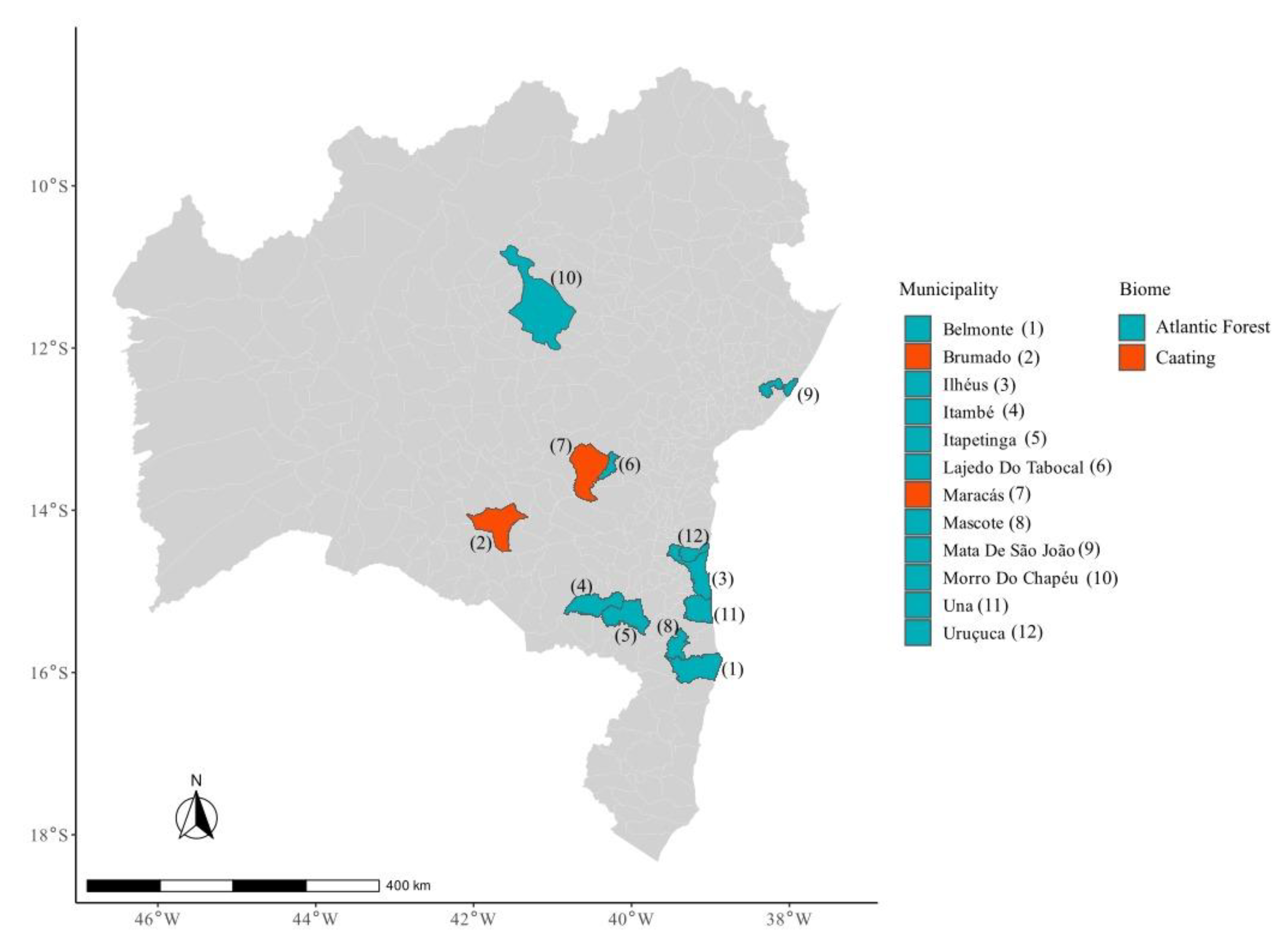
The following components of this study were approved by the Ethics Committee on Animal Use of the State University of Santa Cruz (CEUA-UESC) and the Oswaldo Cruz Institute (IOC): the use of traps to capture wild birds in 2016–2017 (CEUA-UESC protocol code: 014/2014; date of approval: September 2014); the use of traps to capture wild mammals in 2015 (CEUA-UESC protocol code: 003/2013; date of approval: May 2013), 2017 (CEUA-UESC protocol code: 018/2015; date of approval: August 2015), 2018–2020 (CEUA-UESC protocol code: 028/2018; date of approval: November 2018), and 2021 (CEUA-IOC 036/2020; date of approval: March 2021); and the use of traps to capture wild reptiles in 2018 (CEUA-UESC protocol code: 034/16; date of approval: September 2016).

2.2. Study Area

Bahia is located in northeastern Brazil, covering a total area of 564,760,429 km2, with a population of approximately 14,136,417. This study was conducted in two biomes corresponding to 12 municipalities in the state of Bahia, Brazil: the Atlantic Forest (Belmonte, Ilhéus, Itambé, Itapetinga, Lajedo do Tabocal, Maracás, Mascote, Mata de São João, Morro do Chapéu, Una, and Uruçuca) and Caatinga (Brumado and Maracás) biomes (Figure 1). The areas were classified according to their biomes and vegetation types according to the Brazilian Institute of Geography and Statistics [14]. A comprehensive assessment was conducted based on predefined indicators to evaluate the degree of anthropization in each study area. These included land-use intensity, vegetation cover, biodiversity, and the prevalence of human activities. These categories of land cover and land use were obtained using MapBiomas software v7.1 “https://mapbiomas.org/en (accessed on 28 August 2023)”, from raster images of 30 m definition. Their percentages in each buffer were calculated in R software v4.1.1 using the “extract” function of the raster package. A buffer radius of 1 km was used around the collection sample points. The anthropization was considered as the sum of the percentages of the areas with pasture, other cultures, silviculture, and urbanization.

2.3. Specimen Collection

The orders Didelphimorphia and Rodentia were captured with falling traps (bucket size of 60 L and arranged 10 m from each other), Sherman traps (31 × 8 × 9 cm), and Tomahawk traps (46 × 16 × 17 cm) arranged in transects and with bait comprising a mixture of sweet peanut, cornmeal, sardine, and banana. In 2015, the municipalities of Belmonte (June and July, n = 47), Mascote (August, n = 19), and Una (August, n = 15) were sampled for 10 consecutive nights, with a total of 13,020 traps/night. Sampling was conducted in the Itapetinga and Itambé municipalities for seven days each in August 2018 and February 2019, using 7742 traps/night. Similarly, in Lajedo do Tabocal and Maracás, sampling occurred for 10 days each in August 2019 and February 2020, using 6820 traps per night.
After collection for a previous research project, small mammals were euthanized by intramuscular injection of 100 mg/kg ketamine hydrochloride + 5 mg/kg xylazine hydrochloride (rodents) or 30 mg/kg ketamine hydrochloride + 2 mg/kg xylazine hydrochloride (marsupials) in the gluteal region and deposited as witness material in the Alexandre Rodrigues Ferreira collection of mammals belonging to the State University of Santa Cruz (CMARF/UESC). The collected individuals were used for taxonomic and morphological characterization and demonstration of the sexual dimorphism of the species. However, pups and pregnant and lactating females were identified using numbered aluminum earrings and returned to the place of capture.
Samples were collected from golden-headed lion tamarins, Leontopithecus chrysomelas, in 2017 (March, May, June, and July) from Ilhéus (n = 12) and Una (n = 9). The collection method involved the use of Tomahawk traps measuring 48.3 cm long × 15.2 cm wide × 15.2 cm high, baited with bananas. These traps were deployed for approximately 11 h daily, accounting for 22 h per month at each location. After capture, L. chrysomelas were anesthetized with intramuscular ketamine 10 mg/kg and midazolam 0.3 mg/kg.
The Atlantic Forest endemic sloth, Bradypus torquatus, was sampled in 2021 (May and October) in Mata de São João (n = 11) using an active search. When the maned sloth was located, a trained climber accessed the treetops to catch the animals using their hands. During the physical restraint, the claws of B. torquatus were wrapped with Velcro tape to keep them closed. The animal was placed in a cotton bag and lowered to the forest floor using a rope. The animals were sedated with 4.0 mg/kg ketamine hydrochloride and 0.03 mg/kg medetomidine hydrochloride intramuscularly and 0.1 mg/kg atipamezole hydrochloride intravenously.
In the municipalities of Uruçuca, Ilhéus, and Una, wild birds were captured using mist nets, with each net active for approximately 10 h daily and inspected every 40 min. Ilhéus was sampled in 2016 (November) and 2017 (April and June) and Una in 2016 (November) and 2017 (March and August), with a sampling effort of 40,500 h·m2 for five consecutive days. Sampling in May and June 2017 in Uruçuca resulted in a sampling effort of 32,400 h·m2 for four consecutive days.
Manual collection of the Reptilia class was performed using an active search methodology in 2018 (September and October) in Brumado (n = 11), Ilhéus (n = 1), and Morro do Chapéu (n = 4) between 6 p.m. and 11 p.m. without standardization.
Fecal samples were collected using rectal swabs from 674 wild animals (Table S1) captured from forest remnants and adjacent pastures in the Atlantic Forest (n = 548) and Caatinga (n = 126). Wildlife genera and species were defined based on morphological and anatomical criteria. Upon collection, all swab samples were kept in 2 mL microtubes containing 1 mL of buffered peptone water (Liofilchem, Roseto degli Abruzzi, Italy) and promptly refrigerated to maintain their integrity during transportation to the laboratory for further processing.

2.4. Salmonella Isolation and Identification

Salmonella samples from avians used in this study [15] and from mammalian and reptilian samples were obtained as follows. Rectal swab samples underwent a pre-enrichment step, which was performed in 2 mL microtubes containing 1 mL of buffered peptone water (Liofilchem, Roseto degli Abruzzi, Italy) at 37 °C for 24 h. Thereafter, selective enrichment was performed by inoculating 1 mL of the bacterial suspension in 9 mL of Rappaport Vasiliadis broth (Oxoid Ltd., Basingstoke, UK) and incubating at 43 °C for 18–24 h. Differential isolation was then performed by streaking a loopful on xylose-lysine-deoxycholate agar (Acumedia; Neogen Corporation, Lansing, MI, USA) and Hektoen enteric agar (Becton Dickinson, Franklin Lakes, NJ, USA) and incubating at 37 ± 1 °C for 18–24 h. Suspected colonies were inoculated in 1 mL of trypticase soy broth (HiMedia Laboratories Pvt. Ltd., Mumbai, India). The samples were then incubated (37 °C/18–24 h) for presumptive biochemical identification using triple sugar iron agar (HiMedia Laboratories Pvt. Ltd.), lysine iron agar (HiMedia Laboratories Pvt. Ltd.), urease (Acumedia, Neogen Corporation), and strips of oxidase (Laborclin, Pinhais, Brazil).
Presumptive colonies of Salmonella spp. were grown in trypticase soy broth (HiMedia Laboratories Pvt. Ltd.) at 37 °C for 18–24 h before submitting to DNA extraction using a DNA PureLink™ Genomic DNA Mini Kit (Invitrogen, Carlsbad, CA, USA), according to manufacturer instructions, and were confirmed using polymerase chain reaction (PCR), as described below. The total PCR reaction was 25 µL and consisted of 1× PCR buffer (Invitrogen, Carlsbad, CA, USA); 1.25 mM MgCl2 (Invitrogen); 200 µM dNTP; 10 pmol of each primer, ST11 (5′-AGCCAAACCATTGCTAAATTGGCGCA-3′) and ST15 (5′ TTTGCGACTATCAGGTTACCGTGG-3′), which are specific for the Salmonella genus [16]; 1.25 U of Taq DNA polymerase (Invitrogen); and 2 µL of DNA (50 ng/µL) from the suspected colony of the pathogen. Sterile nuclease-free water was used to complete the reaction volume to 25 µL. S. enterica serovar Enteritidis PT1 was used as a positive control, and a DNA-free reaction was used as a negative control. The amplification occurred with an initial denaturation (5 min at 94 °C) followed by an amplification phase of 35 cycles (30 s at 94 °C, 30 s at 62 °C, and 1 min at 72 °C) and a final extension step (7 min at 72 °C) in a Proflex PCR system (Applied Biosystems; Life Technologies, Carlsbad, CA USA). PCR products were visualized on a 1% agarose gel stained with SYBR Green (Invitrogen) and examined under UV light [17]. The PCR-positive samples were further serotyped using serogroup- and serovar-specific antisera obtained from the Enterobacteriaceae Laboratory of the Oswaldo Cruz Foundation in Rio de Janeiro, Brazil.

2.5. Antibiotic Susceptibility Test

Antimicrobial resistance was assessed using the Kirby–Bauer disk-diffusion method on Mueller–Hinton agar (HiMedia Laboratories Pvt. Ltd.), with the bacterial suspension turbidity adjusted to a McFarland scale of 0.5 [18], and following the Clinical Laboratory Standards Institute [19] protocol. The following 15 antimicrobial compounds were used: amikacin (30 µg), amoxicillin/clavulanic acid (20/10 µg), ampicillin with sulbactam (10/10 µg), cefepime (30 µg), cefoxitin (30 µg), ciprofloxacin (5 µg), chloramphenicol (30 µg), gentamicin (10 µg), imipenem (10 µg), lomefloxacin (10 µg), norfloxacin (10 µg), piperacillin with tazobactam (100/10 µg), sulfazotrim (trimethoprim sulfamethoxazole (25 µg)), tobramycin (10 µg), and trimethoprim (5 µg) (LABORCLIN, Produtos para Laboratórios Ltd., Pinhais, Brazil). Escherichia coli ATCC 25922 was used for quality control.

2.6. Statistical Analysis

Due to the limited number of positive samples, only descriptive statistical analyses were performed [20]. In addition, the patterns of wild animals (diet, locomotion, species, biomes, and municipalities) were analyzed using descriptive and inferential statistics [20].

3. Results

Between 2015 and 2021, 674 samples were collected from wild animals in the Atlantic Forest and Caatinga. These samples were obtained from the 12 municipalities studied and were collected from the following animals: 114 from birds, 544 from mammals (317 rodents, 195 marsupials, 21 primates, and 11 sloths), and 16 from reptiles (Table S1). Different study areas showed varying land use and, consequently, different percetanges of anthropization. The forest environments studied showed lower percentages of anthropization (except for Itapetinga and Mascote) when compared with Caatinga (except for Brumado). It was not possible to observe if areas that had anthropization such as the cocoa plantations and agroforestry systems had any influence on biodiversity. Four Salmonella isolates (0.59%) were obtained from three mammals and one bird. These isolates belonged to three distinct serovars: S. enterica subsp. Enterica serovar Agona, Salmonella enterica subsp. enterica O:16, and Salmonella enterica subsp. enterica serovar Muenchen. All positive samples were found in areas of the Atlantic Forest (Table 1) and had percentages of anthropization of 51.8%, 47.0%, 79.2%, and 70.3% (Table S2). All negative Salmonella samples varied in their anthropization percentages (Table 2).
The antimicrobial sensitivity profile of Salmonella enterica subsp. enterica serogroup O:16 isolated from a young female Didelphis albiventris showed resistance to 6 of the 15 antimicrobials tested (amoxicillin/clavulanic acid, ampicillin with sulbactam, cefoxitin, chloramphenicol, trimethoprim, and sulfamethoxazole). The other serovar isolated from an adult female did not exhibit antimicrobial resistance. S. Agona isolated from a female passerine showed intermediate resistance to amikacin and gentamicin. S. Muenchen from a male Cerradomys vivoi demonstrated intermediate resistance to amikacin.
The most frequent family was Cricetidae (282), followed by Didelphidae (197), Pipridae (34), Turdidae (32), Echimidae (30), Dendrocolaptidae (24), Callithrichidae (21), Bradypodidae (11), Sphaerodactylidae (11), Columbidae (6), Thraupidae (5), and Tyraniidae (4). The other families were represented by fewer than 2 exemplars and were thus grouped as others (17).
The most frequently observed species were Didelphis albiventris (95), Necromys lasiurus (68), Calomys expulsus (62), Cerradomys vivoi (44), Marmosops incanus (39), Hylaeamys seuanezi (32), Gracilinanus agilis (31), Rhipidomys mastacalis (23), Wiedomys pyrrhorhinos (23), Leontopithecus chrysomelas (21), Turdus leucomelas (18), Thrichomys aff laurentius (18), Marmosa murina (16), Turdus rufiventris (13), Ceratopipra rubrocapilla (12), Bradypus torquatus (11), Coleodactylus meridionalis (11), Dixiphia pipra (9), Xiphorhynchus fuscus (8), Oligoryzomys nigripes (8), Trinomys albispinus (8), Dendrocincla turdina (7), Manacus manacus (7), Mus musculus (7), Oligoryzomys stramineus (7), Machaeropterus regulus (6) Cryptonanus agricolai (6), Leptotila rufaxilla (5), Glyphorynchus spirurus (4), Trinomys sp (4), and Monodelphis americana (3). Other species, which were represented by fewer than 2 exemplars each or were not identified, were grouped as others (48) (Table 3).
Most of the wild animals studied were from the Atlantic Forest biome (79.5%), were terrestrial (40.5%), and had a mostly fruit-based diet (71.6%), and 56.8% were collected during the rainy season (Table S1). When comparing animal class, season, biome, animal locomotion, and animal diet, most were terrestrial mammals fed a fruit diet (32.9%) (Table S1). A detailed assessment of the diet, locomotion, and biome is provided in the Supplementary Material (Table S2). The municipalities with the highest numbers of animals were Maracás (175), followed by Itapetinga (122), Lajedo do Tabocal (80), Una (72), Ilhéus (62), Itambé (54), Belmonte (47), Mascote (19), Uruçuca (17), Brumado (11), Mata de São João (11), and Morro do Chapéu (4).
The most frequently observed feeding behaviors were frugivore/omnivore (187), frugivore/granivore (174), insectivore/omnivore (105), frugivore/insectivore (46), frugivore (34), insectivore (33), frugivore/seed predator (24), granivore (18), frugivore/invertivore (13), folivore (11), and invertivore (11) (Table S3).

4. Discussion

The results observed in this study may not be directly comparable with those of other studies because the actual prevalence of Salmonella spp. depends on the sampling location and methods applied [21]. Thus, the low prevalence (0.59% in the 6-year convenience sampling) of Salmonella spp. may be attributed to cloaca/rectal swab sampling. However, in a previous Danish study on carcasses, the authors observed a low prevalence of Salmonella spp. in wild birds [22] and boars [9]. Similarly, a low frequency of isolation has been reported in wild birds from a Swiss rehabilitation center (1.08%) [22] and in wild animals in northwestern Italy (4.30%), including canids, mustelids, wild birds, and ungulates [23]. In addition, another study from western Italy reported a frequency of 2.87% in wild animals, such as magpies, foxes, crows, jays, brown hares, hedgehogs, starlings, swans, wild ducks, Porcupine’s green woodpeckers, and Eurasian collared doves [7]. No isolates of Salmonella spp. were obtained in a study on wild rodents (n = 237) in Poland [24]. These results reinforce the fact that the prevalence of Salmonella spp. is usually lower in wild than in domesticated animals [25].
A low frequency of positive results (9.5%) was observed in the wild boar population in São Paulo [9]. These results were attributed to the fact that as the number of human settlements in close vicinity increases, wild animals come into contact with other animal species, raw food, and high temperatures, all of which favor the occurrence of Salmonella spp.; therefore, they can serve as vectors for pathogen transmission [26]. This behavior was observed in a previous study [27], where rodents from urban areas were more prone to carry pathogens. Therefore, as stated by Skov [21], the prevalence of Salmonella spp. in wildlife can vary greatly among studies, and this variation is not associated with sampling methods, study designs, or differences in prevalence between countries.
Although the areas sampled were exposed to human contact, the low occurrence of Salmonella spp. in this study may be attributed to the fact that the areas were highly conserved and animals from preserved areas showed a lower risk of contamination than those with human contact [26]. In addition, the animals presented a mostly terrestrial and frugivorous nature, which may contribute to their occurrence because the behavior and feeding habits of wild animals are known to influence the likelihood of infection with Salmonella [27]. Badgers, red foxes, raptors, and wild boars acquire Salmonella infections by scavenging contaminated carcasses or human leftovers [8]. However, wild animals can be latent carriers and thus contribute to the low frequency of isolation because, during some periods, the microorganisms are located intracellularly and are not released into the environment [28].
All positive samples were obtained from the Atlantic Forest. This result agrees with that of another study on 36 wild mammals from the Atlantic Forest and Amazonia biomes in Brazil, which obtained only one isolate of Salmonella enterica subspecies diarizonae serotype 60:r:e,n,z15, which was isolated from a rodent in Amazonia [2]. In another study of 207 wild rodents from farmland and nonfarmland areas in Sweden, only one Salmonella enterica serovar Typhimurium isolate from a subpopulation of 11 individuals was obtained from a chicken farm [29]. In our study, the samples positive for Salmonella spp. from small mammals were from areas close to a cattle farm, which is a factor alongside the changes made to the forest area as it favors the habit of looking for shelter and food in peridomiciliary areas, thus facilitating the acquisition and dispersion of pathogenic microorganisms [30]. Among the marsupials sampled in this study, two Didelphis albiventris tested positive for Salmonella enterica subsp. enterica O:16. Similar results were found when Didelphis albiventris in a rural area in Argentina (five poultry farms) and eight surrounding forest areas were sampled, and a 4% positivity rate for Salmonella enterica was found. However, the authors found no relationship between age and sex [31]. Additionally, Casagrande [32] isolated Salmonella from 17.0% (18/106) of Didelphis aurita and 17.5% (7/40) of Didelphis albiventris individuals in their study, most of which were wild animals that lived in the forest around a human population. These authors also found no influence of sex or age on the frequency of Salmonella infections.
Resistance to environmental bacteria is a natural biological process, and its capacity for transmission and dissemination is enhanced by multiple drug resistance genes shed in the feces of livestock and humans [33]. However, fragmentation promotes environmental stress in ecosystems, allowing the selective pressure of multiresistant agents on antibiotics [34]. With interactions between humans, domestic animals, wild animals, and the environment, contact has intensified, increasing the chances of transmission of resistant strains [35]. Therefore, free-ranging wild animals that have not been exposed to antibiotics may have high rates of drug resistance owing to environmental contamination [36]. This resistance can also be acquired from consuming contaminated water or food and transported via direct contact with garbage and sewage residues [37].
The detection and analysis of microorganisms, mainly bacteria, can provide information on ecosystem exposure to biological agents [38]. Based on our findings, we propose that the detection of pathogenic Salmonella spp. isolates be used for biomonitoring environmental quality in future wildlife studies. This approach facilitates the assessment of conservation and management practices.

5. Conclusions

In the present study, wild animals assessed in the mesoregions showed a low frequency (4/674 = 0.59%) of Salmonella, which is consistent with previous reports on wild animal populations worldwide. In addition, the isolates were found to be multiresistant, including resistance to extended-spectrum penicillins with beta-lactamase inhibitors. Thus, it is important to consider that specific behavior, diet, and the low percentage of anthropization may be the main factors affecting the studied species in the Atlantic Forest and Caatinga in Bahia, Brazil. Therefore, we propose that the detection of Salmonella spp. can be used for anthropization biomonitoring and as an indicator of environmental preservation in future wildlife studies.

Supplementary Materials

The following supporting information can be downloaded at: https://www.mdpi.com/article/10.3390/ani14010021/s1, Table S1: Absolute and relative frequency of captured and sampled wildlife, characterized by class, season, biome, locomotion, and diet; Table S2: Evaluated parameters of captured and sampled wildlife in the municipalities of Bahia State, Brazil; Table S3: Frequency of the feeding habits of the captured and sampled wildlife.

Author Contributions

For this research article, we relied on working partners who made the following individual contributions: conceptualization, methodology (isolation and identification; antibiotic susceptibility test), and writing—original draft preparation, E.J.E.d.S.; conceptualization, methodology (isolation and identification, antibiotic susceptibility test), and writing—review, A.T.S.L.; methodology (molecular biology), H.F.F.; methodology (isolation and identification), E.A.G.; methodology (specimen collection), J.M.R., P.d.A.B.J., F.C.S.B., T.d.S.O.C., K.M.D.V., L.d.C.O., B.F.R., B.S.d.A., L.M.C.F., E.O.R. and S.S.F.; methodology (Salmonella serotyping), D.d.P.R.; resources, M.R.D.V.A., V.G.D.O., G.R.A., F.R.M., R.P.R. and S.S.d.C.N.; manuscript conceptualization, data analysis, and writing—review and editing, L.J.L.P.; conceptualization, project administration, and writing—review, B.M.M. All authors have read and agreed to the published version of the manuscript.

Funding

This research was funded by Fundação de Amparo à Pesquisa do Estado da Bahia—FAPESB (Grant numbers 013/2014; PNE0001/2014; and 011/2015); Conselho Nacional de Desenvolvimento Científico e Tecnológico—CNPq (Grant numbers Pq-CNPq 303448/2019-9; Pq-CNPq 310467/2017-9; Pq-CNPq 310256/2020-8; and Pq-CNPq 431772/2018-5 CNPq-PPBIO REDE BIOMA 457524/2012-0); Coordenação de Aperfeiçoamento de Pessoal de Nível Superior—CAPES (Grant numbers 001/2017; 4672/2014); and Universidade Estadual de Santa Cruz—UESC (Grant numbers UESC PROPP 00220.1100.1645; UESC PROPP 00220.1100.1905 and SEI 073.11016.2021.0010482-32).

Institutional Review Board Statement

The animal study protocol was approved by the Ethics Committee of the State University of Santa Cruz (protocol code: 014/2014, date of approval: September 2014; protocol code: 003/2013, date of approval: May 2013; protocol code: 018/2015, date of approval: August 2015; protocol code: 028/2018, date of approval: November 2018) and Osvaldo Cruz Institute (protocol code: 036/2020, date of approval: March 2021; protocol code: 034/16, date of approval: September 2016).

Informed Consent Statement

Not applicable.

Data Availability Statement

No new data were created or analyzed in this study. Data sharing is not applicable in this study.

Acknowledgments

The authors thank Rafaela Porto Azevedo for assistance with laboratory tasks, and Anaiá da Paixão Sevá for assistance with the spatial categorical analysis of vegetation cover and land use. The authors would also like to thank the Department of Economics, Science, and Innovation of the Flemish Government of Belgium through the Center for Research and Conservation/Royal Zoological Society of Antwerp.

Conflicts of Interest

The authors declare no conflict of interest.

References

  1. Rock, M.; Buntain, B.J.; Hatfield, J.M.; Hallgrímsson, B. Animal–Human Connections, “One Health”, and the Syndemic Approach to Prevention. Soc. Sci. Med. 2009, 68, 991–995. [Google Scholar] [CrossRef] [PubMed]
  2. De Oliveira Iovine, R.O.; Dejuste, C.; Miranda, F.; Filoni, C.; Bueno, M.G.; de Carvalho, V.M. Isolation of Escherichia coli and Salmonella spp. from Free-Ranging Wild Animals. Braz. J. Microbiol. 2015, 46, 1257–1263. [Google Scholar] [CrossRef] [PubMed]
  3. Tompkins, D.M.; Dunn, A.M.; Smith, M.J.; Telfer, S. Wildlife Diseases: From Individuals to Ecosystems. J. Anim. Ecol. 2011, 80, 19–38. [Google Scholar] [CrossRef] [PubMed]
  4. Siembieda, J.L.; Kock, R.A.; McCracken, T.A.; Newman, S.H. The Role of Wildlife in Transboundary Animal Diseases. Anim. Health Res. Rev. 2011, 12, 95–111. [Google Scholar] [CrossRef]
  5. Hilbert, F.; Smulders, F.J.M.; Chopra-Dewasthaly, R.; Paulsen, P. Salmonella in the Wildlife-Human Interface. Food Res. Int. 2012, 45, 603–608. [Google Scholar] [CrossRef]
  6. López-Islas, J.J.; Méndez-Olvera, E.T.; Martínez-Gómez, D.; López-Pérez, A.M.; Orozco, L.; Suzan, G.; Eslava, C. Characterization of Salmonella spp. and E. coli Strains Isolated from Wild Carnivores in Janos Biosphere Reserve, Mexico. Animals 2022, 12, 1064. [Google Scholar] [CrossRef]
  7. Rubini, S.; Ravaioli, C.; Previato, S.; D’Incau, M.; Tassinari, M.; Guidi, E.; Lupi, S.; Merialdi, G.; Bergamini, M. Prevalence of Salmonella Strains in Wild Animals from a Highly Populated Area of North-Eastern Italy. Ann. Ist. Super. Sanità. 2016, 52, 277–280. [Google Scholar] [CrossRef]
  8. Millan, J.; Aduriz, G.; Moreno, B.; Juste, R.A.; Barral, M. Salmonella Isolates from Wild Birds and Mammals in the Basque Country (Spain). Rev. Sci. Tech. OIE 2004, 23, 905–911. [Google Scholar] [CrossRef]
  9. Carraro, P.E.; Barbosa, F.D.O.; Benevides, V.P.; Casas, M.R.T.; Berchieri Junior, A.; Bürger, K.P. Prevalence and Antimicrobial Resistance of Salmonella spp. Isolated from Free-Ranging Wild Boars in the State of São Paulo, Brazil. Cienc. Rural. 2022, 52, e20210263. [Google Scholar] [CrossRef]
  10. Merkevičienė, L.; Butrimaitė-Ambrozevičienė, Č.; Paškevičius, G.; Pikūnienė, A.; Virgailis, M.; Dailidavičienė, J.; Daukšienė, A.; Šiugždinienė, R.; Ruzauskas, M. Serological Variety and Antimicrobial Resistance in Salmonella Isolated from Reptiles. Biology 2022, 11, 836. [Google Scholar] [CrossRef]
  11. Fluit, A.C.; Schmitz, F.-J. Resistance Integrons and Super-Integrons. Clin. Microbiol. Infect. 2004, 10, 272–288. [Google Scholar] [CrossRef] [PubMed]
  12. Perveen, N.; Muzaffar, S.B.; Al-Deeb, M.A. Exploring Human-Animal Host Interactions and Emergence of COVID-19: Evolutionary and Ecological Dynamics. Saudi J. Biol. Sci. 2021, 28, 1417–1425. [Google Scholar] [CrossRef] [PubMed]
  13. Uelze, L.; Bloch, A.; Borowiak, M.; Grobbel, M.; Deneke, C.; Fischer, M.; Malorny, B.; Pietsch, M.; Simon, S.; Szabó, I.; et al. What WGS Reveals about Salmonella enterica subsp. enterica in Wildlife in Germany. Microorganisms 2021, 9, 1911. [Google Scholar] [CrossRef]
  14. IBGE, Instituto Brasileiro de Geografia e Estatística. Biomas e Sistema Costeiro-Marinho Do Brasil: Compatível Com a Escala 1:250,000; Coordenação de Recursos Naturais e Estudos Ambientais: Rio de Janeiro, Brazil, 2019. [Google Scholar]
  15. Dos Santos, E.J.E.; Azevedo, R.P.; Lopes, A.T.S.; Rocha, J.M.; Albuquerque, G.R.; Wenceslau, A.A.; Miranda, F.R.; Rodrigues, D.d.P.; Maciel, B.M. Salmonella spp. in Wild Free-Living Birds from Atlantic Forest Fragments in Southern Bahia, Brazil. BioMed Res. Int. 2020, 2020, 7594136. [Google Scholar] [CrossRef] [PubMed]
  16. Aabo, S.; Rasmussen, O.F.; Roseen, L.; Sørensen, P.D.; Olsen, J.E. Salmonella Identification by the Polymerase Chain Reaction. Mol. Cel. Probes 1993, 7, 171–178. [Google Scholar] [CrossRef] [PubMed]
  17. Maciel, B.M.; Argôlo Filho, R.C.; Nogueira, S.S.C.; Dias, J.C.T.; Rezende, R.P. High Prevalence of Salmonella in Tegu Lizards (Tupinambis Merianae), and Susceptibility of the Serotypes to Antibiotics. Zoonoses Public Health 2010, 57, e26–e32. [Google Scholar] [CrossRef]
  18. Bauer, A.W.; Kirby, W.M.; Sherris, J.C.; Turck, M. Antibiotic Susceptibility Testing by a Standardized Single Disk Method. Am. J. Clin. Pathol. 1966, 45, 493–496. [Google Scholar] [CrossRef]
  19. CLISI, Clinical and Laboratory Standards Institute. Performance Standards for Antimicrobial Susceptibility Testing; Twenty-Fourth Informational Supplement; CLSI Document M100-S24; CLSI: Wayne, NJ, USA, 2014; Volume 34. [Google Scholar]
  20. Dohoo, I.; Martin, W.; Stryhn, H.E. Veterinary Epidemiologic Research, 2nd ed.; University of Prince Edward Island: Charlottetown, Canada, 2010; ISBN 978-0-919013-60-5. [Google Scholar]
  21. Skov, M.N.; Madsen, J.J.; Rahbek, C.; Lodal, J.; Jespersen, J.B.; Jørgensen, J.C.; Dietz, H.H.; Chriél, M.; Baggesen, D.L. Transmission of Salmonella between Wildlife and Meat-Production Animals in Denmark. J. Appl. Microbiol. 2008, 105, 1558–1568. [Google Scholar] [CrossRef]
  22. Vogler, B.R.; Zurfluh, K.; Mattmann, P.; Schmitt, K.; Albini, S. Low Occurrence of Salmonella spp. in Wild Birds from a Swiss Rehabilitation Centre. Vet. Rec. Open 2021, 8, e17. [Google Scholar] [CrossRef]
  23. Botti, V.; Navillod, F.V.; Domenis, L.; Orusa, R.; Pepe, E.; Robetto, S.; Guidetti, C. Salmonella spp. and Antibiotic-Resistant Strains in Wild Mammals and Birds in North-Western Italy from 2002 to 2010. Vet. Ital. 2013, 49, 195–202. [Google Scholar] [CrossRef]
  24. Skarżyńska, M.; Zając, M.; Kamińska, E.; Bomba, A.; Żmudzki, J.; Jabłoński, A.; Wasyl, D. Salmonella and Antimicrobial Resistance in Wild Rodents—True or False Threat? Pathogens 2020, 9, 771. [Google Scholar] [CrossRef]
  25. Dos Santos, E.J.E.; Lopes, A.T.S.; Maciel, B.M. Salmonella in Wild Animals: A Public Health Concern. In Enterobacteria; Bhonchal Bhardwaj, S., Ed.; IntechOpen: London, UK, 2022; Volume 136, ISBN 978-1-80355-310-8. [Google Scholar]
  26. Ribas, A.; Saijuntha, W.; Agatsuma, T.; Prantlová, V.; Poonlaphdecha, S. Rodents as a Source of Salmonella Contamination in Wet Markets in Thailand. Vector Borne Zoonotic Dis. 2016, 16, 537–540. [Google Scholar] [CrossRef] [PubMed]
  27. Williams, S.H.; Che, X.; Paulick, A.; Guo, C.; Lee, B.; Muller, D.; Uhlemann, A.-C.; Lowy, F.D.; Corrigan, R.M.; Lipkin, W.I. New York City House Mice (Mus Musculus) as Potential Reservoirs for Pathogenic Bacteria and Antimicrobial Resistance Determinants. mBio 2018, 9, e00624-18. [Google Scholar] [CrossRef] [PubMed]
  28. Mares, M. Current Topics in Salmonella and Salmonellosis; IntechOpen: London, UK, 2017; Volume 284, ISBN 978-953-51-3066-6. [Google Scholar]
  29. Backhans, A.; Jacobson, M.; Hansson, I.; Lebbad, M.; Lambertz, S.T.; Gammelgård, E.; Saager, M.; Akande, O.; Fellström, C. Occurrence of Pathogens in Wild Rodents Caught on Swedish Pig and Chicken Farms. Epidemiol. Infect. 2013, 141, 1885–1891. [Google Scholar] [CrossRef]
  30. De Barros, B.D.C.V.; Chagas, E.N.; Bezerra, L.W.; Ribeiro, L.G.; Duarte Júnior, J.W.B.; Pereira, D.; da Penha Junior, E.T.; Silva, J.R.; Bezerra, D.A.M.; Bandeira, R.S.; et al. Rotavirus A in Wild and Domestic Animals from Areas with Environmental Degradation in the Brazilian Amazon. PLoS ONE 2018, 13, e0209005. [Google Scholar] [CrossRef]
  31. Carusi, L.C.P.; Farace, M.I.; Ribicich, M.M.; Villafañe, I.E.G. Reproduction and Parasitology of Didelphis albiventris (Didelphimorphia) in an Agroecosystem Landscape in Central Argentina. Mammalia 2009, 73, 89–97. [Google Scholar] [CrossRef]
  32. Casagrande, R.A.; Lopes, L.F.L.; Reis, E.M.d.; Rodrigues, D.d.P.; Matushima, E.R. Isolation of Salmonella enterica in opossum (Didelphis aurita and Didelphis albiventris) of the São Paulo State, Brazil. Cienc. Rural. 2011, 41, 492–496. [Google Scholar] [CrossRef]
  33. Reinthaler, F.F.; Posch, J.; Feierl, G.; Wüst, G.; Haas, D.; Ruckenbauer, G.; Mascher, F.; Marth, E. Antibiotic resistance of E. coli in sewage and sludge. Water Res. 2003, 37, 1685–1690. [Google Scholar] [CrossRef] [PubMed]
  34. Robinson, T.P.; Bu, D.P.; Carrique-Mas, J.; Fèvre, E.M.; Gilbert, M.; Grace, D.; Hay, S.I.; Jiwakanon, J.; Kakkar, M.; Kariuki, S.; et al. Antibiotic resistance is the quintessential One Health issue. Trans. R. Soc. Trop. Med. Hyg. 2016, 110, 377–380. [Google Scholar] [CrossRef]
  35. Wang, J.; Ma, Z.B.; Zeng, Z.L.; Yang, X.W.; Huang, Y.; Liu, J.H. The role of wildlife (wild birds) in the global transmission of antimicrobial resistance genes. Zool. Res. 2017, 38, 55–80. [Google Scholar] [CrossRef]
  36. Jechalke, S.; Heuer, H.; Siemens, J.; Amelung, W.; Smalla, K. Fate and effects of veterinary antibiotics in soil. Trends Microbiol. 2014, 22, 536–545. [Google Scholar] [CrossRef] [PubMed]
  37. Radhouani, H.; Silva, N.; Poeta, P.; Torres, C.; Correia, S.; Igrejas, G. Potential impact of antimicrobial resistance in wildlife, environment and human health. Front. Microbiol. 2014, 5, 23. [Google Scholar] [CrossRef] [PubMed]
  38. Parmar, T.K.; Rawtani, D.; Agrawal, Y.K. Bioindicators: The Natural Indicator of Environmental Pollution. Front. Life Sci. 2016, 9, 110–118. [Google Scholar] [CrossRef]
Figure 1. Biome of areas where the wildlife was captured and sampled for Salmonella screening from 2015 to 2021 in Bahia State, Brazil.
Animals 14 00021 g001
Table 1. Characterization of the captured and sampled wildlife positive for Salmonella spp. according to their respective location and environment.
BiomeAgeSexMunicipalitySeasonSampling DateAnthropization (%)SerovarsSpeciesClass
Atlantic ForestAdultFIlhéusRainy27 July 201751.8AgonaCeratopipra
rubrocapilla
Avian
Atlantic ForestAdultFItapetingaDry24 August 201847.0O:16Didelphis
albiventris
Mammalia
Atlantic ForestYoungFItapetingaRainy5 February 201979.2O:16Didelphis
albiventris
Mammalia
Atlantic ForestAdultMItambéRainy7 February 201970.3MuenchenCerradomys
vivoi
Mammalia
Table 2. Characterization of the captured and sampled wildlife negative for Salmonella spp. according to their respective location, biome, and anthropization percentages by wildlife family and by municipality.
MunicipalityFamily (n)BiomeAnthropization (%) by FamilyAnthropization (%) by Municipality
BelmonteCricetidae (33) Atlantic Forest39.8 ± 1.5139.9 ± 1.54
Didelphidae (12)40.6 ± 1.62
Sciuridae (2)38.8 ± 0.00
BrumadoSphaerodactylidae (11)Caatinga17.2 ± 0.0017.2 ± 0.00
IlhéusCallithrichidae (12)Atlantic Forest21.7 ± 0.0046.2 ± 12.1
Columbidae (5)52.1 ± 0.62
Dendrocolaptidae (8)52.5 ± 0.72
Fringillidae (1)51.85
Onychorhynchidae (1)51.85
Passerellidae (1)51.85
Pipridae (10)51.8 ± 0.00
Thraupidae (5)51.8 ± 0.00
Trochilidae (2)51.8 ± 0.00
Turdidae (15)52.3 ± 0.70
Typhlopidae (1)45.4
Tyraniidae (1)51.8
ItambéCricetidae (18)Atlantic Forest57.4 ± 12.456.4 ± 11.8
Didelphidae (36)56.2 ± 11.6
ItapetingaCricetidae (38)Atlantic Forest72.1 ± 11.268.9 ± 13.9
Didelphidae (84)67.4 ± 14.8
Lajedo do TabocalCricetidae (56)Atlantic Forest28.7 ± 7.8930.6 ± 8.54
Didelphidae (10)36.4 ± 9.49
Echimidae (14)34.2 ± 7.91
MaracásCricetidae (46)Atlantic Forest58.6 ± 11.164.7 ± 21.4
Cricetidae (77)Caatinga63.5 ± 24.8
Didelphidae (1)Atlantic Forest40.5
Didelphidae (34)Caatinga72.0 ± 21.6
Echimidae (16)76.3 ± 18.0
Muridae (1)Atlantic Forest40.5
MascoteCricetidae (8)Atlantic Forest85.1 ± 11.787.2 ± 9.36
Didelphidae (11)88.7 ± 7.51
Mata de São JoãoBradypodidae (11)Atlantic Forest8.52 ± 2.438.52 ± 2.42
Morro do ChapéuPhyllodactylidae (1)Atlantic Forest41.141.1 ± 0.00
Scincidae (1)41.1
Tropiduridae (2)41.1 ± 0.00
UnaCallithrichidae (9)Atlantic Forest48.4 ± 0.0012.0 ± 14.4
Columbidae (1)5.61
Cricetidae (6)10.3 ± 10.6
Dendrocolaptidae (10)5.61 ± 0.00
Didelphidae (9)11.1 ± 6.84
Furnariidae (1)5.61
Grallariidae (1)5.61
Picidae (1)5.61
Pipridae (23)5.61 ± 0.00
Rhynchocyclidae (1)5.61
Turdidae (9)5.58 ± 0.09
Tyraniidae (1)5.61
UruçucaDendrocolaptidae (6)Atlantic Forest15.4 ± 0.0015.4 ± 0.00
Pipridae (1)15.4 ± 0.00
Turdidae (8)15.4 ± 0.00
Tyraniidae (2)15.4 ± 0.00
Table 3. Frequencies of captured wildlife species at each sample site in Bahia State, Brazil.
Relative Frequency (%)Absolute Frequency (n)SpeciesFamilyClass
14.195Didelphis albiventrisDidelphidaeMammalia
10.168Necromys lasiurusCricetidaeMammalia
9.2062Calomys expulsusCricetidaeMammalia
6.5344Cerradomys vivoiCricetidaeMammalia
5.7939Marmosops incanusDidelphidaeMammalia
4.7532Hylaeamys seuaneziCricetidaeMammalia
4.6031Gracilinanus agilisDidelphidaeMammalia
3.4123Rhipidomys mastacalisCricetidaeMammalia
3.4123Wiedomys pyrrhorhinosCricetidaeMammalia
3.1221Leontopithecus chrysomelasCallithrichidaeMammalia
2.6718Turdus leucomelasTurdidaeAvian
2.6718Thrichomys aff laurentiusEchimidaeMammalia
2.3716Marmosa murinaDidelphidaeMammalia
1.9313Turdus rufiventrisTurdidaeAvian
1.7812Ceratopipra rubrocapillaPipridaeAvian
1.6311Bradypus torquatusBradypodidaeMammalia
1.6311Coleodactylus meridionalisSphaerodactylidaeReptilia
1.349Dixiphia pipraPipridaeAvian
1.198Xiphorhynchus fuscusDendrocolaptidaeAvian
1.198Oligoryzomys nigripesCricetidaeMammalia
1.198Trinomys albispinusEchimidaeMammalia
1.047Dendrocincla turdinaDendrocolaptidaeAvian
1.047Manacus manacusPipridaeAvian
1.047Mus musculusCricetidaeMammalia
1.047Oligoryzomys stramineusCricetidaeMammalia
0.896Machaeropterus regulusPipridaeAvian
0.896Cryptonanus agricolaiDidelphidaeMammalia
0.745Leptotila rufaxillaColumbidaeAvian
0.594Glyphorynchus spirurusDendrocolaptidaeAvian
0.594Trinomys sp.EchimidaeMammalia
0.453Monodelphis americanaDidelphidaeMammalia
7.0148Others-Avian/Mammalia/Reptilia
100674Total
Disclaimer/Publisher’s Note: The statements, opinions and data contained in all publications are solely those of the individual author(s) and contributor(s) and not of MDPI and/or the editor(s). MDPI and/or the editor(s) disclaim responsibility for any injury to people or property resulting from any ideas, methods, instructions or products referred to in the content.

