Article

Masculinization of Red Tilapia (Oreochromis spp.) Using 17α-Methyltestosterone-Loaded Alkyl Polyglucosides Integrated into Nanostructured Lipid Carriers

by
Jakarwan Yostawonkul
1,2,
Sirikorn Kitiyodom
3,
Kittipat Supchukun
1,
Nutthanit Thumrongsiri
2,
Nattika Saengkrit
2,
Komkiew Pinpimai
4,
Amin Hajitou
5,
Kim D. Thompson
6,
Kasem Rattanapinyopituk
3,
Masashi Maita
7,
Manoj Tukaram Kamble
3,
Teerapong Yata
8,* and
Nopadon Pirarat
3,*
1
The International Graduate Course of Veterinary Science and Technology (VST), Faculty of Veterinary Science, Chulalongkorn University, Bangkok 10330, Thailand
2
National Nanotechnology Center (NANOTEC), National Science and Technology Development Agency (NSTDA), Pathumthani 12120, Thailand
3
Wildlife, Exotic and Aquatic Animal Pathology Research Unit, Department of Pathology, Faculty of Veterinary Science, Chulalongkorn University, Bangkok 10330, Thailand
4
Aquatic Resources Research Institute, Chulalongkorn University, Bangkok 10330, Thailand
5
Cancer Phagotherapy, Department of Brain Sciences, Imperial College London, London W12 0NN, UK
6
Moredun Research Institute, Penicuik EH26 0PZ, UK
7
Laboratory of Fish Health Management, Department of Marine Biosciences, Tokyo University of Marine Science and Technology, Konan 4-5-7, Minato, Tokyo 108-8477, Japan
8
Unit of Biochemistry, Department of Physiology, Faculty of Veterinary Science, Chulalongkorn University, Bangkok 10330, Thailand
*
Authors to whom correspondence should be addressed.
Animals 2023, 13(8), 1364; https://doi.org/10.3390/ani13081364
Submission received: 18 February 2023 / Revised: 31 March 2023 / Accepted: 11 April 2023 / Published: 16 April 2023

Abstract

:

Simple Summary

Lipid-based nanocarriers have proved effective for drug and hormone delivery. The androgen hormone 17 alpha-methyltestosterone (MT) is used for sex reversal in tilapia; however, the hormone has biopharmaceutical limitations such as insolubility, low stability, limited intestinal absorption, and poor bioavailability. Incorporating MT into lipid-based nanocarriers to improve hormone uptake in tilapia is an interesting prospect. In the present study, we reported an improved version of alkyl polyglucosides loaded with 17 alpha-methyltestosterone integrated into nanostructured lipid carriers (APG-NLC) for the masculinization of red tilapia (Oreochromis spp.) via oral administration as a feed additive. We demonstrated that nanotechnology-based drug delivery systems represent a novel and promising strategy for improving the oral absorption of these androgen hormones, resulting in a 50% w/w reduction in the hormonal dosage during the sex reversal period. Therefore, we strongly believe that this newly optimized nanotechnology is one of the solutions for sustainable aquaculture production.

Abstract

The aim of the present study was to optimize a masculinization platform for the production of all-male red tilapia fry by oral administration of 30 and 60 ppm of MT and alkyl polyglucoside nanostructured lipid carriers (APG-NLC) loaded with MT, respectively, for 14 and 21 days. The characterization, encapsulation efficiency and release kinetics of MT in lipid-based nanoparticles were assessed in vitro. The results showed that the MT-loaded nanoparticles were spherical, ranging from 80 to 125 nm in size, and had a negative charge with a narrow particle distribution. The APG-NLC loaded with MT provided higher physical stability and encapsulation efficacy than the NLC. The release rate constants of MT from MT-NLC and MT-APG-NLC were higher than those of free MT, which is insoluble in aqueous media. There was no significant difference in survival between the fish administered MT or the those fed orally with MT-APG-NLC fish. According to the logistic regression analysis, the sex reversal efficacy of MT-APG-NLC (30 ppm) and MT (60 ppm), resulted in significantly higher numbers of males after 21 days of treatment compared with the controls. The production cost of MT-APG-NLC (30 ppm) after 21 days of treatment was reduced by 32.9% compared with the conventional MT treatment group (60 ppm). In all the treatments, the length–weight relationship (LWR) showed negatively allomeric growth behavior (b < 3), with a relative condition factor (Kn) of more than 1. Therefore, MT-APG-NLC (30 ppm) would seem to be a promising, cost-effective way to reduce the dose of MT used for the masculinization of farmed red tilapia.

Graphical Abstract

1. Introduction

Red tilapia (Oreochromis spp.) is recognized as an important farmed fish species worldwide because it can easily adapt to the environment and has a fast growth rate [1,2]. However, growth retardation at the onset of sexual maturity and overpopulation by excessive or unwanted reproduction are the major concerns for mixed-sex red tilapia production. Male red tilapia are generally preferred by fish farmers over females because of their faster growth and lower feed conversion ratio, resulting in higher profit and better color [3]. To overcome this problem, sex reversal using 17 alpha-methyltestosterone (MT) is commonly used in red tilapia culture globally to produce all-male fry [4,5]. It is added to the feed at 30–60 mg/kg of the diet, which is then fed to fish daily for 21–60 days post-hatching (dph), resulting in over 90% male sex reversal [6,7]. The effective concentration of MT ranges from 10 µg/L to 1000 µg/L and is administered as a sex reversal treatment during embryogenesis. Furthermore, a sex reversal effect from female to male has been observed in more than 80% of red tilapia treated with the hormone [8]. Although MT has been proven to be an effective hormone, it is a hydrophobic compound with limited intestinal absorption, low bioavailability and the potential to precipitate in the rearing water and the environment [9,10]. The consequential effects of MT on the environment and the consumer are increasing concerns for the public.
Recently, lipid-based nanoparticles (LBN) have been developed and used for drug and hormone delivery [11,12]. The first generation of LBN tended to consist of two types of nanoparticles: solid lipid nanoparticles (SLN) and nanoemulsions (NE) [13]. SLN and NE are typically synthesized with solid and liquid lipids as a lipid core surrounded by a surfactant. The nanostructured lipid carriers (NLC) are an improved version of lipid-based nanocarriers that consist of a mixture of solid and liquid lipids in different ratios, with particle sizes ranging from 20 to 200 nm [14]. The physicochemical characteristics of LBN, such as particle size, shape and charge, can be controlled through the external energy and the components of the formulation. Furthermore, the particles’ surface can be modified to induce the specificity of the target site and to regulate the kinetic release of the hormone [15]. LBN have gained much interest for their low toxicity, high bioavailability and drug solubility when incorporated in nanoparticles to allow the delivery of hydrophobic drugs or lipid compounds to the target sites [16,17,18,19,20]. They offer several benefits, including low cost, large-scale production, high loading capacity and high stability and versatility, which have strengthened research into their application in industrial development.
The potential of lipid-based nanocarriers to enhance the sex reversal efficacy of MT in fish has not been investigated. Designing a lipid-based nanodelivery system containing 17 alpha-methyltestosterone (MT) could offer a promising masculinization platform to induce single-sex red tilapia (Oreochromis spp.) cultures. Therefore, the aim of the present study was to optimize a ready-to-use masculinization nanoparticle that could be delivered orally to red tilapia fry to reduce the dose and duration of MT administered orally to fish. We characterized four classes of lipid-based nanoparticles, including solid lipid nanoparticles (SLN) and nanostructured lipid carriers (NLC) with and without the integration of alkyl polyglucoside (APG), from which we selected a promising nanocarrier platform to use as an alternative for the oral delivery of MT in a sex reversal study in red tilapia fry.

2. Materials and Methods

2.1. Chemicals

The hormone 17 alpha-methyltestosterone was purchased from Sigma (Bangkok, Thailand). Medium-chain triglycerides (MCT) and ethoxydiglycol were purchased from PC Intertrade (Bangkok, Thailand). Span 80, Tween 20, glycerol and Synperonic PE/F68 were purchased from Croda (Bangkok, Thailand). Montanov 82 was purchased from Chemico (Bangkok, Thailand). All chemicals, materials and water were purified and of laboratory grade. Alkyl-poly glucosides (APG) were purchased from Croda (Bangkok, Thailand).

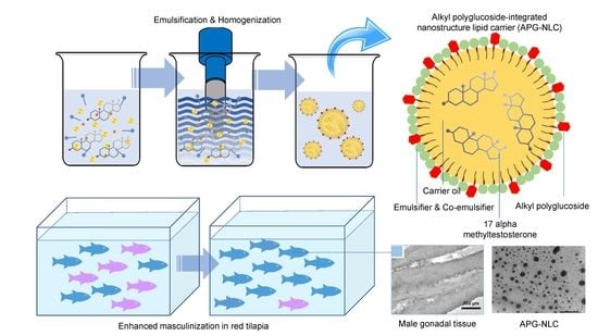
2.2. Formulation and Fabrication of Lipid-Based Nanoparticles

The NLC and SLN, the subtypes of the lipid-based nanoparticles, were fabricated using hot high-energy homogenization techniques [21]. As shown in Table 1, the oil phase was prepared using 17 alpha-methyltestosterone as a solid lipid (200 mg) and MCT as a liquid lipid (5 g only for the fabrication of NLC).
The oil mixture was dissolved and mixed with the oil phase surfactant by stirring at 200 rpm and 70 °C on a hotplate. Ethoxydiglycol (3 g), cetearyl alcohol and cocoglucoside (1 g), and Span 80 (3 g) were added to the oil mixture and stirred until completely dissolved. An aqueous phase mixture was prepared by mixing purified water, Tween 20 (3 g), glycerol (2.5 g) and Synperonic PE/F68 (2 g) on a hotplate stirrer, and the mixture was poured into the lipid phase. A high-speed homogenizer (T25, IKA, Wilmington, NC, USA) at 10,000 rpm was used to prepare the pre-emulsion for 5 min. Furthermore, the mixtures were sonicated in a sonicator unit (Qsonica sonicators, Newtown, CT, USA) at a 20 amp pulse of 30 s on and off at 5 s intervals for 5 min to prepare the testosterone–nanostructure lipid nanocarriers (MT-NLC). Testosterone–solid lipid carriers (MT-SLN) were also fabricated by following the same preparation steps as for NLC by without adding MCT (the liquid lipid). The MT-NLC and MT-SLN were cooled to an ambient temperature and used for subsequent studies. For the APG-integrated lipid-based nanoparticles, APG was added to a lipid mixture to fabricate alkyl-polyglucoside-integrated nanostructured lipid carriers (MT-APG-NLC) and alkyl-polyglucoside-integrated solid lipid nanoparticles (MT-APG-SLN).

2.3. Characterization of the Lipid-Based Nanoparticles

2.3.1. Dynamic Light Scattering

The hydrodynamic diameter, particle distribution and particle surface charge of lipid-based nanoparticles were characterized by dynamic light scattering (DLS) using a zetasizer (Nano ZS, Malvern Instrument, Malvern, Worcs, UK). DLS experiments were performed using a He-Ne laser (λ0 = 633 nm, θ = 173°). Before the measurements, the samples were diluted 50 times in purified water. All reported data were from experiments performed in triplicate at 25 °C. The particles’ stability was detected using changes in the particles’ size and the particles’ phase separation after incubation under various storage conditions (4, 25 and 45 °C) for two months.

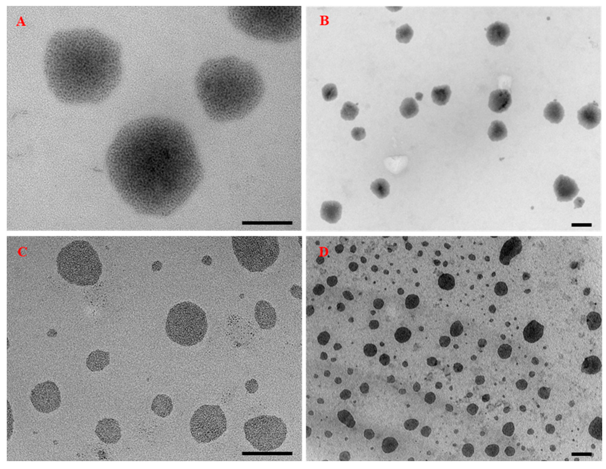
2.3.2. Transmission Electron Microscopy

The particles’ size and morphology were characterized using a transmission electron microscope (TEM; JEOL-2100 Plus, JEOL, Akishima, TYO, Japan). Samples were diluted 50-fold in deionized water, dropped onto a carbon grid, stained with phosphotungsten (1% w/w), dried in a dry cabinet overnight and observed under 100 kV with a magnification of ×25k and ×100k. The particles’ size and morphology were determined for an average of more than 50 particles for each sample.

2.3.3. Encapsulation Efficiency

The encapsulation efficiency (EE) was evaluated using an Amicon centrifuge filter (30 kDa MWCO, cellulose membrane, Millipore, Burlington, MA, USA). The lipid-based nanoparticles (10 mL) were loaded into the filter membrane and centrifuged (MX-307, Tomy, Tokyo, Japan) by following the manufacturer’s protocol. The supernatant was filtrated through a syringe filter (0.22 µm nylon membrane, Millipore, Burlington, MA, USA), referred to as the “non-encapsulated drug concentration”. Furthermore, the encapsulated lipid particles were extracted using the liquid extraction method and filtrated through a syringe filter (0.22 µm nylon membrane, Millipore, Burlington, MA, USA) to be used as the encapsulated drug concentration [22,23]. High-performance liquid chromatography coupled with a photodiode array detector (HPLC-PAD 2998 separation unit and PDA, Water, Milford, MA, USA) was validated and used with the following conditions: an Atlantis C-18 column (4.6 mm × 250 mm, Waters, Milford, MA, USA), a detection wavelength of 240 nm, a mobile phase of acetonitrile: MeOH (60:40, v/v) and a flow rate of 1.0 mL/min. The encapsulation efficiency (EE) of testosterone in lipid-based nanoparticles was calculated by using the following equation [24].
EE (%) = [(CiCf)/Ci)] × 100
where Ci corresponds to the initial concentration of testosterone added to LBN particles and Cf represents the concentration of free testosterone.

2.3.4. Release Kinetics of Testosterone in Lipid-Based Nanoparticles

The release profiles of MT from NLC and APG-NLC were determined and compared with those of free testosterone (diluted in ethanol) using a dialysis membrane (3500 MWCO regenerated cellulose tubular membrane, CelluSep, Seguin, TX, USA). The samples were placed in a dialysis membrane and incubated in the released buffer (PBS buffer pH 7.4, 25 °C). The released buffer was collected and replaced with fresh released buffer at regular intervals. The concentrations of testosterone were determined using HPLC-PAD as described above. Moreover, Avrami’s equation was used to determine the release mechanism and release constant of MT-synthesized-LBN.
R = exp [−(kt)n]
where R corresponds to the retention of MT at time (t), and n and k correspond to the release mechanism and release rate constants, respectively.

2.4. Masculinization of Red Tilapia

2.4.1. Ethical Statement

The protocols for the fish experiments used in this research were approved by the animal ethics committee of Chulalongkorn University Animal Care and Use Committee (CUACUC; approval No. 1931077).

2.4.2. Fish Production, Rearing and Experimental Protocol

The red tilapia fry (8-day-old swim-up (Stage 5)) were obtained from a hatchery unit from a GMP Certified fish farm in Ayutthaya Province, Thailand. Furthermore, the red tilapia swim-up fry were acclimatized (3 days) and randomly divided into 10 treatments (400 fish/treatment). The experimental groups were Control 1 (commercial feed, Charoen Pokphand (CP) 9931, mixed with ethanol (ET) without MT), T1 (30 ppm of MT in the feed for 14 days), T2 (30 ppm of MT in the feed for 21 days), T3 (60 ppm of MT in the feed for 14 days), T4 (60 ppm of MT in the feed for 21 days), Control 2 (commercial feed mixed with APG-NLC without MT), T5 (30 ppm of APG-NLC loaded with MT in the feed for 14 days), T6 (30 ppm of APG-NLC loaded with MT in the feed for 21 days), T7 (60 ppm of APG-NLC loaded with MT in the feed for 14 days) and T8 (60 ppm of APG-NLC loaded with MT in the feed for 21 days) with four replications. The red tilapia fry were distributed into 100 L fiber tanks containing dechlorinated fresh water under continuous aeration. The water (50%) was exchanged daily, and the tanks were replenished with fresh dechlorinated water. The experiment was performed at a controlled temperature within the range of 25–33 °C for air temperature and 25–28 °C for water temperature. The pH and content of dissolved oxygen (DO) were also determined to be within the range of 7.4–8.2 and 5.0–6.0 mg/L, respectively.
To prepare the MT-ET diet, MT was diluted in ethanol, sprayed, mixed with the commercial diet and air-dried. In addition, the MT-APG-NLC diet was prepared by directly mixing the APG-NLC loaded with MT and a commercial diet. Fish in all groups were fed at 10% of body weight five times per day. The fish were fed with a commercial diet until the 45th day of the trial, after completing the sex reversal treatment for 14 days (T1, T3, T5 and T7) and 21 days (Control 1, T2, T4, Control 2, T6 and T8). At the end of 21 days of feeding, the fish in each group were counted, and the body weight was recorded to indicate the growth performance of the red tilapia fry.

2.4.3. Length–Weight Relationship

The length–weight relationship (LWR) between the total length and body weight was expressed as [25]
W = aLb
where W is the body weight of the fish (g), L is the total length (cm), a is the rate of the change in weight with a constant length and b is the weight of one unit of length (slope) estimated from the linear regression equation transformed by taking the natural logarithm (log) of both sides.
Log W = Log a + b. Log L
when b = 3, an isometric pattern of growth occurs but when b is >3 or <3, an allometric pattern emerges, which may be positive when the length increases relative to the body’s thickness or negative when the length increases relative to the body’s thinness [26].

2.4.4. Relative Condition Factor

The relative condition factor (Kn) was developed for assessing the health condition of tilapia under all the treatments.
Kn = Wo/Wc
where Wo is the observed weight and Wc is the calculated weight [27]. Good growth condition of the fish was when Kn > 1, while the organism was in poor growth condition compared with an average individual of the same length when Kn < 1.

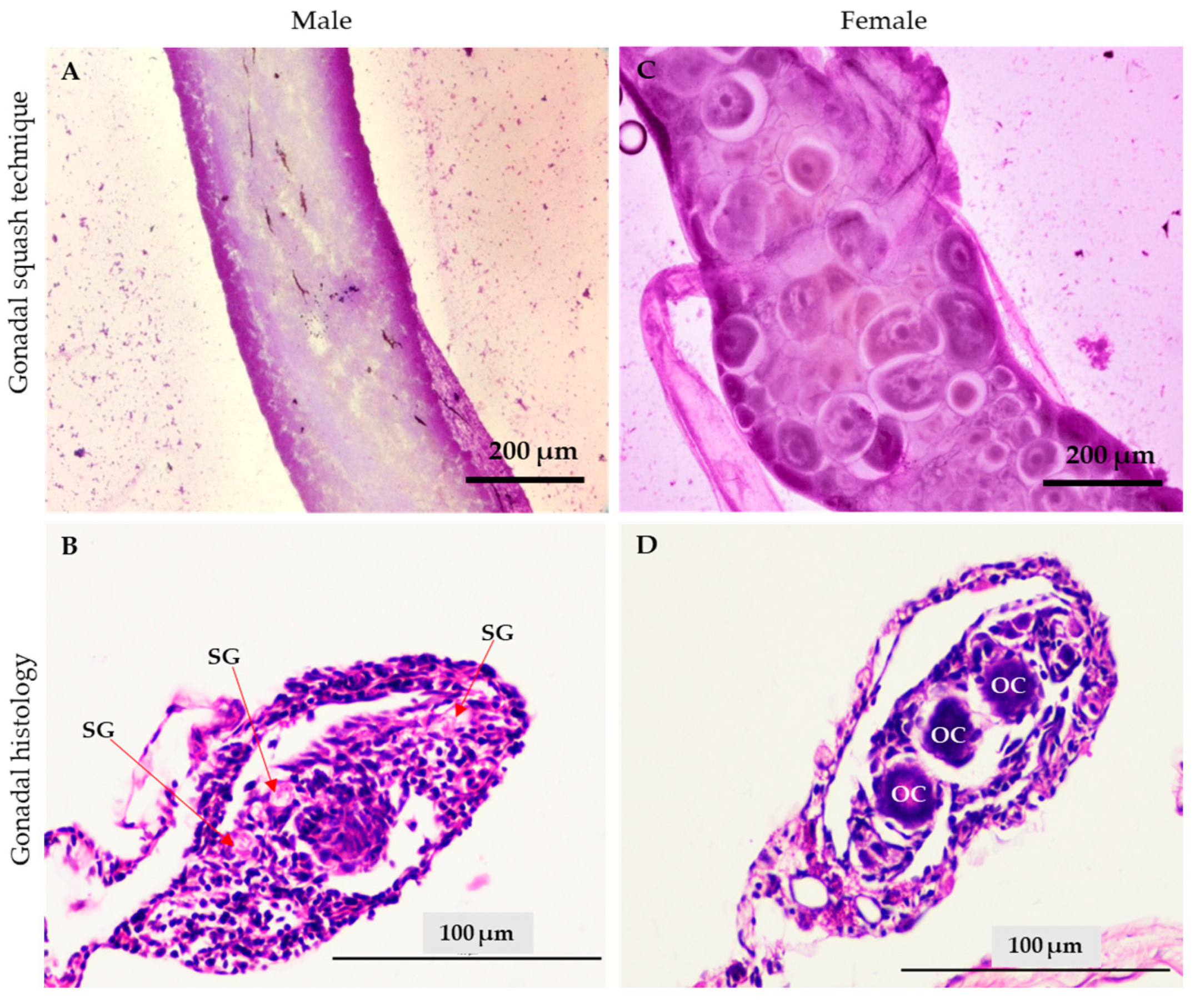
2.4.5. Sex Reversal Ratio Determined by the Gonad Squashing Technique

After 45-days of the experimental period, all the fish in each group were collected and anesthetized using clove oil for sex determination. The gonads of the fish were collected using fine forceps and placed on glass slides. The samples were stained with an aceto-carmine staining solution, squashed with cover slides and examined under a light microscope [28].

2.5. Statistical Analysis

The results were expressed as the means ± SD, and the statistical analysis was conducted by using IBM SPSS statistics software (SPSS Inc., Version 28, Chicago, IL, USA). The effects of the treatments on different parameters were analyzed by one-way analysis of variance (ANOVA), followed by Tukey’s post hoc tests for multiple comparisons. The effect of dietary supplementation with MT and MT-APG-NLC on the sex ratio (0 = male; 1 = female) of red tilapia fry was analyzed using a binary logistic regression model. Linear regression analysis was used to assess the length–weight relationship. A value of p < 0.05 was considered statistically significant.

3. Results

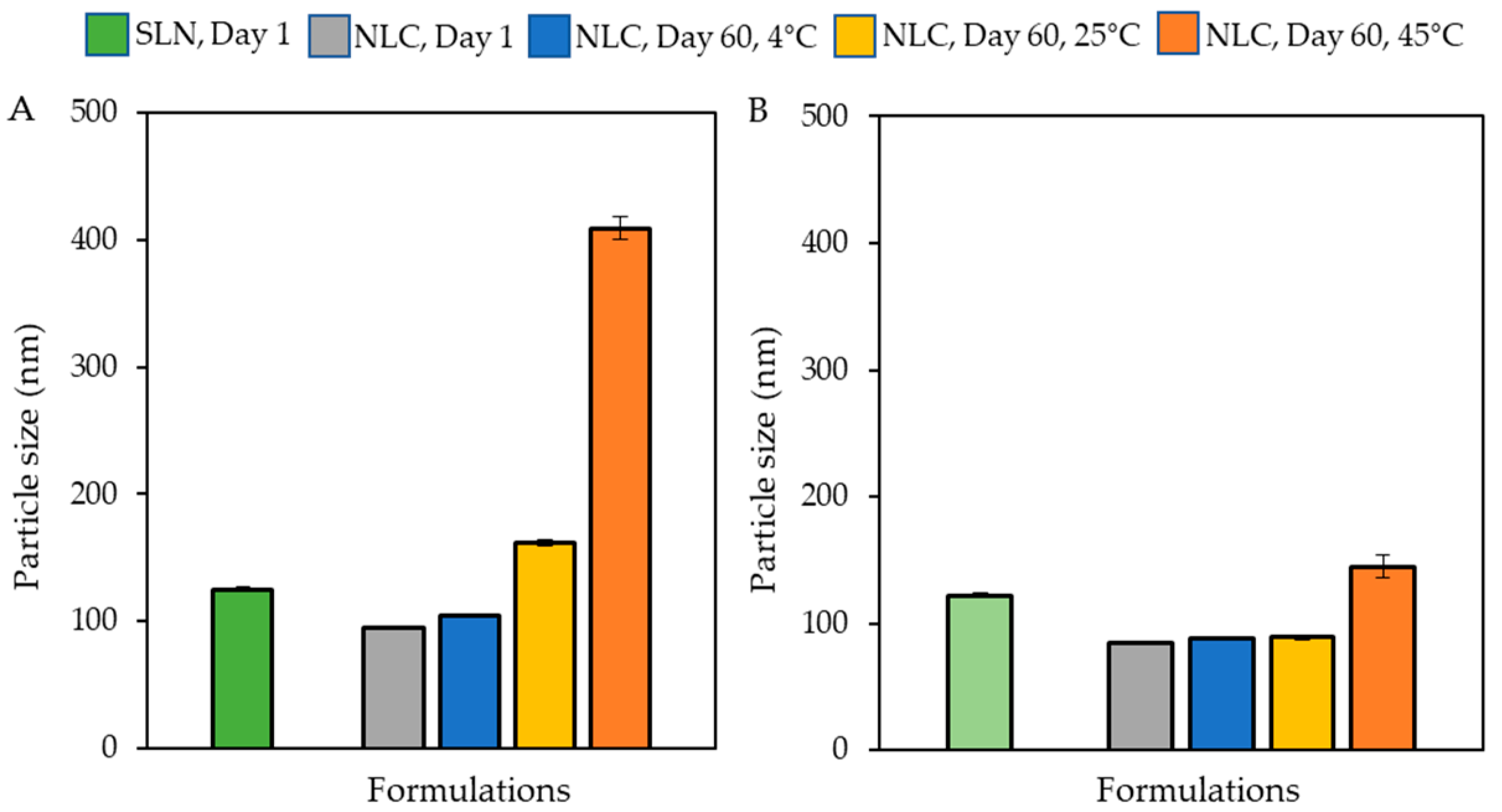
3.1. Characterization of the Lipid-Based Nanoparticles

The size of all four synthesized nanoparticles was lower than 125 nm. APG-SLN (122.23 nm) and APG-NLC (84.21 nm) were both smaller than bare SLN (125.30 nm) and NLC (94.56 nm). The average surface charge and polydispersity index (PDI) of the synthesized nanoparticles were lower than −20 mV and 0.3, respectively, as shown in Table 2 and Figure 1A,B. The stability of the synthesized nanoparticles was observed over time. After 2 months, solid sediments accumulated in the SLN formulation, while the bare NLC and APG-NLC provided higher particle stability without phase separation or solid sedimentation (data not shown). Importantly, APG-NLC had smaller particle sizes immediately after particle synthesis compared with the bare NLC. In terms of the particles’ stability, APG-NLC showed no significant change in the size of particles after being stored under cool (4 °C) and ambient (25 °C) conditions for 2 months.
However, under accelerated (45 °C) conditions, the size of the particles tended to increase to 144.83 nm without phase separation and sedimentation. For the bare NLC particles, the size was increased, and phase separation was observed under accelerated (45 °C) conditions. In addition, TEM images showed the spherical particles of LBNs within the range of 90–120 nm (Figure 2A–D).

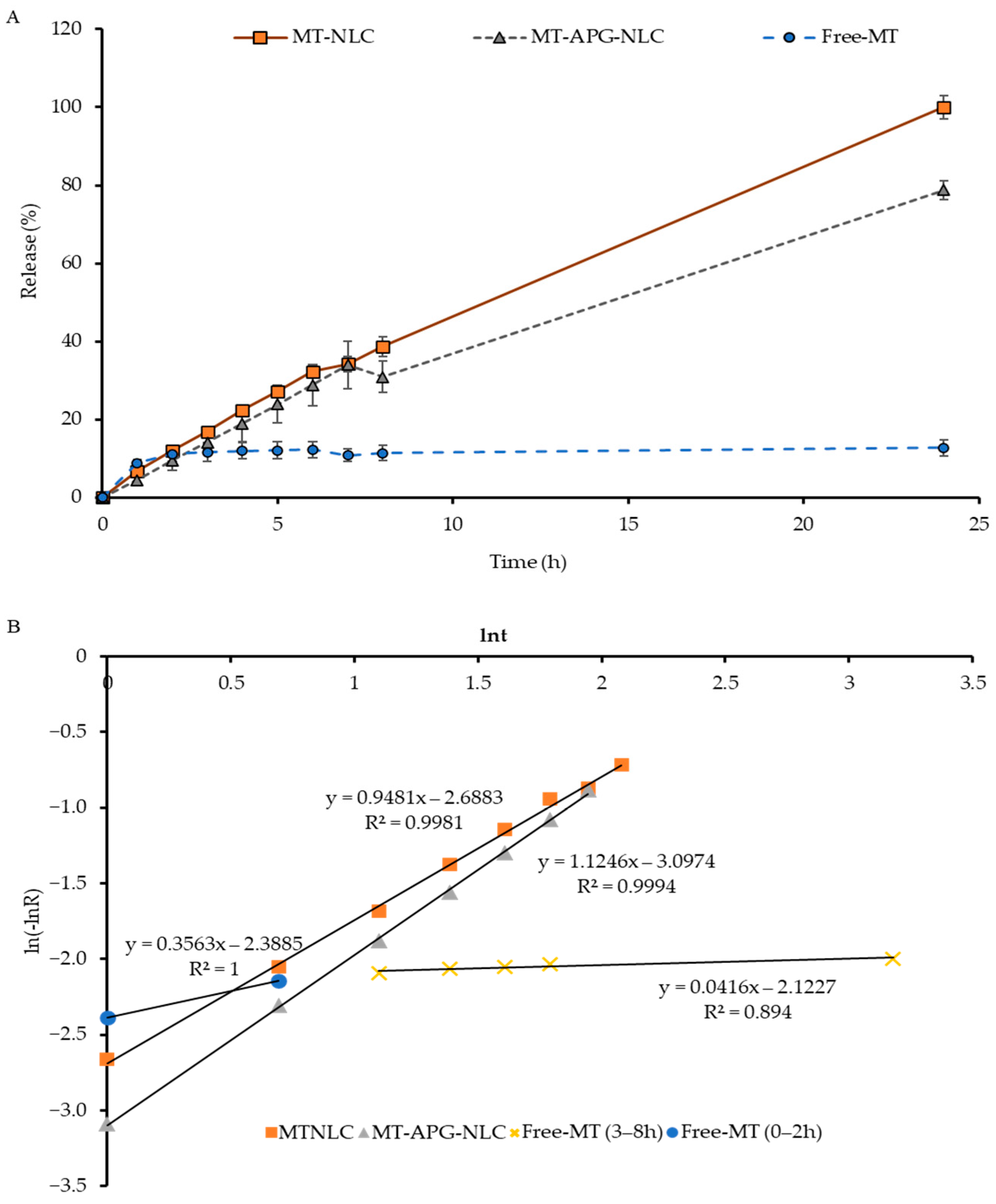
3.2. Encapsulation Efficiency and Study of the In Vitro Release of MT from LBN

Chemical characterization using HPLC demonstrated that the encapsulation efficiency (EE) values of the synthesized lipid-based nanoparticles were 70.52 ± 3.42%, 70.06 ± 2.24%, 83.91 ± 4.01% and 84.75 ± 13.56% for MT-SLN, MT-APG-SLN, MT-NLC and MT-APG-NLC, respectively. On the basis of the stability and encapsulation efficiency of the synthesized particles, MT-NLC, MT-APG-NLC and MT-ET were selected for evaluation in an in vitro release study. During the first three hours, MT released from MT-NLC, MT-APG-NLC and free MT (dissolved in EtOH) showed release percentages of 16.9 ± 1.0%, 14.1 ± 2.0% and 11.6 ± 2.4%, respectively. A gradual release of MT from MT-NLC, MT-APG-NLC and MT-ET was observed, which reached 38.6 ± 2.5, 30.9 ± 4.1 and 11.4 ± 1.8% within 8 h and extended to the maximum in 24 h (Figure 3A).
After 24 h, there was no further release of MT into the receiving buffer, which may be due to MT’s insoluble nature in water, leading to sedimentation in the aqueous media and the inability to diffuse or pass through the dialysis membrane. The release kinetics of the MT loaded in the synthesized LBN applied Avrami’s equation to determine the release mechanism (n) and release rate constant (k) (Table 3 and Figure 3B).
The release mechanisms (n) were found to be close to 1, being 0.95 for MT-NLC and 1.12 for MT-APG-NLC. Simultaneously, the release mechanism (n) of MT-ET was classified in two separate steps: 0.36 for the first two hours and 0.04 after three hours. Moreover, the release rate constant of MT from MT-NLC and MT-APG-NLC was found to be 0.587 × 10⁻¹/h and 0.637 × 10⁻¹/h, respectively. The release constant of MT-ET was lower than 0.01 × 10⁻¹/h, as shown in Table 3 and Figure 3B. Importantly, further analysis was carried out by using APG-NLC based on the particles’ size and stability, the encapsulation efficiency and the results of the release kinetics.

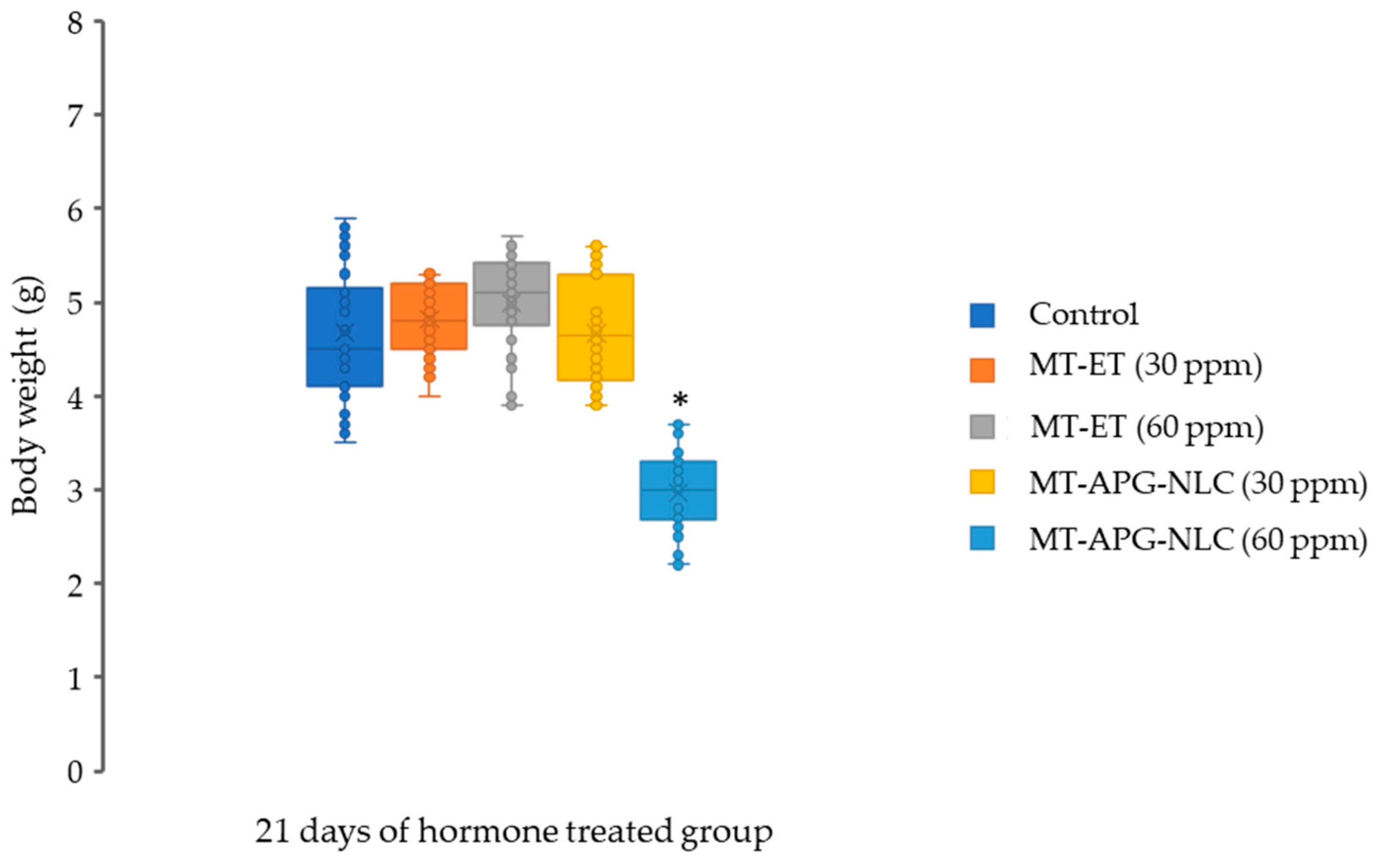
3.3. Red Tilapia Masculinization Using Testosterone Loaded in Lipid-Based Nanoparticles

The sex of the treated red tilapia fry was identified by staining the gonads with acetocarmine and determined on the basis of the morphological characteristics. For the gonad squashing technique, the male gonad was identified by its smooth tubular shape and the absence of an oocyte (Figure 4A), while the female gonad was identified by its round-shaped oocytes (Figure 4C). The gonadal tissue was also confirmed by histological observations, in which the male gonad was identified by the appearance of spermatogonia (Figure 4B) and the female gonadal tissue was characterized by the round shape of the oocyte (Figure 4D).
ANOVA showed a significant interaction (p < 0.01) between the effects of the concentrations of MT and MT-APG-NLC and the duration of the sex reversal period on the male ratio of red tilapia. The logistic regression model found that the male percentage in T1 (b = 0.260, p = 0.017), T2 (b = −0.284, p = 0.009), T4 (b = −2.165, p = 0.000), T5 (b = −1.297, p = 0.000), T6 (b = −4.205, p = 0.000) and T8 (b = −0.698, p = 0.000) was significantly higher than that in Control 1, Control 2, T3 and T7 (Table 4).
The T6 treatment displayed the highest male percentage (98.5%), which was significantly higher (p < 0.05) than that of all the other experimental groups. The chi-square analysis of Control 1 (Wald X2 = 0.012, p = 0.914) and Control 2 (Wald X2 = 2.301, p = 0.129) indicated that males and females were equally represented. Likewise, there was no significant change in the normal 1:1 sex ratio in fish fed with T3 (Wald X2 = 3.389, p = 0.066) and T7 (Wald X2 = 0.047, p = 0.828). All of the studied fish were found to be either male or female.
The survival rate was more than 85% in the MT and MT-APG-NLC groups, which was not significantly decreased (p > 0.05) compared with the control groups (Table 4). The production cost (in Thai baht) of the sex reversal treatments of red tilapia was estimated from the cost of MT, LBN and ethanol used in this experiment, based on equal amounts of the diets and the fish. The production cost was reduced by 32.90% (Table 4) under the T6 treatment compared with the conventional treatment (T4). Furthermore, the final body weight of red tilapia was also measured in the group that received the sex reversal diets for 21 days. The final body weights of red tilapia were similar among the controls, MT-APG-NLC (30 ppm), MT-APG-NLC (60 ppm), MT (30 ppm) and MT (60 ppm) groups (Figure 5). There was a significant decrease in the body weight of red tilapia fed MT-APG-NLC (60 ppm) compared with the other treatment groups.
The length–weight relationship was used to determine the growth performance of hormone-treated tilapia (Table 5). The calculated b-values were lower than 3.0 for all treatment groups, which indicated negatively allomeric growth behavior and implied that the length of tilapia increased relative to body thickness. The higher r2 values (>0.95) obtained in the assessment of LWR under all the treatments suggested a good quality of fit for the predictions of the linear regression. Furthermore, a significant correlation (p < 0.05) was observed between the length and weight under all the treatments. The relative condition factor (Kn) was higher than 1.0, demonstrating that the fish were in good condition during the treatment period.

4. Discussion

Methyl testosterone is unlikely to have a detrimental effect on the flesh of fish after the sex reversal treatment (SRT) has been discontinued; nonetheless, its effects on the major organs of fish, the metabolic profiles and nucleic acids are inconclusive [29]. However, fish treated with synthetic MT had altered enzymatic metabolic pathways [30], as well as a substantial increase in plasma creatine phosphokinase activity and DNA fragmentation in the liver tissue [31]. The dynamics of the ecosystem may be affected if tilapia hatcheries discharge steroids into the aquatic environment [32]. Furthermore, in certain natural populations, this may result in more males than females, leading to long-term reproductive failure [33]. It has been shown that the accumulation of MT in pond sediments and its detection in the soil occurred approximately 3 months after SRT. Importantly, many countries have limited the administration of synthetic hormones and antibiotics in fish farms because of chemical residues and antimicrobial resistance [34]. Therefore, the application of a potential nanocarrier platform as an alternative for the dietary administration of MT for reversing the sex of tilapia species is highly demanded.
Lipid-based nanocarriers have been shown to be a landmark in drug and hormone delivery. The application of LBN loaded with steroid derivatives, such as exemestane, spironolactone or methylprednisolone aceponate, has been developed and approved to enhance their in vivo bioavailability [35,36,37]. Nanoparticles have been integrated into hormone delivery applications such as luteinizing hormone-releasing hormone (LHRH) encapsulated in chitosan–gold conjugated nanoparticles to induce the reproduction efficiency of Cyprinus carpio [38]. In our study, we investigated the efficacy of lipid nanocarriers containing 17 MT for dietary administration as a masculinization agent to enhance the all-male ratio in red tilapia. The average surface charge was lower than −20 mV, and the polydispersity index (PDI) of 0.3 indicated that the MT-SLN, MT-APG-SLN, MT-NLC and MT-APG-NLC were assuredly stable [39]. APG has been previously used as a stabilizer for solid lipid nanoparticles for transdermal and cosmetic applications, which can reduce the particles’ size and induce the physical stability of the nanoparticles [40,41]. In this study, APG also acted as a surfactant in the formulation; therefore, with increasing concentrations, the size of APG nanoparticles decreased [42]. The solid sediments that appeared in the SLN formulation might be attributed to the recrystallization property of MT in the absence of an oil carrier (data not shown) [43]. NLC particles were formulated with an appropriate ratio of the liquid lipid and the solid lipid; it formed an amorphous structure that prevented the expulsion of testosterone to the extracellular matrix or the environment [44,45,46]. In contrast, the crystalline structure of SLN could induce the expulsion and sedimentation of the drug during storage [45,47,48]. The size of the synthesized APG-NLC particles was smaller than that of bare NLC particles and provided greater thermal stability under all storage conditions. This effect can be explained by the fact that APG can act as a non-ionic and biodegradable surfactant [49].
Furthermore, an in vitro investigation showed that the amount of MT released, which was dissolved in ethanol from 3 h to 24 h, could be classified into two separate steps. In both steps, the values of the release mechanism were lower than 1, which suggested that the MT was released through a diffusion mechanism, while the MT loaded in lipid-based nanoparticles was released by first-order release kinetics [50]. The rates of MT released from MT-NLC and MT-APG-NLC were constantly higher than those of MT-ET. The results showed that the release of MT was better in aqueous matrices, which confirmed the benefits of lipid-based nanoparticles as encapsulation agents and for prolonging the release of the androgen hormone.
The conventional sex reversal treatment (MT) has the potential to disrupt the natural differentiation process, even after its initiation, by interfering with normal embryonic gene expression patterns and physiological regulation, resulting in a skewed sex ratio towards males [51]. Moreover, the androgen-induced masculinization of tilapia gonads is correlated with the upregulated expression of dmrt1 [52,53,54]. In the present study, the T4 treatment produced 89.7% sex-reversed males in red tilapia. Our finding is in contrast with previous studies that reported 100% males in commercial tilapia farms [55,56], which was possibly due to the various nursing techniques and climates. Importantly, red tilapia fry in the T6 group produced 98.5% males, which is a significantly higher percentage of male tilapia compared with previous studies that found less than 70% sex-reversed males after the administration of 30 mg/kg of conventional MT for 28 days [6,57]. Our findings also confirmed that lipid nanoparticles could be a promising delivery system for androgen hormones. Furthermore, the current findings were corroborated by the in vitro release results, which revealed that APG-NLC could induce drug solubility and controlled drug release in aqueous media. This indicated that MT-APG-NLC (30 ppm) has the potential to be an alternative for delivering sex-reversal hormones to farmed tilapia. We postulated that APG-NLC increased the solubility of MT, which activates the expression of dmrt1, an important mechanism underpinning gonadal masculinization in red tilapia that requires further investigation. The higher dose of MT-APG-NLC (60 ppm) resulted in a significant decrease in the percentage of males and their growth, which may be due to a reduction in the intake of feed. Moreover, the reduction in the percentage of male tilapia as the concentration of MT-APG-NLC increased could possibly be due to paradoxical feminization and the aromatization of MT to estradiol, which expresses the female sex hormone [4,58,59]. These results showed that a lower dose (30 ppm) of MT-APG-NLC was more effective than the conventional sex reversal treatment currently used in red tilapia.
The conventional sex reversal treatment of tilapia results in a varied survival rate of fish [60,61,62,63]. Factors that affect the survival of fish include the hormones (natural or synthetic), dosage, feeding frequency and the duration of the sex reversal treatment [58]. All sex reversal treatments achieved more than 80% survival, which is consistent with the 80% survival rate obtained in seed production tilapia farms [56]. Furthermore, of the groups examined here, T6 had the lowest production costs, indicating that the MT-APG-NLC treatment is a more cost-effective alternative to the conventional sex reversal treatment for producing all-male red tilapia fry.
The negative allometric growth showed that the LWRs of the control group were in good agreement with those of the MT treatment group. Similarly, Nile tilapia and red tilapia showed negative allometric growth behavior [64,65]. Importantly, a relative condition factor of >1 was found for all the treatments, suggesting good fish health, better feed intake and proper environmental conditions. The results are in accordance with previous studies [65,66].

5. Conclusions

APG-integrated lipid-based nanoparticles are a new LBN platform for delivering the androgen hormone MT as a sex reversal agent to tilapia via dietary administration. APG-NLC achieved high encapsulation efficiency, thermal stability and cost-effectiveness for the masculinization of tilapia with MT when delivered orally at 30 ppm for 21 days. This platform is a promising way to reduce the dose of artificial sex reversal hormones used in red tilapia aquaculture for masculinization.

Author Contributions

Conceptualization, J.Y., N.S., A.H., K.D.T., M.M., T.Y. and N.P.; methodology, J.Y., S.K., N.T., K.P. and N.P.; software, J.Y., K.P., K.R., M.T.K. and K.S.; validation, J.Y., S.K., T.Y., N.T. and N.P.; formal analysis, J.Y., S.K. and K.R.; investigation, J.Y. and N.P.; resources, N.T. and N.P.; data curation, K.S., N.S., K.P., A.H., K.D.T., K.R., M.T.K. and M.M.; writing—original draft preparation, J.Y., K.S. and M.T.K.; writing—review and editing, N.S., A.H., K.D.T., M.M., M.T.K. and N.P.; visualization, N.P.; supervision, T.Y. and N.P.; project administration, T.Y. and N.P.; funding acquisition, N.P. and N.S. All authors have read and agreed to the published version of the manuscript.

Funding

This research was funded by the Second Century Fund (C2F), Chulalongkorn University, the 90th Anniversary of Chulalongkorn University Fund (Ratchadaphiseksomphot Endowment Fund), Thailand Science Research and Innovation (TSRI) under the TRF Research Career Development Grant (grant number: RSA 6280108), the National Science and Technology Development Agency (NSTDA, Thailand), the National Research Council of Thailand (NRCT: N42A650230) and Thailand Science Research and Innovation Fund, Chulalongkorn University (FOOD6631 0013).

Institutional Review Board Statement

All of the protocols for the fish experiments used in this research were approved by the animal ethics committee of Chulalongkorn University Animal Care and Use Committee (CUACUC, approval No. 1931077) and followed all the relevant guidelines and regulations.

Informed Consent Statement

Not applicable.

Data Availability Statement

The data presented in this study are available on request from the corresponding author.

Acknowledgments

The present research was supported by the Second Century Fund (C2F), The Chulalongkorn University, the 90th Anniversary of Chulalongkorn University Fund (Ratchadaphiseksomphot Endowment Fund), Thailand Science Research and Innovation (TSRI) under the TRF Research Career Development Grant (grant number: RSA 6280108), the National Science and Technology Development Agency (NSTDA, Thailand), the National Research Council of Thailand (NRCT: N42A650230) and Thailand Science Research and Innovation Fund, Chulalongkorn University (FOOD6631 0013). The Faculty of Veterinary Science at Chulalongkorn University is also acknowledged.

Conflicts of Interest

The authors declare no conflict of interest. The funders had no role in the design of the study; in the collection, analyses, or interpretation of data; in the writing of the manuscript; or in the decision to publish the results.

References

  1. Omasaki, S.; Charo-Karisa, H.; Kahi, A.; Komen, H. Genotype by environment interaction for harvest weight, growth rate and shape between monosex and mixed sex Nile tilapia (Oreochromis niloticus). Aquaculture 2016, 458, 75–81. [Google Scholar] [CrossRef]
  2. Saha, P.; Hossain, M.E.; Prodhan, M.M.H.; Rahman, M.T.; Nielsen, M.; Khan, M.A. Profit and loss dynamics of aquaculture farming. Aquaculture 2022, 561, 738619. [Google Scholar] [CrossRef]
  3. El-Sayed, A.-F.M.; Abdel-Aziz, E.-S.H.; Abdel-Ghani, H.M. Effects of phytoestrogens on sex reversal of Nile tilapia (Oreochromis niloticus) larvae fed diets treated with 17α-Methyltestosterone. Aquaculture 2012, 360, 58–63. [Google Scholar] [CrossRef]
  4. Karaket, T.; Reungkhajorn, A.; Ponza, P. The optimum dose and period of 17α-methyltestosterone immersion on masculinization of red tilapia (Oreochromis spp.). Aquac. Fish. 2023, 8, 174–179. [Google Scholar] [CrossRef]
  5. Snake, M.; Maluwa, A.; Zidana, H.; Chigwechokha, P.; Simwaka, M. Production of a predominantly male tilapia progeny using two Malawian tilapias, Oreochromis shiranus and Oreochromis karongae. Aquac. Rep. 2020, 16, 100274. [Google Scholar] [CrossRef]
  6. Celik, I.; Guner, Y.; Celİk, P. Effect of orally-administered 17α-Methyltestosterone at different doses on the sex reversal of the Nile tilapia (Oreochromis niloticus, Linneaus 1758). J. Anim. Vet. Adv. 2011, 10, 853–857. [Google Scholar] [CrossRef]
  7. El-Greisy, Z.; El-Gamal, A. Monosex production of tilapia, Oreochromis niloticus using different doses of 17α-methyltestosterone with respect to the degree of sex stability after one year of treatment. Egypt. J. Aquat. Res. 2012, 38, 59–66. [Google Scholar] [CrossRef]
  8. Arriesgado, D.M.; Vicente, H.J.; Vicente, D.A. Production of Male Oreochromis niloticus GET Excel Tilapia by Egg Immersion in Methyl Testosterone Hormone. J. Environ. Aquat. Resour. 2011, 2, 21–33. [Google Scholar] [CrossRef]
  9. Ahmad, A.; Abdullah, S.R.S.; Hasan, H.A.; Othman, A.R.; Ismail, N.I. Aquaculture industry: Supply and demand, best practices, effluent and its current issues and treatment technology. J. Environ. Manag. 2021, 287, 112271. [Google Scholar] [CrossRef]
  10. Ding, J.; Sun, H.; Liang, A.; Liu, J.; Song, L.; Lv, M.; Zhu, D. Testosterone amendment alters metabolite profiles of the soil microbial community. Environ. Pollut. 2021, 272, 115928. [Google Scholar] [CrossRef]
  11. Azar, F.A.N.; Pezeshki, A.; Ghanbarzadeh, B.; Hamishehkar, H.; Mohammadi, M. Nanostructured lipid carriers: Promising delivery systems for encapsulation of food ingredients. J. Agric. Food Res. 2020, 2, 100084. [Google Scholar]
  12. Beloqui, A.; del Pozo-Rodríguez, A.; Isla, A.; Rodríguez-Gascón, A.; Solinís, M.Á. Nanostructured lipid carriers as oral delivery systems for poorly soluble drugs. J. Drug Deliv. Sci. Technol. 2017, 42, 144–154. [Google Scholar] [CrossRef]
  13. Müller, R.; Petersen, R.; Hommoss, A.; Pardeike, J. Nanostructured lipid carriers (NLC) in cosmetic dermal products. Adv. Drug Deliv. Rev. 2007, 59, 522–530. [Google Scholar] [CrossRef]
  14. Müller, R.; Radtke, M.; Wissing, S. Nanostructured lipid matrices for improved microencapsulation of drugs. Int. J. Pharm. 2002, 242, 121–128. [Google Scholar] [CrossRef]
  15. Fang, J.-Y.; Fang, C.-L.; Liu, C.-H.; Su, Y.-H. Lipid nanoparticles as vehicles for topical psoralen delivery: Solid lipid nanoparticles (SLN) versus nanostructured lipid carriers (NLC). Eur. J. Pharm. Biopharm. 2008, 70, 633–640. [Google Scholar] [CrossRef]
  16. Mu, H.; Holm, R.; Müllertz, A. Lipid-based formulations for oral administration of poorly water-soluble drugs. Int. J. Pharm. 2013, 453, 215–224. [Google Scholar] [CrossRef] [PubMed]
  17. Nasirizadeh, S.; Malaekeh-Nikouei, B. Solid lipid nanoparticles and nanostructured lipid carriers in oral cancer drug delivery. J. Drug Deliv. Sci. Technol. 2020, 55, 101458. [Google Scholar] [CrossRef]
  18. Padhye, S.; Nagarsenker, M.S. Simvastatin solid lipid nanoparticles for oral delivery: Formulation development and in vivo evaluation. Indian J. Pharm. Sci. 2013, 75, 591–598. [Google Scholar]
  19. Salah, E.; Abouelfetouh, M.M.; Pan, Y.; Chen, D.; Xie, S. Solid lipid nanoparticles for enhanced oral absorption: A review. Colloids Surf. B Biointerfaces 2020, 196, 111305. [Google Scholar] [CrossRef]
  20. Silva, A.C.; Kumar, A.; Wild, W.; Ferreira, D.; Santos, D.; Forbes, B. Long-term stability, biocompatibility and oral delivery potential of risperidone-loaded solid lipid nanoparticles. Int. J. Pharm. 2012, 436, 798–805. [Google Scholar] [CrossRef]
  21. Gomaa, E.; Fathi, H.A.; Eissa, N.G.; Elsabahy, M. Methods for preparation of nanostructured lipid carriers. Methods 2022, 199, 3–8. [Google Scholar] [CrossRef] [PubMed]
  22. Beloqui, A.; Solinís, M.Á.; des Rieux, A.; Préat, V.; Rodríguez-Gascón, A. Dextran–protamine coated nanostructured lipid carriers as mucus-penetrating nanoparticles for lipophilic drugs. Int. J. Pharm. 2014, 468, 105–111. [Google Scholar] [CrossRef] [PubMed]
  23. Pinheiro, M.; Ribeiro, R.; Vieira, A.; Andrade, F.; Reis, S. Design of a nanostructured lipid carrier intended to improve the treatment of tuberculosis. Drug Des. Dev. Ther. 2016, 10, 2467. [Google Scholar] [CrossRef] [PubMed]
  24. Shimamoto, A.; Liu, J.; Kozawa, S.; Fujimiya, T. Determination of endogenous testosterone in rat tissues following fetal alcohol exposure using HPLC with UV detection. J. Chromatogr. B 2006, 836, 69–73. [Google Scholar] [CrossRef] [PubMed]
  25. Pauly, D. Some Simple Methods for the Assessment of Tropical Fish Stocks; Food & Agriculture Organization: Rome, Italy, 1983. [Google Scholar]
  26. Froese, R. Cube law, condition factor and weight–length relationships: History, meta-analysis and recommendations. J. Appl. Ichthyol. 2006, 22, 241–253. [Google Scholar] [CrossRef]
  27. Le Cren, E.D. The length-weight relationship and seasonal cycle in gonad weight and condition in the perch (Perca fluviatilis). J. Anim. Ecol. 1951, 20, 201–219. [Google Scholar] [CrossRef]
  28. Guerrero III, R.D.; Shelton, W.L. An aceto-carmine squash method for sexing juvenile fishes. Progress. Fish-Cult. 1974, 36, 56. [Google Scholar] [CrossRef]
  29. Aziz, M.A.; Mostary, M.M.; Sume, I.J.; Uddin, M.H.; Khan, M.G.Q.; Alam, M.S.; Islam, M.S. The efficacy of using pine (Pinus massoniana) pollen as an alternative to synthetic steroids in producing monosex male Nile tilapia (Oreochromis niloticus, L.). Aquac. Fish Fish. 2022, 2, 375–383. [Google Scholar] [CrossRef]
  30. Rivero-Wendt, C.L.G.; Miranda-Vilela, A.L.; Domingues, I.; Oliveira, R.; Monteiro, M.S.; Moura-Mello, M.A.; Matias, R.; Soares, A.M.V.M.; Grisolia, C.K. Steroid androgen 17 alpha methyltestosterone used in fish farming induces biochemical alterations in zebrafish adults. J. Environ. Sci. Health Part A 2020, 55, 1321–1332. [Google Scholar] [CrossRef]
  31. Hasheesh, W.S.; Marie, M.-A.S.; Abbas, H.H.; Eshak, M.G.; Zahran, E.A. An evaluation of the effect of 17α-Methyltestosterone hormone on some biochemical, molecular and histological changes in the liver of Nile Tilapia, Oreochromis niloticus. Life Sci. J. 2011, 8, 343–358. [Google Scholar]
  32. Sudtida, P.T.; Chheang, L.; Math, C. Ecological risk of 17α-methyltestosterone contaminated water discharged from a full water recirculating earthen masculinization pond. Hum. Ecol. Risk Assess. Int. J. 2021, 27, 1696–1714. [Google Scholar]
  33. Megbowon, I. Tilapia Production in Nigeria. Fishnetwork 2011, 4, 18–22. [Google Scholar]
  34. Megbowon, I.; Mojekwu, T. Tilapia sex reversal using methyl testosterone (MT) and its effect on fish, man and environment. Biotechnology 2013, 13, 213–216. [Google Scholar] [CrossRef]
  35. Günther, C.; Kecskes, A.; Staks, T.; Täuber, U. Percutaneous absorption of methylprednisolone aceponate following topical application of Advantan® lotion on intact, inflamed and stripped skin of male volunteers. Skin Pharmacol. Physiol. 1998, 11, 35–42. [Google Scholar] [CrossRef] [PubMed]
  36. Kelidari, H.R.; Saeedi, M.; Hajheydari, Z.; Akbari, J.; Morteza-Semnani, K.; Akhtari, J.; Valizadeh, H.; Asare-Addo, K.; Nokhodchi, A. Spironolactone loaded nanostructured lipid carrier gel for effective treatment of mild and moderate acne vulgaris: A randomized, double-blind, prospective trial. Colloids Surf. B Biointerfaces 2016, 146, 47–53. [Google Scholar] [CrossRef]
  37. Singh, A.; Neupane, Y.R.; Mangla, B.; Kohli, K. Nanostructured lipid carriers for oral bioavailability enhancement of exemestane: Formulation design, in vitro, ex vivo, and in vivo studies. J. Pharm. Sci. 2019, 108, 3382–3395. [Google Scholar] [CrossRef]
  38. Rather, M.A.; Sharma, R.; Gupta, S.; Ferosekhan, S.; Ramya, V.; Jadhao, S.B. Chitosan-nanoconjugated hormone nanoparticles for sustained surge of gonadotropins and enhanced reproductive output in female fish. PLoS ONE 2013, 8, e57094. [Google Scholar] [CrossRef]
  39. Rabelo, R.S.; Oliveira, I.F.; da Silva, V.M.; Prata, A.S.; Hubinger, M.D. Chitosan coated nanostructured lipid carriers (NLCs) for loading Vitamin D: A physical stability study. Int. J. Biol. Macromol. 2018, 119, 902–912. [Google Scholar] [CrossRef]
  40. Keck, C.M.; Kovačević, A.; Müller, R.H.; Savić, S.; Vuleta, G.; Milić, J. Formulation of solid lipid nanoparticles (SLN): The value of different alkyl polyglucoside surfactants. Int. J. Pharm. 2014, 474, 33–41. [Google Scholar] [CrossRef]
  41. Von Rybinski, W.; Hill, K. Alkyl polyglycosides—Properties and applications of a new class of surfactants. Angew. Chem. Int. Ed. 1998, 37, 1328–1345. [Google Scholar] [CrossRef]
  42. Qushawy, M. Effect of the surfactant and liquid lipid type in the physico-chemical characteristics of beeswax-based nanostructured lipid carrier (NLC) of metformin. Pharm. Nanotechnol. 2021, 9, 200–209. [Google Scholar] [CrossRef] [PubMed]
  43. Gao, S.; McClements, D.J. Formation and stability of solid lipid nanoparticles fabricated using phase inversion temperature method. Colloids Surf. A Physicochem. Eng. Asp. 2016, 499, 79–87. [Google Scholar] [CrossRef]
  44. Jagdevappa, P.; Prashant, G.; Ravindra, K.; Sachin, J.; Satish, M.; Meghanath, S. Applications of solid lipid nanoparticle in novel drug delivery system. Br. Biomed. Bull. 2013, 1, 103–118. [Google Scholar]
  45. Jaiswal, P.; Gidwani, B.; Vyas, A. Nanostructured lipid carriers and their current application in targeted drug delivery. Artif. Cells Nanomed. Biotechnol. 2016, 44, 27–40. [Google Scholar] [CrossRef] [PubMed]
  46. Kumar, S.; Dilbaghi, N.; Saharan, R.; Bhanjana, G. Nanotechnology as emerging tool for enhancing solubility of poorly water-soluble drugs. Bionanoscience 2012, 2, 227–250. [Google Scholar] [CrossRef]
  47. Cipolla, D.; Shekunov, B.; Blanchard, J.; Hickey, A. Lipid-based carriers for pulmonary products: Preclinical development and case studies in humans. Adv. Drug Deliv. Rev. 2014, 75, 53–80. [Google Scholar] [CrossRef]
  48. Sarathchandiran, I. A review on nanotechnology in solid lipid nanoparticles. Int. J. Pharm. Dev. Technol. 2012, 2, 45–61. [Google Scholar]
  49. Pajić, N.B.; Ilić, T.; Nikolić, I.; Dobričić, V.; Pantelić, I.; Savić, S. Alkyl polyglucoside-based adapalene-loaded microemulsions for targeted dermal delivery: Structure, stability and comparative biopharmaceutical characterization with a conventional dosage form. J. Drug Deliv. Sci. Technol. 2019, 54, 101245. [Google Scholar] [CrossRef]
  50. Phunpee, S.; Saesoo, S.; Sramala, I.; Jarussophon, S.; Sajomsang, W.; Puttipipatkhajorn, S.; Soottitantawat, A.; Ruktanonchai, U.R. A comparison of eugenol and menthol on encapsulation characteristics with water-soluble quaternized β-cyclodextrin grafted chitosan. Int. J. Biol. Macromol. 2016, 84, 472–480. [Google Scholar] [CrossRef]
  51. Singh, A.K. Introduction of modern endocrine techniques for the production of monosex population of fishes. Gen. Comp. Endocrinol. 2013, 181, 146–155. [Google Scholar] [CrossRef]
  52. Kobayashi, T.; Kajiura-Kobayashi, H.; Guan, G.; Nagahama, Y. Sexual dimorphic expression of DMRT1 and Sox9a during gonadal differentiation and hormone-induced sex reversal in the teleost fish Nile tilapia (Oreochromis niloticus). Dev. Dyn. 2008, 237, 297–306. [Google Scholar] [CrossRef] [PubMed]
  53. Bhandari, R.K.; Nakamura, M.; Kobayashi, T.; Nagahama, Y. Suppression of steroidogenic enzyme expression during androgen-induced sex reversal in Nile tilapia (Oreochromis niloticus). Gen. Comp. Endocrinol. 2006, 145, 20–24. [Google Scholar] [CrossRef] [PubMed]
  54. Piferrer, F. Hormonal control of reproduction and growth: Endocrine control of sex differentiation in fish. In Encyclopedia of Fish Physiology; Farrell, A.P., Ed.; Academic Press: San Diego, CA, USA, 2011; pp. 1490–1499. [Google Scholar]
  55. Vinarukwong, N.; Lukkana, M.; Wongtavatchai, J. Decreasing duration of androgenic hormone feeding supplement for production of male monosex in tilapia (Oreochromis spp.) fry. Thai J. Vet. Med. 2018, 48, 375–383. [Google Scholar]
  56. Karaket, T.; Seel-audom, M.; Areechon, N. Analysis and Synthesis of Knowledge from Nile Tilapia Philosophers for Sustainable Culture in the Northern Thailand. Thai Sci. Technol. J. 2021, 29, 454–468. [Google Scholar]
  57. Amer, M.; Zaki, F.M.; Tahoun, A.; Said, M. Effects of 17 α-Methyltestosterone and Tribulus terrestris extract on sex ratio and gonads histology of Red tilapia hybrid. Egypt. J. Nutr. Feed. 2021, 24, 189–198. [Google Scholar] [CrossRef]
  58. Alcántar-Vázquez, J.P. Sex proportion in Nile tilapia Oreochromis niloticus fed estrogen mixtures: A case of paradoxical masculinization. Lat. Am. J. Aquat. Res. 2018, 46, 337–345. [Google Scholar] [CrossRef]
  59. Rinchard, J.; Dabrowski, K.; Garcia–Abiado, M.A.; Ottobre, J. Uptake and depletion of plasma 17α-methyltestosterone during induction of masculinization in muskellunge, Esox masquinongy: Effect on plasma steroids and sex reversal. Steroids 1999, 64, 518–525. [Google Scholar] [CrossRef]
  60. Marín-Ramírez, J.; Alcántar-Vázquez, J.; Antonio-Estrada, C.; Moreno-de la Torre, R.; Calzada-Ruiz, D. Feminization of nile tilapia Oreochromis niloticus (L.) by diethylstilbestrol growth and gonadosomatic index. Ecosist. Recur. Agropecu. 2016, 3, 51–61. [Google Scholar]
  61. Juárez-Juárez, V.; Alcántar-Vázquez, J.; Antonio-Estrada, C.; Marín-Ramírez, J.; Moreno-De La Torre, R. Feminization by 17α-ethinylestradiol of the progeny of XY-female Nile tilapia (Oreochromis niloticus). Effects on growth, condition factor and gonadosomatic index. Turk. J. Fish. Aquat. Sci. 2017, 17, 599–607. [Google Scholar] [CrossRef]
  62. Rima, N.; Rahman, M.; Sarker, M. Optimization of 17-alpha methyltestosterone (MT) hormone dose during masculinization of Nile tilapia (Oreochromis niloticus) fry. J. Noakhali Sci. Technol. Univ. 2017, 1, 35–41. [Google Scholar]
  63. Kamble, M.T.; Chavan, B.R.; Ataguba, G.; Azpeitia, T.; Medhe, S.; Jain, S.; Jadhav, R. Application of Moringa oleifera for development of Sustainable and Biosecure Aquaculture. Aquac. Indones. 2015, 15, 64–73. [Google Scholar] [CrossRef]
  64. Malik, A.; Abbas, G.; Soomro, M.; Shah, S.; Asadullah, A.; Bhutto, A.; Jamali, G.; Roonjho, Z. Length-weight relationship and condition factor of red tilapia (Hybrid) reared in cemented tanks of Sun-bright Red Tilapia and ornamental hatchery-Karachi, Sindh-Pakistan. Sindh Univ. Res. J. (Sci. Ser.) 2017, 49, 159–162. [Google Scholar]
  65. Cishahayo, L.; Yongo, E.; Mutethya, E.; Waithaka, E.; Ndayishimiye, R. Length–weight relationship, condition factor, sex ratio and size at first maturity of the blue-spotted tilapia (Oreochromis leucostictus) in Lake Naivasha, Kenya. Lakes Reserv. Res. Manag. 2022, 27, e12417. [Google Scholar] [CrossRef]
  66. Komba, E.A.; Munubi, R.N.; Chenyambuga, S.W. Comparison of body length-weight relationship and condition factor for Nile tilapia (Oreochromis niloticus) cultured in two different climatic conditions in Tanzania. Int. J. Fish. Aquat. Stud. 2020, 8, 44–48. [Google Scholar]
Figure 1. Size and stability of unmodified lipid-based nanoparticles, including solid lipid nanoparticle (SLN), nanostructured lipid carriers (NLC) (A) and alkyl polyglucoside integrated with solid lipid nanoparticles (APG-SLN) and alkyl polyglucoside integrated with nanostructured lipid carriers (APG-NLC). (B) The samples were stored at 4, 25 and 45 °C for 2 months.
Animals 13 01364 g001
Figure 2. Morphological observations of the nanostructured lipid carriers (NLC) (A,B) and alkyl polyglucoside integrated with nanostructured lipid carriers (APG-NLC) (C,D) loaded with 17 alpha-methyltestosterone using transmission electron microscopy (TEM). Scale bar: 100 nm.
Animals 13 01364 g002
Figure 3. In vitro cumulative release profile of MT from the synthesized LBNs with and without a polyglucoside structure and MT-ET (non-encapsulated MT) using the HPLC-PAD technique (n = 3 for each group) (A) and an evaluation of the release kinetics of MT-NLC, MT-APG-NLC and MT-ET using Avrami’s equation (B).
Animals 13 01364 g003
Figure 4. Gonadal morphology of the red tilapia ovary and testis using the gonad squashing technique (A,C) and histological observations (B,D) at 45 dph after feeding with MT-APG-NLC (60 ppm) for 21 days. SG: spermatogonia; OC: oocytes.
Animals 13 01364 g004
Figure 5. Box plot of the body weight of red tilapia after being fed with the control diet (hormone-free diet), MT-ET (30 and 60 ppm) and MT-APG-NLC (30 and 60 ppm) for 21 days and then continuously fed with control diet until 45 dph. * p < 0.05 indicates significant differences among the different treatment groups.
Animals 13 01364 g005
Table 1. Composition (percent by weight) of four different formulations of MT-LBNs.
Formulations
CompositionSLNAPG-SLNNLCAPG-NLC
17 α-methyltestosterone0.20.20.20.2
Medium chain triglyceride (MCT)0.00.05.05.0
Alkyl-polyglucoside0.02.00.02.0
Span 803.03.03.03.0
Ethoxydiglycol4.04.03.03.0
Tween 203.03.03.03.0
Synperonic PE/F682.02.02.02.0
Glycerol2.52.52.52.5
DI water85.383.381.379.3
Total100.0100.0100.0100.0
SLN: solid lipid nanoparticles; NLC: nanostructured lipid carriers; APG: alkyl polyglucoside.
Table 2. Physical properties of LBNs using the dynamic light scattering technique after loading with 17-alpha methyltestosterone.
FormulationParticle Size (nm)Surface Charge (mV)Polydispersity Index
SLN125.30 ± 1.34−30.4 ± 2.430.211 ± 0.002
APG-SLN122.23 ± 1.50−34.0 ± 1.470.228 ± 0.020
NLC94.56 ± 0.17−28.5 ± 3.500.148 ± 0.010
APG-NLC84.21 ± 0.60−30.9 ± 3.480.123 ± 0.010
MT-ETNDNDND
Values are the mean ± SD of triplicate samples. ND: not detected; SLN: solid lipid nanoparticles; APG-SLN: solid lipid nanoparticles of alkyl polyglucoside; NLC: nanostructured lipid carriers; MT-ET: 17 alpha-methyltestosterone-ethanol.
Table 3. Parameters of the release kinetics (release rate constant (k), release mechanism (n) and R2) of MT from the encapsulation particles after fitting the release profile with Avrami’s equation.
Samplek (10−1/h)nR2
MT-ET (0–2 h)0.0120.35630.9521
MT-ET (3–8 h)0.0000.04160.894
MT-NLC0.5870.94810.9981
MT-APG-NLC0.6371.12460.994
Values are the mean ± SD of triplicate samples. MT-ET: 17 alpha-methyltestosterone–ethanol; MT-NLC: 17 alpha-methyltestosterone– nanostructured lipid carriers; MT-APG-NLC: 17 alpha-methyltestosterone–alkyl polyglucoside–nanostructured lipid carriers.
Table 4. Proportion of males, survival rate and cost of production of red tilapia fry fed with MT and MT-APG-NLC for 14 d and 21 d, followed by a commercial diet for 45 d.
TreatmentsMale
(%)
No. of FishMale: Female
(1:1)
p-Valueb ± SEExponentiation of bWald
X2
Survival
Rate (%)
Cost (THB/Fish)Compared with T4 (%)
Control 150.3 ± 3.2 ab3401:0.990.914−0.012 ± 0.1080.9880.01291.5 ± 2.1--
T143.5 ± 3.8 a3401:1.290.0170.260 ± 0.1091.2975.66291.0 ± 4.10.0011−80.49
T257.1 ± 4.5 b3401:0.760.009−0.284 ± 0.1100.7536.73189.5 ± 2.90.0029−50.00
T355.0 ± 2.1 b3401:0.820.066−0.201 ± 0.1090.8183.38988.8 ± 3.90.0023−60.98
T489.7 ± 3.6 c3401:0.110.000−2.165 ± 0.1780.115147.16088.5 ± 3.70.00590.00
Control 245.9 ± 2.0 a3401:1.190.1290.165 ± 0.1091.1792.30190.3 ± 3.7--
T578.5 ± 4.8 c3401:0.270.000−1.297 ± 0.1320.27396.40491.3 ± 2.10.0015−73.85
T698.5 ± 1.4 d3401:0.020.000−4.205 ± 0.4510.01587.09792.0 ± 2.90.0039−32.98
T750.6 ± 2.8 a3401:0.980.828−0.024 ± 0.1080.9770.04787.3 ± 4.40.0031−47.70
T866.8 ± 1.7 b3401:0.500.000−0.698 ± 0.1150.49836.71185.5 ± 1.70.0079+34.03
The results are expressed as the mean ± SD. Different superscript letters in a column indicate significant differences (p < 0.05) from the control. Chi-square values with p < 0.05 indicate a significant deviation from the expected sex ratio of 1:1. b = regression coefficient; p-value: probability of a male occurring; X2: chi-square values.
Table 5. Length–weight relationship of red tilapia fry fed with MT and MT-APG-NLC for 21 days, followed by a commercial diet for 45 days.
TreatmentsControlMT-ET
(30 ppm)
MT-ET
(60 ppm)
MT-APG-NLC
(30 ppm)
MT-APG-NLC
(60 ppm)
N5050505050
Lmin-max (cm)2.2–8.82.9–7.92.9–8.74.3–8.32.0–7.0
Wmin-max (g)1–7.31.9–6.81.6–7.42.9–6.51.1–5.2
a−0.372−0.258−0.351−0.307−0.363
b1.34231.1961.2861.2251.289
SE (b)0.0310.0380.0280.0180.020
CI (b)1.280–1.4051.119–1.2731.230–1.3411.189–1.2601.249–1.330
r20.9750.9530.9780.9900.988
p0.0000.0000.0000.0000.000
t-test sig0.0000.0000.0000.0000.000
Growth behaviorNegative
allometry
Negative
allometry
Negative
allometry
Negative
allometry
Negative
allometry
Kn1.0031.0021.0011.0001.001
Min-Max0.818–1.4450.830–1.1480.876–1.1020.967–1.0670.926–1.072
SE0.0110.0090.0060.0030.005
N: sample size; L: length (cm); W: weight (g); Min: minimum; Max: maximum; a: intercept; b: slope of the equation; SE: standard error; CI (b): confidence intervals of b; r2: coefficient of determination; p: significance of the regression, with p significant at <0.05; Kn: relative condition factors. Significance was determined by the t-test to verify if b was significantly different from the consensus (b = 3). The growth behavior was based on b.
Disclaimer/Publisher’s Note: The statements, opinions and data contained in all publications are solely those of the individual author(s) and contributor(s) and not of MDPI and/or the editor(s). MDPI and/or the editor(s) disclaim responsibility for any injury to people or property resulting from any ideas, methods, instructions or products referred to in the content.

