Article

Anaplasma Species in Ticks Infesting Mammals of Sardinia, Italy

Dipartimento di Sanità Animale, Istituto Zooprofilattico Sperimentale della Sardegna, Via Vienna 2, 07100 Sassari, Italy
*
Author to whom correspondence should be addressed.
Animals 2023, 13(8), 1332; https://doi.org/10.3390/ani13081332
Submission received: 10 March 2023 / Revised: 5 April 2023 / Accepted: 11 April 2023 / Published: 13 April 2023

Abstract

:

Simple Summary

Bacteria in the Anaplasma genus are intracellular parasites of mammal blood cells transmitted by ticks of genera Amblyomma, Dermacentor, Hyalomma, Ixodes, and Rhipicephalus. In this study, the presence of Anaplasma marginale and A. phagocytophilum in ticks was molecularly confirmed in Rhipicephalus sanguineus s.l. and Rhipicephalus bursa ticks, suggesting that these tick species are of importance in the transmission of potential zoonotic infections. Due to the nature of the Anaplasma species detected here, our results, together with data obtained to date in Sardinia, suggest that from a public health point of view, the potential zoonotic Anaplasma species should be further investigated in the island. Additional studies are needed to clarify whether these tick species can transmit these zoonotic bacteria both to human and animal hosts.

Abstract

Ticks are hematophagous ectoparasites that are recognized for their ability to vector a wide variety of pathogens of viral, bacterial, protozoal, and helminthic nature to vertebrate hosts. Among the different diseases transmitted by ticks, also called “Tick-Borne Diseases” (TBD), many are zoonotic. Pathogens of the genus Anaplasma refer to obligate intracellular bacteria within the Rickettsiales order transmitted mainly through tick bites and considered as well-established threats to domestic animals, livestock, and humans, worldwide. In this retrospective study, 156 ticks collected from twenty goats, one marten, and one cattle from several Sardinian sites, were examined by molecular analyses to detect the presence of Anaplasma species. A total of 10 (10/156; 6.4%) ticks were shown to be Anaplasma-positive by PCR screening. After sequence analyses, A. phagocytophilum was detected in four Rhipicephalus sanguineus s.l. (3.3%) and four Rh. bursa (11%) ticks from goats, while one Rh. sanguineus s.l. (0.8%) and one Rh. bursa (2.8%) collected from the marten and cattle, respectively, exhibited 100% of identity with A. marginale strains. In this study, we provide the first description and molecular detection of A. marginale and A. phagocytophilum in ticks of the Rhiphicephalus genus in Sardinia. Considering the growing impact of tick-borne Anaplasma pathogens on human health, further studies are necessary to monitor the prevalence of these pathogens in Sardinia.

1. Introduction

Ticks are hematophagous ectoparasites that transmit protozoan, bacterial, and viral pathogens of medical and veterinary importance [1]. Among bacterial pathogens, members belonging to the genus Anaplasma (Rickettsiales: Anaplasmataceae) are obligate intracellular organisms that replicate within parasitophorous vacuoles in the cytoplasm of both vertebrate and invertebrate host cells [2].
In the vertebrate hosts, these organisms infect blood cells including erythrocytes (RBCs), monocytes, platelets, and neutrophils and constitute a major public health threat in humans and animals [3]. The different Anaplasma spp. exist in a zoonotic cycle that involve both Ixodidae ticks and vertebrate hosts, which can be reservoirs of infection [4]. Trans-stadial transmission from nymph to adult exists in nature while transovarial infection has been suggested only for few Ixodidae species. Transmission of these pathogens occurs due to the action of ticks during their blood meal on infected animals and they can transmit the agent to other mammals at the next stage. Although Anaplasma spp. are mainly transmitted by tick bites, other modes of transmission such as hematophagous insect bites and exposure to blood-contaminated fomites have been proven [5].
The genus Anaplasma has been especially studied for its pathogenicity in farm animals since various species of Anaplasma such as A. marginale, A. ovis, and A. bovis, along with A. phagocytophilum, are regarded as one of the biggest threats to livestock [6]. In fact, these agents significantly affect animal productivity and cause considerable economic losses to farmers due to the reduction in reproductive performance, decreased milk and meat production, abortion, and death of the infected animal [7]. However, the clinical identification of infected animals is challenging because immunocompetent hosts do not exhibit symptoms while clinical signs ranging from subclinical infections with mild to high fever, anorexia, and respiratory symptoms have been described in immunocompromised hosts [8]. Although anaplasmosis is rarely fatal, leukopenia and impaired neutrophil and lymphocyte function of the bacteria can make animals more susceptible to life-threatening secondary infections [8].
Currently, there is a high diversity of Anaplasma organisms, which includes six validated species (A. phagocytophilum, A. marginale, A. centrale, A. ovis, A. bovis, and A. platys).
Moreover, the genus contains two new species, namely A. odocoilei and A. capra, that have not been cultured yet, as well as species of ‘Candidatus’ status and many other new unclassified Anaplasma genovariants that have been recently detected [9].
Among them, several species that were previously considered non-pathogenic were recently found to be zoonotic and associated with human diseases, suggesting that the number of Anaplasma species that are infecting humans is increasing. Specifically, A. phagocytophilum, the causative agent of tick-borne fever in sheep and granulocytic anaplasmosis in dogs (CGA) and horses (EGA), is also responsible for human infection (HGA) [9]. A. bovis, previously found to infect bovine monocytes, has been recently detected in humans in China [10]. Anaplasma platys, which infects platelets and is the etiological agent of infectious cyclic thrombocytopenia in dogs, has been documented in two women from Venezuela who were exposed to Rhipicephalus sanguineus [11]. A variant of the erythrocytic anaplasmal A. ovis was identified in a Cypriot patient with clinical signs including fever, hepatosplenomegaly, and lymphadenopathy [12]. Finally, Anaplasma capra, a novel, tick-borne pathogen which was detected in China in 2010–2012, causes zoonotic infections and infects many different animal species, including humans [13].
Anaplasma distribution is correlated with the presence of tick vectors, hosts, and competent reservoirs. Therefore, determining the density of ticks and the incidence of the infectious agents they transmit is important to prevent and avoid the transmission of possible diseases to animals and humans. Sardinia is the second biggest island in the Mediterranean Sea covering a surface of 24.090 km2 and with different habitat types. It has an annual mean temperature of 22 °C and a typical Mediterranean climate that allows the survival of ticks during the whole year. Furthermore, the island, which is located approximately halfway between Spain, Italy, and North Africa, is an important stopover area for migratory birds which pose a risk for the introduction and dispersal of ticks and TBD. Specific studies of the prevalence of Anaplasma spp. in Sardinian ticks are limited. It was previously observed that ticks belonging to the Rhipicephalus and Hyalomma genera are the most frequent hosts for A. ovis, A. phagocytophilum, A. platys, and A. platys-like, suggesting that these species could serve as potential vectors for these pathogens [14].
The aim of this study was to verify the distribution of Anaplasma species in ticks collected from mammals from Sardinia, Italy, and provide epidemiological data to develop strategies and control programs for anaplasmosis prevention and monitoring in the island.

2. Materials and Methods

2.1. Tick Collection

In this retrospective study, 156 tick specimens opportunistically removed between June 2011 and October 2013 from 20 goats, 1 cattle, and 1 marten in Sardinia (Italy) were analyzed for the detection of Anaplasma species. The sites belonging to Ogliastra and Sassari provinces were randomly chosen and ticks collected from this geographic area were included in this study. Ticks were removed from each host with tweezers and placed in vials containing 70% ethanol at room temperature. Ticks from one marten that was found dead were provided by hunters who removed specimens from the animal. Morphological identification of the ticks was conducted down to the species level using identification keys [15] with a binocular microscope at a magnification of 50×. Ticks were also sorted by stage and animal host and then stored at −80 °C until further analyses. Details of collection sites, species, and the sex of each tick were collected. The origins and hosts from which each tick was sampled are summarized in Table 1.

2.2. DNA Extraction and PCR

To remove environmental contaminants, the ticks were rinsed with 70% ethanol and then immersed in deionized water to remove the ethanol. The ticks were then longitudinally cut in two equal parts using sterile instruments for each individual dissection, and one half was used for DNA extraction. The half tick was homogenized with a Tissue Lyser (TissueLyser II) in 200 μL of PBS. One hundred microliters of genomic DNA was extracted using QIAgen columns (QIAamp tissue kit; Qiagen, Hilden, Germany, cod.69504), according to the manufacturer’s instructions. PCR amplification of the 16S ribosomal RNA gene was carried out on all genomic DNA samples by using oligonucleotide primer pairs (Eurogentec, Seraing, Belgium), which amplified a 293-bp fragment of Anaplasma species [16]. All reactions and amplification conditions used in this study were confirmed from studies previously published [14]. Negative and positive controls were included in each amplification assay. Eight microliters of each 293-bp PCR product was electrophoresed in 1.5% agarose gel with SYBR™ Safe DNA Gel Stain (Invitrogen, Carlsbad, CA, USA) in one × TAE buffer against a DNA ladder. The gel was then visualized and photographed using Alliance LD2 gel documentation system (UVITEC, Cambridge, UK).
An additional PCR was performed on positive DNA tick samples using 16SANA-F (5′-CAG AGTTTG ATC CTG GCT CAG AAC G-3′) and 16SANA-R (5′-GAGTTT GCC GGG ACT TCT TCT GTA-3′) primers that amplify 16S rRNA gene of Anaplasma spp., as reported in De la Fuente et al., 2005 [17]. The reaction was made up to 25 μL containing 12.5 μL of 2× PCR Master Mix (Quantitect Probe PCR Master Mix; Qiagen, Hilden, Germany), 1 μL of 25 μM of each primer, and 1 uL of template DNA. DNA extracted from uninfected ticks and DNA previously extracted from A. phagocytophilum IFA slides were included in each PCR test as negative and positive controls, respectively. Thermocycler conditions were performed in automated DNA thermal cyclers (GeneAmp PCR Systems 2400 and 9700; Applied Biosystems, Courtaboeuf, France) with the cycling conditions as follows: 95 °C for 15 min, 40 cycles of 94 °C for 30 s, 60 °C for 30 s, and 72 °C for 1 min, with the final elongation step at 72 °C for 5 min. The amplicons were then subjected to electrophoresis in a 1.5% of agarose gel at 110 V for 30 min and visualized using a Syber safe nucleic acid staining solution, under UV light.

2.3. Purification, Sequencing, and Phylogenetic Analyses

The Anaplasma positive samples were selected and purified using the QIAquick Spin PCR purification kit (Qiagen, Hilden, Germany) following the manufacturer’s instructions. The purified PCR products were then sequenced using the 16S primer pairs in both directions on an automated DNA sequencer (ABI-PRISM 3500 Genetic Analyzer; Applied Biosystems, Seevetal, Germany). The DNA sequencing kit (dRhodamine Terminator Cycle Sequencing Ready Reaction; Applied Biosystems) was used according to the manufacturer’s instructions. Chromatograms of nucleotide sequences generated in this study were assembled and edited with ChromasPro software (version 1.34; Technelysium Pty Ltd., Tewantin, Queensland, Australia). Sequences were then aligned with CLUSTALX [18] due to assign them to unique sequence types and checked against the GenBank database by using BLASTn analysis tool [19]. Pairwise/multiple sequence alignments and sequence similarities were calculated using the CLUSTALW [20] and the identity matrix options of Bioedit [21], respectively. The phylogenetic tree was constructed using the neighbor-joining method in MEGA software version 6.0. The distance matrix was calculated by use of Kimura-2 parameters. The statistical analysis was performed using the Bootstrap method with 1000 replicates. The method used to calculate a confidence interval for a proportion is the Wilson score method without continuity correction [22].

3. Results

3.1. Tick Collection

Ticks were morphologically identified at the species level as Rh. sanguineus s.l. (120 specimens) and Rh. bursa (36 specimens). Although Rh. bursa is not included in the Rh. sanguineus group, the species has been differentiated by the shapes of adanal plates for males and by the genital aperture, porose areas in the dorsal surface of basis capituli, spiracle plates, and the presence of dense setae in the spiracle areas for females. Rhipicephalus bursa shows adanal plates guttiform with maximum width at the posterior margin and with quite convex and divergent lateral margins and obtuse and broadly rounded posterior inner angles. Rhipicephalus sanguineus presents adanal plates with rectilinear or weakly inclined posterior margins and posterior inner angles almost right. The genital aperture posterior lips of Rh. bursa have a narrow “V” shape with divaricate and slightly rounded lateral margins, while Rh. sanguineus shows a genital aperture like a broad “U” with divergent lateral margins. Porose areas are nearly circular with a broad distance separating them in Rh. sanguineus and are oval with a narrow separation in Rh. bursa. Rhipicephalus sanguineus shows spiracles plates with narrow tails and the presence of sparse setae in spiracles areas while Rh. bursa have spiracles plates with broad tails and dense setae in spiracles areas. All ticks were adult specimens and no larvae and nymphs were removed from collected animals. Tick species and number, host source, collection sites, stage, and sex of ticks collected from this study are shown in Table 1.

3.2. Molecular Detection of Anaplasma spp.

A total of 10/156 tick samples (6.4%; 95% CI 3.5–11.4) tested positive for Anaplasma DNA using 16S rRNA PCR (Figure 1).
To molecularly determine the identity of Anaplasma spp. detected in ticks from this study, the PCR products of the 10 PCR-positive samples were directly sequenced.
A total of four sequences from Rh. sanguineus s.l. (3.3%; 95% CI 1.3–8.3) and four from Rh. bursa ticks (11.1%; 95% CI 4.4–25.3%), all collected from goats, were readable and chromatograms generated a clear sequencing signal containing an Anaplasma that showed 100% similarity with the 16S ribosomal RNA fragment of A. phagocytophilum strains after BLAST search analyses.
One Rh. sanguineus s.l. from marten (0.83%; 95% CI 0.15–4.57) and one Rh. bursa tick from cattle (2.78%; 95% CI 0.49–14.17) contained Anaplasma DNA that exhibited 100% similarity with A. marginale strains deposited in GenBank. All 16S rRNA PCR product sequences resulted in the establishment of two 16S different genotypes, as shown in Table 2.
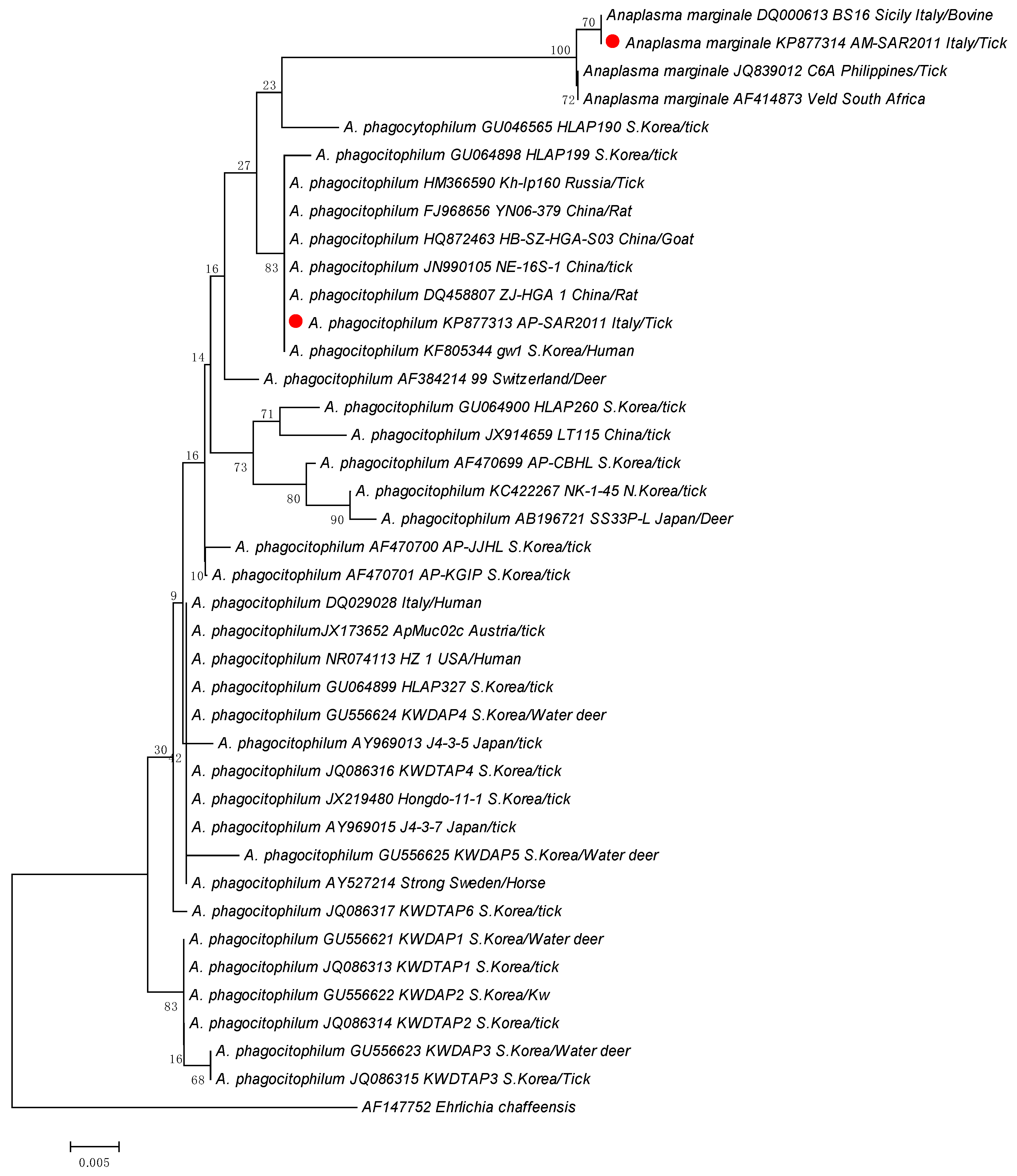
Specifically, eight sequences from ticks sampled from goats were identical to each other and to A. phagocitophilum strains from GenBank, while two sequences from two ticks collected from one marten and one cattle were identical to A. marginale strains isolated worldwide (Table 2). The two different sequence types generated in this study named AP-SAR2011 (Anaplasma phagocitophilum sequence type) and AM-SAR2011 (Anaplasma marginale sequence type) were deposited into GenBank under the accession numbers KP877313 and KP877314, respectively (Table 2). The phylogenetic analysis based on the partial 16S rRNA (Figure 2) showed that the A. phagocitophilum strain AP-SAR2011 found in this study was in the same clade as strains isolated in South Korea, Russia, and China from humans, ticks, rats, and goats. The A. marginale strain AM-SAR2011 was in the same clade as the Italian strain BS16, isolated from bovines, and close to the strains isolated in Philippines and South Africa.

4. Discussion

The rapid identification of different tick species and the bacteria they carry contributes substantially to the clinical diagnosis, treatment, and surveillance of tick-borne diseases. Current knowledge of the incidence of Anaplasma species in Sardinian ticks is limited, and obtained results, based on specific molecular typing, highlighted that Rhipicephalus and Hyalomma ticks harbored several species of Anaplasma (A. ovis, A. platys-like, A. platys and A. phagocytophilum), raising concerns regarding their potential to transmit these pathogens to humans, domestic hosts, and wildlife. The presented data show that within the five monitored sites, two different tick species were identified. Specifically, Rhipicephalus sanguineus s.l. ticks were the predominant identified species documented here, and this result was consistent with data from the previous literature [23]. Rhipicephalus species are widely distributed across the Mediterranean region, as well as in Sardinia where the Dermancentor, Haemaphysalis, and Hyalomma genera are also well represented and adapted to the ecosystem [24]. In this study, the presence of Anaplasma species was recorded in 6.4% of tested ticks. It does not mean that these ticks are competent vectors for the bacteria, as the ticks may have been infected by feeding on bacteremic animals or by cofeeding with tick vectors.
In particular, the obtained results indicated that, like previous studies in which A. marginale was detected in Rh. bursa and Rh. sanguineus s.l. ticks from Italy, Portugal, and Spain [25,26,27], Sardinian tick species are hosts for A. marginale, suggesting that both tick species could serve as potential biological vectors for A. marginale infection. To the best of our knowledge, this is the first molecular evidence of A. marginale in Rh. bursa and Rh. sanguineus s.l. ticks collected from goats and a marten in Sardinia, a region in which species of the Rhipicephalus genus are widely distributed.
Anaplasma marginale, the aetiologic agent of bovine anaplasmosis, represents one of the most important tick-borne diseases in ruminants worldwide, mainly in the tropical and subtropical regions. In the southern regions of Italy (Sicily, Puglia, Campania, and Basilicata), where bovine anaplasmosis is endemic, the presence of A. marginale in Rh. turanicus and Haemaphysalis punctata collected from cattle has been previously reported [27]. This agent infects circulating erythrocytes of domestic and wild ruminants [28]. Infected cattle serve as a reservoir of A. marginale providing a tick blood source for the efficient biological transmission of the pathogen [28]. Although approximately 20 tick species are reported as biological vectors of A. marginale worldwide [9], most of them are able to transmit A. marginale only under experimental conditions, which does not necessarily imply transmission in the field [29]. However, it has been demonstrated that the pathogen can be mechanically transmitted by blood-contaminated mouthparts of biting diptera of the Tabanus and Stomoxys genera, or via fomites [30]. In this study, ticks that tested positive for A. marginale were removed from asymptomatic goats. However, we cannot know the health status of the marten whose ticks tested positive for A. marginale since it was found dead for unknown causes. Results from a previous study conducted in northeastern Hungary highlighted the presence of Anaplasma sp. in spleen and liver samples of the European pine marten in which the zoonotic ecotype I of A. phagocytophilum has been identified [31]. More investigation on A. marginale in Rhipicephalus ticks and in domestic and wild vertebrate hosts could help to highlight the possible role of these ticks as vectors of Anaplasma species. Although phylogenetic analysis based on Anaplasma sequences obtained with 16S rRNA gene amplification revealed that this target gene can be widely used for the identification of Anaplasma species and can be considered a valuable phylogenetic tool, more discriminative genes will be used for the confirmation of these results.
This study also reports the first molecular detection of A. phagocytophilum in Rh. sanguineus and Rh. bursa ticks in Sardinia, indicating a potential role for these tick species in the epidemiology of the disease. Anaplasma phagocytophilum is the cause of granulocytic anaplasmosis in humans (HGA) [32], which severity ranges from asymptomatic infection to mild or severe febrile illness and involvement of multiple organ failure or even death [33]. Therefore, several genetic variants of this pathogen have been determined and all of these differ from each other for the different host specificity, vectors, pathogenicity, and geographical distribution [34]. In fact, all variants can infect different species as demonstrated by experimental studies in which it has been proven that A. phagocytophilum strains isolated from different matrices are not capable of infecting different hosts [4]. The analysis of this genetic variability has been made through molecular methods by using different loci such as 16s rRNA, groESL msp2, msp4, and ankA genes. Thanks to the use of one or more of these molecular markers, A. phagocytphilum has been divided into different genetic variants that can be involved in different epidemiological cycles, distribution, and host spectra [4]. Although 16s gene rRNA is the most used target gene, the phylogenetic study based only on the use of 16S rRNA could be deficient. It is related to the genetic recombination of this gene that undergoes several variations [4]. Moreover, if used alone, it may not be discriminative enough to correctly differentiate the different types of Anaplasma species, although different variants have been identified by using it as a reference point. The analysis of nucleotide sequences using the groESL gene has allowed for the identification of four different ecotypes of A. phagocytophilum in Europe, and different genetic variants adapted to the different hosts and vectors present in a specific geographical area [35]. All these ecotypes can infect both vertebrate and invertebrate hosts. In particular, the ecotype I has a wide host spectrum, being associated to multiple animal species including humans. This ecotype with zoonotic potential has the largest range in wildlife reservoirs but can also infect domestic animals. Specifically, hosts of this pathogen include cattle, sheep, goats, horses, dogs, hares, yaks, and rodents [36] and evidence of the pathogen in several mammalian and invertebrate hosts have been reported in Italy as well [37,38,39]. However, one limitation of this study was the lack of identification of A. phagocytophilum ecotypes, which is essential information for defining the zoonotic relevance. Further studies are needed to better characterize strains by analyzing more discriminative genes and to identify the main vectors implicated in the transmission of Anaplasma species in Sardinia.
Moreover, the A. phagocytophilum strain detected here was close to A. phagocytophilum strains isolated in China, Korea, and Russia. It could be related to anthropogenic activities that contribute directly or indirectly to the emergence and re-emergence of tick-borne pathogens (e.g., animal production, animal–human interfacing, and globalization). Moreover, the role of migratory birds in the spread of ticks and their role in the circulation and dissemination of pathogens in Europe cannot be ruled out. During seasonal migrations, birds that cover short, medium, or long distances within one or more distant geographical regions can carry ticks and related pathogens, introducing ticks and pathogenic species to new areas [40].
Although I. ricinus is the main vector of A. phagocytophylum in Europe [41], the zoonotic pathogen has been also associated with Rhipicephalus and Dermacentor spp. ticks from other parts of the world [42]. However, since the Mediterranean climate could be a limiting factor for Ixodes distribution and it would explain the low population of I. ricinus in Sardinia, the abundance of Rhipicephalus ticks in the Mediterranean basin prompted us to suggest that the Rhipicephalus species can serve as vectors of A. phagocytophilum and may transmit the pathogen to animal hosts. Therefore, the number of ticks was very low and they were collected from the hosts. Therefore, no conclusion can be drawn whatsoever about the circulation of the pathogens within the tick population, as every detection could be the result of infected ingested blood. These results confirmed the presence of A. phagocytophylum in Rhipicephalus ticks feeding in goats. In Sardinia, where ruminant breeding represents a zootechnical reality of primary importance, the increase in the incidence of anaplasmosis transmitted by vectors could represent a serious threat to company profitability. According to the National Italian Database 2020 (BDN) (established by the Ministry of Health at the National Surveillance Centre of the IZS in the Abruzzo and Molise Region), Sardinia has an estimated population of more than 3 million sheep and 0.3 million goats, and losses due to abortion of ruminants are estimated to be around EUR 10 million per year. Veterinarians should not overlook the presence of A. phogocytophilum in Sardinian goats and evaluation of the potential role of Anaplasma species as abortifacient agents should be also taken into account. Improving the entomological surveillance program is necessary to establish and maintain a dialogue with farmers, including listening to and addressing their concerns and sharing an adequate diagnostic and therapeutic path for the good health of farm management through innovative solutions that will reduce the economic losses in this area and ensure the efficiency of vector control interventions. Furthermore, since the risk of transmission of vector-related diseases is extended to the entire population, particular attention must be paid to professional categories who carry out their activities outdoors.

5. Conclusions

The knowledge and characterization of the diversity of Anaplasma strains circulating in the island are fundamental to design epidemiological studies and control strategies for both HGA and bovine anaplasmosis. The results of this study showed that Rh. sanguineus s.l. and Rh. bursa harbor two Anaplasma spp., of which A. marginale was not yet reported in the territory, and support the hypothesis that these tick species could act as vectors for A. marginale and A. phagocytophilum in Sardinia. Further investigation to fully understand a possible role of Rhipicephalus ticks in the A. phagocytophilum and A. marginale strains circulation are warranted. Furthermore, future studies may yield more insight into the seroprevalence of Anaplasma species in ruminants and in the dog population of Sardinia considering their potential role in the transmission of the disease to humans.

Author Contributions

Conceptualization, V.C., S.D.G. and G.M.; methodology, C.F., G.C., S.D.G., V.C., F.P. and V.S.; software, S.D.G.; validation, V.C. and S.D.G.; formal analysis, S.D.G. and V.C.; investigation, V.C.; resources, G.M.; data curation, V.C. and S.D.G.; writing—original draft preparation, V.C. and S.D.G.; writing—review and editing, V.C. and S.D.G.; visualization, V.C.; supervision, V.C. and S.D.G.; project administration, V.C.; funding acquisition, G.M. All authors have read and agreed to the published version of the manuscript.

Funding

This research received no external funding.

Institutional Review Board Statement

Not applicable.

Informed Consent Statement

Not applicable.

Data Availability Statement

Not applicable.

Conflicts of Interest

The authors declare no conflict of interest.

References

  1. Brites-Neto, J.; Duarte, K.M.; Martins, T.F. Tick-borne infections in human and animal population worldwide. Vet. World 2015, 8, 301–315. [Google Scholar] [CrossRef] [Green Version]
  2. Rikihisa, Y. Mechanisms of obligatory intracellular infection with Anaplasma phagocytophilum. Clin. Microbiol. Rev. 2011, 24, 469–489. [Google Scholar] [CrossRef] [Green Version]
  3. Dumler, J.S.; Barbet, A.F.; Bekker, C.P.; Dasch, G.A.; Palmer, G.H.; Ray, S.C.; Rikihisa, Y.; Rurangirwa, F.R. Reorganization of genera in the families Rickettsiaceae and Anaplasmataceae in the order Rickettsiales: Unification of some species of Ehrlichia with Anaplasma, Cowdria with Ehrlichia and Ehrlichia with Neorickettsia, descriptions of six new species combinations and designation of Ehrlichia equi and ‘HGE agent’ as subjective synonyms of Ehrlichia phagocytophila. Int. J. Syst. Evol. Microbiol. 2001, 51, 2145–2165. [Google Scholar]
  4. Battilani, M.; De Arcangeli, S.; Balboni, A.; Dondi, F. Genetic diversity and molecular epidemiology of Anaplasma. Infect. Genet. Evol. 2017, 49, 195–211. [Google Scholar] [CrossRef]
  5. Cabezas-Cruz, A.; Gallois, M.; Fontugne, M.; Allain, E.; Denoual, M.; Moutailler, S.; Devillers, E.; Zientara, S.; Memmi, M.; Chauvin, A.; et al. Epidemiology and genetic diversity of Anaplasma ovis in goats in Corsica, France. Parasit. Vectors 2019, 12, 3. [Google Scholar] [CrossRef] [PubMed] [Green Version]
  6. Lu, M.; Li, F.; Liao, Y.; Shen, J.J.; Xu, J.M.; Chen, Y.Z.; Li, J.H.; Holmes, E.C.; Zhang, Y.Z. Epidemiology and diversity of rickettsiales bacteria in humans and animals in Jiangsu and Jiangxi provinces, China. Sci. Rep. 2019, 9, 13176. [Google Scholar] [CrossRef] [PubMed] [Green Version]
  7. Atif, F.A. Anaplasma marginale and Anaplasma phagocytophilum: Rickettsiales pathogens of veterinary and public health significance. Parasitol. Res. 2015, 114, 3941–3957. [Google Scholar] [CrossRef]
  8. Lysholm, S.; Ådén, F.; Aspán, A.; Högberg, A.; Wensman, J.J.; Omazic, A. Presence of Anaplasma spp. and their associated antibodies in the Swedish goat population. Animals 2023, 13, 333. [Google Scholar] [CrossRef] [PubMed]
  9. Rar, V.; Golovljova, I. Anaplasma, Ehrlichia, and “Candidatus Neoehrlichia” bacteria: Pathogenicity, biodiversity, and molecular genetic characteristics, a review. Infect. Genet. Evol. 2011, 11, 1842–1861. [Google Scholar] [CrossRef] [PubMed]
  10. Lu, M.; Chen, Q.; Qin, X.; Lyu, Y.; Teng, Z.; Li, K.; Yu, L.; Jin, X.; Chang, H.; Wang, W.; et al. Anaplasma bovis Infection in Fever and Thrombocytopenia Patients-Anhui Province, China, 2021. China CDC Wkly. 2022, 4, 249–253. [Google Scholar] [CrossRef]
  11. Arraga-Alvarado, C.M.; Qurollo, B.A.; Parra, O.C.; Berrueta, M.A.; Hegarty, B.C.; Breitschwerdt, E.B. Case report: Molecular evidence of Anaplasma platys infection in two women from Venezuela. Am. J. Trop. Med. Hyg. 2014, 91, 1161–1165. [Google Scholar] [CrossRef]
  12. Chochlakis, D.; Ioannou, I.; Tselentis, Y.; Psaroulaki, A. Human anaplasmosis and Anaplasma ovis variant. Emerg. Infect. Dis. 2010, 16, 1031–1032. [Google Scholar] [CrossRef]
  13. Li, H.; Zheng, Y.C.; Ma, L.; Jia, N.; Jiang, B.G. Human infection with a novel tick-borne Anaplasma species in China, a surveillance study. Lancet Infect. Dis. 2015, 15, 663–670. [Google Scholar] [CrossRef] [PubMed]
  14. Chisu, V.; Zobba, R.; Lecis, R.; Sotgiu, F.; Masala, G.; Foxi, C.; Pisu, D.; Alberti, A. GroEL typing and phylogeny of Anaplasma species in ticks from domestic and wild vertebrates. Ticks Tick. Borne Dis. 2018, 9, 31–36. [Google Scholar] [CrossRef] [PubMed]
  15. Estrada-Peña, A.; Mihalca, A.D.; Petney, T. Ticks of Europe and North Africa. In A Guide to Species Identification; Springer International Publishing: Berlin/Heidelberg, Germany, 2017; p. 404. ISBN 978-3-319-63759-4. [Google Scholar]
  16. Kolbert, C. Detection of the agent of human granulocytic ehrlichiosis. In PCR Protocols for Emerging Infectious Diseases; Persing, D.H., Ed.; American Society for Microbiology: Washington, DC, USA, 1996; pp. 106–111. [Google Scholar]
  17. de la Fuente, J.; Torina, A.; Naranjo, V.; Caracappa, S.; Vicente, J.; Mangold, A.J.; Vicari, D.; Alongi, A.; Scimeca, S.; Kocan, K.M. Genetic diversity of Anaplasma marginale strains from cattle farms in the province of Palermo, Sicily. J. Vet. Med. 2005, 52, 226–229. [Google Scholar] [CrossRef]
  18. Larkin, M.A.; Blackshields, G.; Brown, N.P.; Chenna, R.; McGettigan, P.A.; McWilliam, H.; Valentin, F.; Wallace, I.M.; Wilm, A.; Lopez, R.; et al. Clustal W and Clustal X version 2.0. Bioinformatics 2007, 23, 2947–2948. [Google Scholar] [CrossRef] [PubMed] [Green Version]
  19. Altschul, S.F.; Gish, W.; Miller, W.; Myers, E.W.; Lipman, D.J. Basic Local Alignment Search Tool. J. Mol. Biol. 1990, 215, 403–410. [Google Scholar] [CrossRef] [PubMed]
  20. Thompson, J.D.; Higgins, D.G.; Gibson, T.J. CLUSTALW: Improving the sensitivity of progressive multiple sequence alignment through sequence weighting, positions-specific gap penalties and weight matrix choice. Nucleic Acids Res. 1994, 22, 4673–4680. [Google Scholar] [CrossRef] [Green Version]
  21. Hall, T.A. BioEdit: A user-friendly biological sequence alignment editor and analysis program for Windows 95/98/NT. Nucleic Acids Symp. Ser. 1999, 41, 95–98. [Google Scholar]
  22. Newcombe, R.G. Two-sided confidence intervals for the single proportion: Comparison of seven methods. Stat. Med. 1998, 17, 857–872. [Google Scholar] [CrossRef]
  23. Chisu, V.; Foxi, C.; Mannu, R.; Satta, G.; Masala, G. A five-year survey of tick species and identification of tick-borne bacteria in Sardinia, Italy. Ticks Tick Borne Dis. 2018, 9, 678–681. [Google Scholar] [CrossRef] [PubMed]
  24. Chisu, V.; Loi, F.; Foxi, C.; Chessa, G.; Masu, G.; Rolesu, S.; Masala, G. Coexistence of tick-Borne pathogens in ticks collected from their hosts in Sardinia: An Update. Acta Parasitol. 2020, 65, 999–1004. [Google Scholar] [PubMed]
  25. Ferrolho, J.; Antunes, S.; Santos, A.S.; Velez, R.; Padre, L.; Cabezas-Cruz, A.; Santos-Silva, M.M.; Domingos, A. Detection and phylogenetic characterization of Theileria spp. and Anaplasma marginale in Rhipicephalus bursa in Portugal. Ticks Tick. Borne Dis. 2016, 7, 443–448. [Google Scholar] [CrossRef]
  26. de la Fuente, J.; Vicente, J.; Hofle, U.; Ruiz-Fons, F.; Fernandez De Mera, I.G.; Van Den Bussche, R.A.; Kocan, K.M.; Gortazar, C. Anaplasma infection in free-ranging Iberian red deer in the region of Castilla-La Mancha, Spain. Vet. Microbiol. 2004, 100, 163–173. [Google Scholar] [CrossRef] [Green Version]
  27. de la Fuente, J.; Torina, A.; Caracappa, S.; Tumino, G.; Furla, R.; Almazan, C.; Kocan, K.M. Serologic and molecular characterization of Anaplasma species infection in farm animals and ticks from Sicily. Vet. Parasitol. 2005, 133, 357–362. [Google Scholar] [CrossRef] [PubMed]
  28. Asif, M.; Ben Said, M.; Vinueza, R.L.; Leon, R.; Ahmad, N.; Parveen, A.; Khan, A.; Ejaz, A.; Ali, M.; Khan, A.U.; et al. Seasonal investigation of Anaplasma marginale infection in Pakistani cattle reveals hematological and biochemical changes, multiple associated risk factors and msp5 gene conservation. Pathogens 2022, 11, 1261. [Google Scholar] [CrossRef]
  29. Yu, Z.; Wang, H.; Wang, T.; Sun, W.; Yang, X.; Liu, J. Tick-borne pathogens and the vector potential of ticks in China. Parasit. Vectors 2015, 8, 24. [Google Scholar] [CrossRef] [PubMed] [Green Version]
  30. Scoles, G.A.; Broce, A.B.; Lysyk, T.J.; Palmer, G.H. Relative efficiency of biological transmission of Anaplasma marginale (Rickettsiales: Anaplasmataceae) by Dermacentor andersoni (Acari: Ixodidae) compared with mechanical transmission by Stomoxys calcitrans (Diptera: Muscidae). J. Med. Entomol. 2005, 42, 668–675. [Google Scholar] [CrossRef] [Green Version]
  31. Hornok, S.; Boldogh, S.A.; Takács, N.; Sándor, A.D.; Tuska-Szalay, B. Zoonotic ecotype-I of Anaplasma phagocytophilum in sympatric wildcat, pine marten and red squirrel-Short communication. Acta Vet. Hung. 2022, 26, 215–219. [Google Scholar] [CrossRef]
  32. Ismail, N.; McBride, J.W. Tick-borne emerging infections: Ehrlichiosis and Anaplasmosis. Clin. Lab. Med. 2017, 37, 317–340. [Google Scholar] [CrossRef]
  33. Dumic, I.; Jevtic, D.; Veselinovic, M.; Nordstrom, C.W.; Jovanovic, M.; Mogulla, V.; Veselinovic, E.M.; Hudson, A.; Simeunovic, G.; Petcu, E.; et al. Human Granulocytic Anaplasmosis-A Systematic Review of Published Cases. Mi-Croorganisms 2022, 10, 1433. [Google Scholar] [CrossRef] [PubMed]
  34. Silaghi, C.; Kauffmann, M.; Passos, L.M.F.; Pfister, K.; Zweygarth, E. Isolation, propagation and preliminary characterization of Anaplasma phagocytophilum from roe deer (Capreolus capreolus) in the tick cell line IDE8. Ticks Tick. Borne Dis. 2011, 2, 204–208. [Google Scholar] [CrossRef] [PubMed]
  35. Jahfari, S.; Coipan, E.C.; Fonville, M.; van Leeuwen, A.D.; Hengeveld, P.; Heylen, D.; Heyman, P.; van Maanen, C.; Butler, C.M.; Földvári, G.; et al. Circulation of four Anaplasma phagocytophilum ecotypes in Europe. Parasit. Vectors 2014, 7, 365. [Google Scholar] [CrossRef] [PubMed] [Green Version]
  36. Peng, Y.; Wang, K.; Zhao, S.; Yan, Y.; Wang, H.; Jing, J.; Jian, F.; Wang, R.; Zhang, L.; Ning, C. Detection and phylogenetic characterization of Anaplasma capra: An emerging pathogen in sheep and goats in China. Front. Cell. Infect. Microbiol. 2018, 8, 283. [Google Scholar] [CrossRef] [PubMed] [Green Version]
  37. Lillini, E.; Macrì, G.; Proietti, G.; Scarpulla, M. New findings on anaplasmosis caused by infection with Anaplasma phagocytophilum. Ann. N. Y. Acad. Sci. 2006, 1081, 360–370. [Google Scholar] [CrossRef] [PubMed]
  38. Maioli, G.; Pistone, D.; Bonilauri, P.; Pajoro, M.; Barbieri, I.; Mulatto, P.; Vicari, N.; Dottori, M. Etiological [corrected] agents of rickettsiosis and anaplasmosis in ticks collected in Emilia-Romagna region (Italy) during 2008 and 2009. Exp. Appl. Acarol. 2012, 57, 199–208. [Google Scholar] [CrossRef]
  39. Carpi, G.; Bertolotti, L.; Pecchioli, E.; Cagnacci, F.; Rizzoli, A. Anaplasma phagocytophilum groEL gene heterogeneity in Ixodes ricinus larvae feeding on roe deer in Northeastern Italy. Vector Borne Zoonotic Dis. 2009, 9, 179–184. [Google Scholar] [CrossRef] [Green Version]
  40. Buczek, A.M.; Buczek, W.; Buczek, A.; Bartosik, K. The Potential Role of Migratory Birds in the Rapid Spread of Ticks and Tick-Borne Pathogens in the Changing Climatic and Environmental Conditions in Europe. Int. J. Environ. Res. Public Health 2020, 17, 2117. [Google Scholar] [CrossRef] [Green Version]
  41. Stuen, S.; Granquist, E.G.; Silaghi, C. Anaplasma phagocytophilum-a widespread multi-host pathogen with highly adaptive strategies. Cell Infect. Microbiol. 2013, 3, 1–33. [Google Scholar] [CrossRef] [Green Version]
  42. Chastagner, A.; Bailly, X.; Leblond, A.; Pradier, S.; Vourc’h, G. Single genotype of Anaplasma phagocytophilum identified from ticks, Camargue, France. Emerg. Infect. Dis. 2013, 19, 825–827. [Google Scholar] [CrossRef]
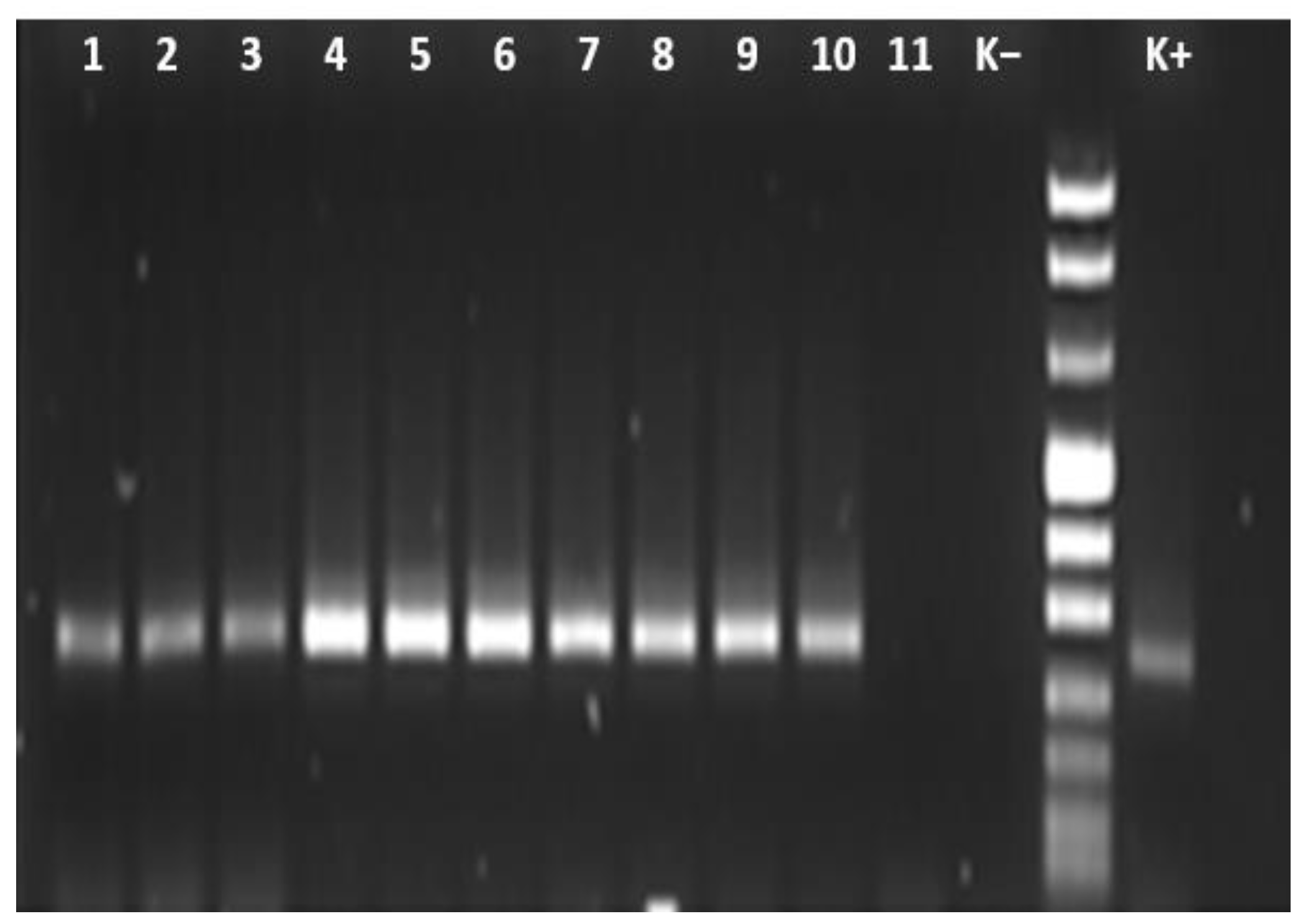
Figure 1. Agarose gel electrophoresis (1.5%) of PCR amplification products of Anaplasma spp. with 16S rRNA primer. Lanes 1–11: numbers of the strains; Lane 11: K(−)-negative control; Lane 12: DNA Size Marker (Marker VIII); Lane 13: K(+)-positive control.
Animals 13 01332 g001
Figure 2. Phylogenetic tree based on 373-bp 16S rRNA gene of A. phagocytophilum and A. marginale strains collected in this study (red dots) compared with Anaplasma species obtained from GenBank database. The tree was constructed using the software MEGA6 and the neighbor-joining method with 1000 bootstrap re-samplings. Ehrlichia chaffeensis was used as outgroup. Country, hosts, and GenBank accession number are also indicated. Scale bar: number of base substitutions per site.
Animals 13 01332 g002
Table 1. Species, sex, hosts, and geographic areas from which ticks were collected.
N. of TicksSexHost (n.)Collection Sites
Ogliastra Province
Rh. sanguineus s.l.9740 males
57 females
Goat (18)Talana
Rh. bursa136 males
7 females
Goat (1)Jerzu
11 maleGoat (1)Talana
Sassari Province
Rh. sanguineus s.l.2319 males
4 females
Marten (1)Bono
Rh. bursa221 male
21 females
Cattle (1)Villanova Monteleone
Table 2. Detection and identification by PCR and sequencing of A. phagocitophilum and A. marginale in ticks collected in Sardinia, Italy.
Tick SpeciesN. of Positive Ticks and Relative SexHostAnaplasma IdentificationStrainGenBank Accession Number
Rh. sanguineus s.l.2♂—2♀GoatA. phagocitophilumAP-SAR2011KP877313
1♂MartenA. marginaleAM-SAR2011KP877314
Rh. Bursa4♀GoatA. phagocitophilumAP-SAR2011KP877313
1♀CattleA. marginaleAM-SAR2011KP877314
Disclaimer/Publisher’s Note: The statements, opinions and data contained in all publications are solely those of the individual author(s) and contributor(s) and not of MDPI and/or the editor(s). MDPI and/or the editor(s) disclaim responsibility for any injury to people or property resulting from any ideas, methods, instructions or products referred to in the content.