Review

Inhibition of Cell Apoptosis by Apicomplexan Protozoa–Host Interaction in the Early Stage of Infection

1
Key Laboratory of Applied Technology on Green-Eco-Healthy Animal Husbandry of Zhejiang Province, Provincial Engineering Research Center for Animal Health Diagnostics & Advanced Technology, Zhejiang International Science and Technology Cooperation Base for Veterinary Medicine and Health Management, China Australia Joint Laboratory for Animal Health Big Data Analytics, College of Animal Science and Technology & College of Veterinary Medicine, Zhejiang A & F University, Hangzhou 311300, China
2
Gansu Polytechnic College of Animal Husbandry and Engineering, Wuwei 733006, China
*
Authors to whom correspondence should be addressed.
These authors contributed equally to this work.
Animals 2023, 13(24), 3817; https://doi.org/10.3390/ani13243817
Submission received: 4 October 2023 / Revised: 30 November 2023 / Accepted: 5 December 2023 / Published: 11 December 2023

Abstract

:

Simple Summary

Protozoa that parasitize within host cells can avoid direct damage from the host immune system. In this review, we have revealed some key factors and signaling pathways that inhibit host cell apoptosis, regulate cell apoptosis signal transduction, and interfere with the molecular mechanisms of cell apoptosis. These findings provide important clues to further understand the infection mechanism and pathogenesis of Apicomplexan protozoa and a theoretical basis for the development of new therapeutic strategies and preventive measures.

Abstract

Apicomplexan protozoa, which are a group of specialized intracellular parasitic protozoa, infect humans and other animals and cause a variety of diseases. The lack of research on the interaction mechanism between Apicomplexan protozoa and their hosts is a key factor restricting the development of new drugs and vaccines. In the early stages of infection, cell apoptosis is inhibited by Apicomplexan protozoa through their interaction with the host cells; thereby, the survival and reproduction of Apicomplexan protozoa in host cells is promoted. In this review, the key virulence proteins and pathways are introduced regarding the inhibition of cell apoptosis by the interaction between the protozoa and their host during the early stage of Apicomplexan protozoa infection. It provides a theoretical basis for the development of drugs or vaccines for protozoal diseases.

1. Introduction

Apicomplexa protozoa, including Toxoplasma gondii (T. gondii), Plasmodium, Cryptosporidium, Neospora caninum (N. caninum), Eimeria, etc., are obligatory intracellular parasites, often zoonotic pathogens, posing a considerable threat to economic development and public health [1,2]. Although apicomplexan parasites can evade direct attacks by the host immune system, infected cells still have the ability to restrain their proliferation and development through apoptosis [3]. The inhibition of apoptosis is, therefore, one of the main mechanisms by which the parasites ensure their growth, development, and reproduction [4]. It has been reported that T. gondii suppresses the apoptosis of T cells and macrophages during infection and, thereby, achieves persistent infection [5]; the Theileria and Cryptosporidium species can also inhibit host cell apoptosis [5]. Similarly, the infection of Eimeria acervulina (E. acervulina) merozoites inhibits the apoptosis of bovine kidney cells [6], and second-generation merozoites of both Eimeria necatrix (E. necatrix) and Eimeria tenella (E. tenella) can inhibit host cell apoptosis by activating the NF-κB signaling pathway [7]. Due to the difference of these parasites in their mechanism of host cell entry and final intracellular localization, the specific mechanism of Apicomplexan parasites’ inhibition of host cell apoptosis remains to be further explored.

2. Apoptosis

Apoptosis is an important host self-defense mechanism that plays key regulatory roles in maintaining normal tissue development, removing damaged and aged cells, and maintaining the functions of the immune system [8]. Apoptosis also plays an important role in the infectious process of pathogens. As a host defense mechanism, it is of great significance, especially in resisting infections by protozoa.
The regulation of cell apoptosis involves multiple signal transduction pathways, mainly the endogenous pathway (mitochondria-mediated pathway) and the extrinsic pathway (death receptor-mediated pathway) [9]. The former is mainly affected by intracellular damage signals and stress factors, while the latter is activated through the binding of extracellular death receptor ligands. The endogenous pathway is the most common and involves the mitochondria [7]. It mainly includes the following steps: firstly, external or damage stimuli induce changes in the expression level of Bcl-2 family proteins inside the mitochondria, leading to the increased permeability of the mitochondrial outer membrane [7] and, thus, to the release of mitochondrial proteins such as cytochrome C into the cytoplasm; secondly, cytochrome C in the cytoplasm binds to an apoptotic protease-activating factor (Apaf-1) to form a complex called the apoptosome, which activates procaspase-9 to be caspase-9 [10,11]; thirdly, the activated caspase-9 further activates other caspases family members (caspase-7, caspase-6, and caspase-3), initiating a cascade in the cell which cleaves specific intracellular proteins (PARP (PolyADP-Ribose Polymerase), GDID4, DFF-45 (DNA fragmentation factor-45), lamin A, keratin 18), ultimately leading to the death of the cell [11]; finally, the condensation of chromatin and DNA fragmentation occur in the nucleus [12]. The exogenous pathway is a process of cell apoptosis mediated by transmembrane receptors such as tumor necrosis factor-α (TNF-α) and the Fas ligand [13]. When there are corresponding ligand molecules outside the cell, the receptor proteins on the cell membrane are recruited together and bind to the ligand, forming a death-inducing signal complex, catalyzing and activating procaspase-8, and, finally, leading to the executive phase when cell apoptosis is activated [14,15]. Caspase activation triggers a series of apoptotic events, including cytoskeleton reorganization, cell membrane changes, enzymatic hydrolysis, and DNA degradation. Eventually, the cells fragment into apoptotic bodies that are taken up and phagocytosed by surrounding macrophages to complete the process of cell clearance [16].

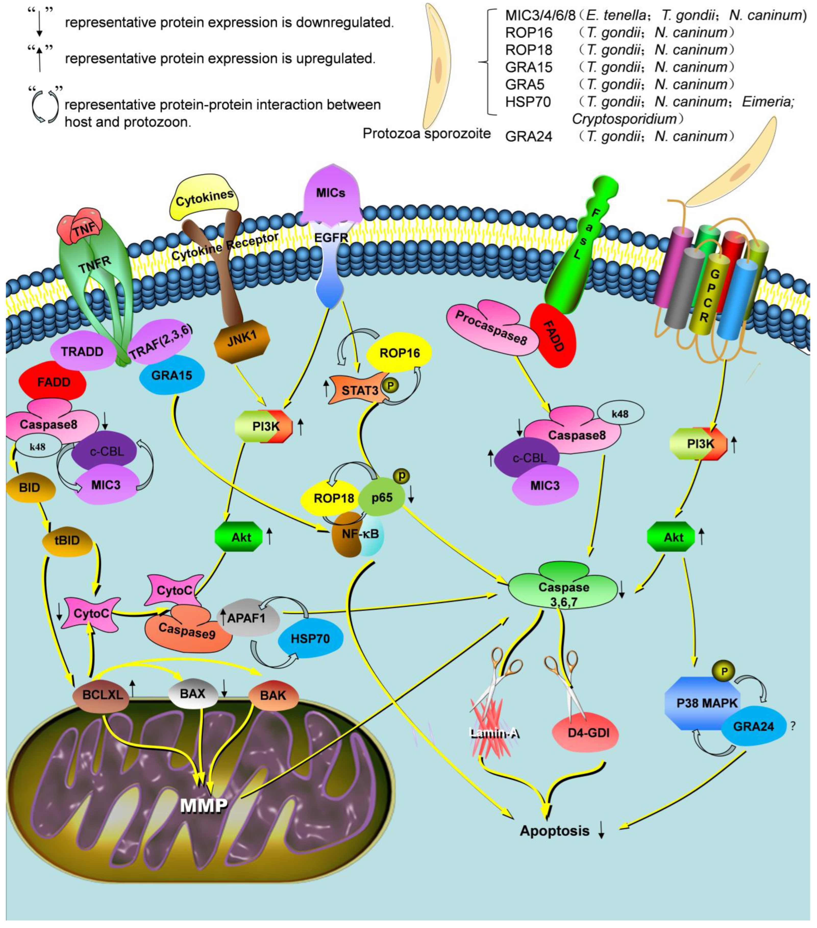
3. Apicomplexan Protozoa Inhibit Cell Apoptosis in the Early Stage of Infection

Plasmodium inhibits host cell apoptosis by Caspase activity, the PI3K signaling pathway, and the regulation of apoptosis-related proteins’ expression [17]. Plasmodium sporozoites pass through and damage a number of hepatocytes before they enter into the last one. This damage leads to the release of HGF, which is the initial reaction protecting the final host cell from spontaneous apoptosis. Since HGF/MET signaling is capable of protecting cells from apoptosis using both the PI3-kinase/Akt and MAPK pathways, the inhibition of HGF/MET signaling induces a specific increase in the apoptosis of infected cells and leads to a great reduction in infection; to counteract this, the infected hepatocytes express the HGF receptor c-met, which ensures the initial survival of the cells [18,19]. Furthermore, apoptosis is induced in mammalian cells in vitro using peroxide treatment or serum, but Plasmodium can inhibit cell apoptosis in vitro with peroxide treatment or serum deprivation and in vivo using TNF-α. This parasite-dependent inhibition of apoptosis is clearly detectable in vitro 24 h p.i. but is more pronounced 48 h p.i. At the latter time point, infected cells have been found to resist prolonged peroxide exposure [20].
This study suggests that C. parvum inhibits apoptosis by activating NF-κB signaling pathway during infection [21]. Interestingly, C. parvum-infected cells exhibit some signs of apoptosis after invasion during the early stages of infection [22]. Together, these observations suggest that C. parvum infection may interfere with the temporal progression of cell apoptosis and delay it sufficiently to support parasite replication.
Several strategies are also adopted to inhibit cell apoptosis for T. gondii, such as the regulation of the NF-κB signaling pathway and mitochondria-mediated apoptotic pathway. As an important anti-apoptotic factor, NF-κB is involved in the regulation of cell survival. T. gondii can inhibit cell apoptosis by activating NF-κB signaling pathway [11,23]. During the early infection of T. gondii, the inhibition of cell apoptosis also results from decreasing the expression of pro-apoptotic proteins and increasing the expression of anti-apoptotic proteins, thereby preventing mitochondria-mediated cell apoptosis [7].
In the early stage of infection of Eimeria species, the opening of mitochondrial permeability transition pore (MPTP) and the decrease in transmembrane potential were activated [24]; the Akt and ERK proteins, key factors of the epidermal growth factor receptor (EGFR) signaling pathway, were upregulated [25]. All these events synergistically contribute to the occurrence of anti-apoptosis.
Apicomplexan protozoa employ different strategies to inhibit apoptosis in order to protect themselves from host immune attacks and achieve survival in host cells. Therefore, these inhibitory mechanisms are important for further understanding the underlying infection process of protozoa.

4. Virulence Factors Associated with Apicomplexan Protozoa Inhibit Host Cell Apoptosis

4.1. Microneme Proteins

Micronemes are the special organelle located at the anterior end of apicomplexan. It secretes a variety of microneme proteins (MICs) and plays a key role in the life cycle of parasites, including invasion, immune evasion, and localization in host cells [26,27]. E. tenella MIC4 protein contains an EGF motif, which can interact with EGFR and activate the EGFR-Akt signaling pathway, finally inhibiting host cell apoptosis [28]. Similarly, E. acervulina MIC 3 protein can inhibit apoptosis of the chicken duodenal epithelial cell by interacting with the casitas B-lineage lymphoma (CBL) protein. The over-expression of E. acervulina MIC 3 protein increased the level of CBL expression, which mediated the ubiquitination of caspase 8 and promoted its degradation. The decrease in caspase 8 expression level finally led to the inhibition of cell apoptosis [6]. It has been shown that T. gondii can activate the EGFR-Akt signaling pathway through the EGF domain-containing MIC3, MIC6, and MIC8, thereby inhibiting host cell apoptosis [29,30,31].

4.2. Rhoptry Proteins

Rhoptry is a unique secretory organelle in protozoa. It has a rod-like outer shape and secretes proteins when the apicomplexan protozoa, such as T. gondii, Eimera, and Cryptosporidium species, invade host cells [1]. According to the localization, the secreted proteins are divided into rhoptry neck proteins (RONs) and rhoptry bulb proteins (ROPs), which are closely related to the invasion and inhibition of cell apoptosis, formation of the parasitophorous vacuole (PV), and formation of the moving junction (MJ) [32].
Rhoptry proteins inhibit apoptosis by affecting the Janus Kinase-signal transducer and activator of transcription (JKT-STAT), NF-κB, and MAPK signaling pathways in host cells [33]. For instance, ROP16 is a soluble rhoptry protein secreted by T. gondii. It was released in the host cell cytoplasm and then translocated into the cell nucleus through its nuclear localization structure (NLS), thereby affecting the expression of host genes [34]. ROP16 has tyrosine kinase activity and can activate the STAT3 and STAT6 signaling pathways by phosphorylating both Tyr705 and Tyr641 of the STAT3 and STAT6 complexes, thereby inhibiting cell apoptosis. In agreement with this idea, ROP16 promotes the synthesis of arginase 1, a major downstream substrate of the STAT6 signaling pathway in host cells [35]. In addition, another rhoptry protein ROP18 is shown to phosphorylate P65 and promote the degradation of p65 by the ubiquitin-dependent degradation pathway, suppress the NF-κB activation, and finally inhibit the production of proinflammatory cytokines and the apoptosis of host cells [36].

4.3. Dense Granule Proteins

Dense granule proteins (GRAs) have a diameter of about 200 nm and are distributed throughout the entire parasite. Approximately 70 GRAs have been reported so far, mainly including the typical GRAs, the inhibitors of STAT transcription (IST), mitochondrial association factors (MAF), and c-Myc regulation (MYR). GRAs related to apoptosis were observed in Toxoplasma-infected cells, such as GRA24 [37,38]. T. gondii GRA24 can induce higher activation of the subunit p38-alpha MAPK.
GRAs can inhibit apoptosis to achieve immune evasion and long-term parasitism [39]. It was shown that IST (a dense granule protein) secreted by T. gondii crossed the parasitophorous vacuole membrane (PVM) and accumulated in the nucleus to block the type I interferon response and promote the infection [40]. GRA5 can inhibit apoptosis of the infected cells to protect the parasites [41]. T. gondii GRA5 deletion mutant protects hosts against T. gondii infection, but the mechanism is unknown. Similarly, GRA24 is secreted by T. gondii into host cell cytoplasm and binds to p38 MAPK, activating GPCR/ PI3K/AKT signaling pathways, which induces p38 phosphorylation. The inhibitory effect of GRA24 on cell apoptosis needs to be further researched [42,43]. GRA15 interacts with TNF receptor-associated factors (TRAFs), which are adaptor proteins functioning upstream of the NF-Κb transcription factor. The deletion of TRAF-binding sites in GRA15 greatly reduces its ability to activate the NF-κB pathway, and TRAF2 knockout cells have impaired GRA15-mediated NF-κB activation [44]. Additionally, previous studies have reported that T. gondii also exploits heterotrimeric G protein-coupled receptor (GPCR)-mediated signaling to activate phosphoinositide 3-kinases (PI3Ks), leading to the phosphorylation of PI3K and MAPK pathways and inhibition of apoptosis [45].

4.4. Heat Shock Proteins

This is an evolutionarily conserved family of heat shock proteins. Based on molecular weight and sequence homology, heat shock proteins (HSPs) are classified into different families, including small HSPs, HSP40, HSP60, HSP70, HSP90, and large HSPs, and each family has a different intracellular distribution and functions [46]. HSPs have a variety of functions in organisms, especially in responding to external stresses, such as high temperature, oxidative stress, radiation, and infection [47]. HSPs also play a crucial role in preventing protein aggregation and promoting proteolysis by catalyzing the refolded damaged or denatured proteins [47]. In addition, they participate in the process of inhibiting cell apoptosis [48]. HSP70 is the main anti-apoptotic protein in T. gondii, which inhibits cell apoptosis via cytochrome c release, anti-apoptotic protein expression, and caspase inhibition [49]. The study demonstrated that the exertion of HSP70 anti-apoptotic effects is directly associated with the pro-apoptotic factors, APAF-1 and Apaf-1. HSP70 can bind to AIF (apoptosis-inducing factor) and apoptosis protease activate factor 1 (APAF-1), overexpress anti-apoptotic Bcl-2, inhibit AIF translocation and cytochrome C redistribution, and then inhibit cell mitochondrial apoptosis [50].
The above research has tried to describe the pathways that are controlled by the parasite to permit its survival inside the host. The main factors participating in the inhibition of cell apoptosis induced by protozoa are members of the MIC, ROP, HSP, and GRA family (Figure 1).

5. Apicomplexan Protozoa Infection Effects on Key Apoptotic Proteins and Pathways in the Host

5.1. Cysteine Protease Family

As a key component of the apoptosis signaling pathway, caspases are one class of cysteine proteases that hydrolyze the ASP-X peptide bond of substrate proteins [8]. They are divided into two groups: apoptotic initiating enzymes, such as Caspase-8 and Caspase-10, and effector enzymes, such as Caspase-3 and Caspase-7. The former initiates a cascade of reactions by activating effector enzymes, which eventually lead to changes in the morphological and biochemical characteristics of cells. However, the latter directly acts on a variety of intracellular substrates, such as nucleases and structural proteins, destroying the integrity and function of cells. Even if the apoptotic pathways are different, the Caspase cascade is a common event that is located downstream of the apoptotic signal transduction [51].

5.2. Bcl-2 Protein Family

Bcl-2 family is a group of proteins that regulate apoptosis and include members that inhibit apoptosis, such as Bcl-2 and Bcl-xL, and members that promote apoptosis, such as Bax and Bak [52]. These proteins are involved in apoptosis by regulating mitochondrial membrane permeability. Therefore, the inhibitors of Bax and Bak can prevent the release of cell death signaling molecules from mitochondria, thereby suppressing apoptosis [52].
Bcl-2 and Bax genes are the upstream factors of the apoptosis pathway. Under normal conditions, both form heterodimers to keep cells in a steady state. When they are stimulated by apoptotic signals, Bax is dissociated from heterodimers and forms the homodimer to induce cell apoptosis. Therefore, the ratio of Bcl-2/Bax in cells is an important factor in determining whether the cells will execute apoptosis after a certain stimulus [50]. The study demonstrated that the extract of Ashwagandha showed that the increase in the Bcl-2/Bax ratio could inhibit the cell apoptosis of splenic lymphocytes of treated mice. Similarly, Eimeria merozoite infection inhibited the host cell apoptosis by regulating the expression of Bcl-2 and Bax [53]. Compared with Bcl-2, Bcl-xl exhibits a stronger anti-apoptotic effect by preventing the release of cytochrome C and other apoptosis-inducing factors from mitochondria. On the contrary, Bid promotes apoptosis by increasing the release of those mitochondrion-derived apoptosis-inducing factors [45]. During Eimeria infection, the inhibition of host cell apoptosis was positively correlated with the ratio of Bcl-xl/Bid [53]. These results suggest that the Eimeria species inhibit apoptosis by preventing the dissociation of both Bcl-xl/Bid and Bcl-2/Bax heterodimers in host cells during infection.
The Bcl-2 gene family, Survivin genes, and microRNAs are involved in regulating host epithelial cell apoptosis induced by C. parvum infection. C. parvum upregulates the expression of anti-apoptotic genes and downregulates the expression of pro-apoptotic genes in the early stage (6–12 h) of infection with HCT-8 cells, while it upregulates the expression of pro-apoptotic genes and downregulates the expression of anti-apoptotic genes in the later stage (24–72 h) of infection. After silencing the Bcl-2 gene, cell apoptosis increases, indicating that the Bcl-2 gene family regulates host epithelial cell apoptosis induced by C. parvum infection [22]. A recent study suggests that in vitro infection of HCT-8 cells with C. parvum can induce the upregulation of the expression of the anti-apoptotic protein Survivin, thereby inhibiting the activities of caspase-3 and caspase-7, and finally weaken cell apoptosis. However, after silencing Survivin expression, cell apoptosis significantly increases and the number of parasites decreases; this indicates that the expression of Survivin can be regulated to inhibit cell apoptosis and promote the development of the parasite itself during C. parvum infection [23].

5.3. Cytochrome-C

Cytochrome-C is an essential component of the cellular respiratory chain and it participates in electron transfer [54]. Cytochrome-C causes cell apoptosis mainly in a Caspases-dependent manner. When cells receive apoptotic signals that cause mitochondrial dysfunction, Cytochrome-C is released from the mitochondrial membrane into the cytoplasm. Subsequently, Cytochrome-C first binds to Apaf-1 and causes conformational changes in Apaf-1, and then seven Apaf-1 molecules aggregate to form an apoptotic complex with the participation of ATP or dATP. This complex activates downstream apoptotic pathways, ultimately leading to cell death [55,56]. It has been found that the level of Cytochrome-C after T. gondii infection is significantly reduced, indicating that T. gondii infection inhibits cell apoptosis via the regulation of Cytochrome-C release (Table 1) [57].
Previous studies have shown that T. gondii RH strain tachyzoites in MEFs (Mouse Embryonic Fibroblasts) and T. gondii NTE strain tachyzoites in human promyelocytic leukemia cells (HL-60) can inhibit the release of mitochondrial cytochrome C by caspases 3 and 9. Further, after T. gondii infects human monocyte-infected cells (TH1), it can upregulate the anti-apoptotic protein Bcl-2 expression and downregulate the expression of pro-apoptotic protein Bax, thereby inhibiting mitochondrial permeability transition pore and blocking the release of cytochrome c from mitochondria.
Cryptosporidium can promote host cell apoptosis by releasing cytochrome C in the later stage of infection. Studies have evaluated the apoptotic changes in Cryptosporidium-infected cells in immunoactive (IC) and immunosuppressive (IS) mice either alone or loaded with silver nanoparticles (AgNPs) and NTZ. The treatment of NTZ loaded with AgNP at different doses showed the highest reduction in egg sac shedding and significant improvement in histopathological changes. Moreover, the levels of cytochrome C and caspase-3 were significantly reduced in IC and IS mice treated with NTZ loaded with AgNP, indicating that NTZ loaded with AgNP can inhibit cell apoptosis infected with Cryptosporidium by blocking the release of cytochrome C, which is a potential target for the treatment of neosporidiosis.

5.4. NF-κB Signaling Pathway

There are five members of the NF-κB family: P50 (NF-κB1), P52 (NF-κB2), P65 (RelA), RelB, and C-Rel [58]. NF-κB is an important transcription factor involved in the regulation of cell survival and apoptotic processes. NF-κB is a dimer of two subunits p50 and p65 [58] and, when not activated, it is bound to IκB in the cytoplasm. In response to certain stimuli, IκB is phosphorylated and degraded, leading to NF-κB transportation into the nucleus and thus the regulation of gene transcription [59].
It has been shown that apicomplexan parasites can inhibit host cell apoptosis by activating the NF-κB signaling pathway [60]. The activation of NF-κB can promote the expression of apoptosis-inhibiting factors and inhibit the production of apoptosis-related factors, thereby inhibiting cell apoptosis. Recently, all the NF-κB, phosphorylated NF-κB (p-IκBα), and Bcl-xL were demonstrated to be highly expressed during E. pestis infection [61], suggesting the activation of NF-κB that protects the infected cells from apoptosis and allows the maturation of the second generation of merozoites [7]. Consistently, when the p65 subunit was deleted, the host cells lost the inhibition of apoptosis during infection [62].
Cells infected with C. parvum activate the NF-κB signaling pathway and are accompanied by the secretion of interleukin-8 (IF-8). Inhibitors MG-132 and SN50 can inhibit the binding of NF-κB to DNA and increase the expression level of IF-8, but significantly promote the H69 cell apoptosis level after C. parvum infection. The results indicate that the cell apoptosis rate after C. parvum infection is influenced by NF-κB. The ability of C. parvum to induce cell apoptosis increases due to the inactivation of the NF-κB signaling pathway. HCT-8 cells apoptosis rates with C. parvum infection in vitro after adding pro-apoptosis drugs were tested. C. parvum significantly reduces the cell apoptosis rate induced by these drugs, indicating that C. parvum can inhibit the cell apoptosis induced by pro-apoptotic factors, which is beneficial for its own proliferation and development.

5.5. Phosphatidylinositol 3-Kinase-Akt Signaling Pathway

Phosphatidylinositol 3-kinase (PI3K) is a large family of lipid kinases, which can be divided into classes I, II, and III according to their structure [63]. Of them, class I is a heterodimer composed of the catalytic subunit (P110) and regulatory subunit (P85). Under normal conditions, the p85-p110 complex is ubiquitously localized in the cytoplasm. After P85 and P110 are activated, they combine to generate a heterodimer, which activates the Akt signaling pathway. Then, the activated Akt inhibits the expression of apoptotic genes and enhances the expression of anti-apoptotic genes via the direct phosphorylation of Forkhead family transcription factors or apoptosis cascade regulatory proteins, or via the upregulation of NF-κB [64]. In macrophages infected with T. gondii, PI3K was demonstrated to be activated by GiPCR signal, leading to PI3K/AKT and MEK1/2 phosphorylation and MEK1/2-mediated ERK1/2 activation. Moreover, T. gondii inhibited crisscrotidyline-induced apoptosis, while PI3K inhibitors LY294002 blocked the ability of T. gondii to inhibit apoptosis, indicating apoptosis inhibition via the PI3K-Akt signaling pathway [55].
After being bitten by infected mosquitoes, the traversal of host hepatocytes by Plasmodium sporozoites (transmission type) leads to the release of hepatocyte growth factor (HGF) and ultimately promotes the invasion of hepatocytes where the parasite will reside, differentiate, and reproduce [65]. PI3K participates in inhibiting liver cell apoptosis in the early stages of Plasmodium parasite infection. HGF/Met signaling inhibits cell apoptosis through PI3K signaling [66]. This inhibitory effect enhances the process of Plasmodium infecting cells, and the use of PI3K inhibitors can lead to cell apoptosis after infection. It has been reported that the overexpression of glutamylcysteine can inhibit TNF-α-induced cell apoptosis and regulate the activation of NF-κB during Plasmodium parasite inhibiting cell apoptosis. It is interesting that the inhibitory effect of infected liver cell apoptosis increases with infection time and shows resistance to cell apoptosis both in vivo and in vitro [67]. The inhibition of cell apoptosis by Plasmodium not only occurs during the replication stage of hepatocytes but also precise control over host cell apoptosis in the final stage of hepatocyte development. It was found that before erythrocyte infection, an increasing number of hepatocytes were filled with merozoites. These isolated hepatocytes exhibit various characteristics of cell apoptosis, including cytoplasmic cytochrome lysis, mitochondrial membrane potential loss, and nuclear coagulation, but lack phosphatidylserine in the outer layer of the plasma membrane. The results show that in the early stages of hepatocytes infection, the inhibition of cell apoptosis is dependent on the induction of non-apoptotic and non-necrotic cell death by merozoites, which promotes the formation of vesicles transporting merozoites, where their release leads to erythrocyte infection.

6. Conclusions

One of the main mechanisms by which Apicomplexan protozoa inhibit cell apoptosis is their ability to delay host cell damage. In the early stages of infection, Apicomplexan protozoa inhibit host cell apoptosis in order to ensure their own growth, development, and reproduction within the cell. The regulation mechanism of apoptosis is inhibited by the interaction between host cells and parasites, including main virulence factors associated with Apicomplexan protozoa that inhibit host cell apoptosis.
Through the study of molecular mechanisms, we have revealed some key factors and signaling pathways of apoptosis inhibition in host cells, which regulate apoptosis signal transduction, and molecular mechanisms that interfere with apoptosis. These findings provide important clues for further understanding the infection mechanism and pathogenesis of Apicomplexan protozoa and a theoretical basis for the development of new therapeutic strategies and preventive measures.
Although we have made some important progress, there are still many questions that need to be further explored. Future research can focus on the molecular mechanisms of apoptosis regulation, the detailed mechanisms of interactions between Apicomplexan protozoa and host cells, and the therapeutic strategies for parasite infections. In summary, studies on the inhibition of apoptosis by the interactions between parasite and host in the early stage of parasite infection provide important scientific evidence for us to understand the mechanism of parasitic infections, develop effective treatments, and prevent and control infectious diseases. In future studies, we will be able to better understand and respond to the challenges posed by Apicomplexan protozoa infections and contribute to human and animal health.

Author Contributions

Writing—original draft, L.L. and X.H.; Formal analysis, Q.S.; Investigation, X.H.; Revision, Q.S., W.L., Y.P. and P.W.; Visualization & Review: Y.C.; Writing—review & editing, Y.P., X.Y. and P.W. All authors have read and agreed to the published version of the manuscript.

Funding

Project support was provided by the Zhejiang Provincial Natural Science Foundation of China (grant No. LY22C180001), ZAFU Talents Starting Program (grant No. 2018FR045 and 203402003002), Research and Training Program for Students of Zhejiang A&F University (grant No. 2020KX0161; S202210341063), NHC Key Laboratory of Echinococcosis Prevention and Control (grant No. 2022WZK1008), and College Students’ Innovation and Entrepreneurship Training Program (grant No. DKDY2022009; DKDY2022005).

Institutional Review Board Statement

Not applicable.

Informed Consent Statement

Not applicable.

Data Availability Statement

No new data were created or analyzed in this study. Data sharing is not applicable to this article.

Conflicts of Interest

The authors declare no conflict of interest.

References

  1. Sparvoli, D.; Lebrun, M. Unraveling the Elusive Rhoptry Exocytic Mechanism of Apicomplexa. Trends Parasitol. 2021, 37, 622–637. [Google Scholar] [CrossRef] [PubMed]
  2. Singh, R.S.; Walia, A.K.; Kanwar, J.R. Protozoa lectins and their role in host-pathogen interactions. Biotechnol. Adv. 2016, 34, 1018–1029. [Google Scholar] [CrossRef]
  3. Guillermo, L.V.; Pereira, W.F.; De Meis, J.; Ribeiro-Gomes, F.L.; Silva, E.M.; Kroll-Palhares, K.; Takiya, C.M.; Lopes, M.F. Targeting caspases in intracellular protozoan infections. Immunopharmacol. Immunotoxicol. 2009, 31, 159–173. [Google Scholar] [CrossRef]
  4. Gervais, O.; Renault, T.; Arzul, I. Molecular and cellular characterization of apoptosis in flat oyster a key mechanisms at the heart of host-parasite interactions. Sci. Rep. 2018, 8, 12494. [Google Scholar] [CrossRef] [PubMed]
  5. Sinai, A.P.; Payne, T.M.; Carmen, J.C.; Hardi, L.; Watson, S.J.; Molestina, R.E. Mechanisms underlying the manipulation of host apoptotic pathways by Toxoplasma gondii. Int. J. Parasitol. 2004, 34, 381–391. [Google Scholar] [CrossRef] [PubMed]
  6. Wang, P.; Jia, Y.; Han, Y.; Wang, W.; Zhu, Y.; Xu, J.; Guan, C.; Ying, J.; Deng, S.; Wang, J.; et al. Eimeria acervulina Microneme Protein 3 Inhibits Apoptosis of the Chicken Duodenal Epithelial Cell by Targeting the Casitas B-Lineage Lymphoma Protein. Front. Vet. Sci. 2021, 8, 636809. [Google Scholar] [CrossRef]
  7. del Cacho, E.; Gallego, M.; López-Bernad, F.; Quílez, J.; Sánchez-Acedo, C. Expression of anti-apoptotic factors in cells parasitized by second-generation schizonts of Eimeria tenella and Eimeria necatrix. Vet. Parasitol. 2004, 125, 287–300. [Google Scholar] [CrossRef]
  8. DosReis, G.A.; Barcinski, M.A. Apoptosis and parasitism: From the parasite to the host immune response. Adv. Parasitol. 2001, 49, 133–161. [Google Scholar] [CrossRef]
  9. Sorice, M. Crosstalk of Autophagy and Apoptosis. Cells 2022, 11, 1479. [Google Scholar] [CrossRef]
  10. Marek, L. The role of the apoptosome in the activation of procaspase-9. Postep. Hig. Med. Dosw. Online 2013, 67, 54–64. [Google Scholar] [CrossRef]
  11. Fan, T.J.; Han, L.H.; Cong, R.S.; Liang, J. Caspase family proteases and apoptosis. Acta Biochim. Biophys. Sin. 2005, 37, 719–727. [Google Scholar] [CrossRef]
  12. Cheng, S.Y.; Leonard, J.L.; Davis, P.J. Molecular aspects of thyroid hormone actions. Endocr. Rev. 2010, 31, 139–170. [Google Scholar] [CrossRef] [PubMed]
  13. Jiang, L.; Zhao, Q.; Zhu, S.; Han, H.; Dong, H.; Huang, B. Establishment of Eimeria tenella (local isolate) in chicken embryos. Parasite 2012, 19, 285–289. [Google Scholar] [CrossRef] [PubMed]
  14. Chen, B.; Fellenberg, J.; Wang, H.; Carstens, C.; Richter, W. Occurrence and regional distribution of apoptosis in scoliotic discs. Spine 2005, 30, 519–524. [Google Scholar] [CrossRef] [PubMed]
  15. Lossi, L. The concept of intrinsic versus extrinsic apoptosis. Biochem. J. 2022, 479, 357–384. [Google Scholar] [CrossRef]
  16. Xu, X.; Lai, Y.; Hua, Z.C. Apoptosis and apoptotic body: Disease message and therapeutic target potentials. Biosci. Rep. 2019, 39, BSR20180992. [Google Scholar] [CrossRef]
  17. Bosurgi, L.; Rothlin, C.V. Management of cell death in parasitic infections. Semin. Immunopathol. 2021, 43, 481–492. [Google Scholar] [CrossRef]
  18. Huh, C.G.; Factor, V.M.; Sánchez, A.; Uchida, K.; Conner, E.A.; Thorgeirsson, S.S. Hepatocyte growth factor/c-met signaling pathway is required for efficient liver regeneration and repair. Proc. Natl. Acad. Sci. USA 2004, 101, 4477–4482. [Google Scholar] [CrossRef]
  19. Leirião, P.; Albuquerque, S.S.; Corso, S.; van Gemert, G.J.; Sauerwein, R.W.; Rodriguez, A.; Giordano, S.; Mota, M.M. HGF/MET signalling protects Plasmodium-infected host cells from apoptosis. Cell Microbiol. 2005, 7, 603–609. [Google Scholar] [CrossRef]
  20. van de Sand, C.; Horstmann, S.; Schmidt, A.; Sturm, A.; Bolte, S.; Krueger, A.; Lütgehetmann, M.; Pollok, J.M.; Libert, C.; Heussler, V.T. The liver stage of Plasmodium berghei inhibits host cell apoptosis. Mol. Microbiol. 2005, 58, 731–742. [Google Scholar] [CrossRef]
  21. Mele, R.; Gomez Morales, M.A.; Tosini, F.; Pozio, E. Cryptosporidium parvum at different developmental stages modulates host cell apoptosis in vitro. Infect. Immun. 2004, 72, 6061–6067. [Google Scholar] [CrossRef] [PubMed]
  22. Chen, X.M.; Levine, S.A.; Splinter, P.L.; Tietz, P.S.; Ganong, A.L.; Jobin, C.; Gores, G.J.; Paya, C.V.; LaRusso, N.F. Cryptosporidium parvum activates nuclear factor kappaB in biliary epithelia preventing epithelial cell apoptosis. Gastroenterology 2001, 120, 1774–1783. [Google Scholar] [CrossRef] [PubMed]
  23. Tenter, A.M.; Heckeroth, A.R.; Weiss, L.M. Toxoplasma gondii: From animals to humans. Int. J. Parasitol. 2000, 30, 1217–1258. [Google Scholar] [CrossRef]
  24. Xu, Z.Y.; Zheng, M.X.; Zhang, Y.; Cui, X.Z.; Yang, S.S.; Liu, R.L.; Li, S.; Lv, Q.H.; Zhao, W.L.; Bai, R. The effect of the mitochondrial permeability transition pore on apoptosis in Eimeria tenella host cells. Poult. Sci. 2016, 95, 2405–2413. [Google Scholar] [CrossRef] [PubMed]
  25. Zhang, X.S.; Zhao, Y.J.; Zhang, Y.; Xu, T.; Cui, K.L.; Duan, B.T.; Lv, X.L.; Zhang, L.; Xu, Z.Y.; Bai, R.; et al. Role of EtMIC4 EGF-like in regulating the apoptosis of Eimeria tenella host cells via the EGFR pathway. Poult. Sci. 2022, 101, 102075. [Google Scholar] [CrossRef]
  26. Carruthers, V.B.; Tomley, F.M. Microneme proteins in apicomplexans. Subcell. Biochem. 2008, 47, 33–45. [Google Scholar] [CrossRef]
  27. Zhang, S.; Wang, F.; Zhang, D.; Liu, D.; Ding, W.; Springer, T.A.; Song, G. Structural insights into MIC2 recognition by MIC2-associated protein in Toxoplasma gondii. Commun. Biol. 2023, 6, 895. [Google Scholar] [CrossRef]
  28. Tomley, F.M.; Billington, K.J.; Bumstead, J.M.; Clark, J.D.; Monaghan, P. EtMIC4: A microneme protein from Eimeria tenella that contains tandem arrays of epidermal growth factor-like repeats and thrombospondin type-I repeats. Int. J. Parasitol. 2001, 31, 1303–1310. [Google Scholar] [CrossRef]
  29. Lu, X.; An, L.; Fan, G.; Zang, L.; Huang, W.; Li, J.; Liu, J.; Ge, W.; Huang, Y.; Xu, J.; et al. EGFR signaling promotes nuclear translocation of plasma membrane protein TSPAN8 to enhance tumor progression via STAT3-mediated transcription. Cell Res. 2022, 32, 359–374. [Google Scholar] [CrossRef]
  30. Muniz-Feliciano, L.; Van Grol, J.; Portillo, J.A.; Liew, L.; Liu, B.; Carlin, C.R.; Carruthers, V.B.; Matthews, S.; Subauste, C.S. Toxoplasma gondii-induced activation of EGFR prevents autophagy protein-mediated killing of the parasite. PLoS Pathog. 2013, 9, e1003809. [Google Scholar] [CrossRef]
  31. Meissner, M.; Reiss, M.; Viebig, N.; Carruthers, V.B.; Toursel, C.; Tomavo, S.; Ajioka, J.W.; Soldati, D. A family of transmembrane microneme proteins of Toxoplasma gondii contain EGF-like domains and function as escorters. J. Cell Sci. 2002, 115 Pt 3, 563–574. [Google Scholar] [CrossRef]
  32. Cova, M.M.; Lamarque, M.H.; Lebrun, M. How Apicomplexa Parasites Secrete and Build Their Invasion Machinery. Annu. Rev. Microbiol. 2022, 76, 619–640. [Google Scholar] [CrossRef]
  33. Wu, L.; Wu, L.; Xi, C.; Liu, Y.; Jiang, X.; Chen, S.; Cao, J. Toxoplasma Gondii Rhoptry Protein 16 (ROP16) Modifies Apoptosis in Human 293T Cells. J. Nanosci. Nanotechnol. 2020, 20, 24–30. [Google Scholar] [CrossRef]
  34. Yamamoto, M.; Standley, D.M.; Takashima, S.; Saiga, H.; Okuyama, M.; Kayama, H.; Kubo, E.; Ito, H.; Takaura, M.; Matsuda, T.; et al. A single polymorphic amino acid on Toxoplasma gondii kinase ROP16 determines the direct and strain-specific activation of Stat3. J. Exp. Med. 2009, 206, 2747–2760. [Google Scholar] [CrossRef]
  35. Butcher, B.A.; Fox, B.A.; Rommereim, L.M.; Kim, S.G.; Maurer, K.J.; Yarovinsky, F.; Herbert, D.R.; Bzik, D.J.; Denkers, E.Y. Toxoplasma gondii rhoptry kinase ROP16 activates STAT3 and STAT6 resulting in cytokine inhibition and arginase-1-dependent growth control. PLoS Pathog. 2011, 7, e1002236. [Google Scholar] [CrossRef] [PubMed]
  36. Du, J.; An, R.; Chen, L.; Shen, Y.; Chen, Y.; Cheng, L.; Jiang, Z.; Zhang, A.; Yu, L.; Chu, D.; et al. Toxoplasma gondii virulence factor ROP18 inhibits the host NF-κB pathway by promoting p65 degradation. J. Biol. Chem. 2014, 289, 12578–12592. [Google Scholar] [CrossRef] [PubMed]
  37. Krishnamurthy, S.; Saeij, J.P.J. Toxoplasma Does Not Secrete the GRA16 and GRA24 Effectors Beyond the Parasitophorous Vacuole Membrane of Tissue Cysts. Front. Cell Infect. Microbiol. 2018, 8, 366. [Google Scholar] [CrossRef] [PubMed]
  38. Ahmadpour, E.; Babaie, F.; Kazemi, T.; Mehrani Moghaddam, S.; Moghimi, A.; Hosseinzadeh, R.; Nissapatorn, V.; Pagheh, A.S. Overview of Apoptosis, Autophagy, and Inflammatory Processes in Toxoplasma gondii Infected Cells. Pathogens 2023, 12, 253. [Google Scholar] [CrossRef]
  39. Thind, A.C.; Mota, C.M.; Goncalves, A.P.N.; Sha, J.; Wohlschlegel, J.A.; Mineo, T.W.P.; Bradley, P.J. The Toxoplasma gondii effector GRA83 modulates the host’s innate immune response to regulate parasite infection. bioRxiv 2023. [Google Scholar] [CrossRef] [PubMed]
  40. Matta, S.K.; Olias, P.; Huang, Z.; Wang, Q.; Park, E.; Yokoyama, W.M.; Sibley, L.D. Toxoplasma gondii effector TgIST blocks type I interferon signaling to promote infection. Proc. Natl. Acad. Sci. USA 2019, 116, 17480–17491. [Google Scholar] [CrossRef]
  41. Ching, X.T.; Lau, Y.L.; Fong, M.Y. Heterologous expression of Toxoplasma gondii dense granule protein 2 and 5. Southeast Asian J. Trop. Med. Public Health 2015, 46, 375–387. [Google Scholar]
  42. Braun, L.; Brenier-Pinchart, M.P.; Yogavel, M.; Curt-Varesano, A.; Curt-Bertini, R.L.; Hussain, T.; Kieffer-Jaquinod, S.; Coute, Y.; Pelloux, H.; Tardieux, I.; et al. A Toxoplasma dense granule protein, GRA24, modulates the early immune response to infection by promoting a direct and sustained host p38 MAPK activation. J. Exp. Med. 2013, 210, 2071–2086. [Google Scholar] [CrossRef] [PubMed]
  43. Mota, C.M.; Oliveira, A.C.; Davoli-Ferreira, M.; Silva, M.V.; Santiago, F.M.; Nadipuram, S.M.; Vashisht, A.A.; Wohlschlegel, J.A.; Bradley, P.J.; Silva, J.S.; et al. Neospora caninum Activates p38 MAPK as an Evasion Mechanism against Innate Immunity. Front. Microbiol. 2016, 7, 1456. [Google Scholar] [CrossRef] [PubMed]
  44. Sangaré, L.O.; Yang, N.; Konstantinou, E.K.; Lu, D.; Mukhopadhyay, D.; Young, L.H.; Saeij, J.P.J. Toxoplasma GRA15 Activates the NF-κB Pathway through Interactions with TNF Receptor-Associated Factors. mBio. 2019, 10, e00808-19. [Google Scholar] [CrossRef] [PubMed]
  45. Kim, L.; Denkers, E.Y. Toxoplasma gondii triggers Gi-dependent PI 3-kinase signaling required for inhibition of host cell apoptosis. J. Cell Sci. 2006, 119, 2119–2126. [Google Scholar] [CrossRef] [PubMed]
  46. Shonhai, A.; Maier, A.G.; Przyborski, J.M.; Blatch, G.L. Intracellular protozoan parasites of humans: The role of molecular chaperones in development and pathogenesis. Protein Pept. Lett. 2011, 18, 143–157. [Google Scholar] [CrossRef] [PubMed]
  47. Hagymasi, A.T.; Dempsey, J.P.; Srivastava, P.K. Heat-Shock Proteins. Curr. Protoc. 2022, 2, e592. [Google Scholar] [CrossRef]
  48. Lee, J.; Roberts, J.S.; Atanasova, K.R.; Chowdhury, N.; Yilmaz, Ö. A novel kinase function of a nucleoside-diphosphate-kinase homologue in Porphyromonas gingivalis is critical in subversion of host cell apoptosis by targeting heat-shock protein 27. Cell Microbiol. 2018, 20, e12825. [Google Scholar] [CrossRef]
  49. Mosser, D.D.; Caron, A.W.; Bourget, L.; Meriin, A.B.; Sherman, M.Y.; Morimoto, R.I.; Massie, B. The chaperone function of hsp70 is required for protection against stress-induced apoptosis. Mol. Cell Biol. 2000, 20, 7146–7159. [Google Scholar] [CrossRef]
  50. Saleh, A.; Srinivasula, S.M.; Balkir, L.; Robbins, P.D.; Alnemri, E.S. Negative regulation of the Apaf-1 apoptosome by Hsp70. Nat. Cell Biol. 2000, 2, 476–483. [Google Scholar] [CrossRef]
  51. Cho, S.G.; Choi, E.J. Apoptotic signaling pathways: Caspases and stress-activated protein kinases. J. Biochem. Mol. Biol. 2002, 35, 24–27. [Google Scholar] [CrossRef]
  52. Bruckheimer, E.M.; Cho, S.H.; Sarkiss, M.; Herrmann, J.; McDonnell, T.J. The Bcl-2 gene family and apoptosis. Adv. Biochem. Eng. Biotechnol. 1998, 62, 75–105. [Google Scholar] [CrossRef]
  53. Cleland, M.M.; Norris, K.L.; Karbowski, M.; Wang, C.; Suen, D.F.; Jiao, S.; George, N.M.; Luo, X.; Li, Z.; Youle, R.J. Bcl-2 family interaction with the mitochondrial morphogenesis machinery. Cell Death Differ. 2011, 18, 235–247. [Google Scholar] [CrossRef]
  54. Noor, M.R.; Soulimane, T. Structure of caa(3) cytochrome c oxidase--a nature-made enzyme-substrate complex. Biol. Chem. 2013, 394, 579–591. [Google Scholar] [CrossRef]
  55. Hu, Y.; Benedict, M.A.; Ding, L.; Nunez, G. Role of cytochrome c and dATP/ATP hydrolysis in Apaf-1-mediated caspase-9 activation and apoptosis. EMBO J. 1999, 18, 3586–3595. [Google Scholar] [CrossRef]
  56. Zhou, M.; Li, Y.; Hu, Q.; Bai, X.C.; Huang, W.; Yan, C.; Scheres, S.H.; Shi, Y. Atomic structure of the apoptosome: Mechanism of cytochrome c- and dATP-mediated activation of Apaf-1. Genes. Dev. 2015, 29, 2349–2361. [Google Scholar] [CrossRef]
  57. Graumann, K.; Schaumburg, F.; Reubold, T.F.; Hippe, D.; Eschenburg, S.; Lüder, C.G. Toxoplasma gondii inhibits cytochrome c-induced caspase activation in its host cell by interference with holo-apoptosome assembly. Microb. Cell 2015, 2, 150–162. [Google Scholar] [CrossRef] [PubMed]
  58. Hoesel, B.; Schmid, J.A. The complexity of NF-kappaB signaling in inflammation and cancer. Mol. Cancer 2013, 12, 86. [Google Scholar] [CrossRef] [PubMed]
  59. Miri, S.; Rasooli, A.; Brar, S.K. Data on changes of NF-κB gene expression in liver and lungs as a biomarker and hepatic injury in CLP-induced septic rats. Data Brief. 2019, 25, 104117. [Google Scholar] [CrossRef] [PubMed]
  60. Herman, R.K.; Molestina, R.E.; Sinai, A.P.; Howe, D.K. The apicomplexan pathogen Neospora caninum inhibits host cell apoptosis in the absence of discernible NF-kappa B activation. Infect. Immun. 2007, 75, 4255–4262. [Google Scholar] [CrossRef] [PubMed]
  61. Hwang, S.B.; Park, J.H.; Kang, S.S.; Kang, D.H.; Lee, J.H.; Oh, S.J.; Lee, J.Y.; Kim, J.Y.; Tchah, H. Protective Effects of Cyclosporine A Emulsion Versus Cyclosporine A Cationic Emulsion Against Desiccation Stress in Human Corneal Epithelial Cells. Cornea 2020, 39, 508–513. [Google Scholar] [CrossRef]
  62. Wang, H.; Zhu, Y.; Xu, X.; Wang, X.; Hou, Q.; Xu, Q.; Sun, Z.; Mi, Y.; Hu, C. Ctenopharyngodon idella NF-kappaB subunit p65 modulates the transcription of IkappaBalpha in CIK cells. Fish. Shellfish. Immunol. 2016, 54, 564–572. [Google Scholar] [CrossRef]
  63. Fruman, D.A.; Chiu, H.; Hopkins, B.D.; Bagrodia, S.; Cantley, L.C.; Abraham, R.T. The PI3K Pathway in Human Disease. Cell 2017, 170, 605–635. [Google Scholar] [CrossRef]
  64. Xu, J.; Wang, K.; Zhang, Z.; Xue, D.; Li, W.; Pan, Z. The Role of Forkhead Box Family in Bone Metabolism and Diseases. Front. Pharmacol. 2021, 12, 772237. [Google Scholar] [CrossRef]
  65. Prudêncio, M.; Rodriguez, A.; Mota, M.M. The silent path to thousands of merozoites: The Plasmodium liver stage. Nat. Rev. Microbiol. 2006, 4, 849–856. [Google Scholar] [CrossRef] [PubMed]
  66. Xiao, G.H.; Jeffers, M.; Bellacosa, A.; Mitsuuchi, Y.; Vande Woude, G.F.; Testa, J.R. Anti-apoptotic signaling by hepatocyte growth factor/Met via the phosphatidylinositol 3-kinase/Akt and mitogen-activated protein kinase pathways. Proc. Natl. Acad. Sci. USA 2001, 98, 247–252. [Google Scholar] [CrossRef]
  67. Sturm, A.; Amino, R.; van de Sand, C.; Regen, T.; Retzlaff, S.; Rennenberg, A.; Krueger, A.; Pollok, J.M.; Menard, R.; Heussler, V.T. Manipulation of host hepatocytes by the malaria parasite for delivery into liver sinusoids. Science 2006, 313, 1287–1290. [Google Scholar] [CrossRef]
Figure 1. Signaling pathways involved in the inhibition of apoptosis by Apicomplexan protozoa. Schematic representation of the proposed apoptosis inhibition mechanisms of Apicomplexan protozoa in host cells. In this model, Apicomplexan protozoa inhibit EGFR, Jun N-terminal kinase (JNK), and p38 phosphorylation (proapoptotic mechanisms) signaling pathway, while activating phosphatidylinositol 3-kinase (PI3K)/Akt (antiapoptotic mechanisms). FADD, Fas-associating protein with a novel death domain; TRADD, TNF receptor-associated death domain; STAT3, signal transducer and activator of transcription 3; NF-κB, nuclear factor kappa-light-chain-enhancer of activated B cells; MMP, mitochondrial membrane potential; APAF-1, apoptotic protease activating factor-1; GPCR, G protein-coupled receptor.
Animals 13 03817 g001
Table 1. The main proteins thus far described participate in the inhibition of apoptosis induced by Apicomplexan protozoa.
Signaling PathwayApicomplexan ProtozoaReferences
MIC6EGFR/PI3K/AktT. gondiiMuniz-Feliciano L et al. (2013) [30]
ROP16JKT-STAT3T. gondiiYamamoto M et al. (2009) [33]
ROP18NF-κBT. gondiiDu J et al. (2014) [35]
GRA15NF-κBT. gondiiSangaré LO et al. (2019) [43]
HSP70Cytochrome-CT. gondiiMosser D et al. (2000) [48]
HSP70APAF-1T. gondiiSaleh A et al. (2000) [49]
MIC3EGFR/PI3K/AktEimeriaMuniz-Feliciano L et al. (2013) [30]
MIC3CBLEimeriaWang P et al. (2021) [6]
GRA5Unknown T. gondiiChing, X.T et al. (2015) [40]
MIC8EGFR/PI3K/AktT. gondiiMeissner M et al. (2002) [31]
MIC4EGFR/PI3K/AktEimeriaXuesong Zhang et al. (2022) [25]
Disclaimer/Publisher’s Note: The statements, opinions and data contained in all publications are solely those of the individual author(s) and contributor(s) and not of MDPI and/or the editor(s). MDPI and/or the editor(s) disclaim responsibility for any injury to people or property resulting from any ideas, methods, instructions or products referred to in the content.

