Article

Determination of Multi-Class Antimicrobial Residues and Antimicrobial Resistance in Cow Milk and Feces Samples during Withdrawal Period

1
Faculty of Veterinary Medicine, Ss. Cyril and Methodius University, Lazar Pop-Trajkov 5-7, 1000 Skopje, North Macedonia
2
Field Veterinary Practice Maksi, Amdi Leshi 21, 1250 Debar, North Macedonia
3
Faculty of Biotechnical Sciences, St. Kliment Ohridski University, Partizanska bb, 7000 Bitola, North Macedonia
4
Faculty of Pharmacy, Ss. Cyril and Methodius University, Majka Tereza 47, 1000 Skopje, North Macedonia
5
International Atomic Energy Agency, Vienna International Centre, P.O. Box 100, 1400 Vienna, Austria
*
Author to whom correspondence should be addressed.
Animals 2023, 13(23), 3603; https://doi.org/10.3390/ani13233603
Submission received: 13 September 2023 / Revised: 12 October 2023 / Accepted: 16 October 2023 / Published: 22 November 2023
(This article belongs to the Special Issue Antibiotic Use in Animals—Second Edition)

Abstract

:

Simple Summary

Antimicrobial resistance is a worldwide problem that involves humans, animals and environmental health. The extensive use of antibiotics in animal production may be a determining factor for the emergence and dissemination of antibiotic resistance genes throughout the animal production chain. The acquisition of the antibiotic resistance genes in commensal and environmental microorganisms, which usually are susceptible to antimicrobials, can contribute to dissemination of antimicrobial resistance throughout the food chain. This study aimed to determine the levels of residues from five different antimicrobial drugs in milk and feces samples during the withdrawal period in dairy cattle administrated with a single dose of the drug, as well as to characterize the antimicrobial resistance patterns of commensal Escherichia coli cultured from feces samples. According to results obtained, the levels of analyzed antimicrobial residues gradually decreased until their elimination (which was on the 7th day after drug administration), and the highest antimicrobial resistance of E. coli (100%) was found in β-lactams. The proper drug dosage adjustment and regular monitoring of antibiotics use in animal production is important for in-time identification of antimicrobial resistance in food-producing animals.

Abstract

The use of antimicrobials in livestock production and their effect on the development of antimicrobial resistance (AMR) is a global health problem for humans, animals and the environment. The aim of this study was to determine antimicrobial residue levels in milk and feces samples during the withdrawal period in dairy cattle administrated with a single dose of the drug, as well as to characterize the antimicrobial resistance patterns of Escherichia coli cultured from feces samples. In the study, dairy cows from three different farms in North Macedonia were included. Raw milk and feces samples were collected before drug administration (0 day) and on the 1st, 2nd, 3rd, 7th and 21st day after drug administration. The antimicrobial residues of oxytetracycline, enrofloxacin, amoxicillin, trimethoprim and procaine-benzylpenicillin were determined using a validated liquid chromatography combined with tandem mass spectrometry (LC-MS/MS) method involving stable isotopes. According to results obtained, the highest levels of analyzed antimicrobial residues were determined on the first day after drug administration, which then gradually decreased until their elimination (7th day). The highest AMR of E. coli (100%) was found in β-lactam antimicrobials. Less exposure to broad-spectrum antimicrobials could be an important factor for reduction of AMR on dairy farms.

1. Introduction

Antimicrobials are used in livestock farming for therapeutic purposes, prophylaxis and growth promotion of animals. The inappropriate use of antimicrobials in livestock production contributes significantly to the development of antimicrobial resistance (AMR), which is a growing threat to public health [1]. Many antimicrobials used in humans either belong to the same classes or have the same mechanism of action as those used for animals [2]. Intensive use of antimicrobials in human and veterinary medicine worldwide has accelerated the selection of antimicrobial resistant bacteria [3].
Maximum residue limits (MRLs) for veterinary drugs in foodstuffs of animal origin are set by the decision of the European Commission in the Regulation EU 37/2010 [4]. However, little attention is often paid to the implication of trace residue levels below the MRL, although a direct relationship between antimicrobial use and subsequent AMR has been described [5,6]. Antimicrobial use does not only affect the target pathogen but also the commensal bacteria. Therefore, the gut microbiota is considered as a potential reservoir of resistance genes [7,8]. About 30–90% of the antimicrobials administered for veterinary use can be excreted through feces or urine, either unchanged or as an active metabolite [9,10].
After application, depending on the substance, antimicrobials may be adsorbed and partially metabolized before they are excreted. Very high concentrations varying from 91 mg kg−1 to 136 mg kg−1 excreted in manure have been reported for antimicrobials such as sulfonamides [11] and tetracyclines [12]. Antimicrobial residues enter the environment either directly by spreading of manure or after collection and storage in the form of sludge. Applied to farmlands, the active ingredients reach the upper soil layer, where they either accumulate or may be rinsed off into surface waters or may leach to groundwater where they can impact both human and environmental health. Massive discharges of antimicrobials into the environment have been reported with high levels in soil [13], sediment [14] as well as surface and groundwater [15]. Such presence is one of the factors contributing to the spread of antibiotic resistance [16].
One of the ways for entrance of antimicrobial residues and antimicrobial resistant bacteria into the environment is application of raw manure on farmlands. The exchange of antimicrobial resistance genes in manure- and soil-associated bacteria can be accomplished by horizontal gene transfer [17]. In soil, antimicrobial residues and resistant bacteria can end up in surface water, through drainage they can contaminate groundwater, and they can also be taken up by crops, thus they present a risk to human and animal health and a risk for the further spread of antimicrobial resistance [18,19,20].
Despite the fact that antimicrobial resistance has a large impact on public health and it is an urgent problem, there is little knowledge about the presence of antimicrobial agents and antimicrobial resistant bacteria in animal manure. The most widely used antimicrobials in cattle production systems are tetracyclines and ionophores. We undertook this research with the following goals: (1) detection and quantification of antimicrobial residues in milk and feces of treated cows; (2) detection and isolation of extended spectrum β-lactamase- (ESBL), AmpC- and carbapenemase-producing Escherichia coli from feces samples of the dairy cows; (3) determination of the minimal inhibitory concentration (MIC) of antimicrobials in feces by broth microdilution; (4) determination of the phenotypic profiles of the isolates using commercial microtiter panels.

2. Materials and Methods

2.1. Animals and Treatment

This study was performed in spring 2021 involving three farms located in western part of North Macedonia. Twenty-five dairy cattle at first lactation, age of two years, were included in the study. They were kept in tie stall housing system. The cattle were chosen randomly, and they were grouped in experimental (n = 20) and control group (n = 5). The animals in experimental group were with indications of infectious disease or established diagnosis confirmed by a veterinarian. Due to differences in herd sizes of the three farms included in this study, the number of animals in the experimental group was different than control group.
The experimental group of animals had been treated with a single dose administration of: Geomicin R (oxytetracycline 200 mg/mL) for the treatment of infections of the genito–urinary tract; Pen-strept (200,000 IU/mL; dihydrostreptomycin sulphate 200 mg/mL) for treatment of mastitis; Trimetosulfon 48% (sulfadiazine sodium 400 mg/mL and trimethoprim 80 mg/mL) for infections of the respiratory system; Amoximed LA (amoxicillin), 150 mg/mL for local bacterial infection; and Enrocin-S 10% (enrofloxacine 100 mg/mL) for gastrointestinal infections. Selection of animals for antimicrobial treatment was done according to routine diagnostic cases and was based on indications of infectious disease or established diagnosis. The control group was composed of healthy animals, which were not treated with antimicrobial drugs.

2.2. Sample Collection

2.2.1. Sample Collection for Antimicrobial Residue Analysis

To assess the concentrations of oxytetracycline, procaine benzylpenicillin, trimethoprim, amoxicillin and enrofloxacin in milk and feces, the samples were collected manually on 0, 1, 3, 5, 7, 14, and 21 days after drug administration. The sample sets consisted of 25 mL of milk and 25 g feces and were collected using sterile plastic screw-caps and then stored at −20 °C until laboratory analysis. Sample collection was done by a veterinarian. Total number of sample sets was 25 (20 sample sets were collected from animals from experimental group, and 5 sets were from animals from control group).

2.2.2. Feces Samples for Antimicrobial Susceptibility Testing (AST)

For detection and isolation of presumable resistant ESBL, AmpC and carbapenemase Escherichia coli, feces samples were collected using sterile medical gloves and sterile cups. The samples were transported to the Laboratory of microbiology of the Food Safety Institute, following guidelines in the ISO/DIS 7218 standard [21] Microbiology of food and feed ((EC) No. 625/2017) [22]. The samples were maintained at 5 ± 3 °C and delivered to the laboratory within 4–6 h. The microbiological examination started on the same day.

2.3. Identification of Commensal E. coli

First, 1 g of fecal sample was incubated for 18 to 22 h at 37 ± 1 °C after being mixed 1:10 with Buffered Peptone Water (BPW) (Merck, Darmstadt, Germany). Subsequently, a 10 μL loop containing the enriched sample was cultivated on TBX agar (Merck, Darmstadt, Germany). A typical E. coli green colony from the TBX plate was subcultured on Nutrient agar (Oxoid, Hampshire, UK) and incubated for 24 h at 37 °C following a 24-h incubation period at 44 °C. Using biochemical assays such the VI-TEK 2 Compact System (BioMérieux, Craponne, France) and indole and oxidase tests (Oxoid, Hampshire, UK), colonies were employed to confirm the presence of commensal E. coli after incubation.

2.4. Isolation and Identification of Presumptive ESBL/AmpC and Carbapenemase-Producing Commensal E. coli

Presumptive ESBL-, AmpC- and carbapenemase-producing Escherichia coli were detected and isolated (EURL-AMR) according to the EU Reference Laboratory for Antimicrobial Resistance’s protocol [22]. For that purpose, 1 g of fecal sample was incubated for 18 to 22 h at 37 ± 1 °C after being mixed 1:10 with Buffered Peptone Water (BPW). The enriched sample was then looped full (10 μL loop) onto MacConkey agar (Oxoid, UK), supplemented with 1 mg/L cefotaxime by streaking and incubated for 18–22 h at 44 ± 0.5 °C. Presumptive ESBL/AmpC-producing E. coli on MacConkey + cefotaxime plates showed up as reddish-purple colonies based on colony morphology and color. Using commercial bi-plate selective chromogenic medium (Chromid Carba Smart; BioMérieux, Craponne, France) and the previously described methodology, carbapenemase-producing commensal E. coli were detected.
Using the same procedure as previously described, commercial bi-plate selective chromogenic medium (Chromid Carba Smart; BioMérieux, Craponne, France) was utilized to discover carbapenemase-producing commensal E. coli [23].

2.5. Antimicrobial Susceptibility Test (AST) Determination by Broth Microdilution

Antimicrobial Susceptibility Testing was successfully conducted by microdilution in broth (ISO 20776-1/2 [24,25]), published by EUCAST (European Committee on Antimicrobial Susceptibility Testing), using commercial plates according to Decision 2020/1729/EU [26].
Two sensititre susceptibility panels were used to test isolates in order to phenotypically categorize presumed ESBL, AmpC, and carbapenemase producers (Thermo Fisher Scientific, Waltham, MA, USA). Ten classes (Ampicillin, Azithromycin, Cefotaxime, Chloramphenicol, Ciprofloxacin, Col-istin, Gentamicin, Meropenem, Nalidixic acid, Sulphametoxasole, Ceftazidime, Tetracyline, Tigecycline, Trimethoprim) of antimicrobial drugs were represented in the first panel, EUVSEC1. EUVSEC2, the second panel, included ten antimicrobial substances: Ertapenem, Cefepime, Cefotaxime, Cefotaxime with Clavulanic acid, Cefoxitin, Imipenem, Meropenem, Ceftazidime, Ceftazidime with Clavulanic acid, and Temocillin. A 50 μL inoculum from Mueller–Hinton Broth of 1 × 10⁵ cfu/mL was inoculated in 96 wells of commercial microtiter panels EUVSEC1 and EUVSEC2. The panels were incubated at 35–37 °C for 16–18 h. The findings were observed following incubation of plates at 35–37 °C for 16–18 h. Epidemiological cutoff values (ECOFFs) provided by the European Committee for Antimicrobial Susceptibility Testing (EUCAST) were used to interpret MIC results in order to define resistance to the tested antibiotic. The synergy, which is necessary for the phenotypic classification of ESBL and/or AmpC synthesis, was tested using clavulanic acid.

2.6. Analytical Standards

The analytical standards were supplied from Sigma-Aldrich (St. Louis, MO, USA) (amoxicillin (99.6%), ampicillin (99.8%), procaine benzylpenicillin (99.3%), cloxacillin (98.7%), oxacillin (98.4%), lincomycin (100.3%), tylosin (87.9%), trimethoprim (99.5%), tetracycline (96.8%), cefapirin (98.5%)) and Dr Ehrenstorfer GmbH (Augsburg, Germany) (ceftiofur (98.01%), cefalexin (96.6%), oxytetracycline (96.5%), enrofloxacin (99.74%), ciprofloxacin (98.0%), sulfadimidine (99.6%), sulfamethoxazole (99.7%), sulfadiazine (99.8%), sulfachlorpyridazine (99.1%) and sulfadimethoxine (99.7%)).

2.7. Isotopically Labelled Internal Standards

Flunixin-d3 (100.0%) and penicillin G-d7 N-ethyl-piperidinium salt (98.1%) were obtained from Sigma-Aldrich (St. Louis, MO, USA).

2.8. Preparation of Stock, Intermediate and Working Standard Solutions

Methanol was used to make stock solutions for individual and internal standards that range in concentration from 0.5 mg/mL to 1.0 mg/mL. A mixture of 10 μg/mL of working solutions from standards and internal standards was made in methanol. Blank bovine feces and blank milk samples were used to create matrix-matched calibration curves. The blank feces and milk samples were homogenized for 5 min on a rotary shaker before fortification with the antimicrobial standards.

2.9. Chemicals and Reagents

LC-MS/MS grade water, acetonitrile, methanol, HPLC grade ammonium acetate, Na2EDTA (p.a.) and disodium hydrogen phosphate dihydrate (p.a.) were obtained from Carlo Erba Reagent S.A.S (Val de Reuil, France). The purchase of formic acid (LC-MS grade), citric acid (99.0%) and trichloroacetic acid (99.5%) was made at Merck (Darmstadt, Germany). Oasis HLB (hydrophilic-lipophilic balanced) 6 cc vac cartridges with 500 mg sorbent per cartridge were procured by Waters (Milford, MA, USA).
In the preparation of McIlvaine-Na2EDTA buffer, 115.65 mL 0.2 M phosphate buffer and 184.65 mL 0.1M citric acid were used to dissolve 11.406 g Na2EDTA. To adjust the pH to 4.0, 0.1 M citric acid or 0.2 M phosphate buffer were added. The phosphate solution (0.2 M) was prepared by dissolving 31.2 g Na2HPO4·2H2O in 1 L of water. Citric acid (0.1 M) solution was prepared by dissolving 19.2 g of citric acid in 1000 mL of water.

2.10. Sample Preparation

The used method was a modification of previous work by Patyra et al. (2020) [27] and Pokrant et al. (2020) [28], but we validated it according to 2002/657/EC [29]. The feces samples were homogenized and 2.0 g accurately weighed and placed in 50 mL plastic tubes. The mixture was shaken on a vortex machine for 1 min and kept for 3 h for equilibration in a dark place. For extraction of the antimicrobial residues, 15 mL of Na2EDTA- McIlvaine buffer and 5 mL acetonitrile were added to the samples, which were then shaken for 45 min on a horizontal shaker and after that, were centrifuged at 4000 rpm for 20 min at room temperature. The supernatant was taken, and the extraction procedure was repeated once more. The two supernatants were combined, followed by solid phase extraction with Oasis HLB cartridges (500 mg/6 mL). Before loading and running the supernatant through the Oasis HLB cartridges, the cartridges were preconditioned with 5 mL of methanol and 5 mL of water. After running the supernatant, the cartridges were rinsed with 5 mL of water and dried 20 min using a manifold pump. 5 mL methanol was used to extract the residues from the columns and the eluate was evaporated in a 35 °C water bath with a gentle flow of nitrogen. Before LC-MS/MS analysis, the residues from antibiotics in the samples were dissolved in 1 mL of the mobile phase and filtered through a 0.45 µm syringe filter.
A 5 mL aliquot of milk was placed in a 50 mL plastic centrifuge tube to prepare the milk samples. 20% aqueous trichloroacetic acid was added at a volume of 2 mL. For five minutes, the samples were shaken. 20 mL of McIlvaine buffer were added after shaking. The samples were centrifuged at +4 °C for 20 min at 4000 rpm after being vortexed for 1 min. The SPE cartridge was immediately loaded with the supernatant. Earlier, the cartridge was activated using 3 methanol and 2 mL of water. Following sample loading, the cartridge was cleaned with 4 mL of water and fully vacuum dried for 20 min. Three milliliters of methanol were used to elute antibiotic residues. The samples were dried by being evaporated to dryness in a nitrogen stream at 35 °C. After being reconstituted in 250 μL of mobile phase (98% mobile phase A and 2% mobile phase B), the dry residues were filtered through a 0.22 μm microfilter. The final extract was injected into the LC-MS/MS apparatus.

2.11. LC-MS/MS Analysis

A LC-MS/MS instrument purchased from Waters (Milford, MA, USA) was used to determine the antimicrobial residues in milk and feces. The LC-MS/MS instrument consisted of an autosampler (thermostatted), column manager, vacuum degasser, binary pump and triple quadrupole detector. A Kinetex C18 column (50 mm × 2.1 mm, 2.6 μm) obtained from Phenomenex (Torrance, CA, USA) was used for the chromatographic separation of antibiotic residues. The LC-MS/MS system was managed by the MassLynx software (version 4.1, Waters), which was also utilized to process the analytical results.
The LC-MS/MS conditions were as previously reported [30]. Briefly, 5 μL of injection volume was used, with a flow rate of 0.2 mL/min, at a column temperature of 40 °C. To separate the antimicrobial residues, the following gradient elution protocol was used: 0–1 min 95%–80% A; 1–4 min 80%–60% A; 4–8 min 60%–0% A; 8–10 min 0% A; 10–10.3 min 0–95% A and 10.3–12 min 95% A. From the six mobile phases that were tested and reported by Hajrulai-Musliu et al. [30], mobile phase A (aqueous 5 mM ammonium acetate, 0.01% formic acid and 0.01% trichloroacetic acid) and mobile phase B (methanol with 0.1% formic acid) were chosen as the optimal mobile phases. Capillary voltage of 3.0 kV, source temperature of 150 °C, desolvation temperature of 400 °C, cone gas of 100 L/h, and desolvation gas of 300 L/h were the ideal MS/MS parameters. The electrospray positive mode (ESI+) was used along with multiple reaction monitoring (MRM) of two transition ions which along with the retention times facilitated identification of the antimicrobial residues.

2.12. Method Validation

The method was validated according to the Commission Decision 2002/657/EC [29] and the International Council for Harmonization of Technical Requirements for Pharmaceuticals for Human Use Q2 (R1) guidelines [31]. The parameters validated were linearity, specificity/selectivity, limit of detection (LOD), limit of quantification (LOQ), decision limit (CCα), detection capability (CCβ), recovery and precision. For linearity matrix-matched calibration, curves in the range from LOQ to 1000 µg/kg were prepared by spiking blank feces and milk samples with the standard solutions. Specificity/selectivity of the method was assessed through the analysis of 20 blank feces samples. The LOD was obtained as the mean value of the lowest concentration of the standard curve (n = 6) plus 3.3 times the calculated standard deviation (SD), while the LOQ was the lowest concentration plus 10 times the calculated SD. The CCα and CCβ were calculated using the criteria prescribed in European Commission Decision 2002/657/EC [29]. Instead of trueness, the recovery was conducted, because certified reference material was not available. For evaluation of the recovery, intra-day and inter-day precision, feces samples were spiked at three concentration levels in six replicates each. For the inter-day precision, the study was repeated on three consecutive days. The calculated concentrations were compared with fortified concentration and recoveries calculated as percentage values. The standards deviations (SD) and coefficient of variation (CV) were used to evaluate intra-day and inter-day precisions.

3. Results

3.1. Detection and Quantification of Antimicrobial Residues in Milk and Feces Samples

3.1.1. LC–MS/MS Optimization

The LC–MS/MS method was used for the simultaneous determination of 20 antibiotic residues in feces and milk samples.
The MS conditions used to monitor each transition are summarized in Table 1. Stock solutions of individual drugs at 1 µg/mL were analyzed to verify MRM transitions and the retention time (tR) selected.

3.1.2. Linearity of the Method

The linearity of the calibration curve of the antibiotic standards used in the study was evaluated by calculating the coefficient of correlation (R2). The results obtained (Table 2) about the R2 were in the range from 0.9929 to 0.9999 for feces and from 0.9951 to 0.9999 for milk. R2 was satisfactory for all the antimicrobial standards used in the study.

3.1.3. Selectivity

The method demonstrated good selectivity as no interferences were observed at the retention times of the target analytes in the 10 blank samples.

3.1.4. Limit of Detection (LOD), Limit of Quantification (LOQ), Decion Limit (CCα) and Detection Capability (CCβ)

For feces samples, the LOD and LOQ ranged from 8.26 µg/kg to 27.60 µg/kg and 21.17 µg/kg to 59.60 µg/kg, respectively, while the CCα and CCβ were within the range of 15.35–51.48 µg/kg and 25.10–66.41 µg/kg, respectively.
For milk samples, the LOD and LOQ ranged from 1.28 µg/L to 47.94 µg/L and 1.76 µg/L to 55.78 µg/L, respectively, while the CCα and CCβ were within the range of 4.66–179.19 µg/L and 5.23–199.07 µg/L, respectively (Table 3).

3.1.5. Accuracy and Precision of the Method

The recovery, repeatability (intra-day precision) and reproducibility (inter-day precision) values are shown in Table 4.
For the feces samples, the recovery ranged from 80.07% to 105.92%; the coefficient of variation (CV) for repeatability ranged from 0.80% to 11.06%, while the CV for reproducibility ranged from 2.58% to 14.99%. The chromatograms from spiked feces samples at concentration level 100 μg/kg are given as Supplementary Materials (Figure S1).
For the milk samples, the recovery ranged from 70.83% to 109.00%; the coefficient of variation (CV) for repeatability ranged from 2.41% to 21.12%, while the CV for reproducibility ranged from 3.48% to 23.44%. The chromatograms from spiked milk samples are given as Supplementary Materials (Figure S2).
The concentrations of antimicrobials oxytetracycline, enrofloxacin, amoxicillin, trimethoprim and procaine-benzylpenicillin were quantified in both milk and feces at each of the sampling days (Table 5). All administered antimicrobials showed the highest concentrations, above respective MRLs (100 µg/kg for enrofloxacin and oxytetracycline; 50 µg/kg for trimethoprim, and 4 µg/kg for amoxicillin and procaine benzylpenicillin), on the first day after drug administration and then they gradually decreased until were eliminated after 7th day of the treatment.
On the third day during the withdrawal period, slightly higher values than their MRLs were noted for procaine benzylpenicillin (4.4 µg/L in milk and 24.88 µg/kg in feces samples) and for amoxicillin residues (27.11 µg/L in milk samples). Concentrations of other residues in analyzed milk and feces samples were below their MRL values (Table 5).
In this study, concentrations of antimicrobial residues have shown a sharp decrease. The concentration of residues was higher in milk samples compared with feces. The exception was amoxicillin residues.
The residue depletion curves for both groups of samples (milk and feces) are shown in Figure 1.

3.2. Isolation of Presumptive ESBL/AmpC- and Carbapenemase-Producing E. coli from Feces Samples

Isolation of ESBL-, AmpC- and carbapenemase-producing E. coli from feces samples was performed using an established protocol (Commission Implementing Decision (EU) 2020/1729). This protocol is able to detect resistant isolates through pre-enrichment phase and inoculation on different selective and chromogenic agar plates.
The phenotypic characterization of commensal E. coli in all of the analyzed feces samples was determined thought VITEK 2 Compact System.
When selective MacConkey agar was inoculated with Cefotaxime, five (25%) presumptive ESBL/AmpC-generating strains of E. coli were found; however, organisms that produced carbapenemases were not identified when tested on a bi-plate selective chromogenic medium.

3.3. Antimicrobial Susceptibility Test (AST) Determination by Broth Microdilution

The broth microdilution method was used for antimicrobial susceptibility testing, using two commercial antibiotic panels, EUVSEC1 and EUVSEC2.
The results of MIC values of the first commercial panel EUVSEC1 are shown in Figure 2. The protocol allows growth of only E. coli resistant to cephalosporin. The results of inoculation of the samples on chromogenic, selective MacConkey agar with Cefotaxime showed that all isolates are resistant to cefotaxime, ampicillin and ceftazidime. The MIC value for ampicillin in all five isolates was above 64 mg/L and for cefotaxime was above 4 mg/L. MIC value for ceftazidime in isolates 1 and 2 was 8 mg/L, in isolates 3 and 4 was 1 mg/L, and in isolate 5 was 2 mg/L (Figure 2).
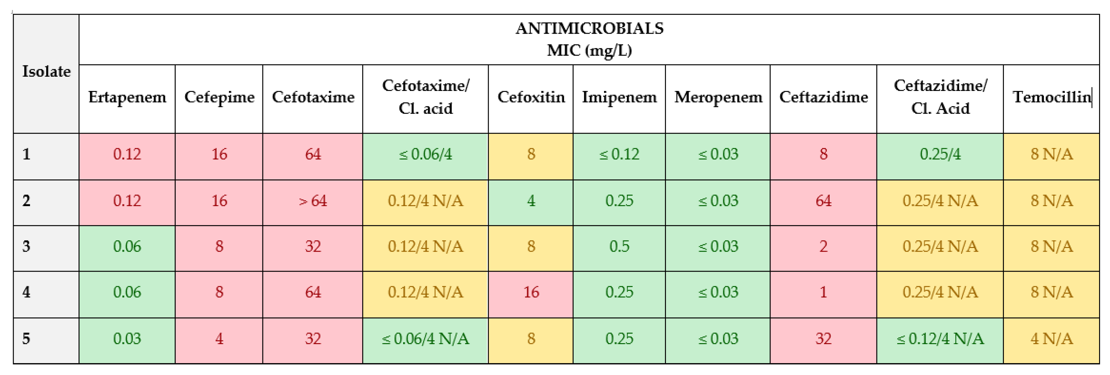
Isolates in which MIC values for cefotaxime and ceftazidime antimicrobials were >1 mg/L were subjected to further phenotypic characterization with a second plate EUVSEC2 (Figure 3).
The MIC values of EUVSEC2 antibiotic panel have shown resistance of isolates to antimicrobials: ertapenem, cefepime, cefotaxime, cefixime and ceftazidime. Isolates 1 and 2 showed resistance to ertapenem with MIC of 0.12 mg/L. All five isolates were resistant to cefepime. Their MIC values were 16 mg/L (1 and 2 isolate), 8 mg/L (3 and 4 isolate) and 4 mg/L (isolate 5). For cefotaxime, antimicrobials recorded high MIC values of 64 mg/L (1, 2 and 4 isolate) and 32 mg/L (3 and 5 isolate). Only one isolate (4) was resistant to cefoxitin with the MIC value of 16 mg/L. The MIC values for ceftazidime were 8 mg/L, 64 mg/L, 2 mg/L, 1 mg/L and 32 mg/L for isolates number 1, 2, 3, 4 and 5, respectively.
Interpretation of phenotypic profiles for determination of resistant ESBL and AmpC E. coli was based on EUCAST guidelines for the detection of resistance mechanisms and specific resistances of clinical and/or epidemiological importance [32]. Briefly, an ESBL phenotype is determined if isolates are resistant to cefotaxime (>1 mg/L) or ceftazidime (>1 mg/L) but susceptible to cefoxitin (≤8 mg/L) and they show clavulanic acid synergy with cefotaxime and/or ceftazidime (≥8-fold reduction of MIC of the cephalosporin combined with 4 mg/L clavulanic acid compared with the MIC of the cephalosporin alone). Isolates are considered to have the AmpC phenotype if they do not show clavulanic acid synergy and are resistant to cefotaxime (>1 mg/L) or ceftazidime (>1 mg/L) and cefoxitin (>8 mg/L). An ESBL+AmpC phenotype is determined if isolates are resistant to cefotaxime (>1 mg/L) or ceftazidime (>1 mg/L), resistant to cefoxitin (>8 mg/L), and they show clavulanic acid synergy with cefotaxime and/or ceftazidime.
In this study, according EUCAST guidelines, four isolates of commensal E coli (1, 2, 3 and 5) were determined as ESBL phenotype and only one (isolate number 4) was determined as ESBL+ AmpC phenotype.

4. Discussion

Livestock and their surroundings appear as significant reservoirs of resistant bacteria, which is a result of the extensive and frequent administration of antimicrobials [33].
There are numerous screening and confirmatory methods for determination of antibiotic residues in milk and tissues. Liquid chromatography combined with tandem mass spectrometry (LC–MS/MS method) is the most frequently applied confirmatory method. This technique enables the identification and determination of multiple classes of antibiotics in milk and tissues [34].
In this study, the LC–MS/MS method was used for simultaneous determination of antimicrobial residues of oxytetracycline, enrofloxacin, amoxicillin, trimethoprim and procaine benzylpenicillin. A closely related method previously developed was used for the detection of 23 veterinary drugs from six different groups in bovine milk [35]. Accuracy and precision of the method comply with the criteria prescribed in Commission Decision 2002/657/EC [29]. According to the validation results, it can be concluded that the applied method is suitable for simultaneous analysis of multi-class and multi-residue antimicrobials in milk and feces samples.
Obtained results from the detection and quantification of antimicrobial residues in milk and feces samples show a sharp decrease in concentrations of antimicrobial residues. Their concentrations started to decrease after the first day of drug administration, and they were eliminated after the 7th day of the treatment. Previous studies have shown similar patterns in residue depletion [36,37]. The minimum withdrawal period for oxytetracycline is 3 days [38], for amoxicillin is 3 days [39] for procaine benzylpenicillin is 5 days [40], for enrofloxacin is 3 days and for trimethoprim is 3.5 day [41].
According to results obtained for MIC values and phenotypic determination of commensal E. coli isolates based on commercial antimicrobial panels, it is important to underline the fact that ESBL-producing E. coli isolates in this study have shown high resistance toward cephalosporins, to the third and fourth generations. Also, high resistance was determined for sulfamethoxazole (SULM) as part of the combined drug therapy of the sulfamethoxazole/trimethoprim that was used during this experiment. No high MIC values were registered toward antimicrobials used in this experiment or to other antimicrobials which were part of commercial plates. Although ertapenem resistance was detected, according to the phenotypic categorization of the isolates, carbapenem phenotype was not determined.
Obtained results in this study are in line with other reported findings from similar studies worldwide. Very similar rates of antimicrobial resistance patterns of Escherichia coli isolates were determined in dairy cattle from small-scale farming systems in Tanzania where the resistant rate to ampicillin and cefotaxime was 96.7%, and 95%, respectively [42], and by Kijima-Tanaka et al. in Japan [43] and Bywater [44] in the European Union determined the rate was about 30%. Meanwhile, compared to other findings in this study, different results were obtained regarding the level of resistance. A study from Korea determined the resistance level was in a range between 0.6–11.7% for cefoxitin and penicillin [45]. In a study from the United Kingdom, the prevalence of resistant E. coli isolates in different cattle groups was as follows: heifers 2.6%, dry cows 3.4% and low-yield milking cows 31.2% [46].
Numerous studies have reported a high prevalence of ESBL-producing E. coli in environmental samples, especially from farm environments. The findings and supporting data imply that dairy cattle farms are a substantial source of ESBL-producing E. coli that play an important role in resistance dissemination across the ‘One Health’ concept [47,48,49].
This type of experiment, which includes a single treatment of animals with different antimicrobials, may have a direct influence or can be related to the emergence or dissemination of antimicrobial resistance between farm animals or into the environment around farms.
Promotion of proper use of antimicrobials in livestock and frequent monitoring play an important role in determination of antimicrobial resistance. The applied LC–MS/MS method together with performed analysis of residue depletion curves in analyzed samples can contribute to the understanding of antimicrobial resistance in food-producing animals and the environment.

5. Conclusions

This study aimed to determine the antimicrobial residue levels in milk and feces samples during the withdrawal period in dairy cattle administrated with a single dose of the drug, as well as to characterize the antimicrobial resistance patterns of commensal isolates of Escherichia coli cultured from cattle feces samples.
The concentrations of antimicrobials oxytetracycline, enrofloxacin, amoxicillin, trimethoprim and procaine benzylpenicillin were quantified in both milk and feces at each of the sampling days. All administered antimicrobials showed the highest concentrations, above their MRLs values, on the first day after drug administration and then they gradually decreased until their elimination (after the 7th day of the treatment).
Antimicrobial pressure on the microbiome in animals or the environment is the result of pressure during a long period of time, through which bacteria ensure their survival, but, obviously, misuse and overuse of semi-synthetic and synthetic antimicrobials play the leading and key role of emerging and dissemination of antimicrobial resistance through bacterial mechanisms.
Regular monitoring would enable timely identification of both emerging and existing forms of antimicrobial resistance in food-producing animals.

Supplementary Materials

The following supporting information can be downloaded at: https://www.mdpi.com/article/10.3390/ani13233603/s1, Figure S1: Chromatograms of spiked feces samples and Figure S2: The chromatograms of spiked milk samples.

Author Contributions

Conceptualization, Z.H.-M. and M.K.; methodology, Z.H.-M., M.K. and J.J.S.; software, R.U. and S.J.; validation, Z.H.-M., R.U., S.J. and M.K.; formal analysis, Z.H.-M., V.S., R.U., S.J., M.K. and D.M.; investigation, Z.H.-M., M.K., R.U., S.J., D.M. and M.A.; resources, Z.H.-M.; data curation, R.U., M.K. and S.J.; writing—original draft preparation, Z.H.-M., M.K. and V.S.; writing—review and editing, M.A.; visualization, M.A.; supervision, Z.H.-M. and J.J.S.; project administration, Z.H.-M. and J.J.S.; funding acquisition, Z.H.-M. All authors have read and agreed to the published version of the manuscript.

Funding

This research was supported by the IAEA project “Integrated radiometric and complementary techniques for mixed contaminants and residues in food” (Grant number D52041).

Institutional Review Board Statement

This study did not require ethical approval because the selection of the animals for antimicrobial treatment was based on indications of infectious disease or established diagnosis confirmed by a doctor of veterinary medicine. Because the treatment of the animals did not differ from the routine veterinary practice and diagnostic procedure, the approval by the Ethic Committee was not required.

Informed Consent Statement

Written informed from the owners of the animal farms consent has been provided.

Data Availability Statement

The data presented in this study are available on request from the corresponding author.

Acknowledgments

The authors would like to acknowledge the support provided by the IAEA project Integrated radiometric and complementary techniques for mixed contaminants and residues in food (D52041).

Conflicts of Interest

The authors declare no conflict of interest.

References

  1. Gemeda, B.A.; Amenu, K.; Magnusson, U.; Dohoo, I.; Hallenberg, G.S.; Alemayehu, G.; Desta, H.; Wieland, B. Antimicrobial Use in Extensive Smallholder Livestock Farming Systems in Ethiopia: Knowledge, Attitudes, and Practices of Livestock Keepers. Front. Vet. Sci. 2020, 7, 55. [Google Scholar] [CrossRef]
  2. Ribeiro da Cunha, B.; Fonseca, L.P.; Calado, C.R.C. Antibiotic discovery: Where have we come from, where do we go? Antibiotics 2019, 8, 45. [Google Scholar] [CrossRef] [PubMed]
  3. Knapp, W.C.; Dolfing, J.; Ehlert, A.I.P.; Graham, W.D. Evidence of increasing antibiotic resistance gene abundances in archived soils since 1940. Environ. Sci. Technol. 2010, 44, 580–587. [Google Scholar] [CrossRef]
  4. Commission Regulation (EU) No 37/2010 of 22 December 2009 on Pharmacologically Active Substances and Their Classification Regarding Maximum Residue Limits in Foodstuffs of Animal Origin (OJ L 15, 20.1.2010, pp. 1–72). Available online: https://eur-lex.europa.eu/legal-content/EN/TXT/?uri=CELEX%3A32010R0037 (accessed on 15 October 2023).
  5. Rice, B.L. The clinical consequences of antimicrobial resistance. Curr. Opin. Microbiol. 2009, 12, 476–481. [Google Scholar] [CrossRef]
  6. Tenover, F.C.; McGowan, J.E. Reasons for the emergence of antibiotic resistance. Am. J. Med. Sci. 1996, 311, 9–16. [Google Scholar] [CrossRef]
  7. Blake, D.P.; Humphry, R.W.; Scott, K.P.; Hillman, K.; Fenlon, D.R.; Low, J.C. Influence of tetracycline exposure on tetracycline resistance and the carriage of tetracycline resistance genes within commensal Escherichia coli populations. J. Appl. Microbiol. 2003, 94, 1087–1097. [Google Scholar] [CrossRef] [PubMed]
  8. Modi, R.S.; Collins, J.J.; Relman, A.D. Antibiotics and the gut microbiota. J. Clin. Investig. 2014, 124, 4212–4218. [Google Scholar] [CrossRef]
  9. Spielmeyer, A. Occurrence and fate of antibiotics in manure during manure treatments: A short review. Sustain. Chem. Pharm. 2018, 9, 76–86. [Google Scholar] [CrossRef]
  10. Filippitzi, M.E.; Devreese, M.; Broekaert, K.; Rasschaert, G.; Daeseleire, E.; Meirlaen, J.; Dewulf, J. Quantitative risk model to estimate the level of antimicrobial residues that can be transferred to soil via manure, due to oral treatments of pigs. Prev. Vet. Med. 2019, 167, 90–100. [Google Scholar] [CrossRef] [PubMed]
  11. Martínez-Carballo, E.; González-Barreiro, C.; Scharf, S.; Gans, O. Environmental monitoring study of selected veterinary antibiotics in animal manure and soils in Austria. Environ. Pollut. 2007, 148, 570–579. [Google Scholar] [CrossRef]
  12. Winckler, C.; Engels, H.; Hund-Rinke, K.; Luckow, T.; Simon, M.; Steffens, G. Verhalten von Tetracyclinen und anderen Veterina rantibiotika in Wirtschaftsdunger und Boden. Ufoplan 2003, 200, 248. [Google Scholar]
  13. Pawelzick, H.T.; Per, H.O.H.; Nau, H.; Hamscher, G. A survey of the occurrence of various tetracyclines and sulfamethazine in sandy soils in northwestern Germany fertilized with liquid manure. In Proceedings of the SETAC Euro 14th Annual Meeting, Prague, Czech Republic, 18–22 April 2004. [Google Scholar]
  14. Rico, A.; Oliveira, R.; McDonough, S.; Matser, A.; Khatikarn, J.; Satapornvanit, K.; Nogueira, A.J.A.; Soares, A.M.V.M.; Domingues, I.; Van Den Brink, P.J. Use, fate and ecological risks of antibiotics applied in tilapia cage farming in Thailand. Environ. Pollut. 2014, 191, 8–16. [Google Scholar] [CrossRef] [PubMed]
  15. Hirsch, R.; Ternes, T.; Haberer, K.; Kratz, K. Occurrence of antibiotics in the aquatic environment. Sci. Total Environ. 1999, 225, 109–118. [Google Scholar] [CrossRef] [PubMed]
  16. Lapara, T.M.; Burch, T.R.; McNamara, P.J.; Tan, D.T.; Yan, M.; Eichmiller, J.J. Tertiary-Treated municipal wastewater is a significant point-source of antibiotic resistance genes into Duluth-superior Harbor. Environ. Sci. Technol. 2011, 45, 9543–9549. [Google Scholar] [CrossRef]
  17. Krapac, I.G.; Koike, S.; Meyer, M.T.; Snow, D.D.; Chou, S.J.; Mackie, R.I.; Roy, W.; Chee, J.C. Long-Term Monitoring of the Occurrence of Antibiotic Residues and Antibiotic Resistance Genes in Groundwater near Swine Confinement Facilities; U.S. Department of Agriculture: Washington, DC, USA, 2004; pp. 158–174. [Google Scholar]
  18. Kivits, T.; Broers, P.H.; Beeltje, H.; Van Vliet, M.; Griffioen, J. Presence and fate of veterinary antibiotics in age-dated groundwater in areas with intensive livestock farming. Environ. Pollut. 2018, 241, 988–998. [Google Scholar] [CrossRef] [PubMed]
  19. Chee-Sanford, J.C.; Mackie, R.I.; Koike, S.; Krapac, I.G.; Lin, Y.F.; Yannarell, A.C.; Maxwell, S.; Aminov, R.I. Fate and Transport of Antibiotic Residues and Antibiotic Resistance Genes following Land Application of Manure Waste. J. Environ. Qual. 2009, 38, 1086–1108. [Google Scholar] [CrossRef] [PubMed]
  20. Boerlin, P.; Travis, R.; Gyles, C.L.; Reid-Smith, R.; Janecko, N.; Lim, H.; Nicholson, V.; Mcewen, S.A.; Friendship, R.; Archambault, M. Antimicrobial Resistance and Virulence Genes of Escherichia coli Isolates from Swine in Ontario. Appl. Environ. Microbiol. 2005, 71, 6753–6761. [Google Scholar] [CrossRef] [PubMed]
  21. ISO/DIS 7218; Microbiology of the Food Chain—General Requirements and Guidance for Microbiological Examinations. ISO: Geneva, Switzerland, 2007.
  22. Regulation (EU) 2017/625 of the European Parliament and of the Council of 15 March 2017 on Official Controls and Other Official Activities Performed to Ensure the Application of Food and Feed Law, Rules on Animal Health and Welfare, Plant Health and Plant Protection Products, Amending Regulations (EC) No 999/2001, (EC) No 396/2005, (EC) No 1069/2009, (EC) No 1107/2009, (EU) No 1151/2012, (EU) No 652/2014, (EU) 2016/429 and (EU) 2016/2031 of the European Parliament and of the Council, Council Regulations (EC) No 1/2005 and (EC) No 1099/2009 and Council Directives 98/58/EC, 1999/74/EC, 2007/43/EC, 2008/119/EC and 2008/120/EC, and Repealing Regulations (EC) No 854/2004 and (EC) No 882/2004 of the European Parliament and of the Council, Council Directives 89/608/EEC, 89/662/EEC, 90/425/EEC, 91/496/EEC, 96/23/EC, 96/93/EC and 97/78/ EC and Council Decision 92/438/EEC (Official Controls Regulation). Available online: https://eur-lex.europa.eu/legal-content/EN/TXT/PDF/?uri=CELEX:32017R0625&from=EN (accessed on 15 October 2023).
  23. Laboratory Protocol Isolation of ESBL-, AmpC- and Carbapenemase-Producing, E. coli from Caecal Samples December 2019 Version 7, Version 7 Was Reviewed and Updated by: Rene, S. Hendriksen and Valeria Bortolaia, Authors of the Document: Henrik Hasman, Yvonne Agersø, Rene Hendriksen, Lina, M. Cavaco (DTU Food) and Beatriz Guerra-Roman. Available online: https://www.eurl-ar.eu/CustomerData/Files/Folders/21-protocols/530_esbl-ampc-cpeprotocol-version-caecal-v7-09-12-19.pdf (accessed on 5 June 2023).
  24. ISO 20776-1:2019; Susceptibility Testing of Infectious Agents and Evaluation of Performance of Antimicrobial Susceptibility Test Devices—Part 1: Broth Micro-Dilution Reference Method for Testing the In Vitro Activity of Antimicrobial Agents against Rapidly Growing Aerobic Bacteria Involved in Infectious Diseases. ISO: Geneva, Switzerland, 2019.
  25. ISO 20776-2:2021; Clinical Laboratory Testing and In Vitro Diagnostic Test Systems—Susceptibility Testing of Infectious Agents and Evaluation of Performance of Antimicrobial Susceptibility Test Devices—Part 2: Evaluation of Performance of Antimicrobial Susceptibility Test Devices against Reference Broth Micro-Dilution. ISO: Geneva, Switzerland, 2021.
  26. Commission Implementing Decision (EU) 2020/1729 of 17 November 2020 on the monitoring and reporting of antimicrobial resistance in zoonotic and commensal bacteria and repealing Implementing Decision 2013/652/EU (notified under document C (2020) 7894). Available online: https://eur-lex.europa.eu/legal-content/EN/TXT/PDF/?uri=CELEX:32020D1729&rid=4 (accessed on 15 October 2023).
  27. Patyra, E.; Kwiatek, K.; Nebot, C.; Gavilán, R.E. Quantification of Veterinary Antibiotics in Pig and Poultry Feces and Liquid Manure as a Non-Invasive Method to Monitor Antibiotic Usage in Livestock by Liquid Chromatography Mass-Spectrometry. Molecules 2022, 25, 3265. [Google Scholar] [CrossRef] [PubMed]
  28. Pokrant, E.; Trincado, L.; Yévenes, K.; Terraza, G.; Maddaleno, A.; San Martín, B.; Zavala, S.; Hidalgo, H.; Lapierre, L.; Cornejo, J. Determination of five antimicrobial families in droppings of therapeutically treated broiler chicken by high-performance liquid chromatography-tandem mass spectrometry. Poultry Sci. 2021, 100, 101313. [Google Scholar] [CrossRef]
  29. 2002/657/EC: Commission Decision of 12 August 2002 Implementing Council Directive 96/23/EC Concerning the Performance of Analytical Methods and the Interpretation of Results (Text with EEA Relevance) (Notified under Document Number C(2002) 3044) (OJ L 221 17.08.2002, p. 8). Available online: http://data.europa.eu/eli/dec/2002/657/oj (accessed on 5 June 2023).
  30. Hajrulai-Musliu, Z.; Uzunov, R.; Jovanov, S.; Jankuloski, D.; Stojkovski, V.; Pendovski, L.; Sasanya, J.J. A new LC–MS/MS method for multiple residues/contaminants in bovine meat. BMC Chem. 2021, 15, 62. [Google Scholar] [CrossRef]
  31. International Conference on Harmonisation of Technical requirements for registration of pharmaceuticals for human use. ICH harmonised tripartite guideline. Validation of analytical procedures: Text and methodology Q2(R1). Current Step 4 version Parent Guideline dated 27 October 1994. Available online: https://database.ich.org/sites/default/files/Q2%28R1%29%20Guideline.pdf (accessed on 15 October 2023).
  32. EUCAST Guidelines for Detection of Resistance Mechanisms and Specific Resistances of Clinical and/or Epidemiological Importance, Version 2.0.1 2017. Available online: https://www.eucast.org/fileadmin/src/media/PDFs/EUCAST_files/Resistance_mechanisms/EUCAST_detection_of_resistance_mechanisms_170711.pdf (accessed on 5 June 2023).
  33. Kimera, Z.I.; Mshana, S.E.; Rweyemamu, M.M.; Mboera, L.E.G.; Matee, M.I.N. Antimicrobial use and resistance in food producing animals and the environment: An African perspective. Antimicrob. Resist. Infect. Control 2020, 9, 37. [Google Scholar] [CrossRef] [PubMed]
  34. Samanidou, V.; Nisyriou, S. Multi-residue methods for confirmatory determination of antibiotics in milk. J. Sep. Sci. 2008, 31, 2068–2090. [Google Scholar] [CrossRef] [PubMed]
  35. Alija, G.; Hajrulai-Musliu, Z.; Uzunov, R. Development and validation of confirmatory LC–MS/MS method for multi-residue analysis of antibiotic drugs in bovine milk. SN Appl. Sci. 2020, 2, 1563. [Google Scholar] [CrossRef]
  36. Chowdhury, S.; Hassan, M.M.; Alam, M.; Sattar, S.; Bari, M.S.; Saifuddin, A.K.M.; Hoque, M.A. Antibiotic residues in milk and eggs of commercial and local farms at Chittagong, Bangladesh. Vet. World. 2015, 8, 467–471. [Google Scholar] [CrossRef] [PubMed]
  37. Mehran, M.A.; Hossein, B.; Masoud, A.; Ashraf-O-Sadat, N.; Mahboob, N. Simultaneous determination of tetracyclines residues in bovine milk samples by solid phase extraction and HPLC-FL method. Adv. Pharm. Bull. 2011, 1, 34–39. [Google Scholar] [CrossRef]
  38. Hassan, B.A.R. HPLC uses and importance in the pharmaceutical analysis and industrial field. Pharm. Anal. Acta 2012, 3, 9. [Google Scholar] [CrossRef]
  39. Martínez-Cortés, I.; Rosiles, R.; Gutierrez, L.; Díaz, D.; Sumano, H. Amoxicillin resides in milk of Holstein cows with double or triple intramammary administration of amoxicillin-potassium clavulanate. Int. J. Appl. Res. Vet. Med. 2014, 12, 186–192. [Google Scholar]
  40. Burmańczuk, A.; Tomasz, G.; Gbylik-Sikorska, M.; Gajda, A.; Kowalski, C. Withdrawal of Amoxicillin and Penicillin G Procaine from Milk after Intramammary Administration in Dairy Cows with Mastitis. J. Vet. Res. 2017, 61, 37–43. [Google Scholar] [CrossRef]
  41. Mahmood, T.; Abbas, M.; Ilyas, S.; Afzal, N.; Nawaz, R. Quantification of fluoroquinolone (enrofloxacin, norfloxacin and ciprofloxacin) residues in cow milk. Int. J. Chem. Biochem. Sci. 2016, 10, 10–15. [Google Scholar]
  42. Azabo, R.R.; Mshana, S.E.; Matee, M.I.; Kimera, S.I. Antimicrobial Resistance Pattern of Escherichia coli Isolates from Small Scale Dairy Cattle in Dar es Salaam, Tanzania. Animals 2022, 12, 1853. [Google Scholar] [CrossRef]
  43. Kijima-Tanaka, M.; Ishihara, K.; Morioka, A.; Kojima, A.; Ohzono, T.; Ogikubo, K.; Takahashi, T.; Tamura, Y. A national surveillance of antimicrobial resistance in Escherichia coli isolated from food-producing animals in Japan. J. Antimicrob. Chemother. 2003, 51, 447–451. [Google Scholar] [CrossRef] [PubMed]
  44. Bywater, R.; Deluyker, H.; Deroover, E.; De Jong, A.; Marion, H.; McConville, M.; Rowan, T.; Shryock, T.; Shuster, D.; Thomas, V.; et al. A European survey of antimicrobial susceptibility among zoonotic and commensal bacteria isolated from food-producing animals. J. Antimicrob. Chemother. 2004, 54, 744–754. [Google Scholar] [CrossRef] [PubMed]
  45. Song, H.J.; Kim, S.J.; Moon, D.C.; Mechesso, A.F.; Choi, J.H.; Kang, H.Y.; Boby, N.; Yoon, S.S.; Lim, S.K. Antimicrobial Resistance in Escherichia coli Isolates from Healthy Food Animals in South Korea. Microorganisms 2022, 10, 524. [Google Scholar] [CrossRef] [PubMed]
  46. Watson, E.; Jeckel, S.; Snow, L.; Stubbs, R.; Teale, C.; Wearing, H.; Horton, R.; Toszeghy, M.; Tearne, O.; Ellis-Iversen, J.; et al. Epidemiology of extended spectrum beta-lactamase E. coli (CTX-M-15) on a commercial dairy farm. Vet Microbiol. 2012, 154, 339–346. [Google Scholar] [CrossRef] [PubMed]
  47. Kerluku, M.; Jankuloski, D.; Manovska Ratkova, M.; Prodanov, M.; Dimzoska Stojanovska, B.; Dodovski, A.; Blagoevska, K. β-Lactamase genes (blaCTX-M, blaSHV, blaTEM, blaOXA1 AND blaOXA2) and phylogenetic groups in ESBL producing commensal Escherichia coli isolated from faecal samples from dairy farm in the Municipality of Debar. Mac. Vet. Rev. 2023, 46, 89–97. [Google Scholar] [CrossRef]
  48. Zhang, Y.L.; Huang, F.Y.; Gan, L.L.; Yu, X.; Cai, D.J.; Fang, J.; Zhong, Z.J.; Guo, H.R.; Xie, Y.; Wang, Z.S.; et al. High prevalence of blaCTX-M and blaSHV among ESBL producing E. coli isolates from beef cattle in China’s Sichuan-Chongqing circle. Sci. Rep. 2021, 11, 13725. [Google Scholar] [CrossRef]
  49. Braun, S.D.; Ahmed, M.F.E.; El-Adawy, H.; Hotzel, H.; Engelmann, I.; Weiß, D.; Monecke, S.; Ehricht, R. Surveillance of extended-spectrum beta-lactamase-producing Escherichia coli in dairy cattle farms in the Nile delta. Egypt. Front. Microbiol. 2016, 7, 1020. [Google Scholar] [CrossRef] [PubMed]
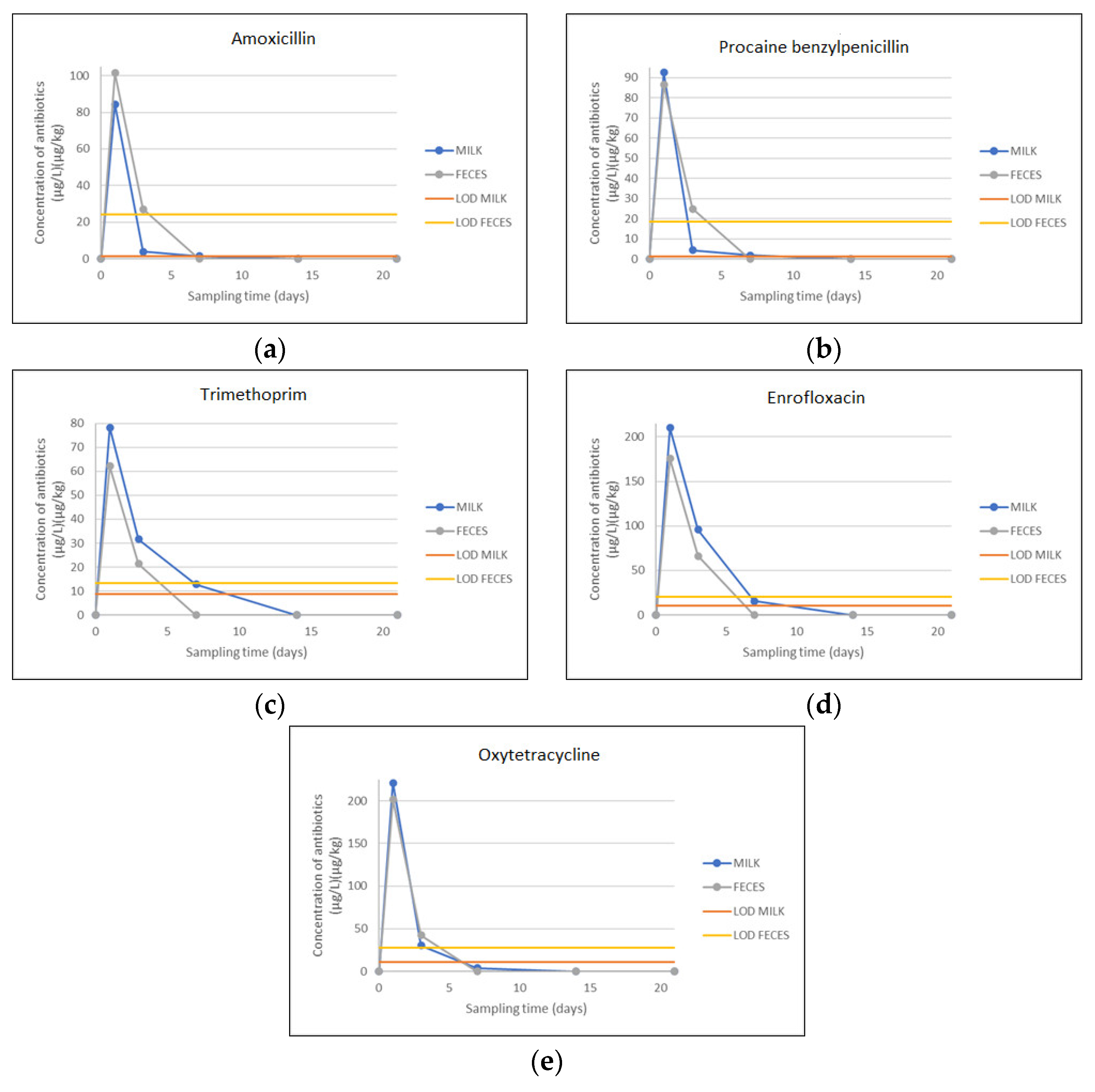
Figure 1. The residue depletion curves for both groups of samples (milk and feces). (a) Amoxicillin residues in milk and feces samples obtained from cattle after single-dose administration with Amoxicillin 150 mg/mL; (b) Procaine benzylpenicillin residues in milk and feces samples obtained from cattle after single-dose administration with procaine-benzyl penicillin G 200,000 IU/mL; (c) Trimethoprim residues in milk and feces samples obtained from cattle after single-dose administration with Trimethoprim 80 mg/mL; (d) Enrofloxacin residues in milk and feces samples obtained from cattle after single-dose administration with Enrofloxacin 100 mg/mL; (e) Oxytetracycline residues in milk and feces samples obtained from cattle after single-dose administration with Oxytetracycline 200 mg/mL.
Animals 13 03603 g001
Figure 2. MIC values (mg/L) on EUVSEC1 commercial panel. Pink squares—resistant isolate (R); Green squares—susceptible isolates (S).
Animals 13 03603 g002
Figure 3. MIC values (mg/L) on EUVSEC2 commercial panel. Pink squares—resistant isolate (R); Green squares—susceptible isolates (S); Yellow squares—N/A (nonapplicable).
Animals 13 03603 g003
Table 1. MRM (Multiple Reaction Monitoring) parameters for each of 20 antimicrobials and two internal standards.
StandardIonisation Mode (ESI)Precursor Ion (m/z)Product Ion (m/z)Collision EnergyCone VoltageRetention Time
Amoxicillin+367.07159.9616285.55
90.8940
Ampicillin+349.97159.9414343.93
105.9520
Procaine benzylpenicillin+334.9990.9642445.52
80.9452
Lincomycin+407.06126.0234222.80
41.7572
Tylosin+916.30100.8852746.31
173.9946
Trimethoprim+290.97229.9424262.90
122.9428
Cephapirin+423.93291.9314422.04
151.8928
Tetracycline+445.03410.0120405.33
153.9034
Cloxacillin+435.94159.9718266.15
276.9614
Oxacillin+402.05159.9610245.95
243.0312
Cefalexin+347.97173.9314302.75
157.868
Ceftiofur+523.96125.1758344.90
241.0016
Enrofloxacin+360.0572.0236363.68
245.0930
Ciprofloxacin+332.01230.9428383.56
245.0540
Oxytetracycline+462.01426.0238363.17
200.9330
Sulfachlorpyridazin+284.9155.9316282.93
91.9334
Sulfadiazine+250.97155.9314281.92
91.9330
Sulfadimethoxine+310.9791.9332364.36
155.9320
Sulfadimidine+278.9591.9336342.71
185.9318
Sulfamethoxazole+253.91155.9416283.01
92.0030
Flunixin–D3+300.03263.9836256.80
Penicillin G–d7+374.03159.9416325.51
Table 2. Linearity of the method.
StandardCalibration RangeR2
MatrixMatrix
FecesMilkFecesMilk
(μg/kg)(μg/L)
Amoxicillin20.0–10001.0–500.99980.9993
Ampicillin20.0–10001.0–500.99890.9963
Procaine benzylpenicillin20.0–10001.0–500.99970.9999
Lincomycin20.0–100050.0–3000.99840.9978
Tylosin20.0–100010.0–1000.99780.9991
Trimethoprim20.0–100010.0–1000.99970.9994
Cephapirin20.0–100010.0–1000.99990.9994
Tetracycline20.0–100010.0–2000.99440.9974
Cloxacillin20.0–100010.0–1000.99610.9954
Oxacillin20.0–100010.0–1000.99840.9951
Cefalexin20.0–100010.0–2000.99290.9976
Ceftiofur20.0–100010.0–2000.99370.9979
Enrofloxacin20.0–100010.0–2000.99990.9981
Ciprofloxacin20.0–100010.0–2000.99490.9997
Oxytetracycline20.0–100010.0–2000.99940.9963
Sulfachlorpyridazin20.0–100010.0–2000.99740.9967
Sulfadiazine20.0–100010.0–2000.99790.9958
Sulfadimethoxine20.0–100010.0–2000.99790.9970
Sulfadimidine20.0–100010.0–2000.99810.9988
Sulfamethoxazole20.0–100010.0–2000.99780.9994
Table 3. Limit of detection (LOD), Limit of quantification (LOQ), Decision limit (CCα) and Detection capability (CCβ).
StandardLODLOQCCαCCβ
MatrixMatrixMatrixMatrix
FecesMilkFecesMilkFecesMilkFecesMilk
(μg/kg)(μg/L)(μg/kg)(μg/L)(μg/kg)(μg/L)(μg/kg)(μg/L)
Amoxicillin24.101.5932.112.5942.365.3452.116.43
Ampicillin20.181.2857.171.7635.144.6649.785.23
Procaine benzylpenicillin18.331.3628.652.1643.184.8056.715.66
Lincomycin14.1047.9435.1755.7827.45179.1937.52199.07
Tylosin12.8010.7134.1213.6220.1255.3131.4066.09
Trimethoprim13.368.6821.1711.0138.2954.7449.4065.31
Cephapirin8.2612.3323.0115.7221.3571.9132.1482.65
Tetracycline21.358.6555.6011.0541.78105.9152.40116.86
Cloxacillin22.1810.2459.6012.9737.5431.7454.2535.93
Oxacillin17.159.2641.1014.0130.1235.1241.3541.32
Cefalexin16.3512.3835.5015.0221.35105.1434.17118.36
Ceftiofur9.1211.2226.4014.6715.35101.6627.14108.76
Enrofloxacin20.4010.5335.4214.1247.80112.3359.71132.14
Ciprofloxacin23.1412.8055.3516.6039.14102.1257.80121.14
Oxytetracycline27.6010.7641.6012.1251.48113.6566.41131.29
Sulfachlorpyridazin14.3510.4732.1813.8822.15116.8240.78142.59
Sulfadiazine21.3010.8955.6013.3927.15121.9558.35152.05
Sulfadimethoxine12.2510.3531.4812.6917.14104.7025.10122.17
Sulfadimidine12.709.0634.1511.4219.48121.3031.40146.93
Sulfamethoxazole15.3010.1348.1213.0323.54103.8137.80119.54
Table 4. Accuracy and precision of the method.
StandardsAddedAverage
Concentration in the Samples (n = 6)
Standard DeviationRecovery (%)RepeatabilityReproducibility
Concentration(μg/kg)(CVr, %)(CVR, %)
MatrixMatrixMatrixMatrixMatrixMatrix
FecesMilkFecesMilk (μg/L)FecesMilk (μg/L)FecesMilkFecesMilkFecesMilk
(μg/kg)(μg/L)(μg/kg)(μg/kg)
Amoxicillin100289.002.183.810.2489.00109.004.2810.9910.6919.37
2504235.714.2610.380.6694.28106.384.4015.599.0218.90
5006464.265.2217.660.6192.8586.973.8011.6114.3714.57
Ampicillin100280.071.743.130.2180.0787.513.9012.0711.3516.64
2504238.593.5611.080.6595.4489.364.6418.2614.8820.18
5006412.295.7112.181.0382.4695.172.9618.0410.4321.35
Procaine benzylpenicillin100283.481.426.370.1483.4870.837.629.5310.4815.99
2504243.113.9213.610.5397.2497.925.6013.596.1718.18
5006473.104.9317.870.3594.6282.083.787.106.6014.23
Lincomycin1007585.5661.256.003.1485.5681.677.015.1314.226.82
250150230.61159.3112.2812.1292.24106.205.327.6112.768.87
500225428.11218.6210.7827.8885.6297.162.5212.766.1615.78
Tylosin1002586.3922.254.632.3086.6989.015.3610.3210.9414.72
25050204.2144.5316.966.5781.6889.078.3014.7614.9917.14
50075488.4074.538.284.8297.6899.381.706.475.6810.84
Tylosin1002597.1121.585.642.7997.1186.315.8012.9413.0315.85
25050263.7944.1712.026.44105.9288.344.5614.598.0317.37
50075524.3073.0719.476.95104.8697.433.719.5111.1011.73
Cephapirin1003097.7426.264.991.2397.7487.525.114.6810.367.31
25060259.8961.1812.126.54103.96101.964.6610.714.8513.06
50090458.1182.2825.581.9991.6291.425.582.4112.103.48
Tetracycline10025102.3244.787.203.82102.3289.577.048.5312.5512.52
25050230.3494.9512.226.6892.1494.955.307.0411.399.80
50075491.56126.8111.558.3098.3184.542.356.558.188.41
Cloxacillin10015105.414.833.771.21105.498.883.588.196.7710.74
25030255.3927.544.952.56102.1691.811.949.283.9812.12
50045488.9239.5111.804.4897.7887.802.4111.335.8917.86
Oxacillin1001589.2914.224.922.4489.2994.845.5117.169.6622.04
25030221.3727.367.983.0888.5591.223.6111.266.0114.33
50045450.8440.176.835.4490.1789.271.2313.544.4416.28
Cefalexin1005085.7341.485.145.2685.7382.966.0012.687.6115.87
250100244.4488.487.197.1197.7788.482.948.048.2110.97
500150484.13123.175.939.2296.8382.111.937.497.3310.02
Ceftiofur1005093.6050.492.133.6793.6100.972.287.275.7314.39
250100246.2394.579.154.3398.4994.573.724.575.127.82
500150480.64132.328.859.2796.1388.211.847.004.309.89
Enrofloxacin1005082.6454.108.607.4782.64108.210.4113.813.3115.92
250100212.5492.5212.4212.0885.0192.525.8413.0614.8114.25
500150432.07141.3627.7417.4886.4198.226.4211.8614.7414.96
Ciprofloxacin1005089.5645.493.243.1489.5690.973.626.918.769.73
250100247.8883.104.7911.699.1583.101.9313.965.6115.46
500150495.32131.553.9816.4699.0687.700.8012.512.1115.98
Oxytetracycline1005089.8240.689.933.2889.8288.0411.067.4514.999.39
250100209.9996.0014.2410.7683.9996.006.7811.2114.7613.22
500150421.17159.9421.6913.3884.23106.635.158.3612.9810.91
Sulfachlorpyridazin1005082.4548.062.527.5182.4596.113.0615.635.2118.96
250100219.0291.054.0115.7187.6191.051.8317.264.6819.98
500150470.61151.3311.6918.2594.12100.892.4812.065.0213.33
Sulfadiazine1005086.0846.894.518.7486.0893.795.2518.636.9021.68
250100246.5991.856.0918.3598.6391.852.4719.983.7720.84
500150488.60141.338.447.3397.7294.211.735.182.588.18
Sulfadimethoxine1005083.6242.421.674.8683.6284.842.0011.463.0914.55
250100226.6887.237.0310.6590.6787.233.1012.216.2013.34
500150467.95123.8113.2112.2193.5982.542.8211.006.6213.30
Sulfadimidine1005097.4042.502.758.9897.4084.992.8321.126.2923.44
250100243.6295.665.6015.6397.4595.662.3016.334.0317.22
500150511.71132.175.5420.01102.3488.111.0815.142.6116.77
Sulfamethoxazole1005085.0546.382.935.8085.0592.753.4512.518.2316.53
250100236.8688.0611.129.6094.7488.064.6910.906.1413.27
500150480.43120.055.7711.4596.0980.031.089.542.6113.70
Table 5. Concentration of antimicrobials (µg/L or µg/kg) in milk and feces analyzed by LC-MS/MS.
AntibioticMatrixSampling Days
01371421LODCCαMRL
Amoxicillin MilkND84.353.801.35<LOD<LOD1.285.344.0
µg/L
FecesND101.3827.11<LOD<LOD<LOD24.1042.36/
µg/kg
Procaine benzylpenicillinMilkND92.504.401.72<LOD<LOD1.364.804.0
µg/L
FecesND86.3024.88<LOD<LOD<LOD18.3343.18/
µg/kg
TrimethoprimMilkND78.3031.6012.80<LOD<LOD8.6854.7450.0
µg/L
FecesND62.1821.30<LOD<LOD<LOD13.3638.29/
µg/kg
EnrofloxacinMilkND210.1095.6015.40<LOD<LOD10.53112.33100.0
µg/L
FecesND175.4866.20<LOD<LOD<LOD20.4047.80/
µg/kg
OxytetracyclineMilkND221.1030.203.90<LOD<LOD10.76113.65100.0
µg/L
FecesND201.3042.45<LOD<LOD<LOD27.6051.48/
µg/kg
Disclaimer/Publisher’s Note: The statements, opinions and data contained in all publications are solely those of the individual author(s) and contributor(s) and not of MDPI and/or the editor(s). MDPI and/or the editor(s) disclaim responsibility for any injury to people or property resulting from any ideas, methods, instructions or products referred to in the content.