Article

Seroprevalence and Risk Factors of Chlamydia Infection in Cattle in Shanxi Province, North China

1
College of Veterinary Medicine, Shanxi Agricultural University, Jinzhong 030801, China
2
Key Laboratory of Veterinary Public Health of Higher Education of Yunnan Province, College of Veterinary Medicine, Yunnan Agricultural University, Kunming 650201, China
3
Veterinary Laboratory, Shanxi Provincial Animal Disease Prevention and Control Center, Taiyuan 030008, China
*
Authors to whom correspondence should be addressed.
These authors contributed equally to this work.
Animals 2023, 13(2), 252; https://doi.org/10.3390/ani13020252
Submission received: 4 December 2022 / Revised: 30 December 2022 / Accepted: 6 January 2023 / Published: 10 January 2023
(This article belongs to the Special Issue Animal Chlamydioses)

Abstract

:

Simple Summary

Chlamydia, an important zoonotic pathogen, poses a serious threat to public health and the development of animal husbandry. Shanxi Province, famous for its Loess Plateau terrain, has been taking cattle and sheep husbandry as an important direction of development, which means that it is necessary to prioritize the detection of pathogens in cattle and sheep. In this study, based on the location and management pattern, the seroprevalence of Chlamydia and C. abortus infection in cattle in Shanxi Province was examined by indirect hemagglutination assay (IHA) and indirect enzyme-linked immunosorbent assay (ELISA), respectively. The results revealed a high (52.29%) overall seroprevalence of Chlamydia in cattle in Shanxi Province. Location was closely related to the prevalence of Chlamydia and C. abortus, and different management patterns also had a certain impact on the prevalence of Chlamydia. This study provided valuable baseline information for the prevention and control of Chlamydia in cattle in Shanxi Province.

Abstract

The information on Chlamydia infection in cattle is limited in Shanxi Province, north China. This study aimed to investigate the seroprevalence and risk factors of Chlamydia and Chlamydia abortus infection in cattle in Shanxi Province. In November 2020, a large-scale investigation of Chlamydia seroprevalence was conducted on 981 cattle serum samples collected from 40 cattle farms in 11 cities of Shanxi Province. The seroprevalence of Chlamydia and C. abortus was examined by indirect hemagglutination assay (IHA) and enzyme-linked immunosorbent assay (ELISA), respectively. The seroprevalence of Chlamydia and C. abortus was 52.29% (513/981) and 2.96% (29/981), respectively, in cattle in Shanxi Province. Location was identified as a risk factor for Chlamydia and C. abortus infection (p < 0.05). Under different management patterns, the seroprevalence of Chlamydia and C. abortus in large-scale animal farming companies was higher than that in household animal farms and animal farming cooperatives, and only the seroprevalence of Chlamydia was significantly different in different management patterns (p < 0.01). The results showed that there was higher seroprevalence of Chlamydia in cattle in Shanxi Province, while C. abortus was not the dominant species. This study provided baseline information on Chlamydia infection in cattle in Shanxi Province, which constitutes valuable data for monitoring livestock health and preventing potential zoonoses.

1. Introduction

Chlamydia organisms are obligate intracellular pathogens distributed globally, known to cause various forms of diseases in humans and other vertebrate animals [1]. To date, more than 15 species of Chlamydia have been identified [2], of which Chlamydia pecorum, Chlamydia abortus and Chlamydia psittaci have been reported in cattle [3]. Chlamydia infection in cattle can cause acute but rare diseases, such as sporadic bovine encephalomyelitis, abortion, enteritis, and pneumonia [3,4]. In addition to the infrequent acute infections, more subclinical infections have been reported in cattle [5,6]. Nevertheless, subclinical infections also have a substantial impact on livestock productivity, including reduced weight gain and infertility [5].
Abortion causes devastating economic losses to the cattle industry, with a total loss of up to GBP 630 per aborted cow [7]. For Iranian Holstein cows, abortion had potential impacts on milk production, fat and protein content in milk of normal calving cows [8]. C. abortus is an important pathogen associated with abortion in cattle [9]. In addition, C. abortus is a zoonotic species of public health importance, which is responsible for abortion and reproductive problems in humans and other animals [10,11,12].
The mountainous area accounts for 80.1% of the total area in Shanxi Province, which is a typical mountain plateau covered by loess. Therefore, the development of animal husbandry based on cattle and sheep has always been of economic importance to Shanxi Province. At present, the management pattern of cattle farming in Shanxi Province is mostly household animal farming, and the development of large-scale animal farming companies is the future development trend of cattle industry in Shanxi Province. Therefore, understanding the potential risk factors of Chlamydia infection is conducive to the transformation of the cattle industry in Shanxi Province.
Serological surveillance of these pathogens in cattle is of great importance. To date, information on the prevalence of Chlamydia and C. abortus in cattle Shanxi Province is very limited. Therefore, the purpose of this study was to investigate the seroprevalence and risk factors of Chlamydia and C. abortus infection in cattle in Shanxi Province, and the study was expected to provide a reference for the prevention and control of these pathogens and the development of the cattle industry.

2. Materials and Methods

2.1. Ethics Approval

The experimental procedures of the study were reviewed and approved by the Animal Research Ethics Committee of Shanxi Agricultural University (Approval No. 2019IACUCSXAU002A01). The animals were handled in accordance with good animal practice as defined by the relevant Animal Ethics Procedures and Guidelines of the People’s Republic of China.

2.2. Study Sites

In this study, serum samples were collected from 11 administrative cities in Shanxi Province, North China. Shanxi Province (34°36′–40°44′ N, 110°15′–114°32′ E), located at the east of the Loess Plateau, has a temperate continental monsoon climate with an average annual temperature of 3–14 °C (Figure 1). In the past ten years, the resident population of Shanxi Province has gradually decreased, and more and more of the rural population choose to live in cities. The number of people working in primary industries (agriculture, forestry, husbandry or fishing) has also dropped sharply (Table 1) (http://tjj.shanxi.gov.cn/tjsj/tjnj/nj2021/zk/indexch.htm, accessed on 18 December 2022).

2.3. Sample Collection

In this study, 981 serum samples of cattle were collected from 40 cattle farms in 11 cities in Shanxi Province in November 2020; of which 267, 360, and 354 samples were collected from northern (Datong (n = 88), Shuozhou (n = 89) and Xinzhou (n = 90)), central (Taiyuan (n = 90), Lvliang (n = 90), Jinzhong (n = 90) and Yangquan (n = 90)), and southern Shanxi Province (Changzhi (n = 90), Jincheng (n = 90), Linfen (n = 84) and Yuncheng (n = 90)), respectively (Figure 1 and Table 2). At least 3 cattle farms were selected for sampling in each city. The 40 cattle farms included 26 household animal farms (n = 567, stock < 200), 5 animal farming cooperatives (n = 100, stock consists of multiple domestic animal cattle farms) and 9 large-scale animal farming companies (n = 314, stock > 900). After standing at room temperature for 2 h, the samples were centrifuged at 3000 g for 10 min to collect the serum and sera were stored at −20 °C for further analysis.

2.4. Serological Tests

Antibodies to Chlamydia were tested by using indirect hemagglutination assay (IHA) with a commercially available Kit (Lanzhou Veterinary Research Institute, Chinese Academy of Agricultural Sciences, Lanzhou, China), and serum samples were considered as positive for Chlamydia if layers of agglutinated erythrocytes were formed in wells at dilutions of 1:16 or greater. Results returning values between 1:4 and 1:16 were considered “suspicious samples” and were tested again. The high sensitivity (100%) and specificity (95%) of this IHA kit guarantee the authenticity of the experimental data [13].
Antibodies to C. abortus were examined by using the ID Screen® C. abortus Indirect Multi-species ELISA kit (Innovative Diagnostics, France). According to the manufacturer’s specifications, the sample was tested and OD value was measured at 450 nm. The ratio was calculated according to the following formula:
The value = (OD sample/OD positive control) × 100
Samples with values greater than 60% were considered as positive; values equal to or less than 50% as negative; values between 50% and 60% were considered suspicious and were tested again.
A second test for Chlamydia (IHA) and C. abortus (ELISA) was considered positive if the results remained suspicious.

2.5. Statistical Analyses

The seroprevalence and the factors (location and management pattern) for Chlamydia and C. abortus infection in cattle in Shanxi Province were evaluated by using the SPSS 26 version software (IBM, Armonk, NY, USA). p < 0.05 was considered statistically significant. The odds ratios and the 95% confidence interval (CI) of each factor were analyzed in this study.

3. Results

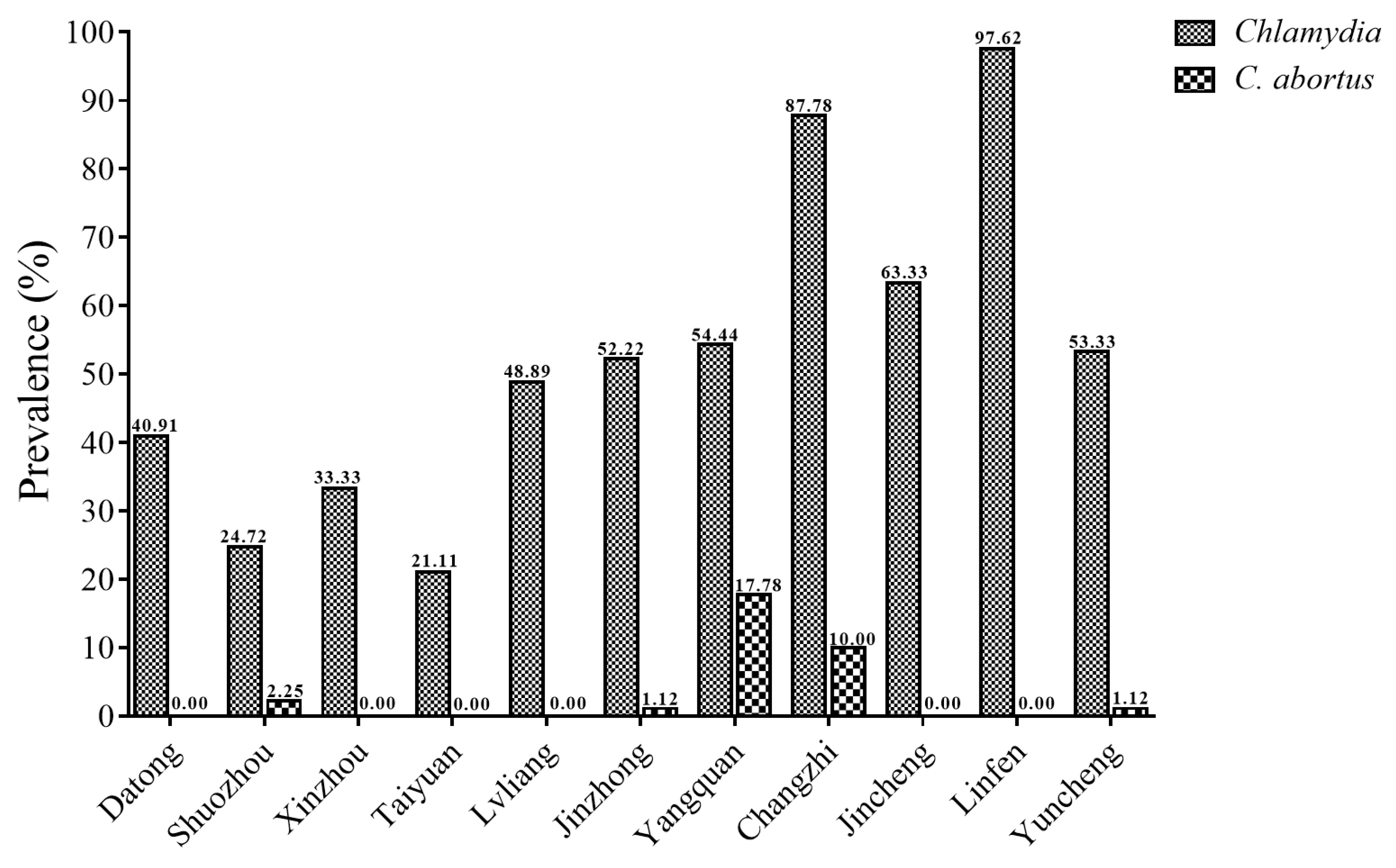
Among 981 cattle serum samples, the total seroprevalence of Chlamydia in Shanxi Province was 52.29% (513/981, 95% CI 49.17–55.42), with IHA titers of 1:16 in 408 cattle sera and 1:64 in 105 cattle sera (Table 2 and Table 3). The seroprevalence of Chlamydia varied greatly from 21.11% in Taiyuan to 97.62% in Linfen (Table 2 and Figure 2). In terms of location, the highest seroprevalence of Chlamydia infection in cattle was found in southern Shanxi Province (75.14%, 95% CI 70.64–79.64), followed by central Shanxi Province (44.17%, 95% CI 39.04–49.30) and northern Shanxi Province (32.96%, 95% CI 27.32–38.60). The seroprevalences of Chlamydia in cattle in the three management patterns (Household animal farm, Animal farming cooperative, and Large-scale animal farming company) were 49.03% (95% CI 44.92–53.14), 47.00% (95% CI 37.22–56.78), and 59.87% (95% CI 54.45–65.29), respectively. The seroprevalences of Chlamydia in cattle in different locations and management patterns were statistically significantly different (p < 0.01) (Table 3).
In this study, the total seroprevalence of C. abortus was 2.96% (29/981, 95% CI 1.90–4.02) in cattle in Shanxi Province (Table 4). Among the different cities, Yangquan had the highest seroprevalence of C. abortus (17.78%), followed by Changzhi (10.00%) and other cities (lower or 0) (Table 2 and Figure 2). There was a statistically significant difference in the seroprevalence of C. abortus between northern, central and southern Shanxi Province (p < 0.05). The highest seroprevalence of C. abortus in cattle was detected in the large-scale animal farming companies (3.82%, 12/314, 95% CI 1.70–5.94), followed by 3.00% (17/567, 95% CI 1.59–4.40) in the household animal farms and 0.00% in animal farming cooperatives. However, the seroprevalence of C. abortus in cattle in this study did not differ significantly between different management patterns (p > 0.05) (Table 4).

4. Discussion

The overall seroprevalence of Chlamydia infection in cattle in Shanxi Province was 52.29%, which was higher than that in Guangzhou (7.25%) [14]. The prevalence of Chlamydia has been reported in many other countries, such as Great Britain (31%) [15], Germany (50.8%) [16] and Poland (36.9%) [17]. The seroprevalence of C. abortus in cattle in Shanxi Province (2.96%) was lower than that in Tibet Autonomous Region (23.81%) [18], Gansu Province (16.22%) [19], Hebei Province (11.80%), and Shandong Province (12.67%) in China [9]. Similarly, C. abortus is widely found in cattle around the world, such as Switzerland (14.6%) [20] and Argentina (4.78%) [21]. The different seroprevalence of Chlamydia and C. abortus both within China and abroad may be due to the differences in national plans to control and eradicate the pathogens, detection methods, susceptibility of different breeds to pathogens, breeding environment, geographic location, and climate in the examined locations.
Given the significant differences in temperature and humidity between the north and south of Shanxi Province, the location was used as a factor to explore Chlamydia infection in cattle in Shanxi Province. As expected, the results showed that location was a significant risk factor for Chlamydia infection in cattle in Shanxi Province (p < 0.01) (Table 3). Compared to cattle in northern Shanxi Province, cattle in central and southern Shanxi Province had 1.61 times (95% CI 1.16–2.24) and 6.15 times (95% CI 4.33–8.73) higher risk of acquiring the infection, respectively. The high seroprevalence of Chlamydia in cattle in southern Shanxi Province may be due to the higher temperature and humidity in this area, and previous studies showed that animals have a higher seroprevalence of Chlamydia in summer or during the wet season [22,23]. It is worth noting that Datong (the northernmost city in Shanxi Province) had lower temperature and humidity than other northern cities, but the seroprevalence of Chlamydia showed the opposite trend. The main cause for this phenomenon may be that Datong is located at the throat of the Shanxi–Hebei–Mongolian Great Wall Golden Triangle cooperation zone, and there may be a certain risk of cross infection among cattle. Within southern Shanxi Province, Chlamydia seroprevalence in cattle in Changzhi and Linfen was higher than in surrounding cities. This may be due to Changzhi being located in the rainy belt of the Taihang Mountains of southeast Shanxi Province, which may have facilitated a high prevalence of Chlamydia. Disruption of the natural environment increases animal exposure to vectors and hosts of unknown pathogenic microorganisms [24]. For Linfen, its air pollution ranks first in Shanxi Province, which may increase the possibility of Chlamydia transmission.
In addition to the immutable factor of location, an essential factor, management pattern, was investigated for its influence on Chlamydia infection in cattle in Shanxi Province. The seroprevalences of Chlamydia in cattle in different management patterns were significantly different (p < 0.01), ranging from 47.00% (95% CI 37.22–56.78) to 59.87% (95% CI 54.45–65.29) (Table 3). This indicated that management pattern was a risk factor for Chlamydia infection in cattle in Shanxi Province. Cattle in large-scale animal farming companies had a risk 1.68 times (95% CI 1.07–2.65) higher of acquiring Chlamydia infection than those in animal farming cooperatives. This may be related to breeding density, breeding environment and grazing conditions. Different breeding environment and grazing conditions may lead to differences in the proportion and types of microorganisms in the digestive system of cattle, leading to low immunity and increasing the incidence of chlamydiosis [25]. In addition, the suitability of breeding density also influences the spread of Chlamydia [26]. Furthermore, the vast majority of cattle farms in Shanxi Province use artificial insemination to breed calves to create greater economic value. Studies have demonstrated the presence of Chlamydia in artificial insemination bull semen, suggesting the possibility of sexual transmission [16]. This may also be one of the reasons why Chlamydia infection persists in cattle in Shanxi Province. For cattle herds, vaccination, such as inactivated vaccines and attenuated vaccines, is the most effective preventive measure, but poor immunogenicity and easy recurrence of virulence are its defects [27,28]. For large-scale Chlamydia infection in cattle, antibiotics are a good choice in terms of use and efficacy; however, antibiotics fail to be used frequently due to bacterial resistance and drug residues [10]. Cattle farms should carry out regular inspection of bulls and take timely measures to prevent the spread of infection once Chlamydia-positive cattle are found. In addition, regular disinfection and ventilation are also effective means to reduce the incidence of Chlamydia infection.
In view of the potential harm that C. abortus can cause to humans along with animals, this study also investigated C. abortus infection in cattle in Shanxi Province by two factors (location and management pattern). The seroprevalence of C. abortus in cattle in different locations was significantly different (p < 0.05). Compared with cattle in northern Shanxi Province (0.75%, 95% CI 0–1.78), cattle in central (4.72%, 95% CI 2.53–6.91) and southern Shanxi Province (2.82%, 95% CI 1.10–4.55) had more than six times (OR = 6.57, 95% CI 1.50–28.67) and nearly four times (OR = 3.85, 95% CI 0.84–17.73) higher risk of acquiring C. abortus infection, respectively (Table 4). These results were consistent with previous reports that location is a risk factor for C. abortus infection, such as in goats [29]. Nevertheless, management pattern was not a risk factor for the prevalence of C. abortus in cattle in Shanxi Province (p > 0.05) (Table 4). Four Chlamydia species were identified in cattle, namely C. psittaci, C. pecorum, C. abortus and C. suis [30]. Chlamydiosis in cattle is associated with a variety of diseases ranging from acute to chronic conditions [31]. C. psittaci causes acute bronchopneumonia after infecting calves and may result in long-term health impairments [32]. Most of the cattle infected with C. pecorum presented the symptoms of late-pregnancy abortion, calf meningoencephalitis and vasculitis [5]. Although C. suis has been found in cattle, no clinical symptoms were described [33]. The reproductive system of cattle infected with C. abortus is affected, with a variety of reproductive problems and miscarriage [34]. Therefore, although C. abortus was not the dominant species in cattle in Shanxi Province, attention still needs to be paid to its pathogenic characteristics and transmission route.
Taken together, Chlamydia had a high seroprevalence (52.29%) in cattle in Shanxi Province, but C. abortus was not the dominant species. In contrast, the prevalence of species within the genus Chlamydia in China pointed to the most adapted species in cattle herds—C. pecorum or the dominant species in the detection of Chlamydia in cattle blood-C. psittaci, but this conjecture needs to be confirmed in cattle in Shanxi Province [33]. The temperature and humidity in southern Shanxi Province may be more suitable for the survival and spread of Chlamydia than in northern and central Shanxi Province. Eliminating the source of infection (e.g., timely elimination of Chlamydia-positive cattle, introduction of breeding bulls from epidemy-free areas) and cutting off routes of transmission (e.g., frequent ventilation to reduce aerosol concentrations) will effectively limit transmission by reducing the risk of exposure to Chlamydia and C. abortus.

5. Conclusions

The present study reveals the seroprevalence and risk factors of Chlamydia and C. abortus in cattle in Shanxi Province. The overall seroprevalence of Chlamydia and C. abortus was 52.29% and 2.96%, respectively. Chlamydia showed a high prevalence in cattle in Shanxi Province, especially in southern Shanxi Province. Location was identified as a risk factor for Chlamydia and C. abortus infection in cattle, whereas management pattern was identified as a risk factor for Chlamydia infection in cattle. This study provided baseline data for the prevention and control of Chlamydia infection in cattle in Shanxi Province.

Author Contributions

Conceptualization, Y.-P.L. and W.-W.G.; Data curation, X.-J.W. and W.-B.Z.; Formal analysis, X.-J.W., Y.-P.L. and W.-W.G.; Funding acquisition, X.-Q.Z.; Investigation, X.-J.W. and W.-W.G.; Methodology, X.-J.W., J.G., Q.Z. and C.-X.L.; Project administration, Y.-P.L. and W.-W.G.; Software, X.-J.W. and J.G.; Supervision, Y.-P.L. and W.-W.G.; Validation, Q.L.; Visualization, X.-J.W.; Writing—original draft, X.-J.W. and J.G.; Writing—review & editing, W.-B.Z., Q.L., Y.-P.L., W.-W.G. and X.-Q.Z. All authors have read and agreed to the published version of the manuscript.

Funding

This research was provided by the Fund for Shanxi “1331 Project” (Grant No. 20211331-13), the Special Research Fund of Shanxi Agricultural University for High-level Talents (Grant No. 2021XG001) and the Yunnan Expert Workstation (Grant No. 202005AF150041). The funders had no role in study design, data collection and analysis, decision to publish, or preparation of the manuscript.

Institutional Review Board Statement

Not applicable.

Informed Consent Statement

We did not ask the farmers to sign the informed consent form, but we obtained verbal consent from each farmer: All farmers agreed that we can perform Chlamydia detection in blood samples of cattle, as well as use and publish the relevant data.

Data Availability Statement

All datasets generated for this study are included in the article.

Conflicts of Interest

The authors declare no conflict of interest.

References

  1. Elwell, C.; Mirrashidi, K.; Engel, J. Chlamydia cell biology and pathogenesis. Nat. Rev. Microbiol. 2016, 14, 385–400. [Google Scholar] [CrossRef] [PubMed]
  2. Chaiwattanarungruengpaisan, S.; Thongdee, M.; Anuntakarun, S.; Payungporn, S.; Arya, N.; Punchukrang, A.; Ramasoota, P.; Singhakaew, S.; Atithep, T.; Sariya, L. A new species of Chlamydia isolated from Siamese crocodiles (Crocodylus siamensis). PLoS ONE 2021, 16, e0252081. [Google Scholar] [CrossRef] [PubMed]
  3. Reinhold, P.; Sachse, K.; Kaltenboeck, B. Chlamydiaceae in cattle: Commensals, trigger organisms, or pathogens? Vet. J. 2011, 189, 257–267. [Google Scholar] [CrossRef] [PubMed]
  4. Borel, N.; Thoma, R.; Spaeni, P.; Weilenmann, R.; Teankum, K.; Brugnera, E.; Zimmermann, D.R.; Vaughan, L.; Pospischil, A. Chlamydia-related abortions in cattle from Graubunden, Switzerland. Vet. Pathol. 2006, 43, 702–708. [Google Scholar] [CrossRef] [PubMed]
  5. Struthers, J.D.; Lim, A.; Ferguson, S.; Lee, J.K.; Chako, C.; Okwumabua, O.; Cuneo, M.; Valle, A.M.; Brower, A. Meningoencephalitis, vasculitis, and abortions caused by Chlamydia pecorum in a herd of cattle. Vet. Pathol. 2021, 58, 549–557. [Google Scholar] [CrossRef]
  6. Poudel, A.; Elsasser, T.H.; Rahman Kh, S.; Chowdhury, E.U.; Kaltenboeck, B. Asymptomatic endemic Chlamydia pecorum infections reduce growth rates in calves by up to 48 percent. PLoS ONE 2012, 7, e44961. [Google Scholar] [CrossRef]
  7. Cabell, E. Bovine abortion: Aetiology and investigations. Pract. 2007, 29, 455–463. [Google Scholar] [CrossRef]
  8. Hossein-Zadeh, N.G.; Ardalan, M. Evaluation of the potential effects of abortion on the productive performance of Iranian Holstein dairy cows. Anim. Sci. J. 2011, 82, 117–121. [Google Scholar] [CrossRef]
  9. Sun, W.W.; Meng, Q.F.; Cong, W.; Shan, X.F.; Wang, C.F.; Qian, A.D. Herd-level prevalence and associated risk factors for Toxoplasma gondii, Neospora caninum, Chlamydia abortus and bovine viral diarrhoea virus in commercial dairy and beef cattle in eastern, northern and northeastern China. Parasitol. Res. 2015, 114, 4211–4218. [Google Scholar] [CrossRef]
  10. Phillips, S.; Quigley, B.L.; Timms, P. Seventy years of Chlamydia vaccine research-limitations of the past and directions for the future. Front. Microbiol. 2019, 10, 70. [Google Scholar] [CrossRef]
  11. Rodolakis, A.; Yousef Mohamad, K. Zoonotic potential of Chlamydophila. Vet. Microbiol. 2010, 140, 382–391. [Google Scholar] [CrossRef] [Green Version]
  12. Wheelhouse, N.; Longbottom, D. Endemic and emerging chlamydial infections of animals and their zoonotic implications. Transbound Emerg. Dis. 2012, 59, 283–291. [Google Scholar] [CrossRef]
  13. Ni, X.; Qin, S.; Lou, Z.; Ning, H.; Sun, X. Seroprevalence and risk factors of Chlamydia infection in domestic rabbits (Oryctolagus cuniculus) in China. BioMed Res. Int. 2015, 2015, 460473. [Google Scholar] [CrossRef] [Green Version]
  14. Zhou, D.H.; Zhao, F.R.; Xia, H.Y.; Xu, M.J.; Huang, S.Y.; Song, H.Q.; Zhu, X.Q. Seroprevalence of chlamydial infection in dairy cattle in Guangzhou, southern China. Ir. Vet. J. 2013, 66, 2. [Google Scholar] [CrossRef] [Green Version]
  15. Velasova, M.; Damaso, A.; Prakashbabu, B.C.; Gibbons, J.; Wheelhouse, N.; Longbottom, D.; Van Winden, S.; Green, M.; Guitian, J. Herd-level prevalence of selected endemic infectious diseases of dairy cows in Great Britain. J. Dairy Sci. 2017, 100, 9215–9233. [Google Scholar] [CrossRef] [Green Version]
  16. Kauffold, J.; Henning, K.; Bachmann, R.; Hotzel, H.; Melzer, F. The prevalence of Chlamydiae of bulls from six bull studs in Germany. Anim. Reprod. Sci. 2007, 102, 111–121. [Google Scholar] [CrossRef]
  17. Didkowska, A.; Klich, D.; Hapanowicz, A.; Orłowska, B.; Gałązka, M.; Rzewuska, M.; Olech, W.; Anusz, K. Pathogens with potential impact on reproduction in captive and free-ranging European bison (Bison bonasus) in Poland-a serological survey. BMC Vet. Res. 2021, 17, 345. [Google Scholar] [CrossRef]
  18. Li, Z.; Cao, X.; Fu, B.; Chao, Y.; Cai, J.; Zhou, J. Identification and characterization of Chlamydia abortus isolates from yaks in Qinghai, China. BioMed Res. Int. 2015, 2015, 658519. [Google Scholar]
  19. Qin, S.Y.; Huang, S.Y.; Yin, M.Y.; Tan, Q.D.; Liu, G.X.; Zhou, D.H.; Zhu, X.Q.; Zhou, J.Z.; Qian, A.D. Seroprevalence and risk factors of Chlamydia abortus infection in free-ranging white yaks in China. BMC Vet. Res. 2015, 11, 8. [Google Scholar] [CrossRef] [Green Version]
  20. Blumer, S.; Greub, G.; Waldvogel, A.; Hässig, M.; Thoma, R.; Tschuor, A.; Pospischil, A.; Borel, N. Waddlia, Parachlamydia and Chlamydiaceae in bovine abortion. Vet. Microbiol. 2011, 152, 385–393. [Google Scholar] [CrossRef] [Green Version]
  21. Rojas, M.D.C.; Fort, M.; Bettermann, S.; Entrocassi, C.; Costamagna, S.R.; Sachse, K.; Rodríguez Fermepin, M. Detection of Chlamydia abortus in bovine reproductive losses in the province of La Pampa, Argentina. Rev. Argent. Microbiol. 2018, 50, 269–274. [Google Scholar] [PubMed]
  22. Zhang, X.X.; Li, R.C.; Liu, G.H.; Cong, W.; Song, H.Q.; Yu, X.L.; Zhu, X.Q. High seroprevalence of Chlamydia infection in sows in Hunan province, subtropical China. Trop. Anim. Health Prod. 2014, 46, 701–704. [Google Scholar] [CrossRef]
  23. Ndengu, M.; Matope, G.; Tivapasi, M.; Scacchia, M.; Bonfini, B.; Pfukenyi, D.M.; de Garine-Wichatitsky, M. Seroprevalence of chlamydiosis in cattle and selected wildlife species at a wildlife/livestock interface area of Zimbabwe. Trop. Anim. Health Prod. 2018, 50, 1107–1117. [Google Scholar] [CrossRef] [PubMed]
  24. Halánová, M.; Petrová, L.; Halán, M.; Trbolová, A.; Babinská, I.; Weissová, T. Impact of way of life and environment on the prevalence of Chlamydia felis in cats as potentional sources of infection for humans. Ann. Agric. Environ. Med. 2019, 26, 222–226. [Google Scholar] [CrossRef] [PubMed]
  25. Barkallah, M.; Jribi, H.; Ben Slima, A.; Gharbi, Y.; Mallek, Z.; Gautier, M.; Fendri, I.; Gdoura, R. Molecular prevalence of Chlamydia and Chlamydia-like bacteria in Tunisian domestic ruminant farms and their influencing risk factors. Transbound. Emerg. Dis. 2018, 65, e329–e338. [Google Scholar] [CrossRef] [Green Version]
  26. Jee, J.; Degraves, F.J.; Kim, T.; Kaltenboeck, B. High prevalence of natural Chlamydophila species infection in calves. J. Clin. Microbiol. 2004, 42, 5664–5672. [Google Scholar] [CrossRef] [Green Version]
  27. Rey-Ladino, J.; Koochesfahani, K.M.; Zaharik, M.L.; Shen, C.; Brunham, R.C. A live and inactivated Chlamydia trachomatis mouse pneumonitis strain induces the maturation of dendritic cells that are phenotypically and immunologically distinct. Infect. Immun. 2005, 73, 1568–1577. [Google Scholar] [CrossRef] [Green Version]
  28. Longbottom, D.; Sait, M.; Livingstone, M.; Laroucau, K.; Sachse, K.; Harris, S.R.; Thomson, N.R.; Seth-Smith, H.M.B. Genomic evidence that the live Chlamydia abortus vaccine strain 1B is not attenuated and has the potential to cause disease. Vaccine 2018, 36, 3593–3598. [Google Scholar] [CrossRef]
  29. Hu, S.F.; Li, F.; Zheng, W.B.; Liu, G.H. Seroprevalence and risk factors of Chlamydia abortus infection in goats in Hunan Province, subtropical China. Vector Borne Zoonotic Dis. 2018, 18, 500–503. [Google Scholar] [CrossRef]
  30. Wang, Q.; Tian, T.; Yao, N.Q.; Chen, Z.Y.; Yang, Y.; Sun, Y.H.; Shi, J.F.; Li, J.M.; Shi, K.; Du, R. A meta-analysis of bovine Chlamydia prevalence in cattle in China from 1989 to 2020. Vector Borne Zoonotic Dis. 2022, 22, 76–92. [Google Scholar] [CrossRef]
  31. Reinhold, P.; Hartmann, H.; Constable, P.D. Characterisation of acid-base abnormalities in pigs experimentally infected with Chlamydia suis. Vet. J. 2010, 184, 212–218. [Google Scholar] [CrossRef]
  32. Ostermann, C.; Rüttger, A.; Schubert, E.; Schrödl, W.; Sachse, K.; Reinhold, P. Infection, disease, and transmission dynamics in calves after experimental and natural challenge with a bovine Chlamydia psittaci isolate. PLoS ONE 2013, 8, e64066. [Google Scholar] [CrossRef]
  33. Li, J.; Guo, W.; Kaltenboeck, B.; Sachse, K.; Yang, Y.; Lu, G.; Zhang, J.; Luan, L.; You, J.; Huang, K.; et al. Chlamydia pecorum is the endemic intestinal species in cattle while C. gallinacea, C. psittaci and C. pneumoniae associate with sporadic systemic infection. Vet. Microbiol. 2016, 193, 93–99. [Google Scholar] [CrossRef]
  34. Orjuela, A.G.; Reyes Castañeda, L.J.; Tobón, J.C.; Parra Arango, J.L.; Guzmán-Barragán, B. Seroprevalence of antibodies to Chlamydia abortus and risk factors in cattle from Villavicencio, Colombia. Heliyon 2022, 8, e09481. [Google Scholar] [CrossRef]
Figure 1. Map showing the geographical locations of 11 administrative cities in Shanxi Province, north China, where cattle blood samples were collected. Sky blue represents north Shanxi Province; grass green represents central Shanxi Province; magenta represents south Shanxi Province. Red pentagram represents Beijing city, the capital of China.
Animals 13 00252 g001
Figure 2. The seroprevalence of Chlamydia and C. abortus infection in cattle in 11 administrative cities in Shanxi Province, north China.
Animals 13 00252 g002
Table 1. Population and employment statistics of Shanxi Province.
YearResident PopulationRuralPrimary Industry
202034.9113.084.24
201934.9713.534.62
201835.0214.064.78
201735.1014.545.00
201635.1415.025.22
201535.1915.535.50
201435.2816.125.95
201335.3516.665.92
201235.4817.276.09
201135.6217.886.50
Unit: million.
Table 2. Seroprevalence of Chlamydia and C. abortus in Cattle in 11 Cities of Shanxi Province.
LocationCityNo. ExaminedChlamydia
No. of Positive (%)
C. abortus
No. of Positive (%)
Northern ShanxiDatong8836 (40.91)0 (0.00)
Shuozhou8922 (24.72)2 (2.25)
Xinzhou9030 (33.33)0 (0.00)
Central ShanxiTaiyuan9019 (21.11)0 (0.00)
Lvliang9044 (48.89)0 (0.00)
Jinzhong9047 (52.22)1 (1.12)
Yangquan9049 (54.44)16 (17.78)
Southern ShanxiChangzhi9079 (87.78)9 (10.00)
Jincheng9057 (63.33)0 (0.00)
Linfen8482 (97.62)0 (0.00)
Yuncheng9048 (53.33)1 (1.12)
Total 981513 (52.29)29 (2.96)
Table 3. Analysis of the related variables of Chlamydia infection in cattle in Shanxi Province.
VariableCategoriesNo.
Examined
No.
Positive
Antibody TitersPrevalence %
(95% CI)
p-ValueOR
(95% CI)
1:161:64
LocationNorthern Shanxi2678883532.96 (27.32–38.60)<0.01Reference
Central Shanxi3601591233644.17 (39.04–49.30) 1.61 (1.16–2.24)
Southern Shanxi3542662026475.14 (70.64–79.64) 6.15 (4.33–8.73)
Management patternHousehold animal farm5672782374149.03 (44.92–53.14)<0.011.09 (0.71–1.66)
Animal farming cooperative1004743447.00 (37.22–56.78) Reference
Large-scale animal farming company3141881286059.87 (54.45–65.29) 1.68 (1.07–2.65)
Total 98151340810552.29 (49.17–55.42)
Table 4. Analysis of the related variables of C. abortus infection in cattle in Shanxi Province.
VariableCategoriesNo.
Examined
No.
Positive
Prevalence %
(95% CI)
p-ValueOR
(95% CI)
LocationNorthern Shanxi26720.75 (0–1.78)<0.05Reference
Central Shanxi360174.72 (2.53–6.91) 6.57 (1.50–28.67)
Southern Shanxi354102.82 (1.10–4.55) 3.85 (0.84–17.73)
Management patternHousehold animal farm567173.00 (1.59–4.40)0.14Reference
Animal farming cooperative10000.00
Large-scale animal farming company314123.82 (1.70–5.94) 1.29 (0.61–2.73)
Total 981292.96 (1.90–4.02)
Disclaimer/Publisher’s Note: The statements, opinions and data contained in all publications are solely those of the individual author(s) and contributor(s) and not of MDPI and/or the editor(s). MDPI and/or the editor(s) disclaim responsibility for any injury to people or property resulting from any ideas, methods, instructions or products referred to in the content.