Article

Novel Quantitative PCR for Rhodococcus equi and Macrolide Resistance Detection in Equine Respiratory Samples

by
Sonsiray Álvarez Narváez
1,*,
Ingrid Fernández
2,
Nikita V. Patel
2 and
Susan Sánchez
2
1
Poultry Diagnostic and Research Center, Department of Population Health, College of Veterinary Medicine, University of Georgia, Athens, GA 30602, USA
2
Athens Veterinary Diagnostic Laboratory, Department of Infectious Diseases, College of Veterinary Medicine, University of Georgia, Athens, GA 30602, USA
*
Author to whom correspondence should be addressed.
Animals 2022, 12(9), 1172; https://doi.org/10.3390/ani12091172
Submission received: 18 March 2022 / Revised: 22 April 2022 / Accepted: 28 April 2022 / Published: 3 May 2022
(This article belongs to the Special Issue Recent Studies on Equine Rhodococcosis)

Abstract

:

Simple Summary

Multidrug-resistant (MDR) Rhodococcus equi (R. equi) is rapidly spreading across the US in equine breeding farms, causing devastating untreatable disease in foals. There is a critical need for new diagnostic tools that can identify R. equi and its antibiotic resistance profile accurately and fast. This project aimed to develop and standardize a novel multiplex quantitative real-time PCR (qPCR) to detect R. equi and its most clinically relevant antimicrobial resistance genes directly from equine respiratory samples. We have designed three pairs of oligos (a.k.a primers or oligonucleotides) to identify R. equi and MLSB (macrolide, lincosamide, streptogramin B) resistance genes erm(46) and erm(51) that can be used in single-plex and multiplex qPCR assays. Furthermore, our new qPCR shows high sensitivity and specificity in in-silico analysis and when tested in mock equine respiratory samples. Therefore, we believe it can be used for a fast-preliminary diagnosis of R. equi and the simultaneous prediction of its most critical resistant profile. The new molecular diagnostic tool presented here will shorten the waiting time from the moment the practitioner sees the equid patient until it is diagnosed and appropriately treated.

Abstract

R. equi is an important veterinary pathogen that takes the lives of many foals every year. With the emergence and spread of MDR R. equi to current antimicrobial treatment, new tools that can provide a fast and accurate diagnosis of the disease and antimicrobial resistance profile are needed. Here, we have developed and analytically validated a multiplex qPCR for the simultaneous detection of R. equi and related macrolide resistance genes in equine respiratory samples. The three sets of oligos designed in this study to identify R. equi housekeeping gene choE and macrolide resistance genes erm(46) and erm(51) showed high analytic sensitivity with a limit of detection (LOD) individually and in combination below 12 complete genome copies per PCR reaction, and an amplification efficiency between 90% and 147%. Additionally, our multiplex qPCR shows high specificity in in-silico analysis. Furthermore, it did not present any cross-reaction with normal flora from the equine respiratory tract, nor commonly encountered respiratory pathogens in horses or other genetically close organisms. Our new quantitative PCR is a trustable tool that will improve the speed of R. equi infection diagnosis, as well as helping in treatment selection.

1. Introduction

R. equi is an animal and human pathogen, well known in veterinary medicine as the leading cause of severe bronchopneumonia in foals [1]. The emergence and spread of MDR R. equi represent a significant threat to the equine industry due to the economic impact associated with this disease, related to animal long-term treatment costs, veterinary and nursing care costs, and lost income from animal mortality. MDR R. equi is also a concern to public health as the antibiotic therapy of choice to treat R. equi infections in horses (a macrolide in combination with rifampin) is also routinely used to treat bacterial infections in humans [2,3].
Our previous investigation determined that in the US, R. equi resistant to macrolides and rifampin mainly cluster in three clonal populations: clone 2287, clone G2016, and clone G2017 [4,5,6]. Clones 2287 and G2016 carry the MLSB resistance gene erm(46) as a part of transposon tnRErm46, and a different rifampin resistance based on point mutations in the rpoB gene at position 531 [4,7]. R. equi clone G2017 carries a different MLSB resistance gene, erm(51) in transposon tnRErm51 [6], and a rpoB mutation in position 531, which confers rifampin resistance [6].
Due to the insidious progression of infection to severe clinical signs, early and accurate diagnosis of foals with R. equi pneumonia is essential. We present a new qPCR approach to quickly and efficiently identify macrolide-resistant R. equi directly from equine respiratory samples.

2. Materials and Methods

2.1. Bacterial Genomes

The 202 R. equi genomes included in our analysis were randomly selected Illumina whole-genome assemblies from isolates characterized in previous studies (see GeneBank accession numbers in Table S1): 105 environmental genomes (n = 35 macrolide susceptible and n = 70 macrolide resistant) from Huber et al., 2020 [6]; 62 clinical isolates (n = 19 macrolide susceptible and n = 43 macrolide resistant) from Álvarez-Narváez et al., 2019 [7]; and 2021 [5]; and 35 macrolide susceptible strains representative of the global genomic diversity of R. equi [8]. The 25 non-R. equi reference genomes were selected based on genetic similarity with R. equi and previous literature [9,10].

2.2. Oligo and Probe Design and In-Silico Validation

Genes choE, erm(46), and erm(51) were extracted from the genomes of R. equi 103S (NCBI accession number NC_014659.1), R. equi PAM2287 (NCBI accession number ASM209440v1), and R. equi lh_50 (NCBI accession number ASM1687895v1) using Artemis software (v18.1.0) [11]. Oligos and probes were designed using the function “find primers” from ApE-A plasmid Editor (v2.0.61, [12]). Geneious mapper (Version 6.0.3, [13]), set up with a high sensitivity method and fine-tuned with up to five iterations, was used to map 202 R. equi genomes (Table S1) and 25 non-R. equi reference genomes (plasmids and chromosomes) to the three sets of oligos and probes designed in this study. Single nucleotide polymorphisms (SNPs) between the oligo sets and the tested sequences were quantified by hand from the sequence alignment. The expected amplification detection was calculated based on QuantiNova DNA polymerase processivity (Taq DNA Polymerase 2–4 kb/min) and PCR elongation time (30 s). This way, amplification would only be detected if the forward and reverse primers are in <2 kb proximity and the corresponding probe is inside that <2 kb DNA fragment.

2.3. Bacterial Strains and Culture Conditions

Three R. equi strains were used in this study: R. equi PAM2287 (NCBI accession number ASM209440v1), a macrolide and rifampin-resistant clinical isolate that carries the virulence plasmid pVAPA, the macrolide resistance gene erm(46), and the rifampin mutation rpoBS53F [7]; R. equi 103-ApraR is a plasmidless derivative strain of R. equi 103 containing the aac(3)IV apramycin resistance cassette integrated into the chromosome [14]; and R. equi lh_50 (NCBI accession number ASM1687895v1) is a macrolide and rifampin-resistant environmental isolate carrying the macrolide resistance gene erm(51), and the rifampin mutation rpoBS531Y [6]. All R. equi strains were routinely cultured in a brain–heart infusion medium (BHI; Difco Laboratories-BD) at 37 °C and 200 RPMs unless otherwise stated. Agar media were prepared by adding 1.6% of bacteriological agar (Oxoid). When required, media was supplemented with antibiotics (erythromycin, 8 µg/mL; and rifampin, 25 µg/mL; Sigma- Aldrich, St. Louis, MO, US). Additionally, qPCR specificity was tested on 12 wildtype R. equi (Table S3) and five clinical isolates frequently isolated from the respiratory tract of healthy and sick horses (Corynebacterium pseudotuberculosis, Streptococcus equi subsp. equi, Streptococcus equi subsp. zooepidemicus, Nocardia asteroides, and Mycobacterium avium subsp. paratuberculosis) from the Athens Veterinary Diagnostic Laboratory (Athens, GA, USA) collection, extracting DNA directly from frozen stocks.

2.4. DNA Extraction and Real-Time qPCR Assay

DNA was extracted from frozen stocks, isolated colonies, and equine nasal swabs using IndiSpin® Pathogen Kit (Indical Bioscience, Orlando, FL, US) following manufacturer’s instructions. qPCRs were carried out using the QuantiNova Pathogen +IC Kit (Qiagen, Germantown, MD, US) in a 7500 Real-Time PCR System thermocycler (Applied Biosystems, Bedford, MA, US). For single qPCRs, a master mix volume of 20 μL per reaction was used mixing 5 μL of DNA template, 5 μL of Quantinova Mix, 0.1 μL of Quantinova Rox, 2 μL of each forward and reverse 10 μM primers (Table 1), 1 μL of corresponding 6 μM probe (Table 1), and 4.90 μL of molecular grade water (Fisher). For multiplex qPCR, a master mix volume of 20 μL per reaction was used mixing 5 μL of DNA template, 5 μL of Quantinova Mix, 0.1 μL of Quantinova Rox, 1 μL of each forward and reverse 10 μM primer combination (n = 6 primers) (Table 1), and 1 μL of each of the three 6 μM probes. PCR reactions were performed in triplicate. The thermocycler conditions used for both singleplex and multiplex were 5 min at 95 °C of initial denaturation, and 40 cycles of amplification with 5 s at 95 °C of denaturation, followed by 30 s of oligonucleotide hybridization at 60 °C, and 30 s of elongation at 68 °C. CT (cycle threshold) values were estimated automatically by QuantStudioTM Design and Analysis Software (v 1.5.1, Applied Biosystems, Bedford, MA, US). CT cut off for all our qPCRs was set at 40 cycles, and CT values below 35 were call positive; CT values between 35 and 40 were called suspect; and Ct values after 40 were considered negative. Cut offs were estimated based on limits of detection (LOD) results. DNA from R. equi 2287, R. equi 103-ApraR, and R. equi lh_50 was used as a positive control for the Rhodo_Dlab set of oligos. R. equi 2287 DNA was used as a positive control for the Erm46_Dlab set and R. equi lh_50 was used as a positive control for the Erm51_Dlab set. The genomes of the three strains have been fully sequenced and are publicly available in NCBI. Water (instead of bacterial DNA) was used as a universal negative control in all assays, and DNA extracted from nasal swabs of healthy horses was additionally used as an extra negative control in the multiplex qPCR in mocking samples.

2.5. Standard Curve Construction

R. equi DNA concentration was measured with a Qubit Fluorometer (ThermoFisher, Waltham, MA, US) and Qubit dsDNA BR Assay Kits (ThermoFisher, Waltham, MA, US) and adjusted to 10–15 ng/μL. Ten-fold serial dilutions were made with R. equi extracted DNA in molecular grade water (Fisher Scientific, Pittsburgh, PA, US). The qPCR assay stated above was used to determine the CT values for each dilution. A standard regression curve was constructed with QuantStudioTM Design and Analysis Software (v 1.5.1, Applied Biosystems, Bedford, MA, US) using linear regression analysis of the log10 sample quantity and the corresponding CT values. Slope and regression equations were calculated for all primer sets individually and in combination. The percentage of efficiency of the qPCR reaction was calculated using the following formula: Efficiency% = (−1 + 10(−1/slope)) × 100.

2.6. Limit of Detection (LOD) Calculation

LOD is presented as the minimum number of target copies in a sample that can be measured accurately [15]. The LOD was calculated with The Rhode Island Genomics and Sequencing Center online calculator (https://cels.uri.edu/gsc/cndna.html, accessed on 22 April 2022, Andrew Staroscik, Kingston, RI, US). This calculator requires the user to input the minimum amount of template detected (MAT) in ng and the length of the template (LT) in bp. With this information the number of copies is calculated with the following formula: number of copies = (MATng × 6.022 × 10²³ molecules/mole)/(LTbp × 1 × 10⁹ ng/g × 650 g/mole of bp). MAT was calculated with the following formula: MAT = DNA concentration of template (ng/µL) × last fold dilution detected × vol. template. R. equi genome size (~5.2 Mbp) was used as LT in all LOD calculations.

3. Results

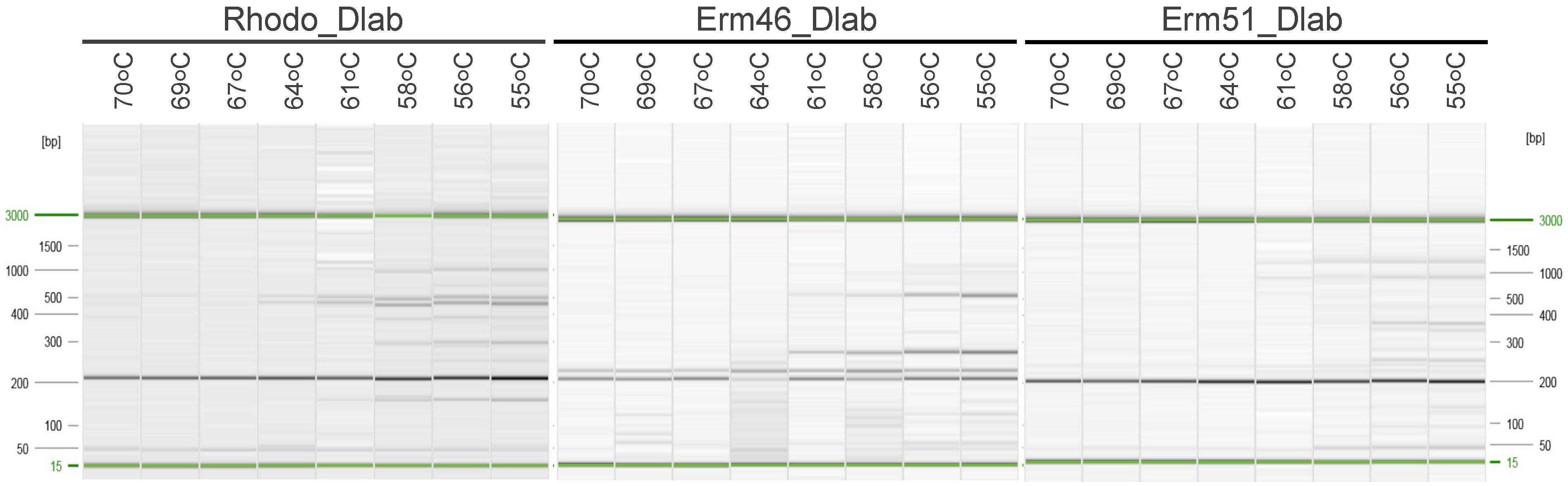
3.1. Oligo Set Design and In-silico Validation

We designed for this study a total of three oligo pairs and their corresponding probes (Table 1) targeting R. equi housekeeping gene choE (Rhodo_Dlab set), and macrolide resistance genes erm(46) (Erm46_Dlab set) and erm(51) (Erm51_Dlab set). We performed a preliminary in-silico validation of the oligos and probes to test their specificity following the inclusivity and exclusivity criteria [16]. We checked in-silico inclusivity by mapping the three primer sets and probes to the whole genome sequences of 202 R. equi (Table S1) isolated from different animal species and presenting multiple susceptibility profiles (Table 2). The Rhodo_Dlab set mapped with the chromosomes of all the 202 R. equi tested at the targeted place. However, three R. equi genomes showed >1 SNP difference with the Rhodo_Dlab set, and to be conservative, we predicted PCR products in 199 out of 202 reactions (98.5%) (Table 2). Erm46_Dlab and Erm51_Dlab sets had perfect matches (zero SNPs) with the genomes of all erm(46)-positive (n = 85) and erm(51)-positive (n = 29) R. equi, respectively, and PCR products were predicted for all reactions (Table 2). Although we observed that the three oligo sets had matches with non-targeted R. equi sequences, amplification was never expected as the combination of the two oligos and corresponding probe was never found in enough proximity to generate a PCR product (Table 2). Additionally, all non-target matches were full of SNPs (data not shown) and that would most probably hamper the alignment between the oligos/probe and the R. equi genome.
We tested the in-silico exclusivity by mapping the three primer sets and probes to the whole genome sequences of 25 non-R. equi reference genomes (plasmids and chromosomes) of bacteria species in close genetic proximity with R. equi and other common respiratory pathogens. Similar to the observations made on the non-targeted R. equi sequences, occasionally, one of the oligos or the probe would align with non-R. equi genomes. (Table S2). Still, detection of amplification was never expected as none of the two primers and corresponding probes align in the same genome simultaneously. (Table 2 and Table S2).

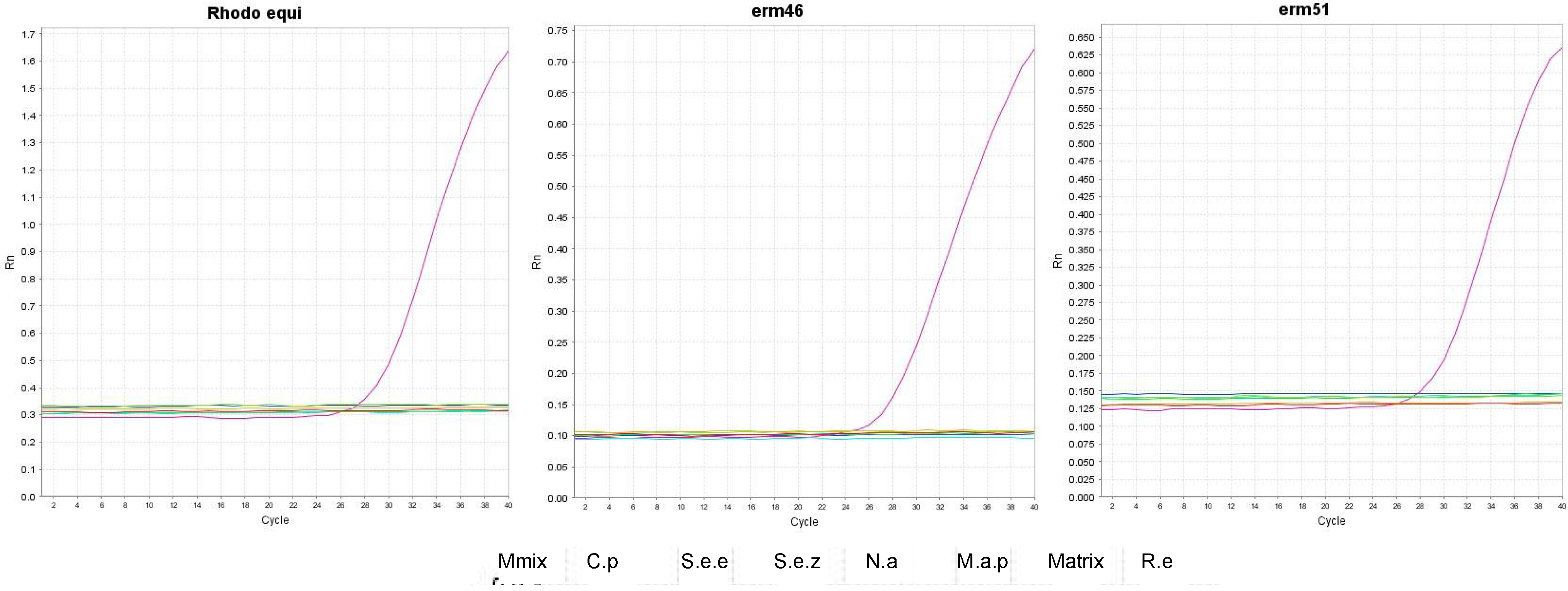
3.2. Testing Oligos and Probes in Singleplex and Multiplex qPCR Assays

We first explored the optimal hybridization temperature for each pair of oligos in a conventional singleplex PCR with a temperature gradient between 55 °C and 70 °C (Figure 1). We observed that the three oligo pairs produced the desired band size (~200 bp) regardless of the hybridization temperature used. Still, their performance was better between 60–70 °C. Most of the qPCR diagnostic tests run in our laboratory use 60 °C as a hybridization temperature. Hence, we decided to use 60 °C as a hybridization temperature in this assay for convenience.
Then we tested the qPCR efficiency and analytic sensitivity (expressed as the limit of detection [LOD]) for each oligo set individually. Macrolide resistant R. equi 2287 was used as a template to characterize the Rhodo_Dlab set and the Erm46_Dlab set, while R. equi lh_50 was used with the Erm51_Dlab group. PCR efficiencies ranged between 104 and 121%, with coefficients of determination (R2) above 0.99 (Table 3. The qPCR detected R. equi (gene choE) even when just as little as 6 × 10⁵ ng of R. equi DNA was present in the sample (Figure 2 and Figure S1), and the LOD for the Rhodo_Dlab set was estimated to be 10.7 complete genome copies per PCR reaction (Table 3). Similarly, the oligo sets designed to identify macrolide resistance genes erm(46) and erm(51) detected the corresponding targets, even when a small load of R. equi DNA was present in the sample (~6 × 10⁶ ng, Figure 2 and Figure S1). The LOD for the Erm46_Dlab and Erm51_Dlab oligo sets was estimated to be 1.18 and 1.07 complete genome copies per PCR reaction, respectively (Table 3).
Finally, we tested the performance of each oligo pair when working together in a multiplex qPCR assay. This time, we decided to use a pool of the three R. equi strains mentioned above (ratio 1:1:1) as a DNA template. We observed that the qPCR efficiency and R2 value decreased for all primers sets (Table 3). No changes in efficiency and sensitivity were observed for the Rhodo_Dlab pair (Table 3). Similarly, a 10-fold decrease in LOD was observed for Erm46_Dlab and Erm51_Dlab oligo sets, and no changes were observed for the Rhodo_Dlab set.

3.3. Testing the Analytic Sensitivity and Specificity in Mocking Equine Respiratory Samples

We extracted and pooled the microbial DNA content of 13 nasopharyngeal swabs from 13 healthy horses (one swab per animal). We used that as representative DNA of the normal equine respiratory flora. We used this DNA extract to dilute R. equi DNA (pool of the three R. equi isolates mentioned above) in 10-fold serial dilutions to have a “mock” respiratory sample containing decreasing amounts of R. equi DNA, and we recalculated the multiplex qPCR efficiency and analytic sensitivity (LOD) (Table 3). We observed that no CT values were recorded for the respiratory microbial DNA when R. equi DNA was not present (Figure 3), demonstrating that our multiplex qPCR is not reactive to normal bacterial communities present in the respiratory tract of horses. Although we found that the LOD slightly decreased in this assay compared to the multiplex qPCR performed on clean R. equi DNA, the amplification efficiency did not significantly change (Table 3).
Additionally, we determined the analytic specificity of the multiplex qPCR by testing its inclusivity, or the ability to detect a wide range of R. equi isolates of the ADVL collection (n = 12, Table S3), and its exclusivity, or lack of interference from three genetically similar organisms C. pseudotuberculosis, N. asteroids, and M. avium subsp. paratuberculosis, and two common equine respiratory pathogens, S. equi subsp. equi, S. equi subsp. zooepidermicus (Figure 3). The multiplex qPCR detected all the R. equi isolates (Table S3). At the same time, no signal (CT values) was observed for the non-R. equi species for any oligo pairs (Figure 3), proving that our strategy is specific for R. equi and not reactive to non-target DNA. All wildtype clinically obtained R. equi tested in this assay were susceptible to macrolides. No signal was detected for the Erm46_Dlab and Erm51_Dlab oligo sets (Table S3), further demonstrating specificity.

4. Discussion

The current diagnosis for R. equi pneumonia in foals generally involves a cytology report describing the presence of pleomorphic rods in respiratory fluids, a preliminary qPCR detecting R. equi directly from the respiratory sample, and subsequent bacteria isolation and antimicrobial susceptibility profiling [17]. Cytology and PCR results are usually available to the clinician in the first 12 h. However, R. equi isolation and corresponding antimicrobial susceptibility testing can take up to 72 h. Due to the insidious nature of this disease [1], fast action is crucial, and clinicians generally start treatment with a macrolide (clarithromycin) and rifampin before the susceptibility profiles are available. This strategy has been effective so far as resistance to macrolides and rifampin was below 5% of all R. equi cases recorded in some studies [5,18]. However, over the last 15 years, the number of resistant isolates has significantly increased, to the point that resistant R. equi to all macrolides and rifampin is being cultured from up to 40% of foals at a farm in Kentucky [19]. The increasing prevalence of MDR R. equi in the US has been recently evidenced by epidemiological studies such as Dr. Huber’s et al. in Kentucky [18,20], and requires the development of new diagnostic tools like the qPCR described here, that account for bacterial detection and clinically relevant resistance identification directly from respiratory samples, fast and accurately.
The dual antimicrobial therapy to treat R. equi infections in horses mentioned above has been used for over 30 years [18,21,22,23]. In the US, resistant R. equi are mainly grouped in three clonal populations with entirely different genetic backgrounds, although they share similar antimicrobial resistance mechanisms [4,5,6]. All resistant clones present a rifampin resistance point mutation at position 531 of the rpoB gene, and they all carry an MLSB resistance erm gene inserted in a transposon [4,5,6]. So far, two different erm genes have been identified in R. equi, erm(46) primarily found in clinical isolates (clones 2287 and G2016) [4,24], and erm(51) mainly associated with environmental samples [6]. Although erm(46) and erm(51) appear to be part of mobilizable elements that could mobilize into other bacteria through horizontal gene transfer [6,7,25], these genes have only been observed in the genomes of R. equi. Additionally, genetic analysis showed that erm(46) and erm(51) share low similarity with each other (~50% at nucleotide level [6]) and with other known erm genes (<68% nucleotide sequence identity to erm(38), erm(39) and erm(40) found in Mycobacterium spp.) [6]. Our multiplex qPCR targets these two erm genes and not the rifampin resistance as punctual mutations are challenging to identify by qPCR, and as epidemiology evidence shows, if an isolate is identified as R. equi and carries an erm gene, it most probably also contains a rifampin resistance mutation [4,5,6].
Like previous approaches, our qPCR strategy targets the R. equi housekeeping gene choE to identify R. equi [10,26,27,28]. Still, we are using a new oligo combination that allows the assay to be run in multiplex with two more sets of primers to detect erm(46) and erm(51) genes. We decided not to target the virulence gene vapA as some did previously [10,26,29,30,31] as vapA is part of a plasmid, and there is a slight chance that this virulence gene is not present in equine R. equi isolates [28,32]. We first performed an in-silico validation of our assay to have preliminary data supporting the specificity of our primers and probes. In-silico validations of molecular-based detection methods are gaining popularity [33,34,35]. However, to our knowledge, this is the first time this approach has been used to develop a qPCR to detect R. equi. The Rhodo_Dlab set showed high in-silico specificity as it mapped with all the chromosomes of the 202 R. equi tested and did not show significant matches with any of the non-R. equi genomes (Table S2).
Still, a small proportion of R. equi genomes (22/202) showed SNPs with the Rhodo_Dlab oligo set: 19 genomes presented only one SNP difference with the Rhodo_Dlab set and three R. equi genomes showed >1 SNP (Table 2). SNPs in primer and probe binding sites can destabilize oligonucleotide binding and reduce target specificity [36]. Yet, PCR performance studies showed that a single SNP does not prevent amplification [37]. Hence, we estimated that the in-silico inclusivity of the Rhodo_Dlab set is 98.5%, and its exclusivity is 100%. Erm46_Dlab and Erm51_Dlab sets only had perfect matches (zero SNPs difference) with the genomes of macrolide-resistant erm(46)-positive and erm(51)-positive R. equi, respectively. They were predicted to have 100% inclusivity and exclusivity (Table 2). Overall, the in-silico validation of the three primer sets designed in this study showed that they are highly specific to R. equi in the case of the Rhodo_DLab set and macrolide resistance genes erm(46) and erm(51) (Erm46_Dlab and Erm51_Dlab oligo sets, respectively). This encourages us to continue testing our qPCR assay in-vitro.
The LOD and amplification efficiency of our choE oligo set (Rhodo_DLab) in singleplex and multiplex assays are comparable to what was previously published [26], and the three oligo sets used in this study showed LODs and amplification efficiencies above what considered an optimal performance. Additionally, we proved the high analytic specificity of this new R. equi multiplex qPCR by testing its inclusivity and exclusivity [16]. In total, 100% of the R. equi strains tested were identified, while none of the non-target species gave a signal in the multiplex. Furthermore, our multiplex qPCR showed not to be reactive to normal bacterial communities present in the respiratory tract of horses, further demonstrating the utility of our assay as a crucial R. equi diagnostic tool. Unfortunately, all the R. equi clinical isolates of the AVDL collection were susceptible to macrolides, so we could not test inclusivity. Future work will focus on validating this qPCR with respiratory samples from horses diagnosed with pneumonia caused by R. equi susceptible and resistant to macrolides and rifampin. Additionally, new resistances to macrolides and rifampin that our test does not detect may develop. Hence, although our qPCR strategy is an excellent tool to provide a fast answer to clinicians, verification based on bacterial culture and susceptibility testing for all samples is paramount, even if tested by this method.

5. Conclusions

This manuscript describes a new multiplex qPCR assay for the simultaneous identification of R. equi and its two most clinically relevant macrolide resistance genes, erm(46) and erm(51). Our qPCR approach is highly specific and sensitive, even when performed directly from equine respiratory samples.

Supplementary Materials

The following supporting information can be downloaded at: https://www.mdpi.com/article/10.3390/ani12091172/s1, Figure S1: Standard curves for qPCR in mocking communities; Table S1: List of R. equi genomes included in the in-silico validation, Table S2: In the in-silico validation of oligos and probes in 25 non-R.equi bacterial genomes (chromosomes and plasmids), Table S3: R. equi clinical isolates tested in this study

Author Contributions

Conceptualization, S.Á.N. and S.S.; methodology, S.Á.N., S.S. and I.F.; software, I.F.; validation, I.F. and N.V.P.; formal analysis, S.Á.N., I.F. and S.S.; investigation, S.Á.N.; resources, S.S.; data curation, S.S.; writing—original draft preparation, S.Á.N.; writing—review and editing, S.Á.N. and S.S.; visualization, S.Á.N. and I.F.; supervision, S.S.; project administration, S.Á.N. and S.S.; funding acquisition, S.S. All authors have read and agreed to the published version of the manuscript.

Funding

This work was supported by grant 1U18FD006157-04 from the U.S. Department of Agriculture.

Institutional Review Board Statement

Not applicable.

Informed Consent Statement

Not applicable.

Data Availability Statement

The datasets analyzed for this study can be found in GenBank (https://www.ncbi.nlm.nih.gov/genbank/, accessed on 22 April 2022).

Acknowledgments

We thank L. Berghaus, K. Hart and the Large Animal Medicine Department from the University of Georgia for facilitating the R. equi strains PAM2287 and 103-ApraR and Laura Huber from the Department of Pathobiology, College of Veterinary Medicine at Auburn University for facilitating R. equi strain lh_50. We also want to thank Noah Cohen from Texas A&M University for advising us about the standard procedures to treat foals with R. equi pneumonia in the clinical setting.

Conflicts of Interest

The authors declare no conflict of interest.

References

  1. Giguère, S.; Cohen, N.; Chaffin, M.K.; Hines, S.; Hondalus, M.; Prescott, J.; Slovis, N. Rhodococcus equi: Clinical Manifestations, Virulence, and Immunity. J. Veter. Intern. Med. 2011, 25, 1221–1230. [Google Scholar] [CrossRef] [PubMed]
  2. Vázquez-Boland, J.A.; Giguère, S.; Hapeshi, A.; MacArthur, I.; Anastasi, E.; Valero-Rello, A. Rhodococcus equi: The many facets of a pathogenic actinomycete. Veter Microbiol. 2013, 167, 9–33. [Google Scholar] [CrossRef] [PubMed]
  3. Prescott, J.F. Rhodococcus equi: An animal and human pathogen. Clin. Microbiol. Rev. 1991, 4, 20–34. [Google Scholar] [CrossRef] [PubMed]
  4. Álvarez-Narváez, S.; Giguère, S.; Cohen, N.; Slovis, N.; Vázquez-Boland, J.A. Spread of Emerging Multidrug-Resistant Rhodococcus equi in USA. Emerg. Infect. Dis. 2021, 27, 529–537. [Google Scholar] [CrossRef] [PubMed]
  5. Álvarez-Narváez, S.; Huber, L.; Giguère, S.; Hart, K.A.; Berghaus, R.D.; Sanchez, S.; Cohen, N.D. Epidemiology and Molecular Basis of Multidrug Resistance in Rhodococcus equi. Microbiol. Mol. Biol. Rev. 2021, 85, e00011-21. [Google Scholar] [CrossRef]
  6. Huber, L.; Giguère, S.; Slovis, N.M.; Álvarez-Narváez, S.; Hart, K.A.; Greiter, M.; Morris, E.R.A.; Cohen, N.D. The novel and transferable erm (51) gene confers macrolides, lincosamides and streptogramins B (MLS B) resistance to clonal Rhodococcus equi in the environment. Environ. Microbiol. 2020, 22, 2858–2869. [Google Scholar] [CrossRef]
  7. Álvarez-Narváez, S.; Giguère, S.; Anastasi, E.; Hearn, J.; Scortti, M.; Vázquez-Boland, J.A. Clonal Confinement of a Highly Mobile Resistance Element Driven by Combination Therapy in Rhodococcus equi. mBio 2019, 10, e02260-19. [Google Scholar] [CrossRef] [Green Version]
  8. Anastasi, E.; MacArthur, I.; Scortti, M.; Alvarez, S.; Giguère, S.; Vázquez-Boland, J.A. Pangenome and Phylogenomic Analysis of the Pathogenic Actinobacterium Rhodococcus equi. Genome Biol. Evol. 2016, 8, 3140–3148. [Google Scholar] [CrossRef] [Green Version]
  9. Arriaga, J.M.; Cohen, N.; Derr, J.N.; Chaffin, M.K.; Martens, R.J. Detection of Rhodococcus equi by Polymerase Chain Reaction Using Species-Specific Nonproprietary Primers. J. Veter. Diagn. Investig. 2002, 14, 347–353. [Google Scholar] [CrossRef] [Green Version]
  10. Halbert, N.D.; Reitzel, R.A.; Martens, R.J.; Cohen, N.D. Evaluation of a multiplex polymerase chain reaction assay for simultaneous detection of Rhodococcus equi and the vapA gene. Am. J. Veter. Res. 2005, 66, 1380–1385. [Google Scholar]
  11. Rutherford, K.; Parkhill, J.; Crook, J.; Horsnell, T.; Rice, P.; Rajandream, M.-A.; Barrell, B. Artemis: Sequence visualization and annotation. Bioinformatics 2000, 16, 944–945. [Google Scholar] [CrossRef] [PubMed] [Green Version]
  12. Davis, M.W.; Jorgensen, E.M. ApE, A Plasmid Editor: A Freely Available DNA Manipulation and Visualization Program. Front. Bioinform. 2022, 2, 818619. [Google Scholar] [CrossRef]
  13. Kearse, M.D.; Sturrock, S.S.; Meintjes, P.L. The Geneious 6.0.3 Read Mapper. 2012. Available online: http://assets.geneious.com/documentation/geneious/GeneiousReadMapper.pdf (accessed on 17 March 2022).
  14. Tripathi, V.N.; Harding, W.C.; Willingham-Lane, J.M.; Hondalus, M.K. Conjugal Transfer of a Virulence Plasmid in the Opportunistic Intracellular Actinomycete Rhodococcus equi. J. Bacteriol. 2012, 194, 6790–6801. [Google Scholar] [CrossRef] [PubMed] [Green Version]
  15. Bustin, S.A.; Benes, V.; Garson, J.A.; Hellemans, J.; Huggett, J.; Kubista, M.; Mueller, R.; Nolan, T.; Pfaffl, M.W.; Shipley, G.L.; et al. The MIQE Guidelines: Minimum Information for Publication of Quantitative Real-Time PCR Experiments. Clin. Chem. 2009, 55, 611–622. [Google Scholar] [CrossRef] [PubMed] [Green Version]
  16. Kralik, P.; Ricchi, M. A Basic Guide to Real Time PCR in Microbial Diagnostics: Definitions, Parameters, and Everything. Front. Microbiol. 2017, 8, 108. [Google Scholar] [CrossRef] [PubMed] [Green Version]
  17. Giguère, S.; Cohen, N.; Chaffin, M.K.; Slovis, N.; Hondalus, M.; Hines, S.; Prescott, J. Diagnosis, Treatment, Control, and Prevention of Infections Caused by Rhodococcus equi in Foals. J. Veter. Intern. Med. 2011, 25, 1209–1220. [Google Scholar] [CrossRef] [PubMed]
  18. Huber, L.; Giguère, S.; Slovis, N.M.; Carter, C.N.; Barr, B.S.; Cohen, N.D.; Elam, J.; Erol, E.; Locke, S.J.; Phillips, E.D.; et al. Emergence of Resistance to Macrolides and Rifampin in Clinical Isolates of Rhodococcus equi from Foals in Central Kentucky, 1995 to 2017. Antimicrob. Agents Chemother. 2019, 63, e01714-18. [Google Scholar] [CrossRef] [PubMed] [Green Version]
  19. Burton, A.J.; Giguère, S.; Sturgill, T.L.; Berghaus, L.J.; Slovis, N.M.; Whitman, J.L.; Levering, C.; Kuskie, K.R.; Cohen, N.D. Macrolide- and Rifampin-Resistant Rhodococcus equion a Horse Breeding Farm, Kentucky, USA. Emerg. Infect. Dis. 2013, 19, 282–285. [Google Scholar] [CrossRef]
  20. Huber, L.; Giguère, S.; Cohen, N.D.; Slovis, N.M.; Hanafi, A.; Schuckert, A.; Berghaus, L.; Greiter, M.; Hart, K.A. Prevalence and risk factors associated with emergence of Rhodococcus equi resistance to macrolides and rifampicin in horse-breeding farms in Kentucky, USA. Veter. Microbiol. 2019, 235, 243–247. [Google Scholar] [CrossRef]
  21. Giguère, S. Treatment of Infections Caused by Rhodococcus equi. Veter. Clin. N. Am. Equine Pract. 2017, 33, 67–85. [Google Scholar] [CrossRef]
  22. Giguère, S.; Berghaus, L.J.; Willingham-Lane, J.M. Antimicrobial Resistance in Rhodococcus equi. Microbiol. Spectr. 2017, 5. [Google Scholar] [CrossRef] [PubMed]
  23. Giguère, S.; Lee, E.; Williams, E.; Cohen, N.D.; Chaffin, M.K.; Halbert, N.; Martens, R.J.; Franklin, R.P.; Clark, C.C.; Slovis, N.M. Determination of the prevalence of antimicrobial resistance to macrolide antimicrobials or rifampin in Rhodococcus equi isolates and treatment outcome in foals infected with antimicrobial-resistant isolates of R equi. J. Am. Veter. Med. Assoc. 2010, 237, 74–81. [Google Scholar] [CrossRef] [PubMed]
  24. Anastasi, E.; Giguère, S.; Berghaus, L.J.; Hondalus, M.K.; Willingham-Lane, J.M.; MacArthur, I.; Cohen, N.D.; Roberts, M.C.; Vazquez-Boland, J.A. Novel transferableerm (46) determinant responsible for emerging macrolide resistance in Rhodococcus equi. J. Antimicrob. Chemother. 2015, 70, 3184–3190. [Google Scholar] [CrossRef] [PubMed] [Green Version]
  25. Álvarez-Narváez, S.; Giguère, S.; Berghaus, L.J.; Dailey, C.; Vázquez-Boland, J.A. Horizontal Spread of Rhodococcus equi Macrolide Resistance Plasmid pRErm46 across Environmental Actinobacteria. Appl. Environ. Microbiol. 2020, 86, e00108-20. [Google Scholar] [CrossRef] [PubMed]
  26. Rodríguez-Lázaro, D.; Lewis, D.A.; Ocampo-Sosa, A.A.; Fogarty, U.; Makrai, L.; Navas, J.; Scortti, M.; Hernández, M.; Vázquez-Boland, J.A. Internally Controlled Real-Time PCR Method for Quantitative Species-Specific Detection and vapA Genotyping of Rhodococcus equi. Appl. Environ. Microbiol. 2006, 72, 4256–4263. [Google Scholar] [CrossRef] [PubMed] [Green Version]
  27. Ladrón, N.; Fernández, M.; Agüero, J.; Zörn, B.G.; Vázquez-Boland, J.A.; Navas, J. Rapid Identification of Rhodococcus equi by a PCR Assay Targeting the choE Gene. J. Clin. Microbiol. 2003, 41, 3241–3245. [Google Scholar] [CrossRef] [Green Version]
  28. Ocampo-Sosa, A.A.; Lewis, D.A.; Navas, J.; Quigley, F.; Callejo, R.; Scortti, M.; Leadon, D.P.; Fogarty, U.; Vázquez-Boland, J.A. Molecular Epidemiology of Rhodococcus equi Based on traA, vapA, and vapB Virulence Plasmid Markers. J. Infect. Dis. 2007, 196, 763–769. [Google Scholar] [CrossRef] [Green Version]
  29. Madrigal, R.; Shaw, S.; Witkowski, L.; Sisson, B.; Blodgett, G.; Chaffin, M.; Cohen, N. Use of Serial Quantitative PCR of the vapA Gene of Rhodococcus equi in Feces for Early Detection of R. equi Pneumonia in Foals. J. Veter. Intern. Med. 2016, 30, 664–670. [Google Scholar] [CrossRef]
  30. Pusterla, N.; Wilson, W.D.; Mapes, S.; Leutenegger, C.M. Diagnostic evaluation of real-time pcr in the detection of Rhodococcus equi in faeces and nasopharyngeal swabs from foals with pneumonia. Veter. Rec. 2007, 161, 272–274. [Google Scholar] [CrossRef] [Green Version]
  31. Harrington, J.R.; Golding, M.C.; Martens, R.J.; Halbert, N.D.; Cohen, N.D. Evaluation of a real-time quantitative polymerase chain reaction assay for detection and quantitation of virulent Rhodococcus equi. Am. J. Veter. Res. 2005, 66, 755–761. [Google Scholar] [CrossRef] [Green Version]
  32. Alvarez-Narvaez, S. Conjugal Transfer of Host-Adaptive Determinants in the Pathogenic Actinobacterium Rhodococcus equi. Ph.D. Thesis, University of Edinburgh, Edinburgh, UK, 2017. [Google Scholar]
  33. Flannery, J.; Rajko-Nenow, P.; Arnold, H.; Van Weezep, E.; van Rijn, P.A.; Ngeleja, C.; Batten, C. Improved PCR diagnostics using up-to-date in-silico validation: An F-gene RT-qPCR assay for the detection of all four lineages of peste des petits ruminants virus. J. Virol. Methods 2019, 274, 113735. [Google Scholar] [CrossRef] [PubMed]
  34. Davi, M.J.P.; Jeronimo, S.M.B.; Lima, J.P.M.S.; Lanza, D.C.F. Design and in-silico validation of polymerase chain reaction primers to detect severe acute respiratory syndrome coronavirus 2 (SARS-CoV-2). Sci. Rep. 2021, 11, 12565. [Google Scholar] [CrossRef] [PubMed]
  35. Nagy, A.; Jiřinec, T.; Jiřincová, H.; Černíková, L.; Havlíčková, M. In-silico re-assessment of a diagnostic RT-qPCR assay for universal detection of Influenza A viruses. Sci. Rep. 2019, 9, 1630. [Google Scholar] [CrossRef] [PubMed]
  36. Lefever, S.; Pattyn, F.; Hellemans, J.; Vandesompele, J. Single-Nucleotide Polymorphisms and Other Mismatches Reduce Performance of Quantitative PCR Assays. Clin. Chem. 2013, 59, 1470–1480. [Google Scholar] [CrossRef] [Green Version]
  37. Letowski, J.; Brousseau, R.; Masson, L. Designing better probes: Effect of probe size, mismatch position and number on hybridization in DNA oligonucleotide microarrays. J. Microbiol. Methods 2004, 57, 269–278. [Google Scholar] [CrossRef]
Figure 1. Conventional PCR with temperature gradient for oligo sets Rhodo_Dlab, Erm46_Dlab, and Erm51_Dlab. Expected band of ~200 bp size.
Animals 12 01172 g001
Figure 2. Testing the analytic sensitivity of Rhodo_Dlab, Erm46_Dlab, and Erm51_Dlab sets in singleplex. qPCR curves for the three oligo sets in singleplex assays. In the Y-exe ΔRn, or fluorescence signal. In the X-exe, PCR amplification cycle. Colors indicate different 10-fold dilution of R.equi DNA (ng/µL).
Animals 12 01172 g002
Figure 3. Testing the analytic specificity of Rhodo_Dlab, Erm46_Dlab, and Erm51_Dlab sets in the multiplex. qPCR curves for the three oligo sets in multiplex assays. In the Y-exe ΔRn, or fluorescence signal. In the X-exe, PCR amplification cycle. Colors indicate different bacteria species and PCR controls: pink- R.equi mix DNA (R.e); purple- nasal swab DNA (matrix); dark blue, M. avium subsp. paratuberculosis (M.a.p); light blue- N. asteorides (N.a); dark green-S. equi subsp. zooepidermicus (S.e.z); light green-S. equi subsp. equi (S.e.e); yellow- C. pseudotuberculosis (C.p); red negative control (no DNA, Mmix).
Animals 12 01172 g003
Table 1. Oligos and probes.
Name SEQUENCE 5’-3’Product SizePurpose
Rhodo_Dlab_FTGTCAACAACATCGACCAGGC200 bpAmplifies choE gene, chromosomal marker in R. equi
Rhodo_Dlab_RGCGTTGTTGCCGTAGATGAC
Rhodo_Dlab_P/56-FAM/CCGCCCAAC/ZEN/GTTCGGGTTTCACAACCGCTT/3IABkFQ/ *
Erm46_Dlab_FGTGGCGCAACGATGATGACT192 bpAmplifies macrolide resistance gene erm46
Erm46_Dlab_RTGAAGACGGTGTGGACGAAG
Erm46_Dlab_P/5HEX/CCGCATCGG/ZEN/CGTTCACACCACGGC/3IABkFQ/ *
Erm51_Dlab_FCTGCCGTTTCACCTGACCAC198 bpAmplifies macrolide resistance gene erm51
Erm51_Dlab_RGGGACGGAAATGTGTGGATG
Erm51_Dlab_P/5Cy5/GCCGGCGTC/TAO/GGTGGTGCCACGATGATGA/3IAbRQSp/ *
* /56-FAM/-excitation 495 emission 520;/5HEX/- excitation 538 emission 555; 5Cy5/-excitation 648 emission 668.
Table 2. In-silico validation of the oligos and probes.
Match with Rhodo_Dlab SetMatch with Erm46_Dlab SetMatch with Erm51_Dlab Set
FWRVProbePCR ProductsFWRVProbePCR ProductsFWRVProbePCR Products
Macrolide Susceptible
[n = 88]
888888871881010880
Macrolide Resistant
erm(46)-positive [n = 85]
858585838585858521850
Macrolide Resistant
erm(51)-positive [n = 29]
2929292902914029292929
Non- R. equi species
[n = 24]
2620463010100
In hard brackets is the number of strains in each resistant genotype category. FW refers to the number of genomes that had a match with the forward oligo of the set. RV refers to the number of genomes that had a match with the reverse oligo of the set. Probe refers to the number of genomes that had a match with the probe of the set. PCR product indicates the number of genomes, in which the PCR set would produce a detectable PCR product (forward and reverse primers are in <2 kb proximity and corresponding probe is inside that <2 kb DNA fragment). This was calculated based on QuantiNova DNA polymerase processivity (Taq DNA Polymerase 2–4 kb/min) and PCR elongation time (30 s).
Table 3. Efficiency, coefficient of determination (R2) and limit of determination (LOD) for each primer set in singleplex and multiplex assays.
Efficiency (%)R2LOD
SingleplexMultiplexMultiplex Mocking *SingleplexMultiplexMultiplex Mocking *SingleplexMultiplexMultiplex Mocking *
Rhodo_Dlab121.1112.8115.50.99760.99870.990410.71110.7
Erm46_Dlab104.7105.5102.80.99990.99940.99761.1811.810.7
Erm51_Dlab120.5105.490.20.99930.99710.99171.0710.7106.9
LOD expressed as the minimal number of complete genome copies detected per PCR reaction. * Multiplex qPCR performed in the mocking equine respiratory samples spiked with R. equi DNA.
Publisher’s Note: MDPI stays neutral with regard to jurisdictional claims in published maps and institutional affiliations.

