
Analgesia during Parturition in Domestic Animals: Perspectives and Controversies on Its Use

 , , , ,
  ,
,  ,
,  ,
,  and
and 
Simple Summary
Abstract
1. Introduction
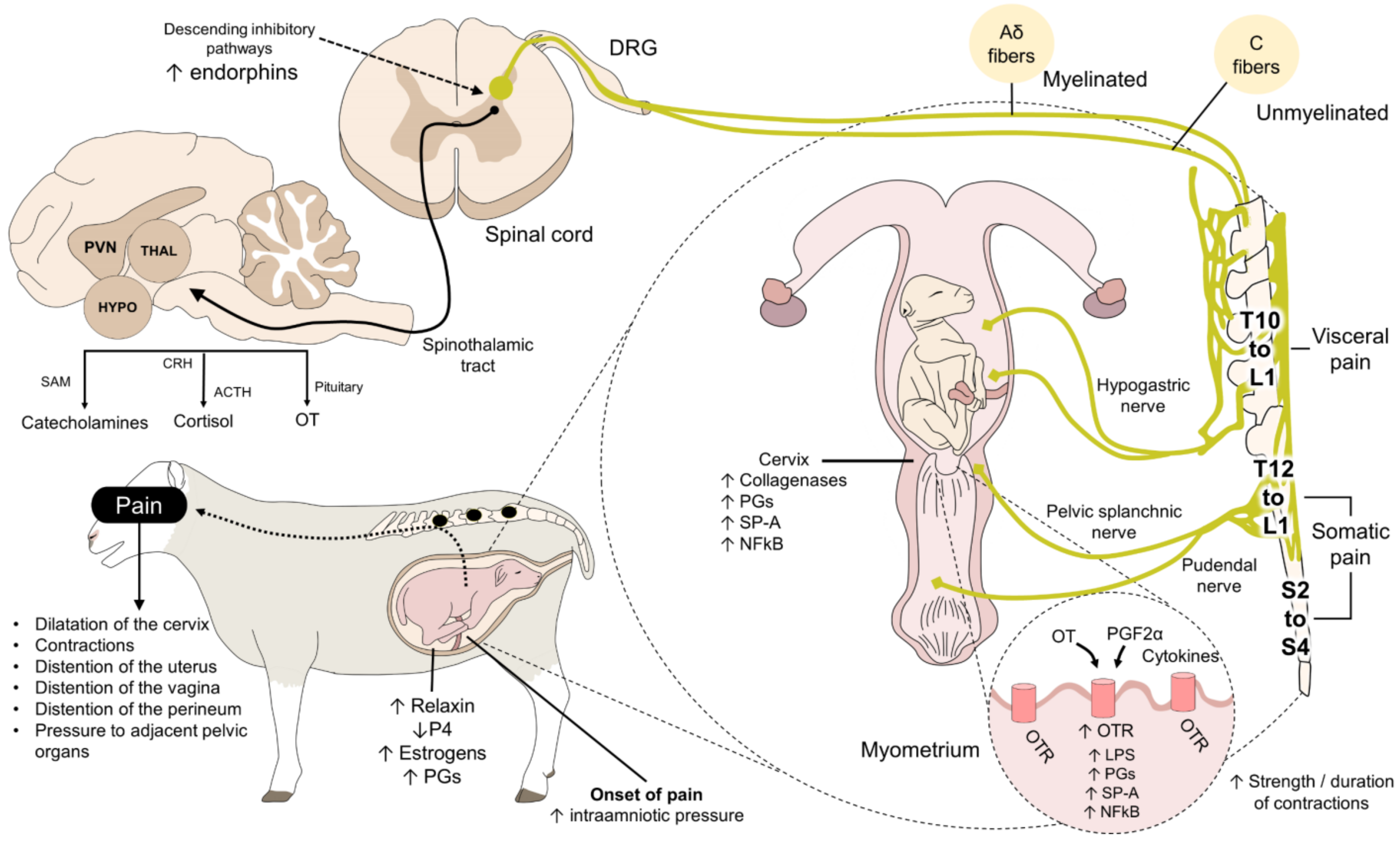
2. Controversy on the Participation of Pain during Parturition
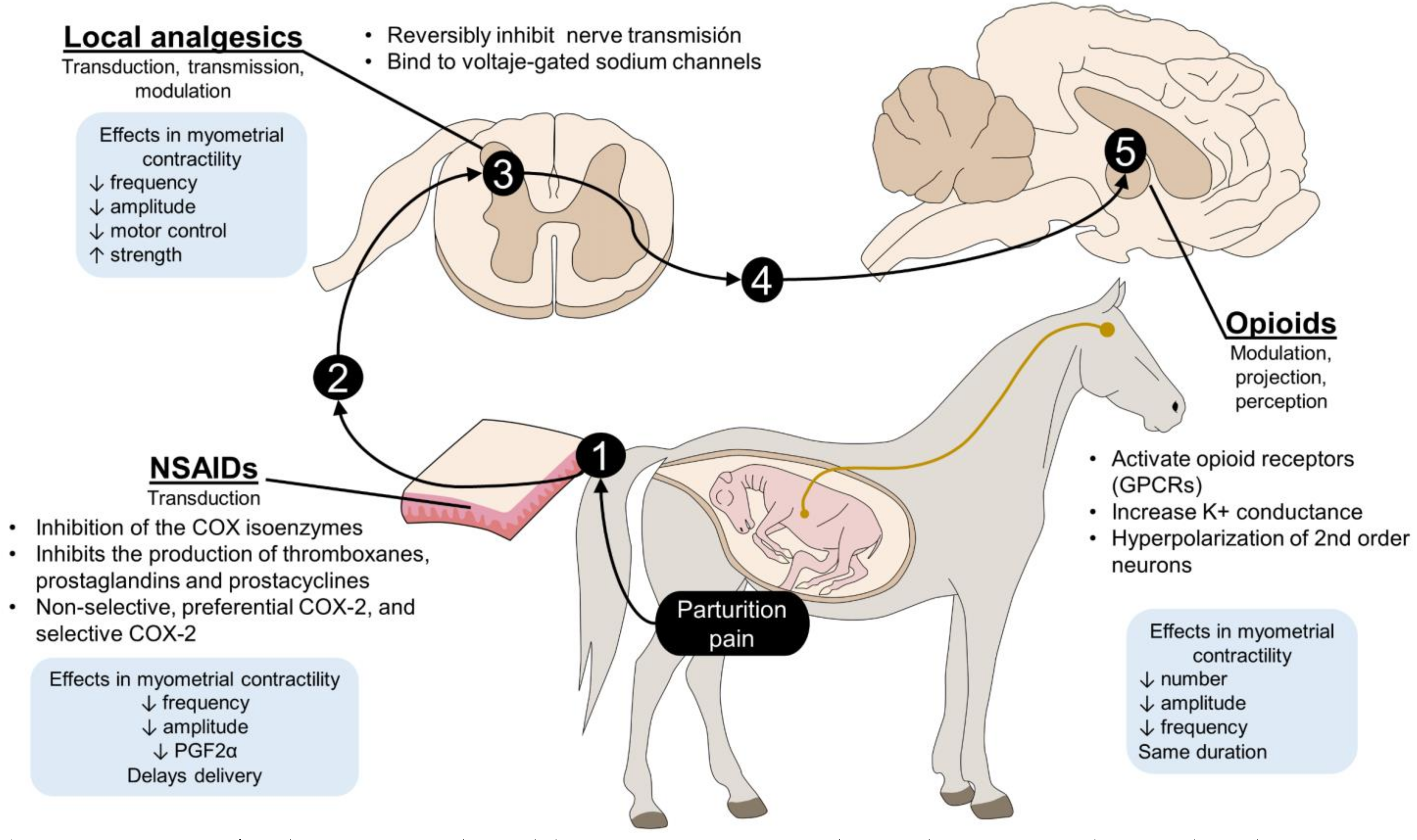
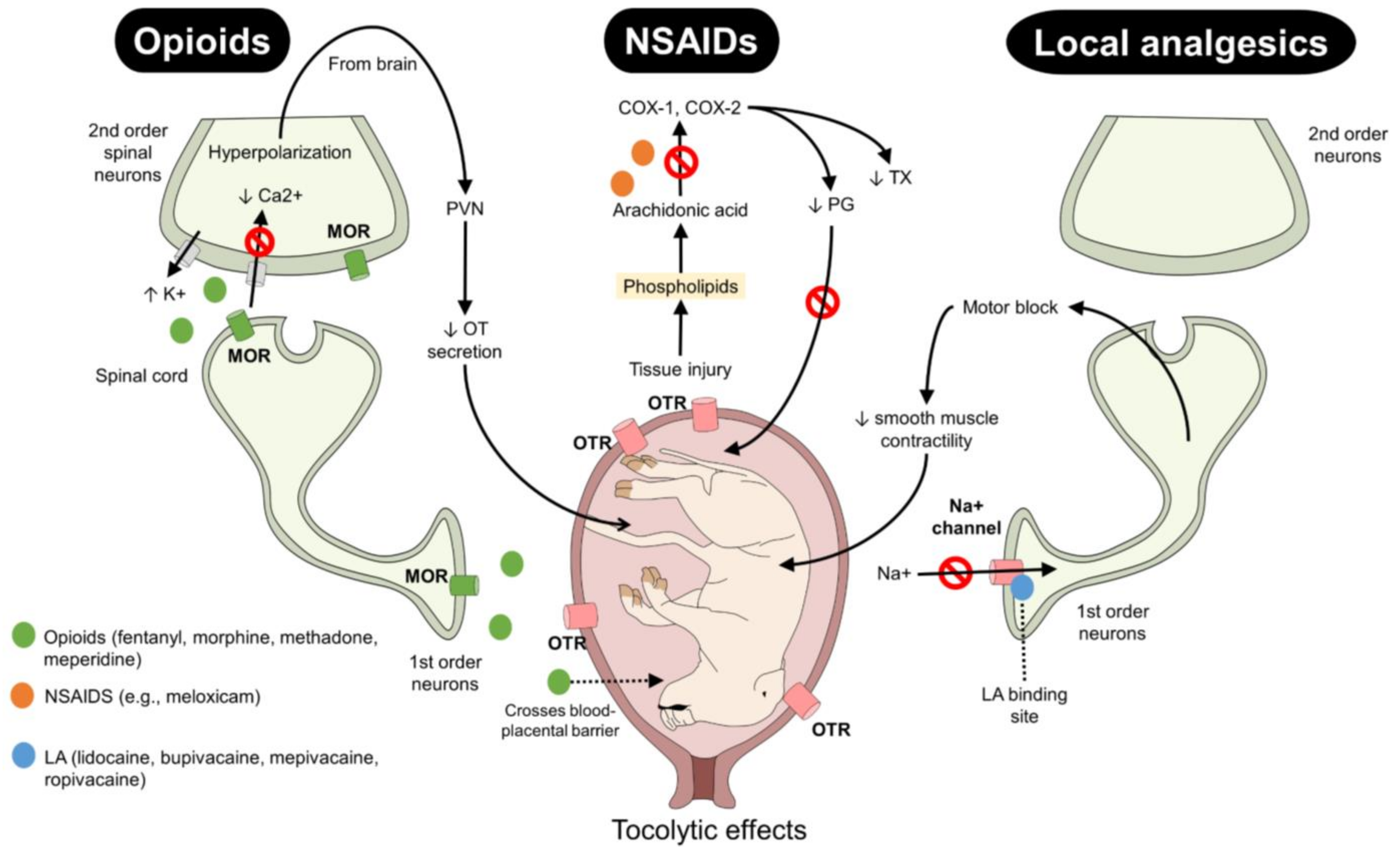
3. Opioids
4. Non-Steroidal Anti-Inflammatory Drugs
5. Local Analgesics
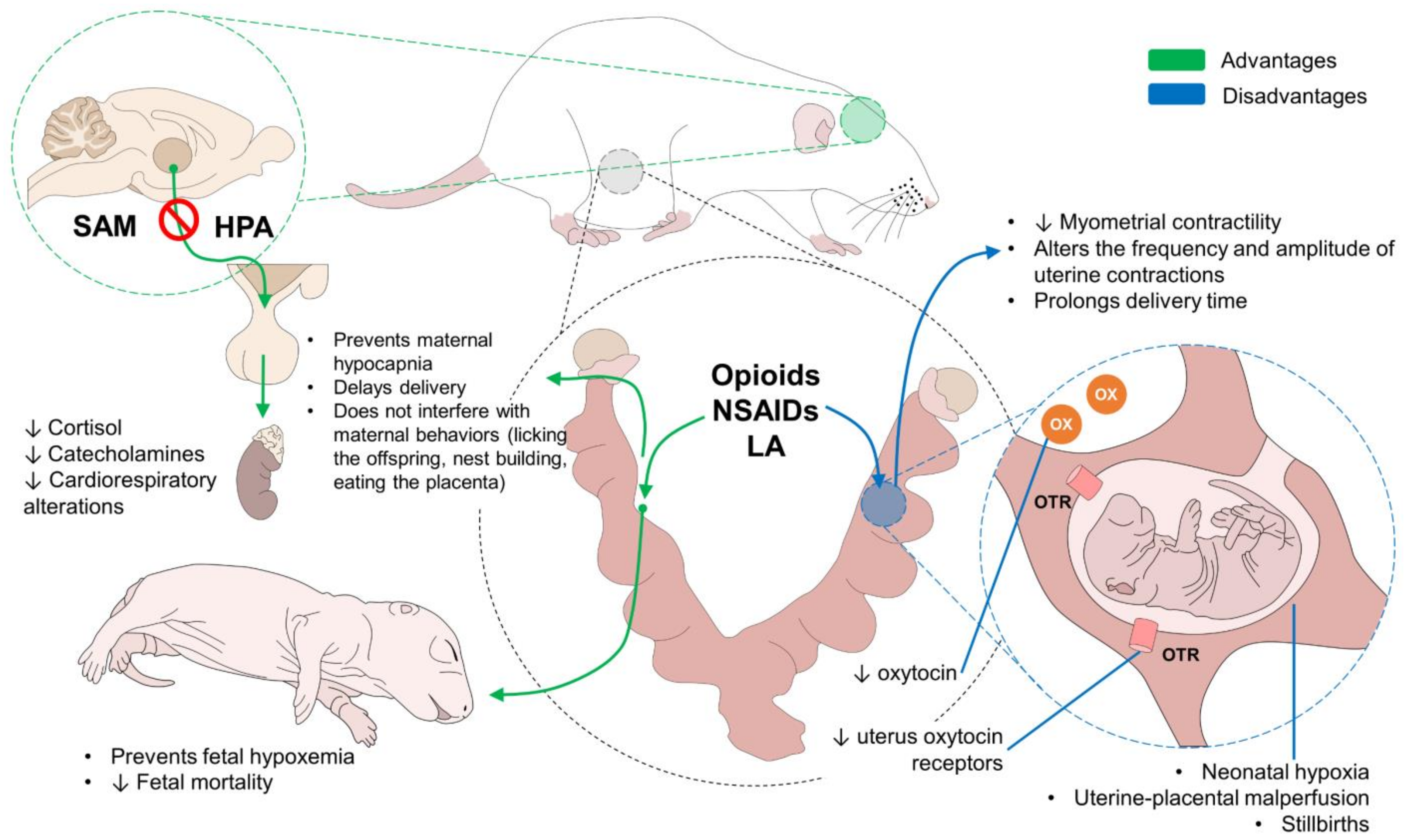
6. Perspectives
7. Conclusions
Author Contributions
Funding
Institutional Review Board Statement
Informed Consent Statement
Data Availability Statement
Conflicts of Interest
References
- Hernández-Avalos, I.; Mota-Rojas, D.; Mora-Medina, P.; Martínez-Burnes, J.; Casas Alvarado, A.; Verduzco-Mendoza, A.; Lezama-García, K.; Olmos-Hernandez, A. Review of different methods used for clinical recognition and assessment of pain in dogs and cats. Int. J. Vet. Sci. Med. 2019, 7, 43–54. [Google Scholar] [CrossRef] [PubMed]
- Hernández-Avalos, I.; Flores-Gasca, E.; Mota-Rojas, D.; Casas-Alvarado, A.; Miranda-Cortés, A.E.; Domínguez-Oliva, A. Neurobiology of anesthetic-surgical stress and induced behavioral changes in dogs and cats: A review. Vet. World 2021, 14, 393–404. [Google Scholar] [CrossRef] [PubMed]
- Shock, D.A.; Renaund, D.L.; Roche, S.M.; Poliquin, R.; Thomson, R.; Olson, M.E. Correction: Evaluating the impact of meloxicam oral suspension administered at parturition on subsequent production, health, and culling in dairy cows: A randomized clinical field trial. PLoS ONE 2018, 13, e0210326. [Google Scholar] [CrossRef] [PubMed]
- Nacitarhan, C.; Sadan, G.; Kayacan, N.; Ertugrul, F.; Arici, G.; Karsli, B.; Erman, M. The effects of opioids, local anaesthetics and adjuvants on isolated pregnant rat uterine muscles. Methods Find. Exp. Clin. Pharmacol. 2007, 29, 273. [Google Scholar] [CrossRef]
- Rac, V.E.; Scott, C.A.; Small, C.; Lee Adamson, S.; Rurak, D.; Challis, J.R.; Lye, S.J. Dose-dependent effects of meloxicam administration on cyclooxygenase-1 and cyclooxygenase-2 protein expression in intrauterine tissues and fetal tissues of a sheep model of preterm labor. Reprod. Sci. 2007, 14, 750–764. [Google Scholar] [CrossRef]
- Alper, M.H.; Brown, W.U.; Ostheimer, G.W.; Scanlon, J.W. Effects of maternal analgesia and anaesthesia on the newborn. Clin. Obstet. Gynaecol. 1975, 2, 661–671. [Google Scholar] [CrossRef]
- Martínez-Burnes, J.; Muns, R.; Barrios-García, H.; Villanueva-García, D.; Domínguez-Oliva, A.; Mota-Rojas, D. Parturition in mammals: Animal models, pain and distress. Animals 2021, 11, 2960. [Google Scholar] [CrossRef] [PubMed]
- Nagel, C.; Aurich, C.; Aurich, J. Stress effects on the regulation of parturition in different domestic animal species. Anim. Reprod. Sci. 2019, 207, 153–161. [Google Scholar] [CrossRef] [PubMed]
- Heavner, J.E.; Cooper, D.M. Pharmacology of analgesics. In Anesthesia and Analgesia in Laboratory Animals, 2nd ed.; Fish, R., Danneman, P., Brown, M., Karas, A., Eds.; Academic Press: San Diego, CA, USA, 2008; pp. 97–123. [Google Scholar]
- Welsh, T.; Johnson, M.; Yi, L.; Tan, H.; Rahman, R.; Merlino, A.; Zakar, T.; Mesiano, S. Estrogen receptor (ER) expression and function in the pregnant human myometrium: Estradiol via ERα activates ERK1/2 signaling in term myometrium. J. Endocrinol. 2012, 212, 227–238. [Google Scholar] [CrossRef] [PubMed][Green Version]
- Katz, N.; Mazer, N.A. The impact of opioids on the endocrine system. Clin. J. Pain 2009, 25, 170–175. [Google Scholar] [CrossRef] [PubMed]
- Vuong, C.; Van Uum, S.H.M.; O’Dell, L.E.; Lutfy, K.; Friedman, T.C. The effects of opioids and opioid analogs on animal and human endocrine systems. Endocr. Rev. 2010, 31, 98–132. [Google Scholar] [CrossRef]
- Wei, J.-S.; Jin, Z.-B.; Yin, Z.-Q.; Xie, Q.-M.; Chen, J.-Q.; Li, Z.-G.; Tang, H.-F. Effects of local anesthetics on contractions of pregnant and non-pregnant rat myometrium in vitro. Acta Physiol. Hung. 2014, 101, 228–235. [Google Scholar] [CrossRef] [PubMed]
- Qian, X.; Wang, Q.; Ou, X.; Li, P.; Zhao, B.; Liu, H. Effects of ropivacaine in patient-controlled epidural analgesia on uterine electromyographic activities during labor. Biomed. Res. Int. 2018, 2018, 7162865. [Google Scholar] [CrossRef] [PubMed]
- Zhao, B.; Qian, X.; Wang, Q.; Ou, X.; Lin, B.; Song, X. The effects of ropivacaine 0.0625% and levobupivacaine 0.0625% on uterine and abdominal muscle electromyographic activity during the second stage of labor. Minerva Anestesiol. 2019, 85, 854–861. [Google Scholar] [CrossRef] [PubMed]
- Mainau, E.; Manteca, X. Pain and discomfort caused by parturition in cows and sows. Appl. Anim. Behav. Sci. 2011, 135, 241–251. [Google Scholar] [CrossRef]
- Alimoradi, Z.; Kazemi, F.; Valiani, M.; Gorji, M. Comparing the effect of auricular acupressure and body acupressure on pain and duration of the first stage of labor: Study protocol for a randomized controlled trial. Trials 2019, 20, 766. [Google Scholar] [CrossRef] [PubMed]
- Hashemi Asl, B.M.; Vatanchi, A.; Golmakani, N.; Najafi, A. Relationship between behavioral indices of pain during labor pain with pain intensity and duration of delivery. Electron. Physician 2018, 10, 6240–6248. [Google Scholar] [CrossRef] [PubMed][Green Version]
- Kidder, G.M.; Winterhager, E. Physiological roles of connexins in labour and lactation. Reproduction 2015, 150, R129–R136. [Google Scholar] [CrossRef] [PubMed]
- Purohit, G. Parturition in domestic animals: A review. WebmedCentral Reprod. 2010, 1, WMC00748. [Google Scholar]
- Mota-Rojas, D.; Villanueva-García, D.; Velazquez-Armenta, E.Y.; Nava-Ocampo, A.A.; Ramírez-Necoechea, R.; Alonso-Sspilsbury, M.; Trujillo, M.E. Influence of time at which oxytocin is administered during labor on uterine activity and perinatal death in pigs. Biol. Res. 2007, 40, 55–63. [Google Scholar] [CrossRef]
- Boll, S.; Almeida de Minas, A.C.; Raftogianni, A.; Herpertz, S.C.; Grinevich, V. Oxytocin and pain perception: From animal models to human research. Neuroscience 2018, 387, 149–161. [Google Scholar] [CrossRef]
- Meddle, S.; Leng, G.; Selvarajah, J.; Bicknell, R.; Russell, J. Direct pathways to the supraoptic nucleus from the brainstem and the main olfactory bulb are activated at parturition in the rat. Neuroscience 2000, 101, 1013–1021. [Google Scholar] [CrossRef]
- Meddle, S.L.; Bishop, V.R.; Gkoumassi, E.; van Leeuwen, F.W.; Douglas, A.J. Dynamic changes in oxytocin receptor expression and activation at parturition in the rat brain. Endocrinology 2007, 148, 5095–5104. [Google Scholar] [CrossRef]
- Ferguson, J.K.W. A Study of the motility of the intact uterus at term. Surg. Gynecol. Obstet. 1941, 73, 359–366. [Google Scholar]
- Murata, T.; Narita, K.; Honda, K.; Matsukawa, S.; Higuchi, T. Differential regulation of estrogen receptor alpha and beta mRNAs in the rat uterus during pregnancy and labor: Possible involvement of estrogen receptors in oxytocin receptor regulation. Endocr. J. 2003, 50, 579–587. [Google Scholar] [CrossRef] [PubMed]
- Brunton, P.J. Endogenous opioid signalling in the brain during pregnancy and lactation. Cell Tissue Res. 2019, 375, 69–83. [Google Scholar] [CrossRef] [PubMed]
- Mazzuca, M.; Minlebaev, M.; Shakirzyanova, A.; Tyzio, R.; Taccola, G.; Janackova, S.; Gataullina, S.; Ben-Ari, Y.; Giniatullin, R.; Khazipov, R. Newborn analgesia mediated by oxytocin during delivery. Front. Cell. Neurosci. 2011, 5, 3. [Google Scholar] [CrossRef] [PubMed]
- Gonzalez-Hernandez, A.; Charlet, A. Oxytocin, GABA, and TRPV1, the analgesic triad? Front. Mol. Neurosci. 2018, 11, 398. [Google Scholar] [CrossRef] [PubMed]
- Street, L.M.; Harris, L.; Curry, R.S.; Eisenach, J.C. Capsaicin-induced pain and sensitisation in the postpartum period. Br. J. Anaesth. 2019, 122, 103–110. [Google Scholar] [CrossRef] [PubMed]
- Nersesyan, Y.; Demirkhanyan, L.; Cabezas-Bratesco, D.; Oakes, V.; Kusuda, R.; Dawson, T.; Sun, X.; Cao, C.; Cohen, A.M.; Chelluboina, B.; et al. Oxytocin modulates nociception as an agonist of pain-sensing TRPV1. Cell Rep. 2017, 21, 1681–1691. [Google Scholar] [CrossRef] [PubMed]
- Dobrinski, I.; Aurich, J.E.; Grunert, E.; Hoppen, H.O. Endogenous opioid peptides in cattle during pregnancy, birth and the newborn period. Deutsche Tierarztliche Wochenschrift 1991, 98, 224–226. [Google Scholar] [CrossRef] [PubMed]
- Douglas, A.J.; Russell, J.A. Chapter 5 Endogenous opioid regulation of oxytocin and ACTH secretion during pregnancy and parturition. In Progress in Brain Research; Russell, J.A., Douglas, R.J., Windle, R.J., Ingram, C.D., Eds.; Elsevier: Amsterdam, The Netherlands, 2001; Volume 133, pp. 67–82. [Google Scholar]
- Hapidou, E.G. Perception of pain during pregnancy and labor. In Pathophysiology of Pain Perception; Lautenbacher, S., Fillingim, R.B., Eds.; Springer: Boston, MA, USA, 2004; pp. 199–214. [Google Scholar]
- Morris, M.S.; Domino, E.F.; Domino, S.E. Review: Opioid modulation of oxytocin release. J. Clin. Pharmacol. 2010, 50, 1112–1117. [Google Scholar] [CrossRef]
- Quaas, L.; Zahradnik, H.P. The effects of α- and β-adrenergic stimulation on contractility and prostaglandin (prostaglandins E2 and F2α and 6-keto-prostaglandin F1α) production of pregnant human myometrial strips. Am. J. Obstet. Gynecol. 1985, 152, 852–856. [Google Scholar] [CrossRef]
- Fuchs, A.-R. Prostaglandin F2alpha and oxytocin interactions in ovarian and uterine function. J. Steroid Biochem. 1987, 27, 1073–1080. [Google Scholar] [CrossRef]
- Sabuncu, A.; Tek, C.; Bademkiran, S.; Kasikci, G.; Horoz Kaya, H.; Senunver, A. Effect of beta adrenergic blocker carazolol on the duration of parturitionin gilts. Indian Vet. J. 2008, 85, 21–22. [Google Scholar]
- Bostedt, H.; Rudloff, P.R. Prophylactic administration of the beta-blocker carazolol to influence the duration of parturition in sows. Theriogenology 1983, 20, 191–196. [Google Scholar] [CrossRef]
- Olmos-Hernández, A.; Trujillo-Ortega, M.E.; Alonso-Spilsbury, M.; Sánchez-Aparicio, P.; Ramírez-Necoechea, R.; Mota-Rojas, D. Foetal monitoring, uterine dynamics and reproductive performance in spontaneous farrowings in sows. J. Appl. Anim. Res. 2008, 33, 181–185. [Google Scholar] [CrossRef]
- Dreiling, M.; Bischoff, S.; Schiffner, R.; Rupprecht, S.; Kiehntopf, M.; Schubert, H.; Witte, O.W.; Nathanielsz, P.W.; Schwab, M.; Rakers, F. Stress-induced decrease of uterine blood flow in sheep is mediated by alpha 1-adrenergic receptors. Stress 2016, 19, 547–551. [Google Scholar] [CrossRef] [PubMed]
- Dreiling, M.; Schiffner, R.; Bischoff, S.; Rupprecht, S.; Kroegel, N.; Schubert, H.; Witte, O.W.; Schwab, M.; Rakers, F. Impact of chronic maternal stress during early gestation on maternal-fetal stress transfer and fetal stress sensitivity in sheep. Stress 2018, 21, 1–10. [Google Scholar] [CrossRef]
- Rakers, F.; Bischoff, S.; Schiffner, R.; Haase, M.; Rupprecht, S.; Kiehntopf, M.; Kühn-Velten, W.N.; Schubert, H.; Witte, O.W.; Nijland, M.J.; et al. Role of catecholamines in maternal-fetal stress transfer in sheep. Am. J. Obstet. Gynecol. 2015, 213, 684.e1–684.e9. [Google Scholar] [CrossRef] [PubMed]
- Mainau, E.; Temple, D.; Llonch, P.; Manteca, X. Pain caused by parturition in sows the farm animal welfare fact sheet farrowing: A painful and stressful process. Farm Anim. Welf. Fact Sheet 2018, 20, 1–2. [Google Scholar]
- Domínguez-Oliva, A.; Casas-Alvarado, A.; Miranda-Cortés, A.E.; Hernández-Avalos, I. Clinical pharmacology of tramadol and tapentadol, and their therapeutic efficacy in different models of acute and chronic pain in dogs and cats. J. Adv. Vet. Anim. Res. 2021, 8, 404. [Google Scholar] [CrossRef] [PubMed]
- De Leon-Casasola, O.A.; Lema, M.J. Postoperative epidural opioid analgesia. Anesth. Analg. 1996, 83, 867–875. [Google Scholar] [CrossRef]
- McKune, C.M.; Murrell, J.C.; Nolan, A.M.; White, K.L.; Wright, B.D. Nociception and pain. In Veterinary Anesthesia and Analgesia, 5th ed.; Grimm, K.A., Laomnt, L.A., Tranquilli, W.J., Greene, S.A., Robertson, S.A., Eds.; John Wiley & Sons: Chichester, UK, 2017; pp. 584–623. [Google Scholar]
- Catheline, G.; Touquet, B.; Besson, J.-M.; Lombard, M.-C. Parturition in the rat. Anesthesiology 2006, 104, 1257–1265. [Google Scholar] [CrossRef] [PubMed]
- Yang, T.; Zhuang, L.; Rei Fidalgo, A.M.; Petrides, E.; Terrando, N.; Wu, X.; Sanders, R.D.; Robertson, N.J.; Johnson, M.R.; Maze, M.; et al. Xenon and sevoflurane provide analgesia during labor and fetal brain protection in a perinatal rat model of hypoxia-ischemia. PLoS ONE 2012, 7, e37020. [Google Scholar] [CrossRef] [PubMed]
- Tong, C.; Conklin, D.R.; Liu, B.; Ririe, D.G.; Eisenach, J.C. Assessment of behavior during labor in rats and effect of intrathecal morphine. Anesthesiology 2008, 108, 1081–1086. [Google Scholar] [CrossRef] [PubMed]
- Javadi-Paydar, M.; Lesani, A.; Vakilipour, R.; Ghazi, P.; Tavangar, S.M.; Hantoushzadeh, S.; Norouzi, A.; Dehpour, A.R. Evaluation of the tocolytic effect of morphine in a mouse model of lipopolysaccharide-induced preterm delivery: The role of nitric oxide. Eur. J. Obstet. Gynecol. Reprod. Biol. 2009, 147, 166–172. [Google Scholar] [CrossRef] [PubMed]
- Kowalski, W.B.; Parsons, M.T.; Pak, S.C.; Wilson, L. Morphine inhibits nocturnal oxytocin secretion and uterine contractions in the pregnant baboon. Biol. Reprod. 1998, 58, 971–976. [Google Scholar] [CrossRef]
- Bricker, L. Parenteral opioids for labor pain relief: A systematic review. Am. J. Obstet. Gynecol. 2002, 186, S94–S109. [Google Scholar] [CrossRef]
- Choi, S.K.; Kim, Y.H.; Kim, S.M.; Wie, J.H.; Lee, D.-G.; Kwon, J.Y.; Song, J.H.; Lee, S.J.; Park, I.Y. Opioid analgesics are the leading cause of adverse drug reactions in the obstetric population in South Korea. Medicine 2019, 98, e15756. [Google Scholar] [CrossRef]
- Lee, H.-L.; Lu, K.-C.; Foo, J.; Huang, I.-T.; Fan, Y.-C.; Tsai, P.-S.; Huang, C.-J. Different impacts of various tocolytic agents on increased risk of postoperative hemorrhage in preterm labor women undergoing Cesarean delivery. Medicine 2020, 99, e23651. [Google Scholar] [CrossRef] [PubMed]
- Jørgensen, J.S.; Weile, L.K.K.; Lamont, R.F. Preterm labor: Current tocolytic options for the treatment of preterm labor. Expert Opin. Pharmacother. 2014, 15, 585–588. [Google Scholar] [CrossRef] [PubMed]
- Bai, Y.H.; Pak, S.C.; Choi, B.C.; Wilson, L. Tocolytic effect of morphine via increased metabolic clearance of oxytocin in the baboon. Yonsei Med. J. 2002, 43, 567–572. [Google Scholar] [CrossRef]
- Wray, S. Insights from physiology into myometrial function and dysfunction. Exp. Physiol. 2015, 100, 1468–1476. [Google Scholar] [CrossRef] [PubMed]
- Musk, G.C.; Catanchin, C.S.M.; Usuda, H.; Woodward, E.; Kemp, M.W. The uptake of transdermal fentanyl in a pregnant sheep model. Vet. Anaesth. Analg. 2017, 44, 1382–1390. [Google Scholar] [CrossRef]
- Varcoe, T.J.; Darby, J.R.T.; Gatford, K.L.; Holman, S.L.; Cheung, P.; Berry, M.J.; Wiese, M.D.; Morrison, J.L. Considerations in selecting postoperative analgesia for pregnant sheep following fetal instrumentation surgery. Anim. Front. 2019, 9, 60–67. [Google Scholar] [CrossRef]
- Hodgkinson, R.; Huff, R.W.; Hayashi, R.H.; Husain, F.J. Double-blind comparison of maternal analgesia and neonatal neurobehaviour following intravenous butorphanol and meperidine. J. Int. Med. Res. 1979, 7, 224–230. [Google Scholar] [CrossRef]
- Court, M.H.; Dodman, N.H.; Levine, H.D.; Richey, M.T.; Lee, J.W.; Hustead, D.R. Pharmacokinetics and milk residues of butorphanol in dairy cows after single intravenous administration. J. Vet. Pharmacol. Ther. 1992, 15, 28–35. [Google Scholar] [CrossRef]
- Arguedas, M.G.; Hines, M.T.; Papich, M.G.; Farnsworth, K.D.; Sellon, D.C. Pharmacokinetics of butorphan ol and evaluation of physiologic and behavioral effects after intravenous and intramuscular administration to neonatal foals. J. Vet. Intern. Med. 2008, 22, 1417–1426. [Google Scholar] [CrossRef]
- Mattingly, J.E.; Alessio, J.D.; Ramanathan, J. Effects of obstetric analgesics and anesthetics a review. Paediatr. Drugs 2003, 5, 615–627. [Google Scholar] [CrossRef]
- Bradbrook, C.A.; Clark, L. State of the art analgesia- recent developments in pharmacological approaches to acute pain management in dogs and cats. Part 1. Vet. J. 2018, 238, 76–82. [Google Scholar] [CrossRef] [PubMed]
- Yakovleva, A.A.; Nazarova, L.A.; Prokopenko, V.M.; Pavlova, N.G. Effect of tramadol on rabbit uterine contractile activity induced in late pregnancy. Bull. Exp. Biol. Med. 2017, 162, 349–352. [Google Scholar] [CrossRef] [PubMed]
- Brideau, C.; Van Staden, C.; Chan, C.C. In vitro effects of cyclooxygenase inhibitors in whole blood of horses, dogs, and cats. Am. J. Vet. Res. 2001, 62, 1755–1760. [Google Scholar] [CrossRef] [PubMed]
- Moeremans, I.; Devreese, M.; De Baere, S.; Croubels, S.; Hermans, K. Pharmacokinetics and absolute oral bioavailability of meloxicam in guinea pigs (Cavia porcellus). Vet. Anaesth. Analg. 2019, 46, 548–555. [Google Scholar] [CrossRef] [PubMed]
- Simmons, D.L.; Botting, R.M.; Hla, T. Cyclooxygenase isozymes: The biology of prostaglandin synthesis and inhibition. Pharmacol. Rev. 2004, 56, 387–437. [Google Scholar] [CrossRef] [PubMed]
- Monteiro-Steagall, B.P.; Steagall, P.V.M.; Lascelles, B.D.X. Systematic review of nonsteroidal anti-inflammatory drug-induced adverse effects in dogs. J. Vet. Intern. Med. 2013, 27, 1011–1019. [Google Scholar] [CrossRef] [PubMed]
- Rac, V.E.; Small, C.; Scott, C.A.; Adamson, S.L.; Rurak, D.; Challis, J.R.; Lye, S.J. Meloxicam effectively inhibits preterm labor uterine contractions in a chronically catheterized pregnant sheep model: Impact on fetal blood flow and fetal-maternal physiologic parameters. Am. J. Obstet. Gynecol. 2006, 195, 528–534. [Google Scholar] [CrossRef]
- Yousif, M.H.; Thulesius, O. Tocolytic effect of the cyclooxygenase-2 inhibitor, meloxicam: Studies on uterine contractions in the rat. J. Pharm. Pharmacol. 2011, 50, 681–685. [Google Scholar] [CrossRef]
- Das, Y.K.; Aksoy, A.; Yavuz, O.; Guvenc, D.; Management, P. Tocolytic effects of meloxicam on isolated cattle myometrium tocolytic effects of meloxicam on isolated cattle myometrium. In Proceedings of the 12th International Congress of the European Association for Veterinary Pharmacology and Toxicology (EAVPT), Holland, The Netherlands, 8–12 July 2012; Volume 2, pp. 8–12. [Google Scholar]
- Verma, A.D.; Panigrahi, M.; Baba, N.A.; Sulabh, S.; Sadam, A.; Parida, S.; Narayanan, K.; Sonwane, A.A.; Bhushan, B. Differential expression of ten candidate genes regulating prostaglandin action in reproductive tissues of buffalo during estrous cycle and pregnancy. Theriogenology 2018, 105, 7–14. [Google Scholar] [CrossRef]
- Li, H.; Zhou, J.; Wei, X.; Chen, R.; Geng, J.; Zheng, R.; Chai, J.; Li, F.; Jiang, S. miR-144 and targets, c-fos and cyclooxygenase-2 (COX2), modulate synthesis of PGE2 in the amnion during pregnancy and labor. Sci. Rep. 2016, 6, 27914. [Google Scholar] [CrossRef]
- Thornton, J.M.; Browne, B.; Ramphul, M. Mechanisms and management of normal labour. Obstet. Gynaecol. Reprod. Med. 2020, 30, 84–90. [Google Scholar] [CrossRef]
- Jaffal, S.M.; Salhab, A.S.; Disi, A.M.; Al-Qaadan, F. Effects of meloxicam on implantation and parturition of rat. Jordan Med. J. 2006, 40, 88–95. [Google Scholar]
- Landsbergen, N.; Pellicaan, C.H.; Schaefers-Okkens, A.C. The use of veterinary drugs during pregnancy of the dog. Tijdschrift voor Diergeneeskunde 2001, 126, 716–722. [Google Scholar] [PubMed]
- Trimboli, F.; Ragusa, M.; Piras, C.; Lopreiato, V.; Britti, D. Outcomes from experimental testing of nonsteroidal anti-inflammatory drug (nsaid) administration during the transition period of dairy cows. Animals 2020, 10, 1832. [Google Scholar] [CrossRef] [PubMed]
- Giammarco, M.; Fusaro, I.; Vignola, G.; Manetta, A.C.; Gramenzi, A.; Fustini, M.; Palmonari, A.; Formigoni, A. Effects of a single injection of Flunixin meglumine or Carprofen postpartum on haematological parameters, productive performance and fertility of dairy cattle. Anim. Prod. Sci. 2018, 58, 322. [Google Scholar] [CrossRef]
- Swartz, T.H.; Schramm, H.H.; Bewley, J.M.; Wood, C.M.; Leslie, K.E.; Petersson-Wolfe, C.S. Meloxicam administration either prior to or after parturition: Effects on behavior, health, and production in dairy cows. J. Dairy Sci. 2018, 101, 10151–10167. [Google Scholar] [CrossRef]
- Castagnetti, C.; Mariella, J. Anti-inflammatory drugs in equine neonatal medicine. Part I: Nonsteroidal Anti-inflammatory Drugs. J. Equine Vet. Sci. 2015, 35, 475–480. [Google Scholar] [CrossRef]
- Okada, C.T.C.; Andrade, V.P.; Freitas-Dell’Aqua, C.P.; Nichi, M.; Fernandes, C.B.; Papa, F.O.; Alvarenga, M.A. The effect of flunixin meglumine, firocoxib and meloxicam on the uterine mobility of equine embryos. Theriogenology 2019, 123, 132–138. [Google Scholar] [CrossRef]
- Newby, N.C.; Leslie, K.E.; Dingwell, H.D.P.; Kelton, D.F.; Weary, D.M.; Neuder, L.; Millman, S.T.; Duffield, T.F. The effects of periparturient administration of flunixin meglumine on the health and production of dairy cattle. J. Dairy Sci. 2017, 100, 582–587. [Google Scholar] [CrossRef]
- Thun, R.; Kündig, H.; Zerobin, K.; Kindahl, H.; Gustafsson, B.K.; Ziegler, W. Uterine motility of cattle during late pregnancy, labor and puerperium. III. Use of flunixin meglumine and endocrine changes. Schweizer Archiv fur Tierheilkunde 1993, 135, 333–344. [Google Scholar]
- Narver, H.L. Oxytocin in the treatment of dystocia in mice. J. Am. Assoc. Lab. Anim. Sci. 2012, 51, 10–17. [Google Scholar] [PubMed]
- Steagall, P.V.M. An update on drugs used for lumbosacral epidural anesthesia and analgesia in dogs. Front. Vet. Sci. 2017, 4, 68. [Google Scholar] [CrossRef] [PubMed]
- Lamont, L.A. Multimodal pain management in veterinary medicine: The physiologic basis of pharmacologic therapies. Vet. Clin. N. Am. Small Anim. Pract. 2008, 38, 1173–1186. [Google Scholar] [CrossRef] [PubMed]
- Kushnir, Y.; Epstein, A. Anesthesia for the pregnant cat and dog. Isr. J. Vet. Med. 2012, 67, 19–23. [Google Scholar]
- Rioja-Garcia, E. Local Anesthetics. In Veterinary Anesthesia and Analgesia, 5th ed.; Grimm, K.A., Laomnt, L.A., Tranquilli, W.J., Greene, S.A., Robertson, S.A., Eds.; John Wiley & Sons: Chichester, UK, 2017; pp. 332–354. [Google Scholar]
- Fanning, R.A.; Campion, D.P.; Collins, C.B.; Keely, S.; Briggs, L.P.; O’Connor, J.J.; Carey, M.F. A Comparison of the inhibitory effects of bupivacaine and levobupivacaine on isolated human pregnant myometrium contractility. Anesth. Analg. 2008, 107, 1303–1307. [Google Scholar] [CrossRef]
- Li, Z.; Zhou, L.; Tang, H. Effects of levobupivacaine and bupivacaine on rat myometrium. J. Zhejiang Univ. Sci. B 2006, 7, 757–762. [Google Scholar] [CrossRef]
- Karsli, B.; Kayacan, N.; Kucukyavuz, Z.; Mimaroglu, C. Effects of local anesthetics on pregnant uterine muscles. Pol. J. Pharmacol. 2003, 55, 51–56. [Google Scholar] [PubMed]
- Arici, G.; Karsli, B.; Kayacan, N.; Akar, M. The effects of bupivacaine, ropivacaine and mepivacaine on the contractility of rat myometrium. Int. J. Obstet. Anesth. 2004, 13, 95–98. [Google Scholar] [CrossRef]
- Weinschenk, F.; Dittrich, R.; Müller, A.; Lotz, L.; Beckmann, M.W.; Weinschenk, S.W. Uterine contractility changes in a perfused swine uterus model induced by local anesthetics procaine, lidocaine, and ropivacaine. PLoS ONE 2018, 13, e0206053. [Google Scholar] [CrossRef]
- Garcia-Pereira, F. Epidural anesthesia and analgesia in small animal practice: An update. Vet. J. 2018, 242, 24–32. [Google Scholar] [CrossRef]
- Greiss, F.C.; Still, J.G.; Anderson, S.G. Effects of local anesthetic agents on the uterine vasculatures and myometrium. Am. J. Obstet. Gynecol. 1976, 124, 889–899. [Google Scholar] [CrossRef]
- Uvnäs-Moberg, K.; Ekström-Bergström, A.; Berg, M.; Buckley, S.; Pajalic, Z.; Hadjigeorgiou, E.; Kotłowska, A.; Lengler, L.; Kielbratowska, B.; Leon-Larios, F.; et al. Maternal plasma levels of oxytocin during physiological childbirth—A systematic review with implications for uterine contractions and central actions of oxytocin. BMC Pregnancy Childbirth 2019, 19, 285. [Google Scholar] [CrossRef] [PubMed]
- Negoita, S.; Gica, N.; Ciobanu, A.M.; Peltecu, G.; Panaitescu, A.M. Anesthesia during pregnancy. Rom. Med. J. 2022, 69, 88–93. [Google Scholar] [CrossRef]
- Bell, A. The neurobiology of acute pain. Vet. J. 2018, 237, 55–62. [Google Scholar] [CrossRef] [PubMed]
- Mansour, C.; Mocci, R.; Santangelo, B.; Sredensek, J.; Chaaya, R.; Allaouchiche, B.; Bonnet-Garin, J.-M.; Boselli, E.; Junot, S. Performance of the Parasympathetic Tone Activity (PTA) index to predict changes in mean arterial pressure in anaesthetized horses with different health conditions. Res. Vet. Sci. 2021, 139, 43–50. [Google Scholar] [CrossRef]
- Hernández-Avalos, I.; Valverde, A.; Antonio Ibancovichi-Camarillo, J.; Sánchez-Aparicio, P.; Recillas-Morales, S.; Rodríguez-Velázquez, D.; Osorio-Avalos, J.; Armando Magdaleno-Torres, L.; Chavez-Monteagudo, J.; Manuel Acevedo-Arcique, C. Clinical use of the parasympathetic tone activity index as a measurement of postoperative analgaesia in dogs undergoing ovariohysterectomy. J. Vet. Res. 2021, 65, 117–123. [Google Scholar] [CrossRef]
- Steagall, P.V.; Benito, J.; Monteiro, B.P.; Doodnaught, G.M.; Beauchamp, G.; Evangelista, M.C. Analgesic effects of gabapentin and buprenorphine in cats undergoing ovariohysterectomy using two pain-scoring systems: A randomized clinical trial. J. Feline Med. Surg. 2018, 20, 741–748. [Google Scholar] [CrossRef]
- Miranda-Cortés, A.E.; Ruiz-García, A.G.; Olivera-Ayub, A.E.; Garza-Malacara, G.; Ruiz-Cervantes, J.G.; Toscano-Zapien, J.A.; Hernández-Avalos, I. Cardiorespiratory effects of epidurally administered ketamine or lidocaine in dogs undergoing ovariohysterectomy surgery: A comparative study. Iran. J. Vet. Res. 2020, 21, 92–96. [Google Scholar]
- Mota-Rojas, D.; Nava-Ocampo, A.A.; Trujillo, M.E.; Velázquez-Armenta, Y.; Ramírez-Necoechea, R.; Martínez-Burnes, J.; Alonso-Spilsbury, Y.M. Dose minimization study of oxytocin in early labor in sows: Uterine activity and fetal outcome. Reprod. Toxicol. 2005, 20, 255–259. [Google Scholar] [CrossRef]
- Mota-Rojas, D.; Villanueva-García, D.; Solimano, A.; Muns, R.; Ibarra-Ríos, D.; Mota-Reyes, A. Pathophysiology of perinatal asphyxia in humans and animal models. Biomedicines 2022, 10, 347. [Google Scholar] [CrossRef]


| Drug | Action Mechanism | Effect | Benefit | Species | References |
|---|---|---|---|---|---|
| Opioids Morphine Fentanyl Tramadol | Agonism to µ, κ, and δ opioid receptors in the cerebral cortex and spinal cord. | Therapeutic: Systemic analgesia. In myometrium: Decreases the amplitude and frequency of uterine contractions. | Modulates and reduces pain perception. | Bovine, ovine, caprine, equid, and leporids. | [4,50,51,66] |
| Non-steroidal anti-inflammatory drugs (NSAIDs) Meloxicam Carprofen Firocoxib Flunixin meglumine | COX inhibitor and nociceptive modulator. | Therapeutic: Systemic analgesia. In myometrium: Tocolytic effect. Diminishes the amplitude and frequency of uterine contractions due to a reduction in PG secretion. | Inhibits the production of COX and proinflammatory mediators. | Bovine, equid, and ovine. | [71,72,83] |
| Local Analgesics Lidocaine Bupivacaine Ropivacaine Levobupivacaine | Sodium channel blockers. Interferes with the transduction and transmission of the nociceptive stimuli. | Therapeutic: Local analgesia. In myometrium: Reduces the amplitude and number of uterine contractions. | Inhibits the transduction and transmission of pain. | Bovine, equid, and pig. | [13,14,93,94] |
Publisher’s Note: MDPI stays neutral with regard to jurisdictional claims in published maps and institutional affiliations. |
© 2022 by the authors. Licensee MDPI, Basel, Switzerland. This article is an open access article distributed under the terms and conditions of the Creative Commons Attribution (CC BY) license (https://creativecommons.org/licenses/by/4.0/).
Share and Cite
Mota-Rojas, D.; Velarde, A.; Marcet-Rius, M.; Orihuela, A.; Bragaglio, A.; Hernández-Ávalos, I.; Casas-Alvarado, A.; Domínguez-Oliva, A.; Whittaker, A.L. Analgesia during Parturition in Domestic Animals: Perspectives and Controversies on Its Use. Animals 2022, 12, 2686. https://doi.org/10.3390/ani12192686
Mota-Rojas D, Velarde A, Marcet-Rius M, Orihuela A, Bragaglio A, Hernández-Ávalos I, Casas-Alvarado A, Domínguez-Oliva A, Whittaker AL. Analgesia during Parturition in Domestic Animals: Perspectives and Controversies on Its Use. Animals. 2022; 12(19):2686. https://doi.org/10.3390/ani12192686
Chicago/Turabian StyleMota-Rojas, Daniel, Antonio Velarde, Míriam Marcet-Rius, Agustín Orihuela, Andrea Bragaglio, Ismael Hernández-Ávalos, Alejandro Casas-Alvarado, Adriana Domínguez-Oliva, and Alexandra L. Whittaker. 2022. "Analgesia during Parturition in Domestic Animals: Perspectives and Controversies on Its Use" Animals 12, no. 19: 2686. https://doi.org/10.3390/ani12192686
APA StyleMota-Rojas, D., Velarde, A., Marcet-Rius, M., Orihuela, A., Bragaglio, A., Hernández-Ávalos, I., Casas-Alvarado, A., Domínguez-Oliva, A., & Whittaker, A. L. (2022). Analgesia during Parturition in Domestic Animals: Perspectives and Controversies on Its Use. Animals, 12(19), 2686. https://doi.org/10.3390/ani12192686