Article

Seasonal Variations of Faecal Cortisol Metabolites in Koalas in South East Queensland

1
Koala Research-Central Queensland and Flora, Fauna and Freshwater Research, School of Health, Medical and Applied Sciences, Central Queensland University, North Rockhampton, QLD 4702, Australia
2
Department of Biomedical Sciences, University of Veterinary Medicine, 1210 Vienna, Austria
3
School of Veterinary Science, The University of Queensland, Gatton, QLD 4343, Australia
*
Author to whom correspondence should be addressed.
Animals 2021, 11(6), 1622; https://doi.org/10.3390/ani11061622
Submission received: 7 May 2021 / Revised: 24 May 2021 / Accepted: 27 May 2021 / Published: 31 May 2021
(This article belongs to the Special Issue Health and Diseases of Koalas)

Abstract

:

Simple Summary

Koala habitat is threatened by urbanisation, agricultural activities and by increased temperatures and droughts caused by climate change. Habitat loss may cause stress in koalas, which, in turn, may exacerbate disease occurrence. Stress is associated with an increase of glucocorticoids, of which cortisol is the main one in most mammals. Cortisol is heavily metabolized and excreted via the faeces, where its metabolites can be measured, without interfering with the animal. However, before a link between events causing stress in koalas can be established, baseline levels in stress-free koalas need to be established. Our study has established which diagnostic procedure is best suited to measure these metabolites and has evaluated the physiological baselines levels in male and female koalas and in koalas during the breeding and non-breeding season.

Abstract

The Koala (Phascolarctos cinereus) is an endemic marsupial inhabiting four states of Australia. Urbanisation, declining habitat, drought and fires are threatening the survival of this flagship species. These threats may cause acute and chronic stress in koalas, which might also be associated with occurrence of infectious diseases in koala populations. Stress may induce an increase in cortisol reflected in increased faecal cortisol metabolite (FCM) values. To be able to use faecal cortisol metabolites to measure stress levels in this species, our aim was to determine baseline values for males and females during breeding and non-breeding season. A total of 351 defecations were collected fortnightly, twice a day, for 12 months from koalas at a wildlife facility in South East Queensland. Samples were analysed with three different enzyme immunoassays (EIAs): a cortisol, 5α-pregnane-3β,11β,21-triol-20-one (37e) and tetrahydrocorticosterone (50c) EIA. The latter, which also reacts with tetrahydrocortisol, the main metabolite in koala faeces, was found to have the highest biological sensitivity and, therefore, is the most suitable EIA to measure stress levels in koalas. Utilising this EIA, we found significant differences (p < 0.05) in FCM values between males and females, breeding and non-breeding season, and between morning and evening samples. Values of faecal cortisol metabolites established in stress-free koalas in this study can serve as a reference for future studies in koalas.

1. Introduction

The Koala (Phascolarctos cinereus) is an endemic marsupial species, whose range spans throughout eastern and south eastern Australia. As many populations are declining, koalas have been listed as vulnerable in Queensland, the Australian Capital Territory and New South Wales under federal and state legislations, but are not listed in Victoria and South Australia [1].
Due to increased urbanisation along the east coast of Australia, anthropogenic activities, causing fragmentation and loss of habitat, are major threats [2,3,4] to this flagship species [5,6]. Moreover, koala populations are increasingly impacted by extensive clearing of habitat due to agriculture activities and mining, as well as droughts exacerbated by climate change [7,8]. Loss of habitat is an important stressor in wildlife often associated with the spread of infectious diseases [9], and this has also been documented in koalas [3]. Management initiatives, such as translocation, are implemented to mitigate the effect of habitat loss [10,11,12]. However, it has been reported that these activities may also potentially be responsible for both acute and chronic stress, increasing the likelihood of disease occurrence such as Chlamydia infection in koalas [13,14,15,16,17,18]. Thus, it is necessary to establish a suitable and reliable method to detect and measure stress levels and ultimately determine the impact of stress on the health of koalas [19,20].
Glucocorticoids (GCs), such as cortisol and corticosterone, are produced by the adrenal cortex and regulate several metabolic and physiological processes [21,22]. During acute or chronic stress events, GCs are part of a cascade of hormones involved in increasing the adrenocorticotropic hormone (ACTH) which, in turn, stimulates their increased release from the adrenal gland into the circulatory system [23,24,25].
Measuring GC values in plasma and saliva or GC metabolites in fur, urine and in faeces are various options to evaluate the stress response in wildlife and domestic animals [26,27,28]. However, blood sampling requires restraining animals and is, per se, a stressful event [23], and sampling saliva, urine and fur are not always practical options when dealing with many wildlife species. In contrast, faeces sampling for the evaluation of faecal GC metabolites, which includes faecal cortisol metabolites (FCMs), has become a popular non-invasive option used for many wildlife species [26,28].
Furthermore, in stress-free animals, physiological GCs concentration in plasma shows variations due to a circadian rhythm, characterised by peaks in the morning for diurnal species and peaks in the evening for nocturnal species, as well as episodic fluctuations may also exist during the day [22,24,26,29,30]. Therefore, using a non-invasive method, such as measuring the concentration of GC metabolites in faeces, ensures that physiological baseline values are reliably detected in stress-free animals. In many wild and domesticated mammalian species, including marsupials, FCM values have been used as a non-invasive method to measure stress [31,32,33].
To determine whether increases in plasma GCs are well reflected in FCMs, ACTH challenges were performed for many species [24,26]. These studies have demonstrated that the increase of GC levels in plasma is well reflected by an increase in FCMs [28,34]. Measuring the concentrations of FCMs represent the cumulative secretion of hormones, thus eliminating the issues of fluctuating values of plasma GCs obtained from blood samples [24,35].
The ACTH challenge has also been used in many studies on mammals [36,37] and marsupials [38,39], including koalas [40,41]. Earlier studies indicated a limited increase in plasma cortisol post challenge in one koala [42] and in a small number of koalas after potential stressful events [43] and, more recent research showed an increase in plasma cortisol levels, but not a corresponding increase of FCMs [44]. However, current studies have revealed an increase in plasma GCs and a correlation with FCM values after ACTH injection [38,39,40,41,45], albeit various levels of success and individual differences between animals.
Most cited studies on marsupials have used cortisol enzyme immunoassays (EIAs) to detect FCMs. However, due to differences in the metabolism and lag-time of excreted CMs among species [26], not one immunoassay can be used successfully to measure CMs in all marsupials, hence it is a prerequisite to thoroughly validate assays used to measure FCMs for each species [26,40,46]. During our previous study [47], we have characterised FCMs found in koalas’ faeces and have found a tetrahydrocorticosterone EIA (5β-pregnane-3α,11β,21-triol-20-one EIA; laboratory code: 50c EIA, first described by Quillfeldt and Möstl [48] and a 3β-allotetrahydrocorticosterone EIA (5α-pregnane-3β,11β,21-triol-20-one EIA; laboratory code 37e EIA; first described by Touma et al. [49]) to be well suited for measuring FCMs in this species.
To determine if FCM levels represent a response to a stressor, physiological baseline levels need to be established [24]. It was, therefore, our aim to determine baseline FCM values for male and female koalas, baseline FCM values during the breeding and non-breeding season of koalas and to explore if a diurnal rhythm in baseline FCM values exists. Based on our previous research [47], we evaluate FCM values using cortisol, 3β-allotetrahydrocorticosterone and tetrahydrocorticosterone EIAs, with the aim to identify which EIA has the highest biological sensitivity and therefore is most suitable to measure physiological baseline levels in koalas.

2. Materials and Methods

2.1. Koalas

This study was conducted at the wildlife park Wildlife HQ located at the Sunshine Coast, South East Queensland, Australia. A total of 8 female and 5 male captive koalas housed in outdoor enclosures were included in this study. Of these, 5 females and 4 males were sampled throughout the year-long project, while 1 male and 3 females were not available for the duration of the whole project and were replaced by other koalas of the same sex and age category. Therefore, defecations of 9 koalas were collected each fortnight. The mean (min–max) age of the koalas at the start of the study was 4.5 years (1.5–13 years). Wildlife HQ koalas are provided daily with freshly cut branches of eucalypt fodder of the same species and quality obtained locally from areas where wild koalas also live and water was provided ad libitum. The health of the koalas was monitored according to procedures of Wildlife HQ.

2.2. Faecal Sample Collection Regime

We placed rubber hollowed mats at the base of the tree stumps, where koalas were sitting, to separate the pellets from soil and to avoid contamination with urine. Fresh faecal pellets were collected from the animals immediately after each defecation. A total of 351 fresh defecations were collected fortnightly over a 12-month period (September 2019–August 2020). To ensure that pellets were collected fresh from each animal, researchers observed each koala until defecations were completed. Defecations were collected between 7:00 and 9:30 and between 15:00 and 18:00. Our previous study [47] showed that, after a cortisol injection, FCMs were excreted with a delay of around 10 h in both females and males. Therefore, FCM values obtained from pellets collected between 7:00 and 9:30 reflected evening plasma cortisol values, and FCM values obtained from pellets collected between 15:00 and 18:00 represented morning plasma cortisol values.
Due to disruptions caused by COVID-19, the number of fortnight samplings were reduced from 26 to 23 and one morning collection was missed for all koalas. A few collections were also missed for individual koalas. Fresh pellets were placed into sample tubes and stored at −20 °C in a portable freezer for transport and then stored at −80 °C until sample preparation and further analysis.

2.3. Sample Preparation, Extraction and EIAs

We followed the extraction procedure described in Palme et al. [50]. We used pestle and mortar to crush 2 frozen (wet) pellets, then placed 500 mg into a 10 mL tube and added 5 mL of 80% methanol. Samples were shaken for 30 min with an orbital rotator shaker, vortexed for 2 min on a hand vortex and centrifuged at 2500 RPM for 15 min. Completely dried down aliquots (0.25 mL) of the extracts in 1 mL Eppendorf tubes, sealed with paraffin film, were shipped to the University of Veterinary Medicine (Austria) for analysis. After redissolving the extracts in 80% methanol and a further dilution step with assay buffer (1 + 9), aliquots were analysed in duplicate with three EIAs. Table 1 shows information on these EIAs and the specific structures measured by each.
Details of the assays, including cross-reactions, have been published previously [47,49,50]. The 3 EIAs were selected based on the findings of Santamaria et al. [47] and used here to validate their biological sensitivity in evaluating FCM changes between breeding and non-breeding seasons, as well as between morning and afternoon. Intra-assay and inter-coefficients of variation (CVs) were below 10% and 15%, respectively, for a high and low concentration pool sample in all three assays. FCM levels are expressed as ln-transformed ng/g wet faeces.

2.4. FCM Variation within Defecations

In addition, between 7 and 13 single pellets from separate defecations of 10 koalas were analysed using all three EIAs to establish whether concentrations of FCM were uniform across pellets of each defecation. Coefficient of variations (CVs) for each defecation and median CV for each EIA were calculated.

2.5. Data Analyses

For each of the 3 (cortisol, 37e, 50c) EIAs, descriptive statistics were produced for FCM values, which included histograms for all samplings, boxplots for individual koalas, boxplots by age, sex and breeding season and line graphs showing the observed FCM mean (with 95% percentiles) per month. Normality of FCM values was assessed by visually examining the histograms and by using the Shapiro–Wilk test, followed by a log-transformation of the FCM values for further statistical analysis.
Mixed effect linear regression [52] was used to explore the association between log- transformed FCM values and predictor variables. Predictors included months of sampling, season (breeding season: September–January; non-breeding season: February–August), sex (male; female), age (categorised as: up to 2-years-old; between 2 years and 5 years; older than 5 years); and sampling time (morning; evening). The koala ID was included in the mixed effect linear regression model as a random effect to account for clustering of observations by koala. Model results were displayed as coefficients with 95% confidence intervals and the p-value. For categorical predictors with more than 2 levels, Wald-test p-values were calculated.
To explore seasonality, we decomposed the FCM values as times series. As our data represented a short time series with only one observation per seasonal period, we used Fourier terms to approximate the seasonal patterns and decompose the time series [53]. We then used the Augmented Dickey–Fuller test [54] to test the significance of the seasonal effect. A p-value less than 0.05 indicates that the time series is stationary, signifying that the seasonal effect is minimal.
The descriptive and regression ana lysis was performed in STATA 16.1 (StataCorp LLC, 4905 Lakeview Drive, College Station, TX 77845, USA), while the time series analysis was conducted in R version 4.0.2 (R Core Team, 2020). [55].

3. Results

3.1. FCM Variation within Defecations

The analyses of all pellets for 10 separate defecations showed a CV between 9.7% and 28.7% (median: 17.4%) with cortisol EIA, 8.4% and 17.7% (11.6%) with 37e EIA and 12.3% and 24.3% (17.6%) with 50c EIA.

3.2. Overview of FCM Values Measured by EIAs

The box whisker plots indicating the variation of FCM values for each of the 13 koalas, measured with the cortisol, 37e, and 50c EIAs are shown in Figure S1 (Supplementary Materials).
Histograms of the distribution of FCM values measured with the cortisol, 37e and 50c EIAs are shown in Figure 1. The mean, median, 25th, 75th percentile, interquartile range and minimum and maximum values for FCMs using a cortisol, 37e and 50c EIAs are shown in Table 2.
The measured FCM values were skewed for all three EIAs, with lowest median values for cortisol, followed by 50c and 37e. The widest interquartile range of values were detected with the 50c EIA, followed by 37e and cortisol EIAs.

3.3. FCMs by Sex, Age Categories and Time of Sampling

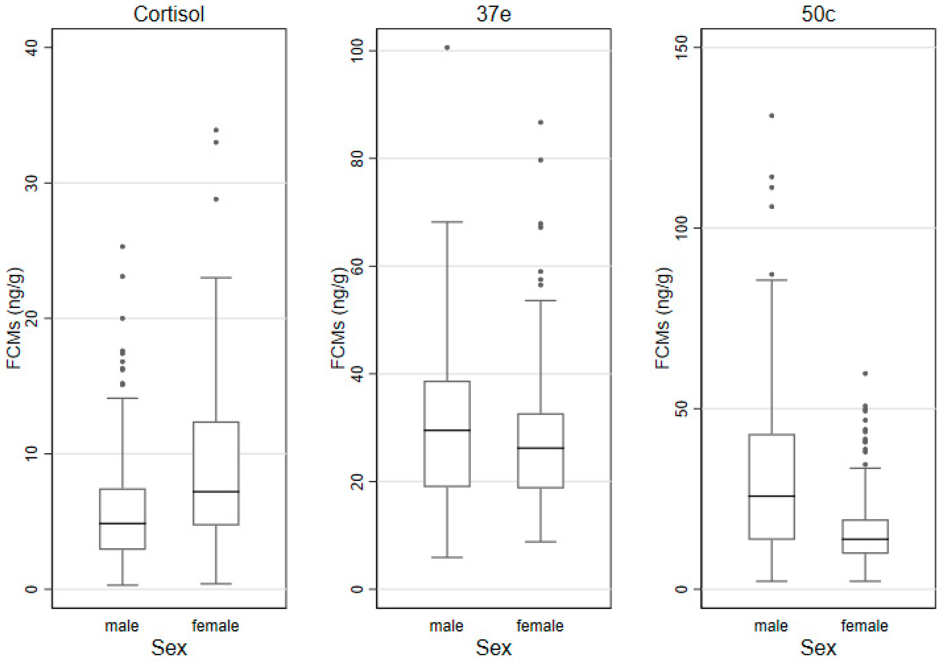
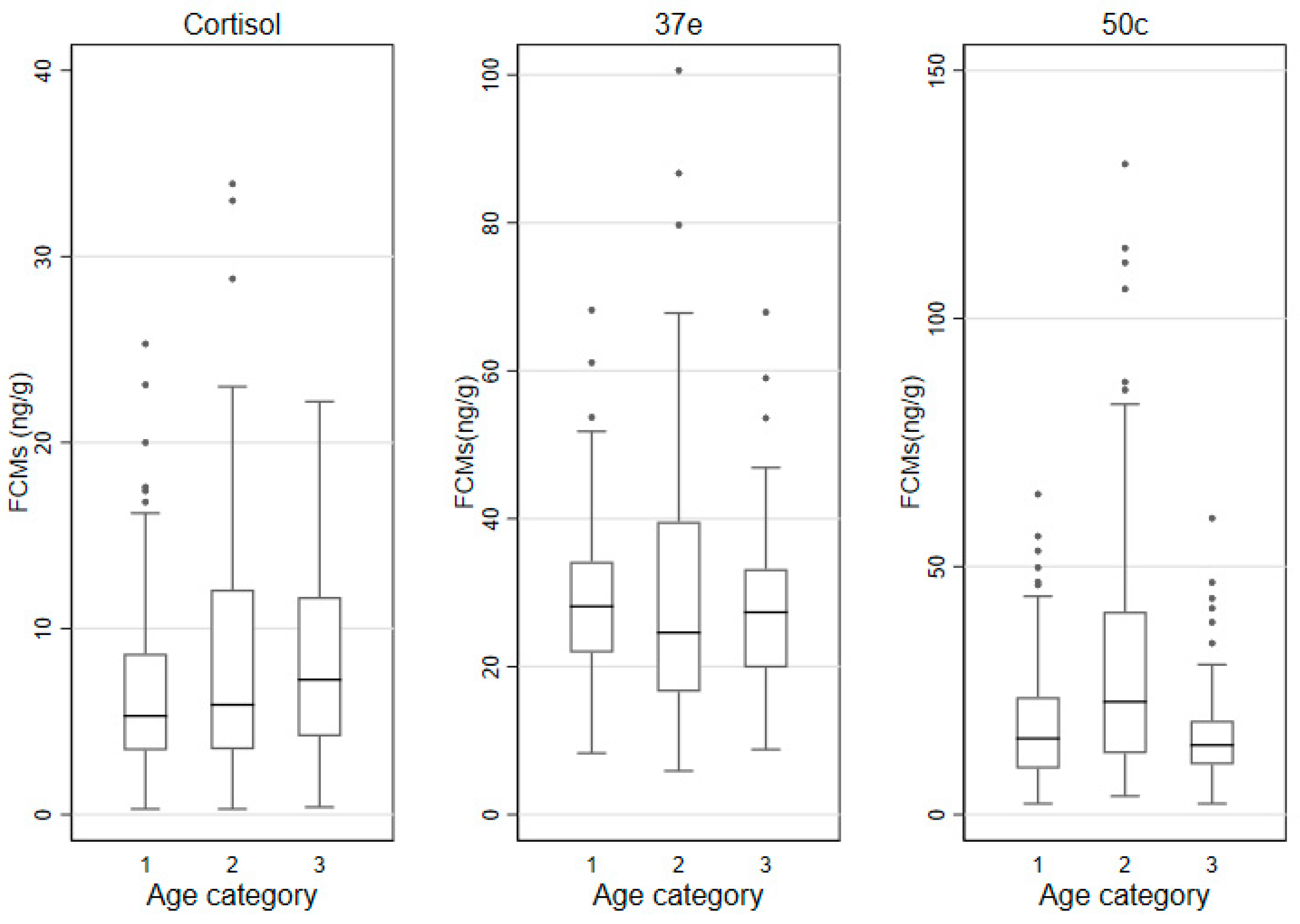
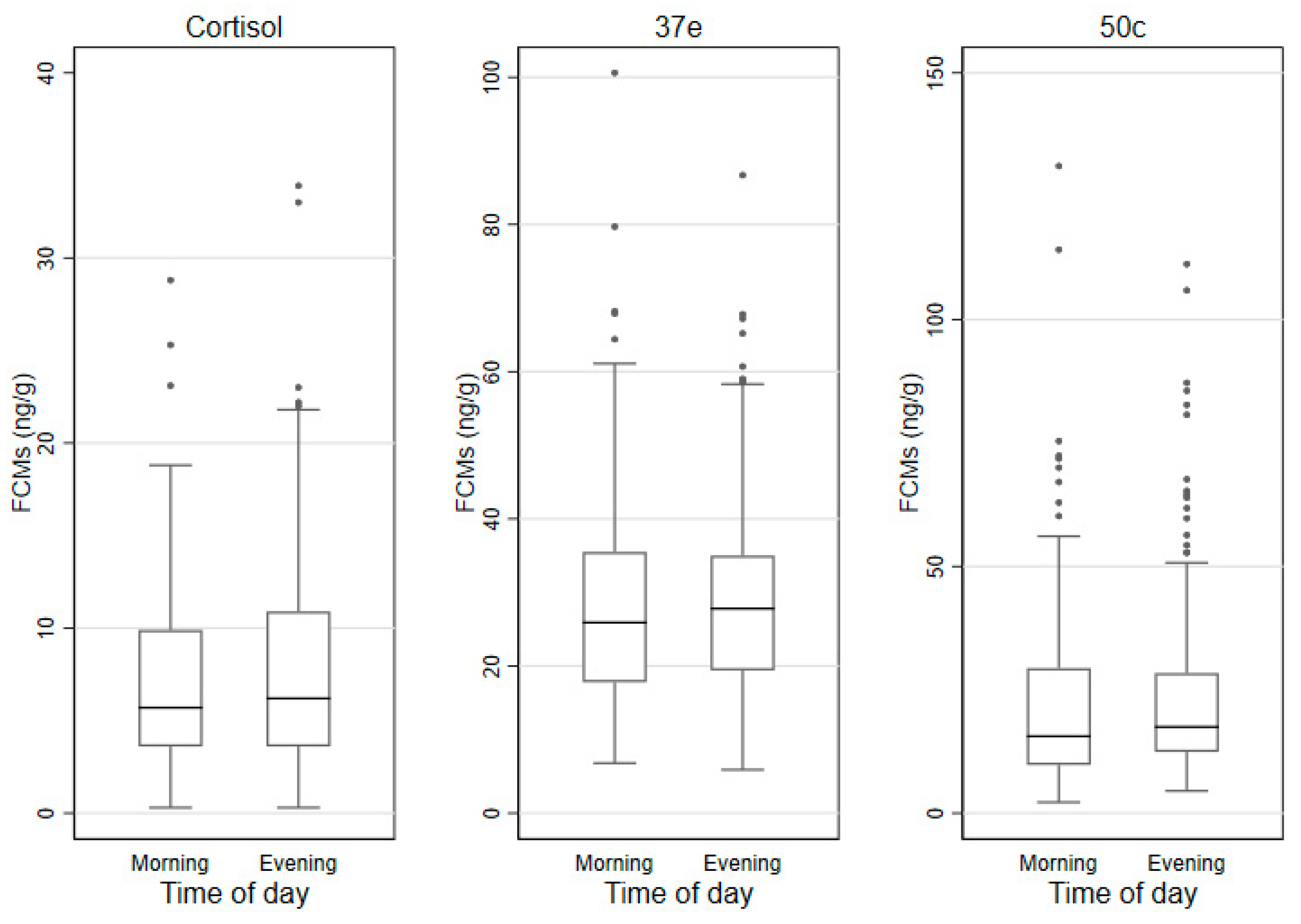
The median (IQR) FCM values measured by the cortisol, 37e and 50c EIAs by sex, age category and time of sampling are shown in Table 3 and the box and whisker plots for these groups are shown in Figure 2, Figure 3 and Figure 4. In males, lowest/highest FCM values (ng/g) detected by the cortisol, 37e and 50c EIAs were 0.3/25.3; 5.9/100.6 and 2.2/131.1, respectively. In females, those lowest/highest FCM values were 0.4/33.9; 8.8/86.7 and 2.2/59.7, respectively.

3.4. Seasonality of FCM Values

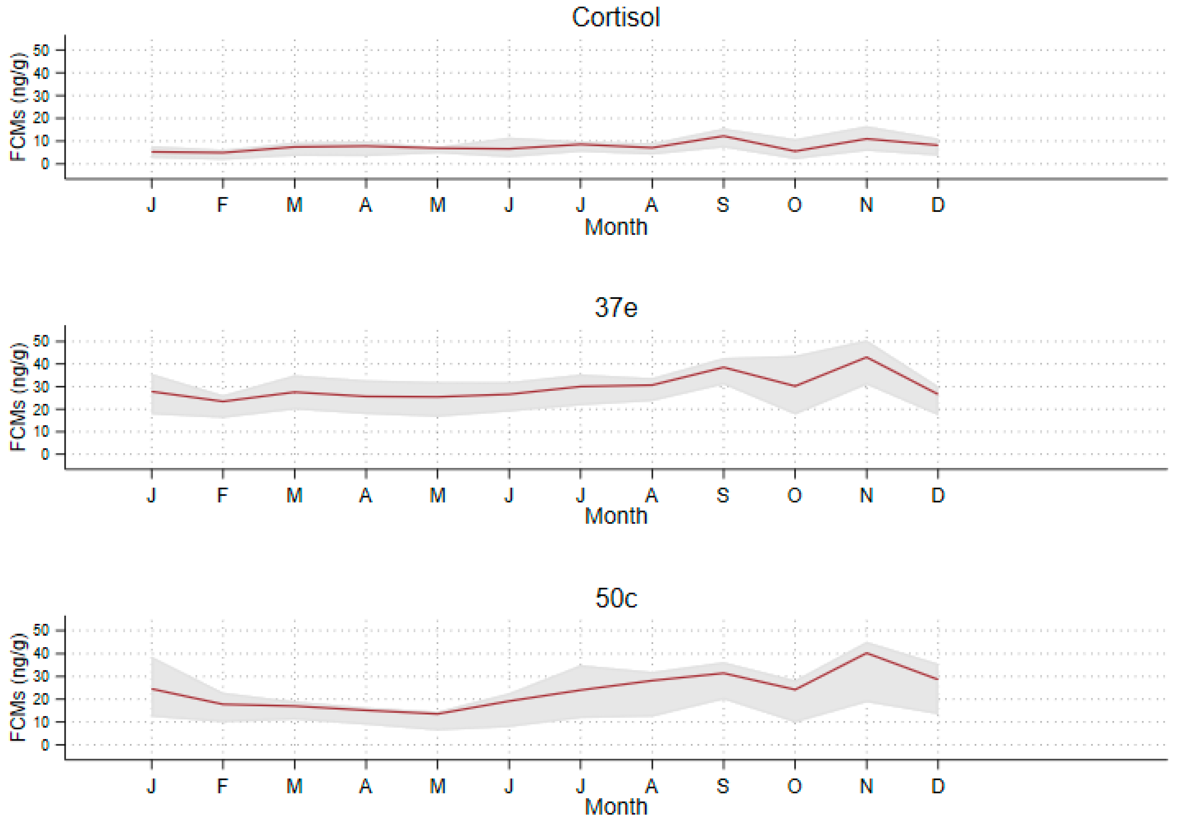
The mean (with 95% percentiles) FCM values measured per month are shown in Figure 5.
It is noticeable that there was a strong increase in FCM values detected by the three EIAs from September until January, coinciding with the breeding season of koalas (Figure 5). FCM values measured for each sampling interval are also shown in Figure S2a–c.
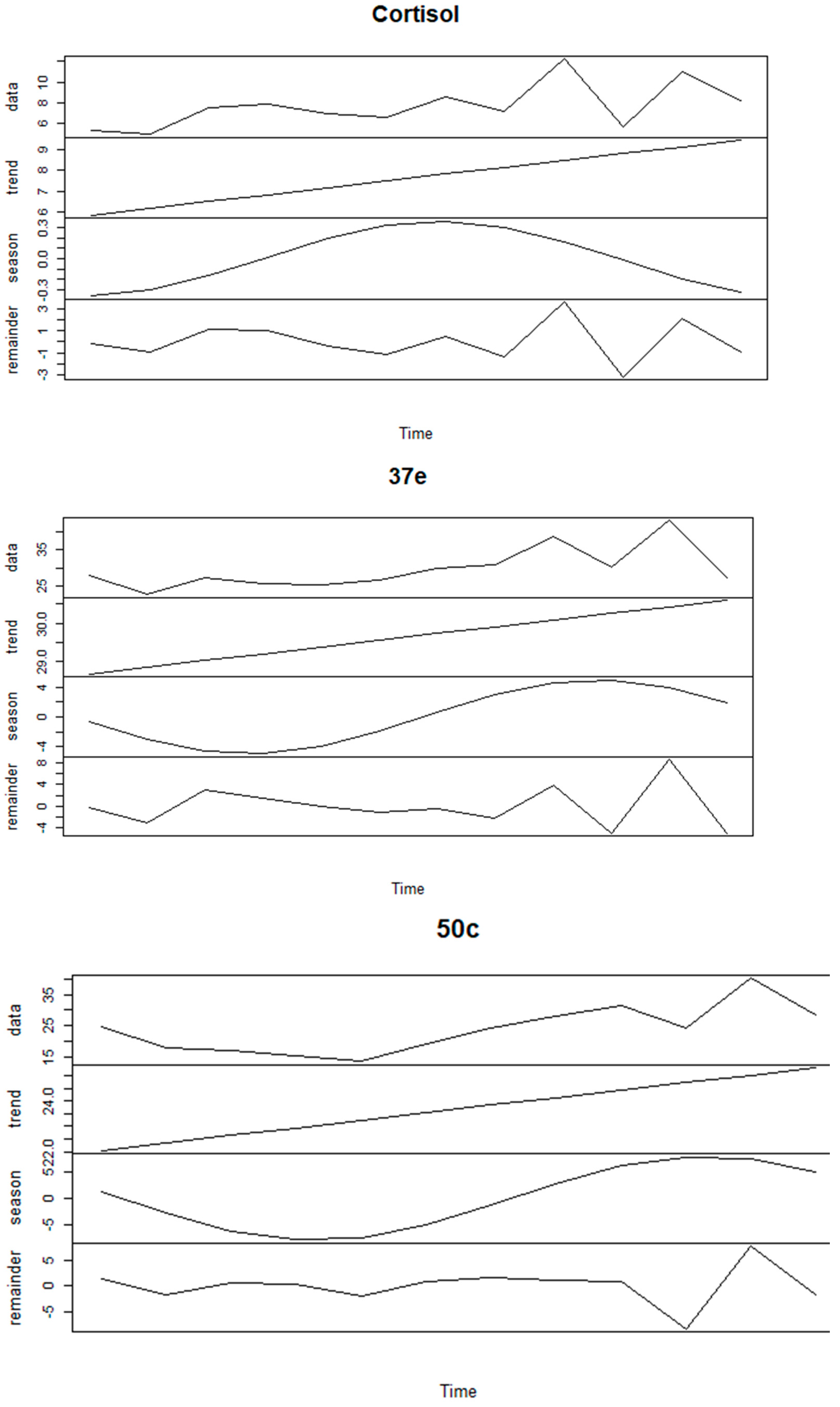
We explored the seasonality of FCM values in more detail using time series analysis (Figure 6).
The decomposition of the time series dataset highlighted a strong seasonal trend for 37e (Augmented Dickey–Fuller test = −1.4224, p-value = 0.7924 for hypothesis of stationary time series) and 50c (Augmented Dickey–Fuller test = −2.1821, p-value = 0.503), but less so for cortisol (Augmented Dickey–Fuller test = −3.391, p-value = 0.079).
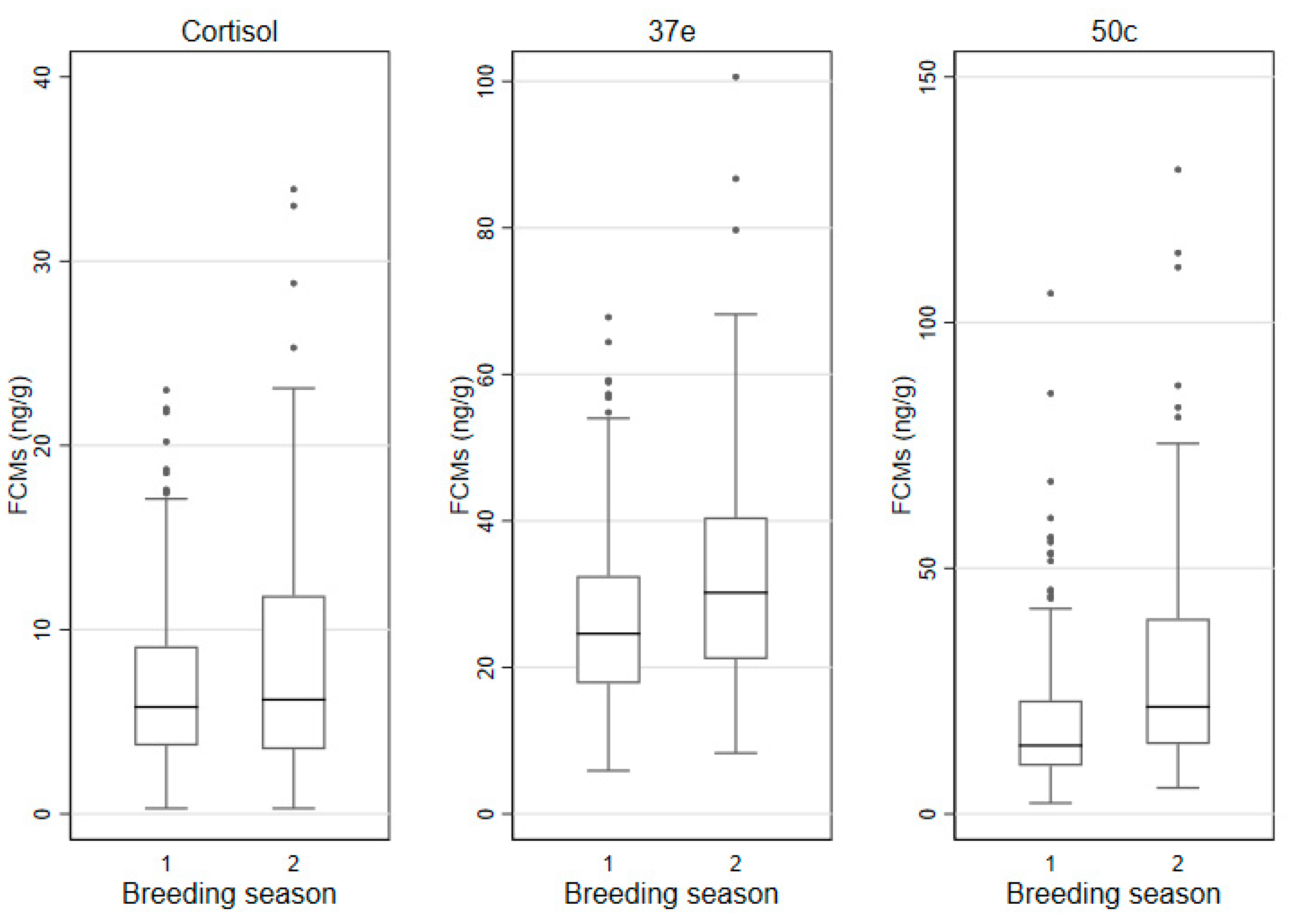
The FCM values were then grouped by breeding and non-breeding season and displayed in box andwhisker plots (Figure 7), while the descriptive statistics are shown in Table 4.

3.5. Correlation of FCM Values between EIAs

Scatterplots were used to display the bivariate correlations between groups of two EIAs (Figure 8).
The Pearson Correlation coefficient for 37e vs. cortisol was 0.2426 (p < 0.0001), for 50c vs. cortisol was −0.0039 (p = 0.9423) and for 37e vs. 50c was 0.6776 (p < 0.0001). Thus, a strong, significant correlation was only observed for the relationship between FCM concentrations measured with the 37e and 50c EIAs.

3.6. Factors Affecting FCM Values

The impact of predictors on the log-transformed FCM values was then evaluated in mixed effect linear regression models. In the univariate analysis, sex of the koalas affected log-transformed FCM values when measured with the 50c EIA: values of males were significantly higher (p = 0.022) than those of females. Sex differences were not detected with the cortisol or 37e EIAs. Age was not a factor affecting the values of log-transformed FCMs measured with any of the three EIAs. Log-transformed FCM values were significantly higher in the evening samples, when measured with the 50c EIA (p = 0.036). However, such differences were not detected with the other two EIAs. Log-transformed FCM values were significantly higher in the breeding season compared to the non-breeding season, when measured with the 50c EIA (p < 0.001). This association was also identified with the 37e EIA (p = 0.001), but not with the cortisol EIA. The univariate and multivariable analysis results are displayed in Table S1 (Supplementary material). There was no significant interaction between breeding season and sex of koalas in the multivariable model for the 50c EIA.

4. Discussion

Non-invasive methods for measuring FCMs are frequently utilized to investigate stress in animals [35]. However, not all EIAs can successfully measure FCMs in all species [40,46], hence biological performance was a priority here, to ensure the selection of the most appropriate EIA for koalas as the target species [26]. Whereas our previous study [47], identified FCMs and validated three EIAs for their measurement in koalas, the current one has tested the biological sensitivity of those three EIAs over a period of one year and determined the most suitable EIA for future studies on stress in koalas.
However, before monitoring stress, establishing baseline levels of cortisol metabolites is of paramount importance. As we assumed that FCM values would likely differ during the year, we conducted a longitudinal study on adrenocortical activity involving a confined koala population and have established FCM baseline values and the effect of time of day, the animals’ sex and age on these values and the seasonal FCM patterns during the non-breeding and breeding season of koalas. FCM baseline values were measured in stress-free koalas adapted to the presence of humans, kept in a constant environment with minimal external disturbance and consistent access to food and water. We only entered the enclosures to access the freshly defecated faecal pellets. We chose to carry out our study with a captive cohort to exclude all the factors that may be cause of stress in wild koalas and potentially increase the levels of FCMs.
The concept of baseline, when referring to glucocorticoid values, has been given different meaning depending on the type of research and the intent of the researchers [41,56,57], and in many cases refers to a point-in-time value [39,40,41] using a limited number of animals. Admittedly, the main intent of most of these studies was to physiologically validate EIAs before and after an ACTH challenge; however, some authors compare obtained FCM values to those of ‘stressed’ animals [41,58,59] without taking into consideration the physiological changes due to period of day, sex and season. Other year-long studies on marsupials were carried out on numbats and wombats also in captivity [38,60]. Yet, only the study on numbats [38] evaluated the variations of FCM values during breeding and non-breeding season, as well as differences between females and males. While we can draw some parallels with those studies, the term baseline is not comparable to ours, as human intervention during those research projects may have caused baseline values to be altered.
We found significantly higher FCM baseline values in males than in females when using the 50c EIA, but we did not detect differences between females and males with neither the cortisol nor the 37e EIAs. A previous study [41] on captive koalas over a 20-day period during the breeding season, found that there were sex differences in FCM values using a cortisol EIA. Yet, it appears that these values were obtained from various groups of males and females (handled, non-handled and with diverse reproductive status) and, therefore, it is not clear if these differences represented undisturbed stress-free koalas. Another study [58] on wild koalas reports no sex differences in FCM values with a similar type of EIA. The year-long study on numbats [38] using a cortisol EIA, reported higher FCM values for males during the breeding season, but not for females, despite a marginal increase. These animals were often handled and, therefore, the results may have been different if they had been left undisturbed. In our study, we did not find any significant relationship between FCM values during the breeding season and the sex of koalas.
Similarly, 50c was the only EIA detecting significant differences between morning and evening samples. We didn’t expect to detect any differences in circadian values due to the length of the caecum of koalas (2 m long or 23% of the intestinal length) where the broken down digested material is further metabolized by a large variety of bacteria [61]. In this section of the intestine, cortisol metabolites could be spread throughout the digested matter, potentially levelling out any peak values that may exist between morning and evening as Touma and Palme [24] found in the case of hind gut fermenters. They suggested that when collecting faecal samples of long gut passage fermenters, the time of collection needs to be considered as it may be otherwise difficult obtaining circadian values. However, there is the possibility that the different circadian values of FCM in this study may originate from the addition of cortisol (metabolites) secreted by the large intestine (colon) located after the caecum. A study [62] found that the epithelium of the colon is also capable of synthesizing GC in response to particular immune responses. While we cannot assert that the colon of koalas is also a source of cortisol metabolism, we suggest that this is something to be further investigated.
Our study did not find any differences in FCM values between the three age categories with any of the EIAs. There are no comparable studies on koalas, nevertheless, research on other marsupials (tammar wallabies) [63] showed no differences in blood cortisol values related to age in captured animals. Also, although physiologically different to marsupials, storks [64] show no age-related differences in baseline GC values. Yet, a study on dingoes [65] found that puppies had higher salivary cortisol values than adults. Therefore, we may speculate that age related differences in FCM values are likely to be species-specific.
Establishing average, minimum and maximum FCM baseline values was the main scope of this research as behavioural changes, during the breeding time as well as pregnancy itself [66], can lead to increased stress. In turn, higher levels of stress have been shown to have a negative effect on gonadotropin secretion, compromising reproduction, in many animal species [67], including humans. Baseline values need to be taken into account together with all the other parameters [24], because evaluating stress without previously establishing these baseline levels may potentially lead to errors in assessment.
Koalas’ breeding season may vary slightly across their northern–southern latitudinal range. Some authors have found that in South East Queensland the koala breeding season is between October and March and non-breeding is between April and September [68], while others found that females showed behavioural signs of oestrus mainly between September and April [69]. In particular, koalas at Wildlife HQ, where this study was conducted, mate during “September through to early/mid-February” (T. Maxwell personal communications). When we analysed monthly mean FCM values throughout the year we noticed that all three EIAs could detect an increase during the breading season (September–January). Yet, statistical analysis, in particular the time series analysis, revealed that only 37e and 50c EIAs could detect considerable differences between breeding and no-breeding season and we also identified a strong correlation of FCM values between these two EIAs. The 37e and 50c EIAs were originally designed to target glucocorticoid metabolites with a 5α-3β,11β-diol and 5β-3α,11β-diol molecular structure and were validated for rodents and birds, respectively [48,49]. Therefore, two metabolites, also found in the koala faeces, namely 3β-allotetrahydrocortisol and tetrahydrocortisol (THF), can also be measured by the 37e and 50c EIA, respectively [47]. Our study has demonstrated that these group-specific EIAs are better suited in measuring FCM values than the cortisol EIA. However, we have shown here that the 50c EIA has the best discerning ability and the capability of detecting a broader range of FCM values than the 37e EIA. Therefore, given the ability of the 50c EIA to detect THF (the main FCM in koalas) and its broader dynamic range of detection, we propose the use of the 50c EIA as the “koala-specific” assay.
We also observed some variation in FCM values for the three EIAs between individual pellets of one defecation. These CV values were in the range or somewhat higher than those for the pool sample extracts analysed in the EIAs. Therefore, whenever possible, we would advise to collect and homogenise at least two pellets from one defection to lower the influence of any potential variation. Faecal decay [70] and stability of FCMs [26,50] need to also be considered in future studies, especially when samples from unknown time of defecation are collected in the wild.

5. Conclusions

With this study, we found the tetrahydrocorticosterone (lab code: 50c) EIA best suited among three previously validated EIA for koalas. We have determined FCM baseline levels during the breeding and non-breeding season, differences between sexes and between morning and evening. Future studies on stress in koalas may now choose to compare their findings to our baseline values using this group-specific EIA, which can give consistent results.

Supplementary Materials

The following are available online at https://www.mdpi.com/article/10.3390/ani11061622/s1, Figure S1: Box and whisker plots of 13 koalas sampled for FCM levels (ng/g) using three EIAs (Cortisol, 37e, 50c). Figure S2: Fortnightly mean (red line) and 95% percentile (shaded area) FCM values (ng/g) measured with the cortisol, 37e and 50c EIA. Table S1: Results of mixed effect linear regression models (with koala ID as random effect) to explore the association between log-transformed FCM values for the cortisol, 37e and 50c EIA, respectively, and predictors (month, breeding season, time of day, sex, age category).

Author Contributions

Conceptualization, F.S.; methodology, F.S., R.S., R.P., J.H.; validation, R.P.; formal data analysis, J.H.; FCM analyses, E.K.-R., R.P.; investigation, F.S., R.S, RP.; resources F.S., R.P.; data curation, F.S., J.H.; writing—original draft preparation, F.S., R.S., R.P., J.H.; writing—review and editing, F.S., R.P., R.S., J.H.; visualization, J.H., F.S., R.P.; supervision, F.S.; project administration, F.S., R.S.; funding acquisition, F.S. All authors have read and agreed to the published version of the manuscript.

Funding

This research was funded by the Central Queensland University Internal Grant Program. Grant number RSH/5453.

Institutional Review Board Statement

This research received an ethics exemption (0000021903) from the CQUniversity’s Animal Ethics Committee due to the collection of faecal pellets without interfering with the koalas.

Data Availability Statement

Raw data are available from corresponding author upon reasonable request.

Acknowledgments

We thank the management of Wildlife HQ (Sunshine Coast, Queensland) for allowing us access to the koalas and the Koala Keepers for their continuous support during the difficult times during COVID-19. We also thank the following people for their support at various stages of this project: A. Schlagloth-Santamaria, L. Valenza, A. Valenza, A. Watkins. A special thank you to Judy Couper for her technical support and kind assistance. Thank you also to Tamara Keeley for initial advice on potential methodology.

Conflicts of Interest

The authors declare no conflict of interest. The funders had no role in the design of the study; in the collection, analyses, or interpretation of data; in the writing of the manuscript, or in the decision to publish the results.

References

  1. Koala (Phascolarctos cinereus) Listing. Available online: https://www.environment.gov.au/biodiversity/threatened/species/koala (accessed on 11 May 2020).
  2. Gonzalez-Astudillo, V.; Allavena, R.; McKinnon, A.; Larkin, R.; Henning, J. Decline causes of Koalas in South East Queensland, Australia: A 17-year retrospective study of mortality and morbidity. Sci. Rep. 2017, 7, srep42587. [Google Scholar] [CrossRef] [Green Version]
  3. Koala Expert Panel Interim Report. Available online: http://www.ehp.qld.gov.au/wildlife/koalas/review-conservation-measures.html (accessed on 2 October 2020).
  4. Henning, J.; Hannon, C.; McKinnon, A.; Larkin, R.; Allavena, R. The causes and prognoses of different types of fractures in wild koalas submitted to wildlife hospitals. Prev. Veter Med. 2015, 122, 371–378. [Google Scholar] [CrossRef] [PubMed]
  5. Schlagloth, R.; Santamaria, F.; Golding, B.; Thomson, H. Why is it important to use flagship species in community education? The Koala as a case study. Anim. Stud. J. 2018, 7, 127–148. [Google Scholar]
  6. Penn, A.M.; Sherwin, W.B.; Gordon, G.; Lunney, D.; Melzer, A.; Lacy, R.C. Demographic Forecasting in Koala Conservation. Conserv. Biol. 2000, 14, 629–638. [Google Scholar] [CrossRef] [Green Version]
  7. Seabrook, L.; McAlpine, C.; Baxter, G.; Rhodes, J.; Bradley, A.; Lunney, D. Drought-driven change in wildlife distribution and numbers: A case study of koalas in south west Queensland. Wildl. Res. 2011, 38, 509–524. [Google Scholar] [CrossRef]
  8. Reckless, H.J.; Murray, M.; Crowther, M. A review of climatic change as a determinant of the viability of koala populations. Wildl. Res. 2017, 44, 458–470. [Google Scholar] [CrossRef]
  9. McCallum, H.; Dobson, A. Disease, habitat fragmentation and conservation. Proc. R. Soc. Lond. Ser. B Biol. Sci. 2002, 269, 2041–2049. [Google Scholar] [CrossRef] [Green Version]
  10. Considine, M.-L. Moving on: Relocating species in response to climate change. ECOS 2011, 160, 11. [Google Scholar]
  11. Gallagher, R.V.; Makinson, R.O.; Hogbin, P.M.; Hancock, N. Assisted colonization as a climate change adaptation tool. Austral Ecol. 2014, 40, 12–20. [Google Scholar] [CrossRef]
  12. Minteer, B.A.; Collins, J.P. Move it or lose it? The ecological ethics of relocating species under climate change. Ecol. Appl. 2010, 20, 1801–1804. [Google Scholar] [CrossRef] [Green Version]
  13. Chipman, R.; Slate, D.; Rupprecht, C.; Mendoza, M. Downside Risk of Wildlife Translocation. Available online: https://digitalcommons.unl.edu/icwdm_usdanwrc/1896?utm_source=digitalcommons.unl.edu%2Ficwdm_usdanwrc%2F1896&utm_medium=PDF&utm_campaign=PDFCoverPages (accessed on 10 December 2020).
  14. Waugh, C.; Hanger, J.; Timms, P.; Polkinghorne, A. Koala translocations and Chlamydia: Managing risk in the effort to conserve native species. Biol. Conserv. 2016, 197, 247–253. [Google Scholar] [CrossRef]
  15. Boyce, W.M.; Weisenberger, M.E.; Penedo, M.C.T.; Johnson, C.K. Wildlife translocation: The conservation implications of pathogen exposure and genetic heterozygosity. BMC Ecol. 2011, 11, 5. [Google Scholar] [CrossRef] [PubMed] [Green Version]
  16. Dickens, M.J.; Delehanty, D.J.; Romero, L.M. Stress and translocation: Alterations in the stress physiology of translocated birds. Proc. R. Soc. Lond. Ser. B Biol. Sci. 2009, 276, 2051–2056. [Google Scholar] [CrossRef] [PubMed] [Green Version]
  17. Short, J. The Characteristics and Success of Vertebrate Translocations within Australia, Perth, and Australian Government Department of Agriculture, Fisheries and Forestry; Wildlife Research and Management Pty Ltd: Canberra, Australia, 2009. [Google Scholar]
  18. Santamaria, F.; Schlagloth, R. The effect of Chlamydia on translocated Chlamydia-naïve koalas: A case study. Aust. Zool. 2016, 38, 192–202. [Google Scholar] [CrossRef]
  19. McAlpine, C. Relationships between human-induced habitat disturbance, stressors and disease in Koala. In Proceedings of the Koala Research Network Disease Workshop, Queensland University of Technology, Brisbane, Australia, 19 September 2011. [Google Scholar]
  20. Brearley, G.; Rhodes, J.; Bradley, A.; Baxter, G.; Seabrook, L.; Lunney, D.; Liu, Y.; McAlpine, C. Wildlife disease prevalence in human-modified landscapes. Biol. Rev. 2012, 88, 427–442. [Google Scholar] [CrossRef] [PubMed]
  21. Björntorp, P. International Textbook of Obesity; Wiley: Chichester, UK, 2001. [Google Scholar]
  22. Timmermans, S.; Souffriau, J.; Libert, C. A General Introduction to Glucocorticoid Biology. Front. Immunol. 2019, 10, 1545. [Google Scholar] [CrossRef] [Green Version]
  23. Palme, R.; Rettenbacher, S.; Touma, C.; El-Bahr, S.M.; Möstl, E. Stress Hormones in Mammals and Birds: Comparative Aspects Regarding Metabolism, Excretion, and Noninvasive Measurement in Fecal Samples. Ann. N. Y. Acad. Sci. 2005, 1040, 162–171. [Google Scholar] [CrossRef]
  24. Touma, C.; Palme, R. Measuring Fecal Glucocorticoid Metabolites in Mammals and Birds: The Importance of Validation. Ann. N. Y. Acad. Sci. 2005, 1046, 54–74. [Google Scholar] [CrossRef]
  25. Spiga, F.; Walker, J.J.; Terry, J.R.; Lightman, S.L. HPA axis-rhythms. Compr. Physiol. 2011, 4, 1273–1298. [Google Scholar]
  26. Palme, R. Non-invasive measurement of glucocorticoids: Advances and problems. Physiol. Behav. 2019, 199, 229–243. [Google Scholar] [CrossRef]
  27. Palme, R. Monitoring stress hormone metabolites as a useful, non-invasive tool for welfare assessment in farm animals. Anim. Welf. 2012, 21, 331–337. [Google Scholar] [CrossRef]
  28. Sheriff, M.J.; Dantzer, B.; Delehanty, B.; Palme, R.; Boonstra, R. Measuring stress in wildlife: Techniques for quantifying glucocorticoids. Oecologia 2011, 166, 869–887. [Google Scholar] [CrossRef] [PubMed]
  29. Weitzman, E.D.; Fukushima, D.; Nogeire, C.; Roffwarg, H.; Gallagher, T.F.; Hellman, L. Twenty-four Hour Pattern of the Episodic Secretion of Cortisol in Normal Subjects. J. Clin. Endocrinol. Metab. 1971, 33, 14–22. [Google Scholar] [CrossRef] [PubMed]
  30. De Lacerda, L.; Kowarski, A.; Migeon, C.J. Integrated Concentration and Diurnal Variation of Plasma Cortisol1. J. Clin. Endocrinol. Metab. 1973, 36, 227–238. [Google Scholar] [CrossRef]
  31. Hunninck, L.; May, R.; Jackson, C.R.; Palme, R.; Røskaft, E.; Sheriff, M.J. Consequences of climate-induced vegetation changes exceed those of human disturbance for wild impala in the Serengeti ecosystem. Conserv. Physiol. 2020, 8, coz117. [Google Scholar] [CrossRef] [PubMed] [Green Version]
  32. Merl, S.; Scherzer, S.; Palme, R.; Möstl, E. Pain causes increased concentrations of glucocorticoid metabolites in horse feces. J. Equine Veter Sci. 2000, 20, 586–590. [Google Scholar] [CrossRef]
  33. Fureix, C.; Benhajali, H.; Henry, S.; Bruchet, A.; Prunier, A.; Ezzaouïa, M.; Coste, C.; Hausberger, M.; Palme, R.; Jego, P. Plasma cortisol and faecal cortisol metabolites concentrations in stereotypic and non-stereotypic horses: Do stereotypic horses cope better with poor environmental conditions? BMC Veter Res. 2013, 9, 3. [Google Scholar] [CrossRef] [Green Version]
  34. Dalerum, F.; Ganswindt, A.; Palme, R.; Bettega, C.; Delgado, M.D.M.; Dehnhard, M.; Freire, S.; González, R.G.; Marcos, J.; Miranda, M.; et al. Methodological Considerations for Using Fecal Glucocorticoid Metabolite Concentrations as an Indicator of Physiological Stress in the Brown Bear (Ursus arctos). Physiol. Biochem. Zool. 2020, 93, 227–234. [Google Scholar] [CrossRef] [Green Version]
  35. Möstl, E.; Palme, R. Hormones as indicators of stress. Domest. Anim. Endocrinol. 2002, 23, 67–74. [Google Scholar] [CrossRef]
  36. Palme, R.; Robia, C.; Messmann, S.; Hofer, J.; Möstl, E. Measurement of faecal cortisol metabolites in ruminants: A non-invasive parameter of adrenocortical function. Wien. Tierärztliche Mon. 1999, 86, 237–241. [Google Scholar]
  37. Palme, R. Measuring Fecal Steroids: Guidelines for Practical Application. Ann. N. Y. Acad. Sci. 2005, 1046, 75–80. [Google Scholar] [CrossRef]
  38. Hogan, L.A.; Lisle, A.T.; Johnston, S.D.; Robertson, H. Non-invasive assessment of stress in captive numbats, Myrmecobius fasciatus (Mammalia: Marsupialia), using faecal cortisol measurement. Gen. Comp. Endocrinol. 2012, 179, 376–383. [Google Scholar] [CrossRef] [PubMed]
  39. Narayan, E.J.; Evans, N.; Hero, J.-M. Monitoring physiological stress in semi-free ranging populations of an endangered Australian marsupial, the Greater Bilby (Macrotis lagotis). Eur. J. Wildl. Res. 2014, 60, 727–735. [Google Scholar] [CrossRef]
  40. Davies, N.; Gillett, A.; McAlpine, C.; Seabrook, L.; Baxter, G.; Lunney, D.; Bradley, A. The effect of ACTH upon faecal glucocorticoid excretion in the koala. J. Endocrinol. 2013, 219, 1–12. [Google Scholar] [CrossRef] [Green Version]
  41. Narayan, E.J.; Webster, K.; Nicolson, V.; Mucci, A.; Hero, J.-M. Non-invasive evaluation of physiological stress in an iconic Australian marsupial: The Koala (Phascolarctos cinereus). Gen. Comp. Endocrinol. 2013, 187, 39–47. [Google Scholar] [CrossRef] [Green Version]
  42. Weiss, M.; Richards, P.G. Adrenal Steroid Secretion in The Koala (Phascolarctos cinereus). J. Endocrinol. 1970, 48, 145–146. [Google Scholar] [CrossRef]
  43. McDonald, I.; Than, K.; Handasyde, K.; Michaelides, J. Factors Affecting Plasma Adrenocortical Hormone Concentration of Koalas. In Biology of the Koala; Lee, A.K., Handasyde, K.A., Sanson, G.D., Eds.; Surrey Beatty and Sons: Sydney, Australia, 1990; pp. 289–294. [Google Scholar]
  44. Johnston, S.; Booth, R.; Pyne, M.; Keeley, T.; Mackie, J.; Hulse, L.; Ellis, W. Preliminary study of faecal cortisol and corticosterone as an index of acute cortisol secretion in the koala (Phascolarctos cinereus). Aust. Veter J. 2013, 91, 534–537. [Google Scholar] [CrossRef]
  45. Keeley, T.; O’Brien, J.; Fanson, B.; Masters, K.; McGreevy, P. The reproductive cycle of the Tasmanian devil (Sarcophilus harrisii) and factors associated with reproductive success in captivity. Gen. Comp. Endocrinol. 2012, 176, 182–191. [Google Scholar] [CrossRef] [PubMed]
  46. Fanson, K.V.; Best, E.C.; Bunce, A.; Fanson, B.G.; Hogan, L.A.; Keeley, T.; Narayan, E.; Palme, R.; Parrott, M.L.; Sharp, T.M.; et al. One size does not fit all: Monitoring faecal glucocorticoid metabolites in marsupials. Gen. Comp. Endocrinol. 2017, 244, 146–156. [Google Scholar] [CrossRef] [PubMed]
  47. Santamaria, F.; Barlow, C.K.; Schlagloth, R.; Schittenhelm, R.B.; Palme, R.; Henning, J. Identification of koala (Phascolarctos cinereus) faecal cortisol metabolites using liquid chromatography-mass spectrometry and enzyme immunoassays. Metabolites 2021, 11, 393. [Google Scholar] [CrossRef]
  48. Quillfeldt, P.; Möstl, E. Resource allocation in Wilson’s storm-petrels Oceanites oceanicus determined by measurement of glucocorticoid excretion. Acta Ethol. 2003, 5, 115–122. [Google Scholar] [CrossRef]
  49. Touma, C.; Sachser, N.; Möstl, E.; Palme, R. Effects of sex and time of day on metabolism and excretion of corticosterone in urine and feces of mice. Gen. Comp. Endocrinol. 2003, 130, 267–278. [Google Scholar] [CrossRef]
  50. Palme, R.; Touma, C.; Arias, N.; Dominchin, M.F.; Lepschy, M. Steroid extraction: Get the best out of faecal samples. Wien. Tierärztliche Mon. 2013, 100, 238–246. [Google Scholar]
  51. Palme, R.; Möstl, E. Measurement of cortisol metabolites in faeces of sheep as a parameter of cortisol concentration in blood. Z. Säugetierkunde Int. J. Mamm. Biol. 1997, 62, 192–197. [Google Scholar]
  52. Ian, D.; Wayne, M.; Stryhyn, H. Veterinary Epidemiologic Research, 2nd ed.; Charlotte, P.E.I., Ed.; VER Inc.: Phoenix, AZ, USA, 2009. [Google Scholar]
  53. Rob, J.H. Seasonal Decomposition of Short Time Series. Available online: https://robjhyndman.com/hyndsight/tslm-decomposition/ (accessed on 5 February 2021).
  54. Cheung, Y.-W.; Lai, K.S. Lag order and critical values of the augmented Dickey-Fuller test. J. Bus. Econ. Stat. 1995, 13, 277–280. [Google Scholar]
  55. R Core Team. A Language and Environment for Statistical Computing; R Foundation for Statistical Computing: Vienna, Austria, 2020. [Google Scholar]
  56. Dupoué, A.; Rutschmann, A.; Le Galliard, J.F.; Clobert, J.; Blaimont, P.; Sinervo, B.; Miles, D.B.; Haussy, C.; Meylan, S. Reduction in baseline corticosterone secretion correlates with climate warming and drying across wild lizard populations. J. Anim. Ecol. 2018, 87, 1331–1341. [Google Scholar] [CrossRef] [PubMed]
  57. Bryan, H.M.; Adams, A.G.; Invik, R.M.; Wynne-Edwards, K.; Smits, J. Hair as a Meaningful Measure of Baseline Cortisol Levels over Time in Dogs. J. Am. Assoc. Lab. Anim. Sci. 2013, 52, 189–196. [Google Scholar]
  58. Davies, N.A.; Gramotnev, G.; McAlpine, C.; Seabrook, L.; Baxter, G.; Lunney, D.; Rhodes, J.R.; Bradley, A. Physiological Stress in Koala Populations near the Arid Edge of Their Distribution. PLoS ONE 2013, 8, e79136. [Google Scholar] [CrossRef]
  59. Narayan, E. Physiological stress levels in wild koala sub-populations facing anthropogenic induced environmental trauma and disease. Sci. Rep. 2019, 9, 6031. [Google Scholar] [CrossRef] [Green Version]
  60. Hogan, L.A.; Johnston, S.D.; Lisle, A.T.; Keeley, T.; Wong, P.; Nicolson, V.; Horsup, A.B.; Janssen, T.; Phillips, C.J. Behavioural and physiological responses of captive wombats (Lasiorhinus latifrons) to regular handling by humans. Appl. Anim. Behav. Sci. 2011, 134, 217–228. [Google Scholar] [CrossRef]
  61. Flanagan, C. Koala Rehabilitation Manual; Koala Preservation Society Austaralia: Port Macquarie, NSW, Australia, 2015. [Google Scholar]
  62. Noti, M.; Sidler, D.; Brunner, T. Extra-adrenal glucocorticoid synthesis in the intestinal epithelium: More than a drop in the ocean? Semin. Immunopathol. 2009, 31, 237–248. [Google Scholar] [CrossRef] [Green Version]
  63. McKenzie, S.; Deane, E.M. The effects of age, season, and gender on serum cortisol levels in the tammar wallaby, Macropus eugenii. Gen. Comp. Endocrinol. 2003, 133, 273–278. [Google Scholar] [CrossRef]
  64. Blas, J.; Baos, R.; Bortolotti, G.R.; Marchant, T.A.; Hiraldo, F. Age-related variation in the adrenocortical response to stress in nestling white storks (Ciconia ciconia) supports the developmental hypothesis. Gen. Comp. Endocrinol. 2006, 148, 172–180. [Google Scholar] [CrossRef]
  65. Smith, B.; Flavel, M.; Simpson, B. Quantification of salivary cortisol from captive dingoes (Canis dingo) in relation to age, sex, and breeding season: Implications for captive management. Aust. Mammal. 2016, 38, 21. [Google Scholar] [CrossRef]
  66. Edwards, P.D.; Boonstra, R. Glucocorticoids and CBG during pregnancy in mammals: Diversity, pattern, and function. Gen. Comp. Endocrinol. 2018, 259, 122–130. [Google Scholar] [CrossRef] [PubMed]
  67. Maeda, K.-I.; Tsukamura, H. The Impact of Stress on Reproduction: Are Glucocorticoids Inhibitory or Protective to Gonadotropin Secretion? Endocrinology 2006, 147, 1085–1086. [Google Scholar] [CrossRef] [Green Version]
  68. Rhodes, J.R.; Beyer, H.; Preece, H.; McAlpine, C. South East Queensland Koala Population Modelling Study; UniQuest: Brisbane, Australia, 2015. [Google Scholar]
  69. Ballantyne, K.; Lisle, A.; Mucci, A.; Johnston, S.D. Seasonal oestrous cycle activity of captive female koalas in south-east Queensland. Aust. Mammal. 2015, 37, 245. [Google Scholar] [CrossRef]
  70. Cristescu, R.H.; Goethals, K.; Banks, P.B.; Carrick, F.N.; Frere, C. Experimental Evaluation of Koala Scat Persistence and Detectability with Implications for Pellet-Based Fauna Census. Int. J. Zool. 2012, 2012, 631856. [Google Scholar] [CrossRef] [Green Version]
Figure 1. Histograms of FCM (faecal cortisol metabolite) values (ng/g) measured with the cortisol, 37e (5α-pregnane-3β,11β,21-triol-20-one) and 50c (tetrahydrocorticosterone) EIAs (enzyme immunoassays).
Animals 11 01622 g001
Figure 2. Box and whisker plots for FCM (faecal cortisol metabolite) levels (ng/g) measured with the cortisol, 37e (5α-pregnane-3β,11β,21-triol-20-one) and 50c (tetrahydrocorticosterone) EIAs (enzyme immunoassays), as grouped by sex (defecations: males n= 164; females n= 187).
Animals 11 01622 g002
Figure 3. Box and whisker plots for FCM (faecal cortisol metabolite) levels (ng/g) measured with the cortisol, 37e (5α-pregnane-3β,11β,21-triol-20-one) and 50c (tetrahydrocorticosterone) EIAs (enzyme immunoassays), as grouped by age categories (1: ≤2 years-; 2: >2–5 years-; 3: >5 years; defecations n= 124, n= 159 and n= 68 respectively).
Animals 11 01622 g003
Figure 4. Box and whisker plots for FCM (faecal cortisol metabolite) levels (ng/g) measured with the cortisol, 37e (5α-pregnane-3β,11β,21-triol-20-one) and 50c (tetrahydrocorticosterone) EIAs (enzyme immunoassays) in morning and evening samples (defecations: n = 182 and n = 169 respectively).
Animals 11 01622 g004
Figure 5. Monthly mean (red line) and 95% percentile (shaded area) FCM (faecal cortisol metabolite) values (ng/g) measured with the cortisol, 37e (5α-pregnane-3β,11β,21-triol-20-one) and 50c (tetrahydrocorticosterone) EIAs (enzyme immunoassays) (defecations: n = 351).
Animals 11 01622 g005
Figure 6. Time series for FCM (faecal cortisol metabolite) values measured with the cortisol, 37e (5α-pregnane-3β,11β,21-triol-20-one) and 50c (tetrahydrocorticosterone) EIAs (enzyme immunoassays) over a 12-month period.
Animals 11 01622 g006
Figure 7. Box and whisker plots for FCM (faecal cortisol metabolite) values (ng/g) measured with the cortisol, 37e (5α-pregnane-3β,11β,21-triol-20-one) and 50c (tetrahydrocorticosterone) EIAs (enzyme immunoassays) (1: non-breeding season (February–August); 2: breeding season (September–January); defecations: n = 199 and n = 152 respectively).
Animals 11 01622 g007
Figure 8. Scatterplots correlating FCM (faecal cortisol metabolite) values (ng/g) measured with cortisol, 37e (5α-pregnane-3β,11β,21-triol-20-one) and 50c (tetrahydrocorticosterone) EIAs (enzyme immunoassay) conducted over a period of 12 months (defecations: n = 351).
Animals 11 01622 g008
Table 1. Details of the EIAs (enzyme immunoassays) utilized in the current study.
EIA CodeDetailsDescription
CortisolStandard4-pregnene-11β,17α,21-triol-3,20-dione(cortisol)
Targeted structure11β,17α,21-triol-20-one
Antibody againstcortisol-3-CMO:BSA
Labelcortisol-3-CMO-DADOO-biotin
ReferencePalme and Möstl [51]
37eStandard5α-pregnane-3β,11β,21-triol-20-one
(3β-allotetrahydrocorticosterone)
Targeted structure5α-3ß,11ß-diol
Antibody against5α-pregnane-3β,11β,21-triol-20-one-CMO-BSA
Label5α-pregnane-3β,11β,21-triol-20-one-CMO-biotinyl-LC
ReferenceTouma et al. [49]
50cStandard5β-pregnane-3α,11β,21-triol-20-one(tetrahydrocorticosterone)
Targeted structure5β-3α,11β-diol
Antibody against5β-pregnane-3α,11β,21-triol-20-one-CMO-BSA
Label5β-pregnane-3α,11β,21-triol-20-one-21-HS-biotinyl-LC
ReferenceQuillfeldt and Möstl [48]
Table 2. Summary statistics for FCM (faecal cortisol metabolite) levels (ng/g) measured by cortisol, 37e and 50c EIAs (enzyme immunoassays).
EIAMeanMedianp25p75IQRMinMax
Cortisol7.65.93.610.67.00.333.9
37e29.327.318.835.316.55.9100.6
50c23.316.511.129.418.32.2131.1
p = percentiles, IQR = Inter Quartile Range; Min = minimum, Max = maximum.
Table 3. Median (IQR) for FCM (faecal cortisol metabolites) values measured by cortisol, 37e and 50c EIAs (enzyme immunoassays) by sex, age category and time of sampling.
VariableSub-GroupsFCM (ng/g)
Cortisol EIA37e EIA50c EIA
SexMale5.9 (4.6)31.0 (19.8)31.1 (29.4)
Female9.0 (7.7)27.9 (14.0)16.4 (9.6)
Age≤2 years6.5 (5.2)28.8 (12.3)18.4 (14.4)
>2–5 years8.0 (8.6)30.2 (23.0)29.9 (28.6)
>5 years8.5 (7.5)27.8 (13.6)16.6 (8.9)
TimeEvening8.1 (7.3)29.5 (15.6)24. 1 (16.0)
of dayMorning7.1 (6.3)29.1 (17.7)22.5 (19.6)
37e (5α-pregnane-3β,11β,21-triol-20-one) and 50c (tetrahydrocorticosterone).
Table 4. Summary statistics for FCM (faecal cortisol metabolites) values measured with the cortisol, 37e and 50c EIAs (enzyme immunoassays) by breeding season.
EIAMeanMedianp25p75IQR
Non-breeding (February–August)
Cortisol7.05.83.79.15.4
37e26.824.617.832.514.7
50c18.713.99.823.113.3
Breeding (September–January)
Cortisol8.26.23.511.98.4
37e32.730.221.140.519.4
50c29.221.814.239.725.6
p = percentiles, IQR = Inter Quartile Range; Min = minimum, Max = maximum.
Publisher’s Note: MDPI stays neutral with regard to jurisdictional claims in published maps and institutional affiliations.