Skin Diseases in Donkeys and Mules—An Update

 , , , , and
Abstract
Simple Summary
Abstract
1. Introduction
2. Relationship between Disease Occurrence and Type of Donkey and Mule Farming
2.1. Overview of Donkey and Mule Farming in Brazil
2.2. Disease Profile of Donkeys and Mules
2.3. Skin Diseases Diagnosed in Donkeys and Mules
3. Equine Dermatopathies in Northeast Brazil
4. Conclusions
Author Contributions
Funding
Conflicts of Interest
References
- The Food and Agriculture Organization-FAO. 2016. Available online: http://www.fao.org/faostat/en/#home (accessed on 23 April 2020).
- The Food and Agriculture Organization-FAO. 2018. Available online: http://www.fao.org/faostat/en/#data/QA (accessed on 23 April 2020).
- Luís, C.; Bastos-Silveira, C.; Cothran, E.G.; Oom, M.D.M. Iberian origins of New World horse breeds. J. Hered. 2006, 97, 107–113. [Google Scholar] [CrossRef] [PubMed]
- Baker, M. Sob a Pele-O Comércio Emergente de Pele de Asno e Suas Implicações Para o Bem-Estar e os Meios de Subsistência dos Asnos, 1st ed.; The Donkey Sanctuary: Sidmouth, UK, 2017; Available online: https://www.thedonkeysanctuary.org.uk/sites/uk/files/201711/under_the_skin_report_portuguese.pdf (accessed on 10 July 2020). (In Portuguese)
- Carneiro, G.F.; Cavalcante Lucena, J.E.; de Oliveira Barros, L. The current situation and trend of the donkey industry in South America. J. Equine Vet. Sci. 2018, 65, 106–110. [Google Scholar] [CrossRef]
- Köhle, N. Feasting on Donkey Skin. In Conspicuous Consumption, 1st ed.; Smith, C.A., Kohle, N., Jaivin, L., Eds.; ANU Press: Canberra, Australia, 2018; ISBN (ebook): 9781760462031. [Google Scholar]
- Lara, M.C.C.S.H.; Villalobos, E.M.C.; Cunha, S.E.M.; Oliveira, J.V.; Castro, C.; Nassar, A.F.C.; Silva, L.M.P.; Okuda, L.H.; Romaldini, A.H.C.N.; Cunha, M.S.; et al. Occurrence of viral diseases in donkeys (Equus asinus) in São Paulo State, Brazil. Braz. J. Vet. Res. An. Sci. 2017, 54, 154–158. [Google Scholar] [CrossRef][Green Version]
- García-Bocanegra, I.; Arenas-Montes, A.; Jaén-Téllez, J.A.; Napp, S.; Fernández-Morente, M.; Arenas, A. Use of sentinel serosurveillance of mules and donkeys in the monitoring of West Nile virus infection. Vet. J. 2012, 194, 262–264. [Google Scholar] [CrossRef]
- Lima, T.S. Caracterização Clínico-patológica e Epidemiológica das Dermatopatias de Ruminantes no Agreste da Paraíba. Master’s Dissertation, Universidade Federal da Paraíba, Areia, Paraíba, 2019. Available online: https://repositorio.ufpb.br/jspui/handle/123456789/14355 (accessed on 10 July 2020).
- Assis-Brasil, N.D.; Marcolongo-Pereira, C.; Stigger, A.L.; Fiss, L.; Santos, B.L.; Coelho, A.C.B.; Sallis, E.S.V.; Fernandes, C.G.; Schild, A.L. Dermatopatias em equinos na região sul do Rio Grande do Sul: Estudo de 710 casos. Ciên. Rural. 2015, 45, 519–524. (In Portuguese) [Google Scholar] [CrossRef]
- Pessoa, A.F.A.; Pessoa, C.R.M.; Miranda Neto, E.G.; Riet-Correa, F. Doenças de asininos e muares no semiárido brasileiro. Pesq. Vet. Bras. 2014, 34, 1210–1214. (In Portuguese) [Google Scholar] [CrossRef]
- Pessoa, A.F.A.; Pessoa, C.R.M.; Miranda Neto, E.G.; Dantas, A.F.M.; Riet-Correa, F. Doenças de pele em equídeos no semiárido brasileiro. Pesq. Vet. Bras. 2014, 34, 743–748. (In Portuguese) [Google Scholar] [CrossRef][Green Version]
- McLean, A.K.; Navas Gonzalez, F.J. Can scientists influence donkey welfare? Historical perspective and a contemporary view. J. Equine Vet. Sci. 2018, 65, 25–32. [Google Scholar] [CrossRef]
- Miranda, A.L.S.; Palhares, M.S. Muares: Características, origem e particularidades clínico-laboratoriais. Rev. Cient. Med. Vet. 2017, 1–8. Available online: http://faef.revista.inf.br/imagens_arquivos/arquivos_destaque/qqT3b4TLANsOU7k_2018-6-30-10-43-55.pdf (accessed on 10 July 2020). (In Portuguese).
- Tesfaye, A.; Tekle, Y.; Taddele, H.; Gezahagn, K.; Yihdego, H. Survey of Common Skin Problem of Working Equines in and Around Mekelle, North Ethiopia. Acad. J. Anim. Dis. 2015, 4, 30–38. [Google Scholar] [CrossRef]
- Davis, E. Donkey and Mule Welfare. Vet. Clin. N. Am. Equine Pract. 2019, 35, 481–491. [Google Scholar] [CrossRef] [PubMed]
- Pierezan, F.; Rissi, D.R.; Rech, R.R.; Fighera, R.A.; Brum, J.S.; Barros, C.S.L. Achados de necropsia relacionados com a morte de 335 eqüinos: 1968–2007. Pesq. Vet. Bras. 2009, 29, 275–280. (In Portuguese) [Google Scholar] [CrossRef]
- Lucena, R.B.; Pierezan, F.; Kommers, G.D.; Irigoyen, L.F.; Fighera, R.A.; Barros, C.S.L. Doenças de bovinos no Sul do Brasil: 6.706 casos. Pesq. Vet. Bras. 2010, 30, 428–434. (In Portuguese) [Google Scholar] [CrossRef]
- Marques, A.L.A.; Aguiar, G.M.N.; Lira, M.A.A.; Miranda Neto, E.G.; Azevedo, S.S.; Simões, S.V.D. Enfermidades do sistema digestório de bovinos da região semiárida do Brasil. Pesq. Vet. Bras. 2018, 38, 407–416. [Google Scholar] [CrossRef]
- Barrandeguy, M.E.; Carossino, M. Infectious diseases in donkeys and mules: An overview and update. J. Equine Vet. Sci. 2018, 65, 98–105. [Google Scholar] [CrossRef]
- Mavrouli, M.; Vrioni, G.; Kapsimali, V.; Tsiamis, C.; Mavroulis, S.; Pervanidou, D.; Billinis, C.; Hadjichristodoulou, C.; Tsakris, A. Reemergence of West Nile Virus Infections in Southern Greece, 2017. Am. J. Trop. Med. Hyg. 2019, 100, 420–426. [Google Scholar] [CrossRef]
- Souza, T.M.; Brum, J.S.; Fighera, R.A.; Brass, K.E.; Barros, C.S.L. Prevalência dos tumores cutâneos de equinos diagnosticados no Laboratório de Patologia Veterinária da Universidade Federal de Santa Maria, Rio Grande do Sul. Pesq. Vet. Bras. 2011, 31, 379–382. (In Portuguese) [Google Scholar] [CrossRef][Green Version]
- Marcolongo-Pereira, C.; Estima-Silva, P.; Soares, M.P.; Sallis, E.S.V.; Grecco, F.B.; Fernandes, C.G.; Raffi, M.B.; Schild, A.L. Doenças de equinos na região Sul do Rio Grande do Sul. Pesq. Vet. Bras. 2014, 34, 205–210. (In Portuguese) [Google Scholar] [CrossRef]
- Miller, M.A.; Moore, G.E.; Bertin, F.R.; Kritchevsky, J.E. What’s New in Old Horses? Postmortem Diagnoses in Mature and Aged Equids. Vet. Pathol. 2015, 53, 390–398. [Google Scholar] [CrossRef]
- Elsheikha, H.M.; Schares, G.; Paraschou, G.; Sullivan, R.; Fox, R. First record of besnoitiosis caused by Besnoitia bennetti in donkeys from the UK. Parasites Vectors 2020, 13, 1–10. [Google Scholar] [CrossRef]
- Radwan, A.M.; Ahmed, N.E.; Elakabawy, L.M.; Ramadan, M.Y.; Elmadawy, R.S. Prevalence and pathogenesis of some filarial nematodes infecting donkeys in Egypt. Vet. World 2016, 9, 888–892. [Google Scholar] [CrossRef] [PubMed]
- White, S.D.; Bourdeau, P.J.; Brement, T.; Vandenabeele, S.I.; Haspeslagh, M.; Bruet, V.; Van Oldruitenborgh-Oosterbaan, M.M.S. Skin disease in donkeys (Equus asinus): A retrospective study from four veterinary schools. Vet. Dermatol. 2019, 30, 247–276. [Google Scholar] [CrossRef] [PubMed]
- Knottenbelt, D.C. Skin Disorders of the Donkey and Mule. Vet. Clin Equine. 2019, 35, 493–514. [Google Scholar] [CrossRef] [PubMed]
- Alvarez, J.A.C.; Socarras, T.O.; Tous, M.G. Dermopatias en burros de trabajo (Equus asinus) en areas rurales de Cordoba (Colombia). Rev. Med. Vet. 2017, 34, 81–91. (In Spanish) [Google Scholar] [CrossRef]
- Meselu, D.; Abebe, R.; Mekibib, B. Prevalence of Epizootic Lymphangitis and bodily distribuition of lesions in cart-mules in Behair Dar Town, Northwest Ethiopia. J. Vet. Sci. Technol. 2018, 9, 1–4. [Google Scholar] [CrossRef]
- Mahendra, P. Occurrence of Cutaneous Epizootic Lymphangitis in Working Donkeys in Debre Zeit, Ethiopia. EC Microbiol. 2019, 15, 382–384. [Google Scholar]
- Mota, R.A.; Oliveira, A.A.F.; Pinheiro Junior, J.W.; Silva, L.B.G.; Brito, M.F.; Rabelo, S.S.A. Glanders in donkeys (Equus asinus) in the state of Pernambuco, Brazil: A case report. Braz. J. Microbiol. 2010, 41, 146–149. [Google Scholar] [CrossRef]
- Rabelo, S.S.A.; Soares, P.C.; Silva, L.B.G.; Cunha, A.P.; Nascimento Sobrinho, E.; Pinheiro Junior, J.W.; Barbosa, M.A.G.; Mota, R.A. Indicadores Clínicos em Muares Naturalmente Infectados pela Burkholderia mallei. Vet. Zootec. 2006, 13, 54–62. Available online: https://docplayer.com.br/71952462-Indicadores-clinicos-em-muares-naturalmente-infectados-pela-burkholderia-mallei-resumo.html (accessed on 10 July 2020). (In Portuguese).
- Mota, R.A.; Brito, M.F.; Castro, F.J.C.; Massa, M. Mormo em eqüídeos nos estados de Pernambuco e Alagoas. Pesq. Vet. Bras. 2000, 20, 155–159. (In Portuguese) [Google Scholar] [CrossRef]
- Ghahvei, Y.; Mirzaei, M.; Hashemnia, S.; Golchin, M.; Kheirandish, R.; Uni, S.; Endoza-Roldan, J.A.; Otranto, D.; Sazmand, A. Scanning electron microscopy of Onchocerca fasciata (Filarioidea: Onchocercidae) adults, microfilariae and eggs with notes on histopathological findings in camels. Parasit. Vectors 2020, 13, 1–10. [Google Scholar] [CrossRef]
- Maia, L.A.; Olinda, R.G.; Araújo, T.F.; Firmino, P.R.; Nakazato, L.; Miranda Neto, E.G.; Riet-Correa, F.; Dantas, A.F.M. Cutaneous pythiosis in a donkey (Equus asinus) in Brazil. J. Vet. Diagn. Invest. 2016, 28, 436–439. [Google Scholar] [CrossRef] [PubMed]
- Tabosa, I.M.; Medeiros, V.T.; Dantas, A.F.M.; Azevedo, E.O.; Maia, J.C. Pitiose cutânea em equinos no semi-árido da Paraíba. ABMVZ 1999, 51, 27–30. (In Portuguese) [Google Scholar]
- Santos, C.E.P.; Santurio, J.M.; Colodel, E.M.; Juliano, R.S.; Silva, J.A.; Marques, L.C. Contribuição ao Estudo da Pitose Cutânea em Equídeos do Pantanal Norte, Brasil. ARS Vet. 2011, 27, 134–140. Available online: http://arsveterinaria.org.br/ars/article/download/310/337 (accessed on 10 July 2020). (In Portuguese).
- Leite, N.M.; Rocha, M.V.; Souza, K.M.; Vago, P.B. Linfangite ulcerativa em equino: Relato de caso. PUBVET 2019, 13, 1–6. (In Portuguese) [Google Scholar] [CrossRef]
- Sureshjani, M.H.; Atyabi, N.; Tazikeh, A.; Falahatipour, S.K.; Hashemian, M. Isolation of Rhodococcus equi from a mule with cutaneous wound. Comp. Clin. Pathol. 2014, 1–3. [Google Scholar] [CrossRef]
- Prescott, J.F. Rhodococcus equi: An animal and human pathogen. Clin. Microbiol. Rev. 1991, 4, 1–20. [Google Scholar] [CrossRef]
- Guimarães, J.H.; Papavero, N.; Prado, A.P. As miíases na região neotropital (identificação, biologia, bibliografia). Rev. Bras. Zool. 1982, 1, 239–416. [Google Scholar] [CrossRef]
- Davis, C.R.; Valentine, B.A.; Gordon, E.; McDonough, S.P.; Schaffer, P.A.; Allen, A.L.; Pesavento, P. Neoplasia in 125 donkeys (Equus asinus): Literature review and a survey of five veterinary schools in the United States and Canada. J. Vet. Diagn. Investig. 2016, 28, 662–670. [Google Scholar] [CrossRef]
- Alberti, T.S.; Zamboni, R.; Venancio, F.R.; Scheid, H.V.; Bermann, C.S.; Raffi, M.B.; Sallis, E.S.V. Melanoma anaplásico em equino de pelagem tordilha com metástase em osso e músculo. Cien. Anim. 2019, 29, 129–134. (In Portuguese) [Google Scholar]
- Rayner, E.; Airikkala-Otter, I.; Susheelan, A.; Gibson, A.; Itaba, R.; Mayani, T.; Mellanby, R.J.; Gamble, L. Prevalence of skin wounds in working donkeys in Bukombe, Tanzania. Vet. Rec. 2019, 186, 1–3. [Google Scholar] [CrossRef]
- Knupp, S.N.R.; Knupp, L.S.; Riet-Correa, F.; Lucena, R.B. Plants that cause photosensitivity in ruminants in Brazil. Semin. Ciênc. Agrár. 2016, 37, 2009–2020. [Google Scholar] [CrossRef]
- Amado, G.P.; Silva, C.C.B.; Barbosa, F.M.S.; Nascimento, H.H.L.; Malta, K.C.; Azevedo, M.V.; Lacerda-Lucena, P.B.; Lucena, R.B. Surtos de fotossensibilização e dermatite alérgica em ruminantes e equídeos no Nordeste do Brasil. Pesq. Vet. Bras. 2018, 38, 889–895. (In Portuguese) [Google Scholar] [CrossRef]
- Silva, T.I.B.; Melchior, L.A.K.; Baptista Filho, L.C.F.; Fernandes, A.C.C.; Silva, L.G.; Vasconcelos, K.F.; Revorêdo, R.G.; Silva, D.D.; Melo, L.E.H. Dermatite alérgica à picada de Culicoides em muar: Relato de caso. Arq. Bras. Med. Vet. Zootec. 2017, 69, 1407–1412. (In Portuguese) [Google Scholar] [CrossRef]
- Schaffartzik, A.; Hamza, E.; Janda, J.; Crameri, R.; Marti, E.; Rhyner, C. Equine insect bite hypersensitivity: What do we know? Vet Immunol. Immunopathol. 2012, 147, 113–126. [Google Scholar] [CrossRef] [PubMed]
- Corrêa, T.G.; Ferreira, J.M.; Riet-Correa, G.; Ruas, J.L.; Schild, A.L.; Riet-Correa, F.; Guimarães, A.; Felippe-Bauer, M.L. Seasonal allergic dermatitis in sheep in southern Brazil caused by Culicoides insignis (Diptera: Ceratopogonidae). Vet. Parasitol. 2007, 10, 181–185. [Google Scholar] [CrossRef] [PubMed]
- Monteiro, G.A.; Souza, M.V.; Conceição, L.G.; Balbi, C.L.; Borba, R.; Moreira, M.A.S. Pênfigo foliáceo em um equino. Ciência Rural 2007, 37, 594–598. (In Portuguese) [Google Scholar] [CrossRef][Green Version]


| Disease | Gross Pathology | Histopathology | Clinical | References |
|---|---|---|---|---|
| Dermatophilosis (Dermatophilus congonlensis) | Lesions are usually crusted, alopecic, circumscribed and with marked agglutination of hair, or spread diffusely. | These areas characterize an exudative dermatitis with the formation of crusts interspersed with layers of exudate. | In general, muzzle, face, eyes, limbs and back are the main affected areas, however they can manifest in a widespread way. | [12,28,29] |
| Dermatophytosis | Unique, multifocal areas slightly elevated and with regular edges, accompanied by alopecia, flaking and grayish crusts. | This lesion represents hyperplastic dermatitis with suppurative folliculitis, hyperkeratosis, epidermal acanthosis and microabscesses. | The lesions are generally not itchy and start in areas of abrasions with loins, rump and head, but which can expand to the back and flank. | [12,28,29] |
| Epizootic lymphangitis (Histoplasma farciminosum) | Single or multiple nodular areas, of slow growth, which ulcerate and drain purulent content. There is usually granulation tissue surrounding these lesions. | Generally granulomatous lesions with adjacent granulation tissue and intrahistiocytic and extracellular yeasts, stained positively with Grocott’s methenamine silver and Periodic acid-Schiff stains; that can even show budding. | It can occur on lymphatic lines of the legs, neck region, on the skin or in the nasolacrimal region, limbs (in particular after localized trauma). | [27,28,30,31] |
| Glanders (Burkholderia mallei) | Lesions range from nodular swelling in lymph vessels (rosary beads) to abscesses, alopecia, ulcerations and edema. | These lesions are irregular and characterized by a necrosis center surrounded by a granulomatous to pyogranulomatous infiltrate and adjacent fibrous connective tissue. | The nodules usually follow the distribution of the lymphatic vessels, but are observed particularly in the limbs and flank, head and neck, and can “float” on palpation. | [32,33,34] |
| Disease | Gross Pathology | Histopathology | Clinical | References |
|---|---|---|---|---|
| Habronemiasis (Habronema sp.) | Small, crusted nodular lesions, which progressively increase in volume and acquire a spongy and reddish appearance. | These nodulations represent severe eosinophilic dermatitis and panniculitis, with fibroplasia and the presence of intralesional larvae. | It can occur on the skin of the limbs, withers, penis or in the ocular conjunctiva. In conjunctival form it accompanies ocular discharge. | [12,27,29] |
| Filariosis (Onchocerca sp.; Setaria equina; Parafilaria multipapillosa) | Nodular swelling, of variable size, which may ulcerate. | These lesions correspond to a granulomatous inflammation that usually forms in response to the larvae. | Skin infections are often associated with Oncocerca cervicalis, in the nuchal ligament but can affect tendons and ligaments of the limbs. | [26,28,35] |
| Pythiosis (Pythium insidiosum) | Ulcerated nodules and drain sero-bloody secretion. Accompanied by fibrous, whitish and shiny fabric, interspersed by kunkers. | Areas of necrosis and eosinophilic infiltrate, surrounded by granulation tissue and fibrosis, with negative images of intralesional hyphae; Grocott-Gomori methenamine silver stain positive. | Lesions are seen in the limbs, ventral abdominal region, chest, neck, face, lips, breast and genitals, and can be itchy, predisposing to self-mutilation. | [12,28,36,37,38] |
| Besnoitiosis (Besnoitia spp.) | They may appear as small, multiple, round, yellowish-white, punctate lesions, with thickening, peeling, formation of wrinkles/folds and lichenification. | These lesions represent a mixed inflammatory infiltrate involving the cysts of Besnoitia spp. In addition, hypeceratosis, scales and crusts can be observed accompanying dermatitis. | Lesions can be seen in the neck, head, limbs and perineum are particularly affected. The lesions can also appear in areas that have suffered previous trauma or in self-inflicted trauma. | [25,27] |
| Disease | Gross Pathology | Histopathology | Clinical | References |
|---|---|---|---|---|
| Sarcoids | Nodulations are classified based on morphological patterns, being of the type, fibroblastic, verrucous and mixed. | These lesions are characterized as proliferative masses that have two histological constituents: an epithelial tissue and a dermal connective tissue with marked disorganized proliferation of connective tissue. | Commonly affected sites include ears, labial commissures, ventral trunk and feet. | [43,44] |
| Squamous cell carcinoma | The lesions are nodular, expansive, usually firm and sessile, which gradually increase in volume, ulcerating, with easy bleeding and crusting. | These nodulations correspond to infiltrative and irregular masses with cells arranged in cords or nests whose center may contain aggregates of keratin (corneal pearls) according to the degree of differentiation. | Its occurrence is often attributed to sun exposure in anatomical regions unprotected from pigmentation or hair, particularly in eyelids, ears, snout, perineum and udder. | [12] |
| Papilloma | The lesions are often arborescent or filiform, with a dry surface and that detach from the skin under traction. | This proliferation is benign and corresponds to an epithelial hyperplasia, forming papillary projections, with moderate supporting connective tissue. | The anatomical location is variable but can be seen particularly in the head, neck, belly, limbs, face, penis and base of the tail. They are usually benign and self-limiting. | [12,43] |
| Melanoma | Melanocytic tumors occur as single or multiple, shiny masses, usually blackened and multilobulated, with a high rate of metastasis. | Neoplastic melanocytes can exhibit intense pleomorphism with high mitotic activity, and are arranged in nests. | They are commonly seen in old, light-haired equines, particularly in the perineum, base of the tail and external genitalia. | [12,44] |
| Disease | Gross Pathology | Histopathology | Clinical | References |
|---|---|---|---|---|
| Photosensitization | The lesions are characterized by erosions and crusts accompanied by hyperemia, serous exudate, and, subsequently, to cracks and cutaneous detachment. | Microscopically, there are hyperkeratosis, ulcers in the epidermis, crusting and infiltration in the dermis that varies from polymorphonuclear to mononuclear cells. | It particularly affects depigmented areas such as the snout, udder, back and vulva. This condition can occur primary or secondary. The animal exhibits intense itching, contributing to self-mutilation. | [46,47] |
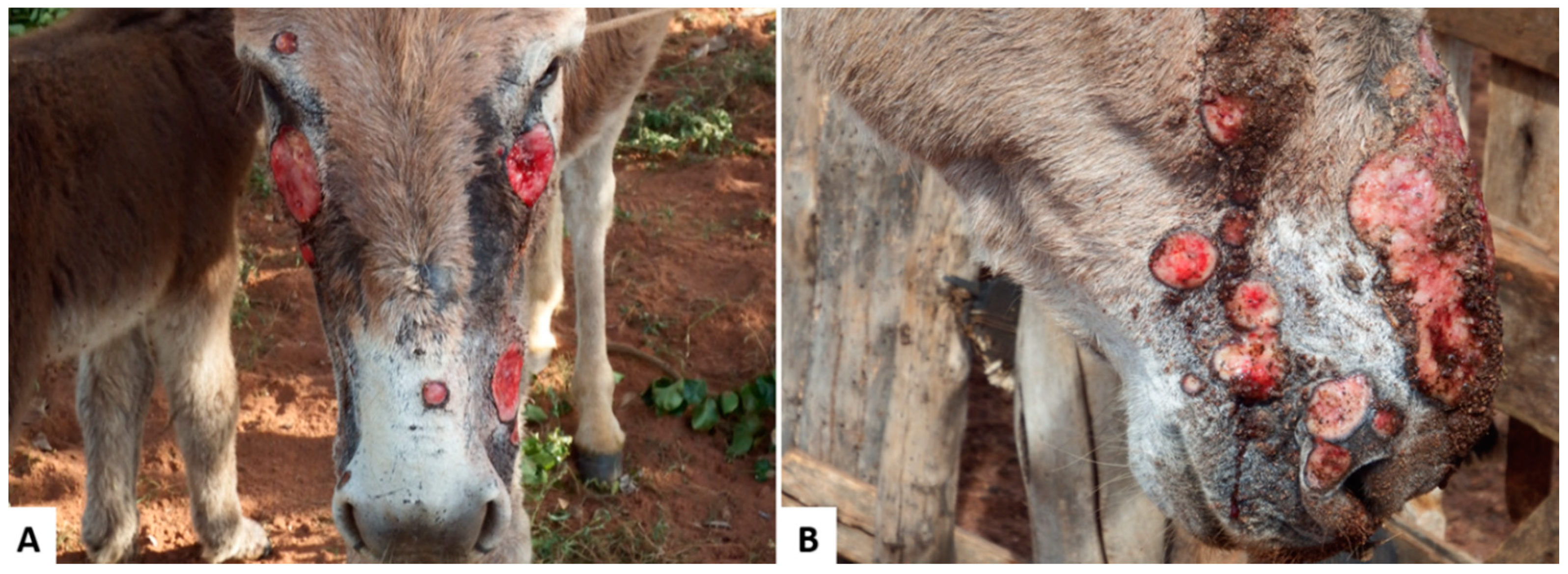
| Wounds, exuberant granulation tissue | Initial wounds can be of varied causes, leading to ulcerative and crusted lesions. These lesions can evolve into the exuberant granulation tissue becoming spongy, irregular, with no evident exudation. | In these lesions, there is a marked proliferation of fibrous connective tissue, neovascularization, fibroplasia, in addition to a chronic active inflammatory infiltrate, depending on whether the pathogenic stimulus persists or not. | Varied location, usually associated with the use of ropes, saddles and whips for containment, being commonly observed in the neck, limbs, back and tail. | [12,28,45] |
Publisher’s Note: MDPI stays neutral with regard to jurisdictional claims in published maps and institutional affiliations. |
© 2020 by the authors. Licensee MDPI, Basel, Switzerland. This article is an open access article distributed under the terms and conditions of the Creative Commons Attribution (CC BY) license (http://creativecommons.org/licenses/by/4.0/).
Share and Cite
Lima, T.S.; Silva, R.A.F.; Pereira, R.M.F.; Soares, K.L.; Santos, N.T.A.; Sousa, M.S.; Mendonça, F.S.; Lucena, R.B. Skin Diseases in Donkeys and Mules—An Update. Animals 2021, 11, 65. https://doi.org/10.3390/ani11010065
Lima TS, Silva RAF, Pereira RMF, Soares KL, Santos NTA, Sousa MS, Mendonça FS, Lucena RB. Skin Diseases in Donkeys and Mules—An Update. Animals. 2021; 11(1):65. https://doi.org/10.3390/ani11010065
Chicago/Turabian StyleLima, Telma S., Raquel A. F. Silva, Raquel M. F. Pereira, Karoline L. Soares, Nayadjala T. A. Santos, Mônica S. Sousa, Fábio S. Mendonça, and Ricardo B. Lucena. 2021. "Skin Diseases in Donkeys and Mules—An Update" Animals 11, no. 1: 65. https://doi.org/10.3390/ani11010065
APA StyleLima, T. S., Silva, R. A. F., Pereira, R. M. F., Soares, K. L., Santos, N. T. A., Sousa, M. S., Mendonça, F. S., & Lucena, R. B. (2021). Skin Diseases in Donkeys and Mules—An Update. Animals, 11(1), 65. https://doi.org/10.3390/ani11010065