Article

Effects of Aging on Expression of Mic60 and OPA1 and Mitochondrial Morphology in Myocardium of Tibetan Sheep

1
Gansu Key Laboratory of Herbivorous Animal Biotechnology, Faculty of Animal Science and Technology, Gansu Agricultural University, Lanzhou 730070, China
2
School of Biotechnology and Food, Shangqiu Normal University, Shangqiu 476000, China
*
Authors to whom correspondence should be addressed.
Animals 2020, 10(11), 2160; https://doi.org/10.3390/ani10112160
Submission received: 11 November 2020 / Accepted: 18 November 2020 / Published: 20 November 2020
(This article belongs to the Special Issue Immunohistochemical and Physiological Research on Farm Animals)

Abstract

:

Simple Summary

Mitochondria play a crucial role in the adaptation to high altitude hypoxia environment in Tibetan sheep, and the changes of its morphology and structure directly affect its function. OPA1 and Mic60 are important mitochondria-shaping proteins that work together to regulate the morphology of mitochondrial inner membrane and cristae. It has been shown that aging affects the expression of OPA1 and Mic60 in mice, but it has not been investigated in sheep and hence it is not known whether it might affect the ultrastructure of mitochondria. In this context, reverse transcription-quantitative PCR (RT-qPCR), enzyme-linked immunosorbent assay (ELISA) and immunohistochemistry method were used to measure the expression of Mic60 and OPA1 genes and proteins in myocardium of adult and aged Tibetan sheep, and the ultrastructure of mitochondria were compared by transmission electron microscope. The results suggest that aging can reduces the expression of Mic60 and OPA1 genes and OPA1 protein, which can affect the mitochondrial function.

Abstract

In order to investigate the effects of aging on the expression of Mic60 and OPA1 and mitochondrial morphology in plateau animals, the expression of Mic60 and OPA1 genes and proteins, and the morphology of mitochondria in the myocardium of adult and aged Tibetan sheep were investigated. The expression of Mic60 and OPA1 genes and OPA1 protein were higher (p < 0.05) in the myocardium of adult Tibetan sheep than in those of the aged ones. The number of mitochondrial cristae in the myocardium of adult was higher than that in aged (p < 0.05). The density of mitochondria in the myocardium of adult was higher than that in aged (p < 0.01). Compared with the adult Tibetan sheep, the mitochondrial crista of aged were relatively sparse, the crista membrane was wide, and the mitochondria were not closely linked, showing fragmentation. These results suggest that the myocardial mitochondria of the adult have better energy supply ability, indicating that aging can lead to the weakening of oxygen supply in the myocardial mitochondria of Tibetan sheep.

1. Introduction

Tibetan sheep live in the Qinghai-Tibet plateau and its adjacent areas at an altitude of 3000 to 5000 m above sea level. Generation reproduction under the altitude environment makes Tibetan sheep well adapted to the low oxygen concentration. It also makes it a good model experimental animal for cardiovascular research. The heart is one of the most important organs in animals, which is the power center for the supply of oxygen in whole body tissues. Under increased metabolism and high oxygen consumption, the heart can provide pressure to increase the blood flow into all parts of the body. As an aerobic tissue, the energy of myocardial metabolism is almost entirely dependent on the oxidative phosphorylation of mitochondria. Therefore, the quantity, function and structure of mitochondria in cardiomyocytes determine the energy supply of the heart.
Mitochondria are the main sites for aerobic respiration of eukaryotic cells and play an important role in the maintenance of calcium homeostasis and the regulation of apoptosis [1,2]. The morphology of mitochondria is regulated by fusion and fission [3,4]. The complete morphological structure is the basis for the function of mitochondria and the integrity of the crista membrane is particularly important [5]. Mitochondrial crista membrane is the main site of oxidative phosphorylation. More than 90% of mitochondrial oxidative phosphorylation complex III, ATP synthase and many proteins and enzymes associated with the oxidative phosphorylation are located in crista [6,7,8].
Mitochondria-shaping proteins Mic60 (Mitofilin) and OPA1 (Dominant Optic Atrophy), which are located in the mitochondrial membrane space, play an important role in the maintenance of mitochondrial morphology and crista membrane structure [9,10]. The nuclear gene OPA1 is widely expressed in many organs, but the expression level is different. The main functions of OPA1 is to promote mitochondrial fusion [11,12]. Inhibition of OPA1 gene expression or inactivation of OPA1 protein will lead to the disruption of mitochondrial network fusion [13]. The loss of OPA1 subtype will cause damage to the crista membrane structure and seriously affects cell proliferation [14,15]. Mic60, as the core component of MINOS (Mitochondrial Contact Site) complex [16], plays an important role in maintaining the morphology and structure of mitochondrial crista [17]. Interfering with the expression of Mic60 not only leads to mitochondrial malformation, but also inhibits the mitochondrial oxidative phosphorylation activity and the normal function of mitochondria [18]. Recent studies showed that Mic60 was crucial for the formation of mitochondrial crista junctions [19,20].
The change of heart function is closely related to age [21]. The age-related changes of cardiac myocardium focus on three main aspects: myocardial cells, mitochondria related proteins and enzymes, and collagen fibers. In this study, the effects of aging on the expression of Mic60 and OPA1 genes and proteins and the mitochondrial morphology in myocardial of Tibetan sheep were studied in combination with the observation of molecular regulation mechanism, protein expression, and mitochondrial ultrastructure.

2. Materials and Methods

All animal work in this study was conducted according to the guidelines of the care and use of experimental animals established by the Ministry of Science and Technology of the People’s Republic of China (Approval number 2006-398), and was approved by the Animal Care Committee of Gansu Agricultural University.

2.1. Materials

Six ewes of Tibetan sheep in Gannan Luqu (the average altitude is 3500 m) were selected randomly, three adult (2 years old) and three aged (6 years old) each. All ewes were slaughtered according to the conventional methods, and the same site of myocardial tissue was immediately collected. Of these, 200 mg was immediately used for the isolation of the mitochondria. The sample for light microscopy was cut into pieces of 1 × 1 × 1 cm, washed with PBS and fixed in a 4% paraformaldehyde fixative solution for later use. Transmission electron microscope samples were cut into strips of 1 × 1 × 2 mm, washed with PBS and fixed in 3% glutaraldehyde phosphate buffer (pH 7.2–7.4, 4 °C). The rest of the samples was rapidly frozen by liquid nitrogen and then stored in the refrigerator at −80 °C for the determination of Mic60 and OPA1 genes and proteins expression.

2.2. Isolation of the Myocardial Mitochondria

Mitochondria in myocardial tissue were isolated according to the Biovision mitochondrial isolation kit (Mammalian Mitochondria Isolation Kit for Tissue and Cultured Cells, K288). The whole process of isolation was carried out at low temperature to ensure the complete structure and function of mitochondria. The isolated mitochondria were fixed with 3% glutaraldehyde phosphate buffer (pH 7.2–7.4, 4 °C).

2.3. Extraction of Tissue RNA

The total RNA in myocardium was extracted according to the RNA extraction kit (RNA isolator Total RNA Extraction Reagent, Vazyme, Nanjing, China). The quality of RNA integrity was tested by 1.5% agarose gel electrophoresis. Next, the OD260/OD280 value and its concentration were measured by ultramicro spectrophotometer, and the qualified RNA was preserved in the freezer at −80 °C. Reverse transcription of RNA was completed by using reverse transcription kit (HiScript ⅡQ RT SuperMix for qPCR, +gDNA wiper, Vazyme, Nanjing, China). The extracted cDNA was diluted into 1:9 solution and stored at −20 °C.

2.4. Design and Synthesis of Primers

The complete sequence of sheep Mic60 and OPA1 genes published by GenBank were used to design PCR primers by Primer Premier 5.0 software (PREMIER Biosoft, San Francisco, CA, USA). The β-actin gene was used as the reference gene, and the designed primers were tested for its specificity by Blast. Primers were synthesized by BGI Liuhe Company (Beijing, China). The detail information of primers is shown in Table 1.

2.5. RT-qPCR Reaction

The cDNA working fluid was carried out by SYBR Green I chimeric fluorescence method using ChamQ Universal SYBR qPCR Master Mix (Q711-02/03, Vazyme, Nanjing, China) for RT-qPCR reaction. The 20 μL reaction containing 2 μL of cDNA working fluid, 0.4 μL of upstream and downstream primers (10 μM), respectively, 2× ChamQ Universal SYBR qPCR Master Mix (10 μL) and RNase-free dd H2O (7.2 μL). The optimal conditions of qPCR amplification procedure were as follows: Pre-denaturation at 94 °C for 4 min; Cyclic reaction, 95 °C 10 s, 60 °C 30 s, 40 cycles; Dissolving curve, 95 °C 15 s, 60 °C 60 s, 95 °C 15 s. The specificity of the PCR product and the presence of primer dimers were determined according to the resulting dissolution curve. Data were collected and gene expression was calculated [22] and analyzed for variance using SPSS software (SPSS, Chicago, IL, USA).

2.6. Immunohistochemistry and ELISA

The samples of cut sections were fixed with 4% paraformaldehyde fixative (4 °C, 24 h) and then cut into 5 μm thick sections by routine paraffin embedding. Cut sections were deparaffinized to water for antigen retrieval (Citrate Buffer-Microwave Heat Recovery Method). After rinsed in PBS, the cut sections were blocked (37 °C, 10 min) by adding 3% H2O2 (SP KIT), rinsed in PBS, placed in wet box and added blocking solution (SP KIT A) on it (at room temperature, 10 min), and then incubated overnight at room temperature with primary antibody (Rabbit Anti-Mitofilin antibody and Rabbit Anti-OPA1 antibody, 1:200, Bioss, Beijing, bs-11764R and bs-1824R). The sections were rinsed by PBS, and then added the secondary antibody (SP KIT B) on it (37 °C, 15 min). The sections were rinsed by PBS and the third antibody (SP KIT C) was added (37 °C, 15 min). At last, the sections were stained by DAB, slightly stained by hematoxylin and fixed by neutral balata. An Olympus BX61 positive universal microscope (Olympus, Tokyo, Japan) was used to observe the sections. The Mic60 and OPA1 proteins in the myocardium were detected by ELISA. To measure this, 100 mg of fresh tissue was triturated using a homogenizer (Biospec, Bartlesville, OK, USA) and mixed with 500 µL PBS. The solution was then centrifuged for 10 min at 3000× g at 4 °C and the supernatant collected. The protein was measured and a solution containing 100 µg proteins was analyzed according to the manufacturer’s instructions (Mic60 and OPA1 ELISA Kit, Bio-Swamp, Wuhan Beinglay Biotech Co., LTD, Wuhan, China). The PBS solution used for grinding tissue was analyzed as blank control and the absorbance was measured at 450 nm. The concentrations of Mic60 and OPA1 were calculated from corresponding with the Mic60 and OPA1 standard curves.

2.7. Transmission Electron Microscopy Preparation

The tissue and isolated mitochondria for cut sections were fixed with 3% glutaraldehyde, rinsed twice with 0.2 mmol/L PB (10 min/time), fixed with 1% osmium tetroxide (4 °C, 2 h), and rinsed three times with PB (10 min/time). Then, the samples were dehydrated by gradient alcohol (50, 60, 70, 80, and 90% ethanol for 10 min and 100% acetone dehydration twice for 10 min each). The samples were soaked in the mixture of epoxy resin (SPION-PON812) with acetone (1:1) for 5 h (room temperature), embedded with epoxy resin and dried (45 °C, 12 h; 65 °C, 48 h). Semi-thin sections were cut before making ultrathin sections (70 nm). The ultrathin sections were stained by uranium and lead (Uranium acetate stained for 30 min and lead citrate stained for 10 min). The ultrathin sections were observed using a JEM-1230 transmission electron microscope (Jeol, Nihon Denshi, Japan).

2.8. Statistical Analyses

To measure the number of mitochondrial crista and density of mitochondria, 50 microscopic fields (10 cut sections were made for each animal and 5 microscopic fields were randomly selected from each cut section, 20,000×) were analyzed from each animal included in the study. All data in this study were analyzed by Independent Samples t-test using SPSS 19.0 (SPSS, Chicago, IL, USA) and were expressed as the mean ± SD. All p-values were considered statistically significant when p < 0.05.

3. Results

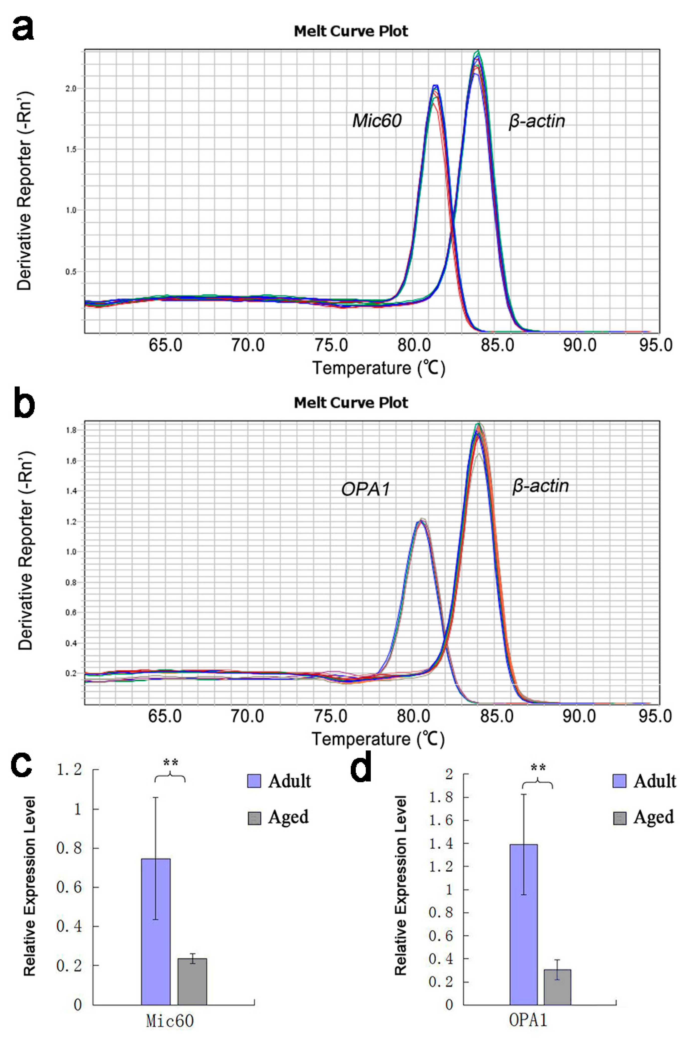
3.1. Analysis of the Result of RT-qPCR

The expression of Mic60 and OPA1 genes in myocardium of adult and aged Tibetan sheep was quantitatively analyzed by RT-qPCR. The results showed that both the Mic60 and OPA1 genes were expressed in the myocardium of adult and aged Tibetan sheep, but the expression levels were different. The Tm values of the two gene amplification products were uniform, and the single peak melt curve indicated that both primer sets had good specificity in the RT-qPCR reaction, and no non-specific amplification and primer dimers were observed (Figure 1a,b). The relative expression levels of Mic60 and OPA1 genes in the myocardium of adult Tibetan sheep were 0.7471 ± 0.3105 and 1.3922 ± 0.43547, respectively, which were significantly higher than those in aged Tibetan sheep (0.2366 ± 0.02534 and 0.307 ± 0.08628, respectively) (p < 0.01) (Figure 1c,d).

3.2. Expression and Analysis of Mic60 and OPA1 Proteins

ELISA method was used to locate and quantify Mic60 and OPA1 proteins. The results from the immunohistochemical photographs showed that Mic60 protein expressed in the myocardium of both adult and aged Tibetan sheep. OPA1 protein expressed in the myocardium of adult Tibetan sheep, but hardly expressed in the aged sheep (Figure 2a,b,d,e). ELISA results showed that the concentrations of Mic60 protein in the myocardium had no significant difference between adult (153.27 ± 26.28 ng/mL) and aged (150.24 ± 25.98 ng/mL) Tibetan sheep (p > 0.05) (Figure 2g). The concentrations of OPA1 protein in the myocardium had significant difference between adult (49.77 ± 16.45 ng/mL) and aged (34.06 ± 12.29 ng/mL) Tibetan sheep (p < 0.05) (Figure 2g).

3.3. Mitochondrial Morphological Structure Comparison

The mitochondrial ultrastructure of myocardium was observed by transmission electron microscopy, and the morphological and the difference of myocardial mitochondria between adult and aged Tibetan sheep were compared. The average number of mitochondrial crista and the density of mitochondria isolated were calculated. The mitochondria in the myocardium of both adult and aged Tibetan sheep had clear double membranes and tubular cristae, and their structures were complete (Figure 3a,b). The mitochondrial density in the myocardium of adult Tibetan sheep was 57.4 ± 6.07/100 μm2, which was significantly higher than that of the older age group (38.6 ± 4.51/100 μm2) (p < 0.01) (Figure 4b). The number of mitochondria cristae in the myocardium of adult Tibetan sheep was 21.5 ± 2.65, which was denser than that of 16.5 ± 3.11 in aged Tibetan sheep (p < 0.05) (Figure 4a). The mitochondria of adult were closely related but were not closely related in the aged Tibetan sheep, being slightly fragmented, and the cristae membrane were slightly swelled (Figure 3a,b).

4. Discussion

The expression of mitochondrial inner membrane proteins Mic60 and OPA1 may be related to aging. Studies found that with the Mic60 heterozygous deletion, the expression of senescence-associated protein p21 in the myocardium of mice was significantly increased, indicated that the mitochondrial inner membrane protein Mic60 may be related to cardiac senescence in mice [23]. Eckert et al. found that the expression of OPA1 in mice was decreased with age [24]. A similar result was obtained by Xu et al., the expression of OPA1 in Drosophila was significantly decreased with age [25]. In this study, the expression levels of Mic60 and OPA1 genes in the myocardium of aged Tibetan sheep were significantly lower than that of adult, which were 0.32 and 0.22 times of that of adult Tibetan sheep, respectively, which indicated that the expression of Mic60 and OPA1 genes in the myocardium of Tibetan sheep would reduce with the age. The expression of OPA1 protein in the aged Tibetan sheep was significantly lower than that of adult. In spite of this, the expression of Mic60 protein in the myocardium of aged Tibetan sheep was similar to that of adult, indicating that the expression of Mic60 protein may affect by other factors, which deserved further investigation.
In this study, the density of myocardial mitochondria in aged Tibetan sheep was significantly lower than that of adult, which indicated that myocardial mitochondrial density was age-dependent. Preston et al. reported that the adult rat heart mitochondrial density was higher than that of older rat [26]. The similar phenomenon was found in the studies of yak heart, which showed that mitochondrial density of heart gradually decreased with age, and the concentration of mitochondrial protein gradually increased [27]. Studies on the heart of yak from six months to two years old showed that the density of myocardial mitochondria gradually decreased with age [28], which was consistent with this research.
The expression of Mic60, OPA1 genes and OPA1 protein were significantly lower in aged Tibetan sheep myocardium than that of adult in this study. Besides, the number of mitochondrial crista was significantly lower, the crista membrane was wide and the mitochondria were not closely linked, showing fragmentation in aged Tibetan sheep. Von et al. reported that absence of fcj1 (Mic60 in mammals) would lead to the mitochondrial crista membrane becoming hypertrophic and stacked, the crista junction being lost and the crista separating from the inner boundary membrane in the yeast cells [16]. Studies found that the oligomerization of OPA1 in starvation cells was significantly increased, while the crista width of the mitochondria in the starving cells were narrowed obviously and OPA1 had a regulatory effect on mitochondrial crista independent of its fusion effect [29]. OPA1 protein was also found to be related to the tubular network structure of mitochondria, and inactivation of OPA1 would make the network cracked and aggravate mitochondrial fragmentation [30]. Some studies have shown that the expression of OPA1 was positively correlated with the number of mitochondria crista. Researchers found that a large number of crista structures disappeared in mitochondria when the OPA1 protein was inhibited in Hela cells [15]. Studies also showed that the interaction between OPA1 and Mic60 regulated the morphology of mitochondrial [31]. Therefore, the mitochondrial morphology of aged Tibetan sheep is different from the adult Tibetan ones, which may be influenced by the regulation of Mic60 and OPA1 genes and proteins expression, with OPA1 possibly playing a more important role.
Mitochondria cristae can greatly increase mitochondrial inner membrane surface area and provide more attachment sites for oxidative phosphorylation-related proteins and enzymes. The more mitochondria cristae there are, the stronger oxidative phosphorylation ability they have, and the higher metabolic activity and energy consumption. The mitochondrial of the adult Tibetan sheep has a larger crista membrane area compared with the aged sheep. This structural feature indicates that the mitochondria of adult Tibetan sheep have stronger oxidative phosphorylation ability.

5. Conclusions

In conclusion, our study investigated the expression of Mic60 and OPA1 genes and proteins and the morphology of mitochondria in the myocardium of adult and aged Tibetan sheep. Our data showed that the expression of Mic60, OPA1 genes and OPA1 protein were significantly lower in aged Tibetan sheep myocardium than that of adult in this study. Besides, the number of mitochondrial crista was significantly lower, the crista membrane was wide and the mitochondria were not closely linked, showing fragmentation in aged Tibetan sheep. These results suggest the expression of Mic60 and OPA1 genes and OPA1 protein will reduce, while reducing the capacity of myocardial mitochondria in Tibetan sheep with age.

Author Contributions

G.W. and Y.L. conceived and designed the experiments; G.W., J.H., J.W., X.L. and S.L. performed the experiments; G.W. analyzed the data; G.W. and Y.L. wrote the manuscript. All authors reviewed and commented on the manuscript. All authors have read and agreed to the published version of the manuscript.

Funding

This work was financially supported by Discipline Construction Fund Project of Gansu Agricultural University (GAU-XKJS-2018-029).

Conflicts of Interest

The authors declare no conflict of interest.

References

  1. Dimmer, K.S.; Scorrano, L. (De) constructing mitochondria: What for? Physiology 2006, 21, 233–241. [Google Scholar] [CrossRef] [PubMed]
  2. Frezza, C.; Cipolat, S.; Brito, O.M.D.; Micaroni, M.; Beznoussenko, G.V.; Rudka, R.T.; Bartoli, D.; Polishuck, R.S.; Danial, N.N.; Strooper, B.D.; et al. OPA1 Controls Apoptotic Cristae Remodeling Independently from Mitochondrial Fusion. Cell 2006, 126, 177–189. [Google Scholar] [CrossRef] [PubMed] [Green Version]
  3. Okamoto, K.; Shaw, J.M. Mitochondrial Morphology and Dynamics in Yeast and Multicellular Eukaryotes. Annu. Rev. Genet. 2005, 39, 503–536. [Google Scholar] [CrossRef] [PubMed]
  4. Chan, D.C. Mitochondrial Fusion and Fission in Mammals. Annu. Rev. Cell. Dev. Bi. 2006, 22, 79–99. [Google Scholar] [CrossRef] [Green Version]
  5. Cogliati, S.; Frezza, C.; Soriano, M.E.; Varanita, T.; Quintana-Cabrera, R.; Corrado, M.; Cipolat, S.; Costa, V.; Casarin, A.; Gomes, L.C.; et al. Mitochondrial Cristae Shape Determines Respiratory Chain Supercomplexes Assembly and Respiratory Efficiency. Cell 2013, 155, 160–171. [Google Scholar] [CrossRef] [Green Version]
  6. Gilkerson, R.W.; Selker, J.M.L.; Capaldi, R.A. The cristal membrane of mitochondria is the principal site of oxidative phosphorylation. FEBS Lett. 2003, 546, 355–358. [Google Scholar] [CrossRef] [Green Version]
  7. Chaban, Y.; Boekema, E.J.; Dudkina, N.V. Structures of mitochondrial oxidative phosphorylation supercomplexes and mechanisms for their stabilisation. BBA-Bioenerg. 2014, 1837, 418–426. [Google Scholar] [CrossRef] [Green Version]
  8. Vogel, F.; Bornhovd, C.; Neupert, W.; Reicher, A.S. Dynamic subcompartmentalization of the mitochondrial inner membrane. J. Cell Biol. 2006, 175, 237–247. [Google Scholar] [CrossRef]
  9. Carelli, V.; Morgia, C.L.; Iommarini, L.; Carroccia, R.; Mattiazzi, M.; Sangiorgi, S.; Farne, S.; Maresca, A.; Foscarini, B.; Lanzi, L.; et al. Mitochondrial Optic Neuropathies: How Two Genomes may Kill the Same Cell Type? Biosci. Rep. 2007, 27, 173–184. [Google Scholar] [CrossRef]
  10. Gieffers, C.; Korioth, F.; Heimann, P.; Ungermann, C.; Frey, J. Mitofilin is a transmembrane protein of the inner mitochondrial membrane expressed as two isoforms. Exp. Cell. Res. 1997, 232, 395–399. [Google Scholar] [CrossRef]
  11. Olichon, A.; Elachouri, G.; Baricault, L.; Delettre, C.; Belenguer, P.; Lenaers, G. OPA1 alternate splicing uncouples an evolutionary conserved function in mitochondrial fusion from a vertebrate restricted function in apoptosis. Cell Death Differ. 2007, 14, 682–692. [Google Scholar] [CrossRef] [PubMed] [Green Version]
  12. Youle, R.J.; van der Bliek, A.M. Mitochondrial Fission, Fusion, and Stress. Science 2012, 337, 1062–1065. [Google Scholar] [CrossRef] [PubMed] [Green Version]
  13. Spinazzi, M.; Cazzola, S.; Bortolozzi, M.; Baracca, A.; Loro, E.; Casarin, A.; Solaini, G.; Sgarbi, G.; Casalena, G.; Cenacchi, G.; et al. A novel deletion in the gtpase domain of opa1 causes defects in mitochondrial morphology and distribution, but not in function. Hum. Mol. Genet. 2008, 17, 3291–3302. [Google Scholar] [CrossRef] [PubMed] [Green Version]
  14. Griparic, L.; Kanazawa, T.; Van Der Bliek, A.M. Regulation of the mitochondrial dynamin-like protein Opa1 by proteolytic cleavage. J. Cell Biol. 2007, 178, 757–764. [Google Scholar] [CrossRef] [Green Version]
  15. Griparic, L.; Van Der Wel, N.N.; Orozco, I.J.; Peters, P.J.; Van Der Bliek, A.M. Loss of the intermembrane space protein Mgm1/OPA1 induces swelling and localized constrictions along the lengths of mitochondria. J. Biol. Chem. 2004, 279, 18792–18798. [Google Scholar] [CrossRef] [Green Version]
  16. Von der Malsburg, K.; Mueller, J.M.; Bohnert, M.; Oeljeklaus, S.; Kwiatkowska, P.; Becker, T.; Loniewska-Lwowska, A.; Wiese, S.; Rao, S.; Milenkovic, D.; et al. Dual Role of Mitofilin in Mitochondrial Membrane Organization and Protein Biogenesis. Dev. Cell 2011, 21, 694–707. [Google Scholar] [CrossRef] [Green Version]
  17. Head, B.P.; Zulaika, M.; Ryazantsev, S.; van der Bliek, A.M. A novel mitochondrial outer membrane protein, MOMA-1, that affects cristae morphology in Caenorhabditis elegans. Mol. Biol. Cell 2011, 22, 831–841. [Google Scholar] [CrossRef]
  18. Yang, R.F.; Sun, L.H.; Zhang, R.; Zhang, Y.; Luo, Y.X.; Zheng, W.; Zhang, Z.Q.; Chen, H.Z.; Liu, D.P. Suppression of Mic60 compromises mitochondrial transcription and oxidative phosphorylation. Sci. Rep. 2015, 5, 7990. [Google Scholar] [CrossRef] [Green Version]
  19. Koerner, C.; Barrera, M.; Dukanovic, J.; Eydt, K.; Harner, M.; Rabl, R.; Vogel, F.; Rapaport, D.; Neupert, W.; Reichert, A.S. The C-terminal domain of Fcj1 is required for formation of crista junctions and interacts with the TOB/SAM complex in mitochondria. Mol. Biol. Cell 2012, 23, 2143–2155. [Google Scholar] [CrossRef]
  20. John, G.B.; Shang, Y.; Li, L.; Renken, C.; Mannella, C.A.; Selker, J.M.L.; Rangell, L.; Bennett, M.J.; Zha, J. The mitochondrial inner membrane protein mitofilin controls cristae morphology. Mol. Biol. Cell 2005, 16, 1543–1554. [Google Scholar] [CrossRef]
  21. Chantler, P.D.; Goldspink, D.F.; Clements, R.E.; Sharp, L.; Schlosshan, D.; Tan, L.B. Congestive heart failure: Extent of cardiac functional changes caused by aging and organ dysfunction. Heart 2006, 92, 686–688. [Google Scholar] [CrossRef] [PubMed] [Green Version]
  22. Livak, K.; Schmittgen, T. Analysis of Relative Gene Expression Data Using Real-Time Quantitative PCR and the 2-△△Ct Method. Methods 2000, 25, 402–408. [Google Scholar] [CrossRef] [PubMed]
  23. Wang, C.L.; Sun, L.H.; Yue, Y.S. Correlation between Mic60 haploid insufficiency and cardiac aging in mouse. Chin. J. Pathol. 2017, 46, 406–410. [Google Scholar]
  24. Eckert, G.P.; Schiborr, C.; Hagl, S.; Abdel-Kader, R.; Muller, W.E.; Rimbach, G.; Frank, J. Curcumin prevents mitochondrial dysfunction in the brain of the senescence-accelerated mouse-prone 8. Neurochem. Int. 2013, 62, 595–602. [Google Scholar] [CrossRef] [PubMed]
  25. Xu, C.; Wang, H.; Wang, Z.; Wang, Q.; Zhang, S.; Deng, Y.; Fang, Y. In vivo imaging reveals mitophagy independence in the maintenance of axonal mitochondria during normal aging. Aging Cell 2017, 16, 1180–1190. [Google Scholar]
  26. Preston, C.C.; Oberlin, A.S.; Holmuhamedov, E.L.; Gupta, A.; Sagar, S.; Syed, R.H.K.; Siddiqui, S.A.; Raghavakaimal, S.; Terzic, A.; Jahangir, A. Aging-induced alterations in gene transcripts and functional activity of mitochondrial oxidative phosphorylation complexes in the heart. Mech. Ageing Dev. 2008, 129, 304. [Google Scholar] [CrossRef] [Green Version]
  27. Simonson, T.S.; Yang, Y.; Huff, C.D.; Yun, H.; Qin, G.; Witherspoon, D.J.; Bai, Z.; Lorenzo, F.R.; Xing, J.; Jorde, L.B.; et al. Genetic evidence for high-altitude adaptation in Tibet. Science 2010, 329, 72. [Google Scholar] [CrossRef] [Green Version]
  28. He, Y.; Yu, S.; Hu, J.; Cui, Y.; Liu, P. Changes in the Anatomic and Microscopic Structure and the Expression of HIF-1α and VEGF of the Yak Heart with Aging and Hypoxia. PLoS ONE 2016, 11, e0149947. [Google Scholar] [CrossRef]
  29. Patten, D.A.; Wong, J.; Khacho, M.; Soubannier, V.; Mailloux, R.J.; Pilon-Larose, K.; MacLaurin, J.G.; Park, D.S.; McBride, H.M.; Trinkle-Mulcahy, L.; et al. OPA1-dependent cristae modulation is essential for cellular adaptation to metabolic demand. Embo J. 2014, 33, 2676–2691. [Google Scholar] [CrossRef] [Green Version]
  30. Meeusen, S.; DeVay, R.; Block, J.; Cassidy-Stone, A.; Wayson, S.; McCaffery, J.M.; Nunnari, J. Mitochondrial inner-membrane fusion and crista maintenance requires the dynamin-related GTPase Mgm1. Cell 2006, 127, 383–395. [Google Scholar] [CrossRef] [Green Version]
  31. Darshi, M.; Mendiola, V.L.; Mackey, M.R.; Murphy, A.N.; Koller, A.; Perkins, G.A.; Ellisman, M.H.; Taylor, S.S. ChChd3, an Inner Mitochondrial Membrane Protein, Is Essential for Maintaining Crista Integrity and Mitochondrial Function. J. Biol. Chem. 2011, 286, 2918–2932. [Google Scholar] [CrossRef] [PubMed] [Green Version]
Figure 1. Melting curves of Mic60, OPA1 and β-actin genes and their relative expression in adult and aged Tibetan sheep cardiomyocytes. Note: (a,b): Melting curve of Mic60, OPA1 and β-actin genes. (c,d): Relative expression of Mic60 and OPA1 genes. All data showed as means ± SD (n = 3). ** p < 0.01.
Animals 10 02160 g001
Figure 2. Expression of Mic60 and OPA1 proteins in cardiomyocytes. Note: (ac): Expression of Mic60 protein in adult and aged Tibetan sheep myocardium and blank control. (df): Expression of OPA1 protein in adult and aged Tibetan sheep myocardium and blank control. (g): Protein concentration of Mic60 and OPA1. Data show means ± SD (n = 3). * p < 0.05.
Animals 10 02160 g002
Figure 3. Ultrastructure observation of myocardial mitochondria in adult and aged Tibetan sheep. Note: (a,b): Adult and aged Tibetan sheep myocardial mitochondria, bar = 0.5 μm. (c,d): Mitochondria isolated from adult and aged Tibetan Sheep myocardium, bar = 0.2 μm.
Animals 10 02160 g003
Figure 4. The number of mitochondrial crista (a) and density of mitochondria (b) in the myocardium of adult and aged Tibetan Sheep. Data show means ± SD. * p < 0.05, ** p < 0.01.
Animals 10 02160 g004
Table 1. Primer information for Tibetan sheep MIC60, OPA1 and β-actin genes.
GeneGenBank Accession No.Primer Sequence (5′–3′)Primer LengthProduct Length
Mic60XM_012169573F:TTGAGATGGTCCTTGGTT18136
R:TTGTTTCTGAGGTGGTGAG19
OPA1XM_012140446F:ATGAAATAGAACTCCGAATG20112
R:GTCAACAAGCACCATCCT18
β-actinNM_001009784F: AGCCTTCCTTCCTGGGCATGGA22113
R:GGACAGCACCGTGTTGGCGTAGA23
Publisher’s Note: MDPI stays neutral with regard to jurisdictional claims in published maps and institutional affiliations.

