Article

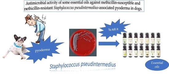
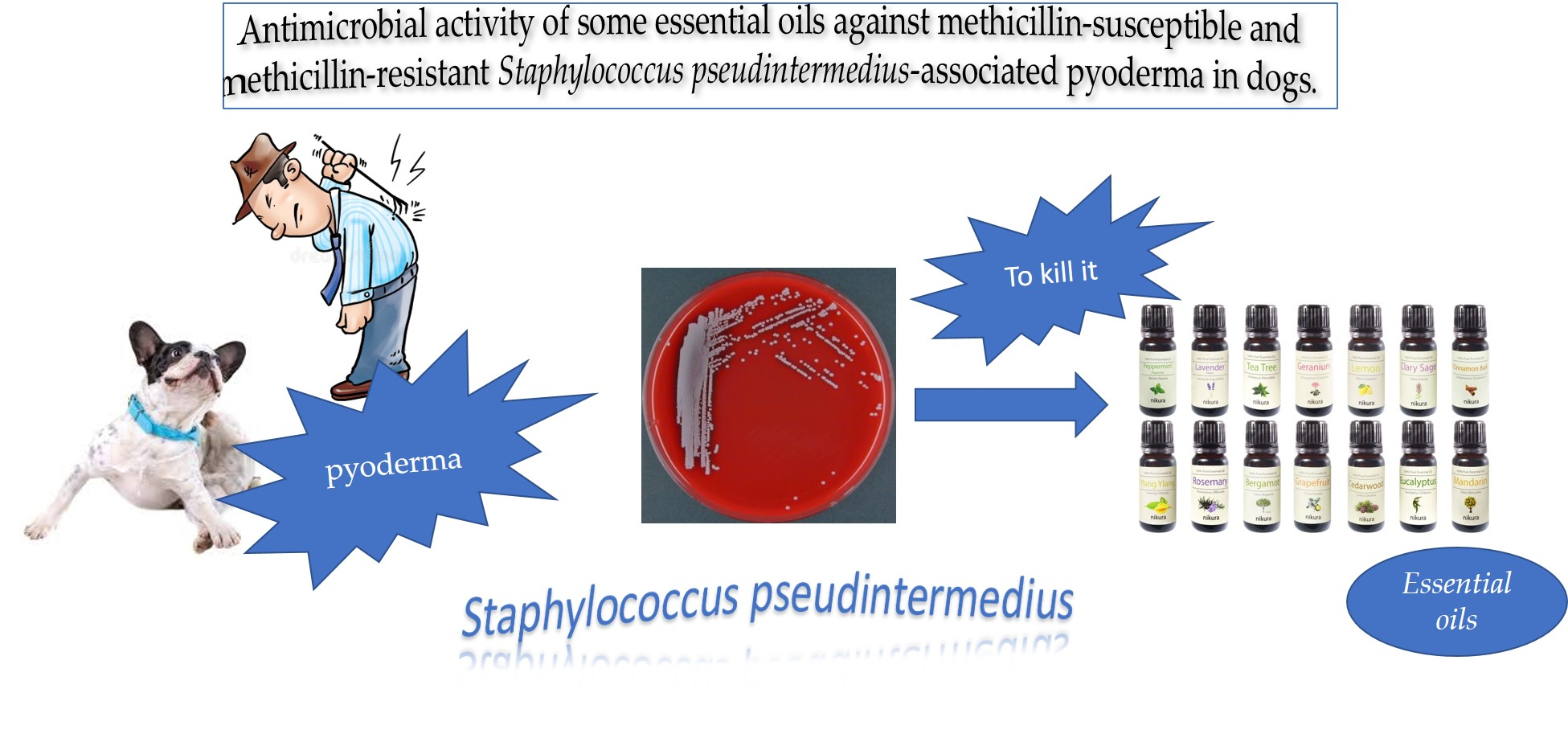
Antimicrobial Activity of Some Essential Oils against Methicillin-Susceptible and Methicillin-Resistant Staphylococcus pseudintermedius-Associated Pyoderma in Dogs

1
Department of Veterinary Medicine and Animal Production, Via Delpino 1, University of Naples “Federico II”, 80137 Naples, Italy
2
Department of Veterinary Sciences, Viale delle Piagge 2, University of Pisa, 56124 Pisa, Italy
3
Department of Pharmacy, Via Bonanno 6, University of Pisa, 56126 Pisa, Italy
*
Author to whom correspondence should be addressed.
These authors contributed equally to this work.
Animals 2020, 10(10), 1782; https://doi.org/10.3390/ani10101782
Submission received: 10 September 2020 / Revised: 21 September 2020 / Accepted: 25 September 2020 / Published: 1 October 2020

Abstract

:

Simple Summary

Pyoderma is one of the most common diseases in dogs, and Staphylococcus pseudintermedius, a Gram-positive coagulase-positive bacterium, represents the most common infectious agent causing canine pyoderma. Since multidrug-resistant S. pseudintermedius strains have become a relevant threat in veterinary medicine, this study aimed to test the antimicrobial properties of some essential oils (EOs) against S. pseudintermedius strains isolated from dogs suffering from pyoderma. The obtained findings demonstrated a clear in vitro efficacy of some tested EOs against clinical methicillin-resistant and methicillin-sensible S. pseudintermedius strains. The applicability and efficacy of EOs in cases of canine pyoderma supported by S. pseudintermedius could be beneficial for both dogs and pet owners, who are inevitably exposed to this zoonotic bacterium.

Abstract

This study aimed to test in vitro the antimicrobial activity of 11 essential oils (EOs) against four methicillin-resistant Staphylococcus pseudintermedius (MRSP) and four methicillin-susceptible S. pseudintermedius (MSSP) clinical isolates. The obtained findings demonstrated a clear in vitro efficacy of some tested EOs against both MRSP and MSSP strains. Particularly, modal minimum inhibitory concentration (MIC) values ranging from 1:2048 v/v for Melissa officinalis against an MSSP strain to 1:256 v/v for Cymbopogon citratus against all MRSP strains were observed. The best results, highlighting a modal MIC value of 1:1024 v/v for all tested isolates, was provided by Cinnamomum zeylanicum. Intriguingly, Cinnamomum zeylanicum showed, in many cases, a correspondence between minimum bactericidal concentration (MBC) and MIC values, indicating that the inhibiting dose is also often bactericidal. Moreover, a mild antibacterial and bactericidal activity against both MRSP and MSSP isolates was detected for the other tested EOs. Considering the zoonotic potential of S. pseudintermedius and the increased dissemination of multidrug-resistant strains, the employment of EOs could be useful for the treatment of canine pyoderma. Since antibiotic resistance has become the most urgent issue, from the perspective of the One Health initiative, alternative therapeutic approaches are desirable to limit the use of antibiotics or to improve the efficacy of conventional therapies.

Graphical Abstract

1. Introduction

In recent years, alternative treatments, including essential oils (EOs), have become very popular as natural remedies in human and veterinary medicine. The establishment of new approaches to conventional therapies, using selected EOs, for the treatment of canine skin disorders was the objective of this study.
Skin disorders are very common in pet animals, and the most frequent causes are allergies from parasites such as fleas, environmental allergies, and adverse food reactions. However, all alterations of the skin surface microenvironment promote bacterial multiplication [1]. It is known that Staphylococcus pseudintermedius is the staphylococcal species most frequently isolated from dogs suffering from pyoderma. This coagulase-positive bacterium is an opportunistic canine skin pathogen that inhabits healthy dogs, and its nasal carriage was also demonstrated in healthy pet-owning household members [2].
In the past, S. pseudintermedius isolates were generally susceptible to β-lactam antibiotics; however, since over a decade, methicillin-resistant strains (MRSP; methicillin-resistant S. pseudintermedius) have emerged as a significant health problem in pet animals. Over the years, MRSP has been reported with increasing frequency [3,4,5]. Furthermore, MRSP strains often show multidrug resistance profiles worldwide, including resistance to several classes of antimicrobial drugs [6].
In recent years, several studies were carried out both in vivo and in vitro on the efficacy of some EOs against the etiological agents of pyoderma in dogs [7,8,9]. Many EOs can be used in these skin disorders; however, thanks to their bioactive chemical compounds, some of them are effective tools especially against Gram-positive bacteria [10]. In particular, several EOs derived from plants belonging to the Lamiaceae family have shown a significant antibacterial activity [11]. Moreover, EOs characterized by high percentages of thymol and carvacrol show a remarkable membrane-damaging activity in bacteria. In this work, the EOs of savory, lemon balm, and basil were selected as representatives of this important family of medicinal plants. The other EOs selected for this research were obtained from plants whose antibacterial activity has been less studied than that of the botanical species belonging to the Labiatae family.
With regard to the antibacterial activity of manuka essential oil, not many data are available; however, some recent researches reported good activity against Staphylococcus spp. and in general against Gram-positive bacteria, thanks to the presence of some compounds such as leptospermone and isoleptospermone [8,12]. In particular, one study analyzed the efficacy of manuka EO against S. pseudintermedius isolated from canine pyoderma and otitis samples, highlighting its excellent activity against all these bacterial isolates [13].
Few scientific works reported the antibacterial activity of some resins such as myrrh, although many important biological activities are traditionally recognized [14,15]. Cinnamon EO is effective against many Gram-positive and Gram-negative bacteria and it is also used in the food industry with considerable results [16]. The antibacterial activity of eucalyptus and lemongrass EOs was reported in numerous studies available in the literature [17,18]. On the other hand, less experimental evidence is available to demonstrate the antibacterial efficacy of verbena EO [19,20]. Recent studies supported the antibacterial effectiveness of the EOs obtained from many citrus fruits, including Citrus aurantium, even though they did not show a particularly high activity [21,22].
The antibacterial activity of Cannabis sativa EO is one of the aspects considered most recently, since other biological activities of this plant have received more attention from the scientific world. A recent study conducted in Italy showed how the presence of some compounds, such as α- and β-pinene, β-myrcene, and β-caryophyllene, promote the antibacterial activities of essential oils derived from Cannabis sativa against different microorganisms [23].
The topical application of EOs could be a promising alternative therapeutic tool in dog skin disorders, such as pyoderma. For this reason, the main purpose of this research was to evaluate the inhibitory and bactericidal activity of different commercially available EOs potentially viable in therapy against methicillin-susceptible and methicillin-resistant S. pseudintermedius isolates from canine pyoderma.

2. Materials and Methods

2.1. Essential Oils

The EOs of Citrus aurantium L. (Ca), Ocimum basilicum L. (Ob), Eucalyptus globulus Labill. (Eg), Aloysia triphylla (L’Hér.) Britton (At), Cymbopogon citratus (DC.) Stapf (Cc), Melissa officinalis L. (Mo), Cinnamomum zeylanicum Blume (Cz), Commiphora molmol (Engl) Engl. ex Tschirch (Cm), Satureja montana L. (Sm), and Leptospermum scoparium J.R. Forst and G. Forst (Ls) were purchased directly from the market (FLORA®, Pisa, Italy); Cannabis sativa L. (Cs) EO was purchased from the company GADOI® (Badia Calavena, Verona, Italy). According to the indications on the label, EOs were obtained by steam distillation, except for the Citrus aurantium L. EO, which was obtained by cold pressing.

2.2. Chemical Composition of the Tested EOs

A chemical characterization of the EOs was carried out by GC-EIMS (Gas chromatography coupled with electron impact mass spectrometry) at the Department of Pharmacy, University of Pisa (Pisa, Italy). Each EO was diluted to 5% in HPLC-grade n-hexane and then injected into a GC-EIMS apparatus. GC-EIMS analyses were performed with an Agilent 7890B gas chromatograph (Agilent Technologies Inc., Santa Clara, CA, USA) equipped with an Agilent HP-5MS (Agilent Technologies Inc., Santa Clara, CA, USA) capillary column (30 m × 0.25 mm; coating thickness 0.25 μm) and an Agilent 5977B single-quadrupole mass detector (Agilent Technologies Inc., Santa Clara, CA, USA). Analytical conditions were as follows: injector and transfer line temperatures of 220 °C and 240 °C, respectively; oven temperature programmed from 60 °C to 240 °C at 3 °C/min; carrier gas helium at 1 mL/min; injection of 1 μL; split ratio 1:25. The acquisition parameters were the following: full scan; scan range: 30–300 m/z; scan time: 1.0 s. Identification of the constituents was based on a comparison of the retention times with those of the authentic samples, comparing their linear retention indices relative to a series of n-hydrocarbons. Computer matching was also used against commercial (NIST 14 and ADAMS) and laboratory-developed mass spectral libraries built up from pure substances and components of known oils and MS literature data [24,25,26,27,28,29]. EOs were stored at 4 ± 2 °C in the dark until their use.

2.3. Phenotypic and Genotypic Identification of Bacterial Isolates

Eight veterinary clinical isolates, named from 1 to 8, using four MRSP (methicillin-resistant S. pseudintermedius) and four MSSP (methicillin-sensible S. pseudintermedius) strains, were selected from the bacterial stocks stored at −80 °C in Microbank™ vials (Pro-lab Diagnostics, Richmond Hill, ON, Canada) belonging to Microbiology Laboratory of the Department of Veterinary Medicine and Animal Production of the University of Naples Federico II (Naples, Italy). Briefly, from dogs, attending the Veterinary University Teaching Hospital of Naples, skin samples were collected to perform bacteriological analysis and antimicrobial susceptibility tests. Upon arrival at the laboratory, specimens were cultured on Columbia Nalidixic Acid agar (CNA) with 5% sheep blood and on mannitol salt agar (MSA) plates (Oxoid, Milan, Italy) and incubated aerobically at 37 °C for 24 h. Staphylococcus spp. presumptive colonies were subjected to a first identification using standard techniques: colony morphology, Gram staining, and coagulase and catalase tests. Then, all the isolates were identified by matrix-assisted laser desorption ionization time-of-flight mass spectrometry (MALDI-TOF-MS) (Bruker Daltonik, Germany) using fresh colonies grown on Columbia CNA agar. Specifically, the bacterial colony was first inoculated in the plate for mass spectrometry and, then, 1 µL of the organic matrix, cinnamic acid, was added to the sample. Afterward, the plate was placed in the equipment for MALDI-TOF-MS analysis. The identification was based on the score value released by the manufacturer’s instructions. Values from 2.3 to 1.9 indicated the best identification of genus and species [30].
For the molecular characterization of the stored strains, each S. pseudintermedius isolate was cultured again on MSA plates with incubation at 37 °C overnight. The bacterial DNA extraction of the isolates was carried out by using the commercial Isolate II Genomic DNA kit (Bioline, London, UK) and following the manufacturer’s instructions. The obtained bacterial DNA was stored at −20 °C.
All isolates were tested by polymerase chain reaction (PCR) for the species-specific nuc and hlb genes (Table 1) to further confirm the proteomic identification by MALDI-TOF-MS. S. pseudintermedius ATCC® 49444TM was used as positive control. Indeed, to distinguish the species belonging to the Staphylococcus intermedius group (SIG), a species-specific multiplex PCR as a function of the thermo nuclease (nuc) gene was generally performed [31].
S. pseudintermedius constitutively produces β-hemolysin. On the basis of the S. pseudintermedius ED99 complete genome, deposited in Genbank, a new pair of primers for hlb gene, which enable the analysis of S. pseudintermedius strains, were designed [32]. These investigations allow better identifying S. pseudintermedius and distinguishing it from the other members of the SIG group.

2.4. Genotypic and Phenotypic Antibiotic Resistance of Isolates

The bacterial DNA was also tested for the presence of the mecA gene [33] (Table 1), which was validated using in-house positive and negative control strains, for which both phenotypic and genomic data were available. The veterinary clinical MRSP and MSSP isolates were further analyzed using the Kirby-Bauer disc diffusion susceptibility method for the occurrence of antibiotic resistance profiles. All isolates were assessed for their susceptibility to the following panel of antimicrobials: amoxicillin-clavulanate (AMC, 20/10 μg), ampicillin (AMP, 10 μg), ceftriaxone (CRO, 30 μg), clindamycin (CD, 2 μg), ciprofloxacin (CIP, 5 μg), erythromycin (E, 15 μg), enrofloxacin (ENR, 5 μg), gentamicin (CN, 10 μg), imipenem (IMI, 10 μg), linezolid (LNZ, 30 μg), oxacillin (OX, 1 μg), penicillin (P, 10 IU), streptomycin (S, 10 μg), sulfamethoxazole-trimethoprim (SXT, 1.25/23.75 μg), tetracycline (TE, 30 μg), tobramycin (TOB, 10 μg), and vancomycin (VA, 30 μg). The isolated strains were classified as susceptible (S) or resistant (R) according to the Clinical and Laboratory Standards Institute [34] and European Committee on Antimicrobial Susceptibility Testing [35] guidelines. For streptomycin and vancomycin breakpoints, those recommended by the French Society for Microbiology (http://www.sfm-microbiologie.fr) were employed.

2.5. Minimum Inhibitory Concentration (MIC) and Minimal Bactericidal Concentration (MBC) Determinations

Minimal inhibitory concentration (MIC) was determined using a twofold serial microdilution method, as previously described [36], at the Department of Veterinary Sciences, University of Pisa (Pisa, Italy). Ninety-five microliters of BHI (Brain Hearth Infusion, Thermo Fischer, Milan, Italy) broth was distributed in a 96-well microtiter plate; the EO dilution stock was prepared in BHI broth with dimethyl sulfoxide (DMSO) added to a final ratio of 1:3:4 (EO:DMSO:BHI, v/v/v). Ninety-five microliters of EO dilution was dispensed in the first well of each series, and then twofold dilutions were performed. Bacterial suspensions, adjusted to 0.5 on the McFarland standard turbidity scale (approximately 1.5 × 10⁸ colony-forming units (CFU)/mL), were added to each well to reach a final volume of 100 μL. Wells containing bacterial suspension and BHI or BHI alone were employed as positive and negative controls, respectively. Microplates were incubated at 37 °C for 24 h in a humid chamber. EO MIC determinations were performed in triplicate.
Minimal bactericidal concentration (MBC) was determined by streaking one drop from each well that showed a concentration of EO equal to or higher than the MIC value on TSA (Trypticase Soy Agar, Thermo Fischer Scientific, Milan, Italy). TSA plates were incubated at 37 °C for 24 h. MBC values were determined as the lowest concentrations that did not allow colonies growth.

3. Results

3.1. S. pseudintermedius Strain Identification

The eight isolated strains were identified, with a log (score) of ≥2.0, as S. pseudintermedius by MALDI-TOF-MS. Moreover, all isolates harbored the species-specific nuc and hlb genes, thus confirming the proteomic identification by MALDI-TOF-MS.

3.2. Antibiotic Resistance Patterns of the S. pseudintermedius Isolates

Four isolates were MRSP strains carrying the mecA gene. Interestingly, they also displayed multidrug-resistant profiles, showing resistance to at least three different antibiotic classes. In fact, MRSP antimicrobial susceptibility results (Table 2), obtained from Kirby-Bauer disc diffusion testing, showed a complete resistance to amoxicillin-clavulanate, ampicillin, ceftriaxone, ciprofloxacin, erythromycin and sulfamethoxazole-trimethoprim (100%). The MSSP isolates displayed broad resistance to ampicillin and penicillin (100%) but revealed broad susceptibility to the other tested antibiotics, as shown in Table 2. No resistance was observed to vancomycin and linezolid for both MRSP and MSSP isolates.

3.3. Essential Oil Composition

The percentage of identified compounds ranged between 87.6% of Leptospermum scoparium to 100% of Citrus aurantium (Table 3). Limonene was the main compound identified in Citrus aurantium with a percentage of 92.6% followed by 1,8-cineole (84.2%) in Eucalyptus globulus and by trans-cinnamaldehyde (63.2%) in Cinnamomum zeylanicum.
Other compounds characterized by considerable antibacterial activity found in good amount were the following: myrcene (16.1%) and β-caryophyllene (20.8%) in Cannabis sativa EO, and curzerene (17.5%) in Commiphora molmol EO. Other compounds found in significant quantities with documented antibacterial properties were carvacrol (45.4%) in Satureja montana, geranial (40.1%) and neral (32.6%) in Cymbopogon citratus. Significant amounts of these antibacterial compounds were also found in Melissa officinalis EO with 36.5% and 29% for geranial and neral, respectively. A high percentage of limonene (31.1%) was also evidenced in Aloysia triphylla EO. Furanoeudesma-1,3-diene, a particular compound with antimicrobial activity [37], was also present in high percentage in Commiphora molmol EO (33.7%). The compounds most represented in Leptospermum scoparium EO, which have demonstrated a remarkable antibacterial activity against several bacterial isolates, were cis-calamenene (22.7%) and leptospermone (19.2%), as already reported in a previous study [38].

3.4. Antibacterial Activity of the Tested Essential Oils

MIC and MBC values are reported in Table 4 and Table 5, respectively. Among the tested EOs the best results against all strains of S. pseudintermedius were provided by Cinnamomum zeylanicum, Melissa officinalis, Leptospermum scoparium, Satureja montana, and Cymbopogon citratus EOs with modal MIC values ranging from 1:2048 v/v for Melissa officinalis EO against an MSSP isolate (isolate number 8) to 1:256 v/v for Cymbopogon citratus EO against all MRSP isolates. The other tested EOs showed instead a mild antibacterial and bactericidal activity against both MRSP and MSSP isolates with MIC modal values ranging from 1:256 v/v for Commiphora molmol EO against all MRSP isolates to 1:16 v/v for Cannabis sativa EO against all MRSP isolates.
Specifically, Cinnamomum zeylanicum EO provided the best results, highlighting a modal MIC value of 1:1024 v/v for all tested isolates, both MRSP and MSSP. With regard to this EO, another important aspect is that, in many cases (5/8), the value of MBC also corresponded to the value of MIC, indicating that the inhibitory dose is often also bactericidal. Melissa officinalis EO showed similar antibacterial activity with MIC values of 1:1024 v/v with the exception of one MRSP isolate (number 2) for which the value was 1:512 v/v and one MSSP isolate (number 8) for which the value was 1:2048 v/v. The bactericidal activity was more effective against MSSP isolates (in three cases out of four, the value of MBC was 1:1024 v/v, isolate numbers 5, 7, and 8) compared to MRSP isolates for which the values of MBC were in all cases equal to 1:512 v/v.
In spite of the susceptibility of the isolates, Leptospermum scoparium EO showed good antibacterial and bactericidal activities with MIC values ranging from 1:512 v/v to 1:1024 v/v and MBC values ranging from 1:256 v/v to 1:512 v/v. Furthermore, in the case of Satureja montana EO, higher inhibitory activity was highlighted against MSSP isolates compared to MRSP with MIC values ranging from 1:512 v/v to 1:1024 v/v for the former versus MIC values ranging from 1:256 v/v to 1:512 v/v for the latter. A similar trend was shown for bactericidal activity.
A remarkable difference in inhibitory activity between MRSP and MSSP isolates was found in Cymbopogon citratus EO. In this case, the MIC values for the MRSPs were 1:256 v/v, which can be interpreted as a modest inhibitory activity, while, for the MSSPs, the MIC values were 1:1024 v/v. However, it should be noted that, for the MRSP isolates, the values of MBC corresponded to the values of MIC.

4. Discussion

Canine bacterial skin infections represent the main reason behind presentation in small animal practice. S. pseudintermedius, a normal inhabitant of the skin and mucosa of dogs, is the major causative agent of superficial pyoderma [4]. The increasing spread of multidrug-resistant S. pseudintermedius strains has become a relevant challenge in veterinary medicine [4]. Repeated antibiotic treatments may then increase the risk of selecting for multidrug-resistant bacteria, one of the most relevant current threats to public health. The close contact between animals and their owners provides opportunities for bacterial transmission, including MRSP strains [39].
Studies on alternative nonantibiotic substances need to be explored in order to carry out new therapies for disease treatments. In the present paper, the obtained promising in vitro results demonstrated a clear efficacy of some EOs against canine MRSP and MSSP. Particularly, some tested EOs demonstrated a relevant antibacterial activity against all tested strains. Precisely, Cinnamomum zeylanicum EO provided the best results against both MRSP and MSSP, showing almost always a concordance in MBC and MIC values. This study finding confirms the efficacy of Cinnamomum zeylanicum EO, whose antibacterial activity was already reported against bacterial isolates from human orofacial infections [40] and against the food-borne pathogens Staphylococcus aureus and Escherichia coli [41]. Moreover, in vivo studies also reported the activity of Cinnamomum zeylanicum EO against both planktonic and biofilm forms of Gram-positive and Gram-negative bacteria [42].
Herein, Melissa officinalis EO showed similar antibacterial activity against both MRSP and MSSP, and a more effective bactericidal activity against MSSP isolates. Melissa officinalis EO properties are already known in veterinary medicine. Indeed, Ehsani et al. [43] reported the possible appropriate application of Melissa officinalis EO in the food industry, due to its antioxidant and antibacterial properties against four important food-borne bacteria (Salmonella typhimurium, Escherichia coli, Listeria monocytogenes, and Staphylococcus aureus). Furthermore, a strong antimicrobial activity of Melissa officinalis EO against bacterial microflora isolated from fish was also described [44]. However, in this study, we also obtained good results for Leptospermum scoparium, Satureja montana, and Cymbopogon citratus EOs against all selected S. pseudintermedius strains.
Since this preliminary investigation highlighted that some of the tested EOs proved to be valuable tools in pyoderma therapy, it seems desirable to continue to perform further studies on EOs, in order to assess their efficacy in not only in vitro but also in vivo trials. Particularly, Cinnamomum zeylanicum, Melissa officinalis, Cymbopogon citratus, and Satureja montana EOs may represent promising and valid candidates for in vivo use. Interestingly, the efficacy demonstrated by Melissa officinalis EO makes it the best prospect for in vivo use.
However, it is also necessary to remember that the yield in essential oil from this plant is extremely low, often below 0.1%; thus, for this essential oil, it would be absolutely desirable to use it in a mixture with other oils [45].
From some of the tested EOs, we could have expected a greater effectiveness in antibacterial action in view of the data reported in literature; however, the differences among the compounds are probably linked to their different biological activities [46]. Hence, mixtures of the EOs could also be considered to determine their potential synergistic action. The extremely low dosages needed for EOs allow minimizing any adverse effects, giving effective alternatives to topical treatment with antibiotics. It is worth noting that these nonantibiotic treatment strategies might help to reduce the severity of canine S. pseudintermedius infections and to limit further colonization, thereby also preserving the health of pet owners.

5. Conclusions

In our knowledge, the present study revealed for the first time the antimicrobial properties of our selected EOs against both MRSP and MSSP strains isolated from dogs suffering from pyoderma. In particular, Cinnamomum zeylanicum and Melissa officinalis showed the strongest antibacterial activity. Our results underline that EOs may be considered promising therapeutic agents to treat infections caused by zoonotic multidrug-resistant S. pseudintermedius strains, which are becoming more and more difficult to manage.

Author Contributions

Conceptualization, F.F. (Filippo Fratini), L.P., and L.D.M.; methodology, F.P.N. and S.M.; investigation, F.P.N., S.M., B.N., F.B., and A.D.F.; supervision, L.D.M. and F.F. (Filippo Fratini); writing—original draft preparation, F.P.N., L.D.M., and F.F. (Filippo Fratini); writing—review and editing, L.D.M., F.P.N., F.F. (Filomena Fiorito), and F.F. (Filippo Fratini). All authors read and agreed to the published version of the manuscript.

Funding

This research received no external funding.

Conflicts of Interest

The authors declare no conflicts of interest.

References

  1. Mason, I.S.; Lloyd, D.H. The role of allergy in the development of canine pyoderma. J. Small Anim. Pract. 1989, 30, 216–218. [Google Scholar] [CrossRef]
  2. Gómez-Sanz, E.; Torres, C.; Ceballos, S.; Lozano, C.; Zarazaga, M. Clonal dynamics of nasal Staphylococcus aureus and Staphylococcus pseudintermedius in dog-owning household members. Detection of MSSA ST(398). PLoS ONE 2013, 8, e69337. [Google Scholar] [CrossRef] [PubMed] [Green Version]
  3. Loeffler, A.; Linek, M.; Moodley, A.; Guardabassi, L.; Sung, J.M.L.; Winkler, M.; Weiss, R.; Lloyd, D.H. First report of multiresistant, mecA-positive Staphylococcus intermedius in Europe: 12 cases from a veterinary dermatology referral clinic in Germany. Vet. Dermatol. 2007, 18, 412–421. [Google Scholar] [CrossRef] [PubMed]
  4. van Duijkeren, E.; Catry, B.; Greko, C.; Moreno, M.A.; Pomba, M.C.; Pyorala, S.; Ruzauskas, M.; Sanders, P.; Threlfall, E.J.; Torren-Edo, J.; et al. Review on methicillin-resistant Staphylococcus pseudintermedius. J. Antimicrob Chemother. 2011, 66, 2705–2714. [Google Scholar] [CrossRef] [Green Version]
  5. Kasai, T.; Saegusa, S.; Shirai, M.; Murakami, M.; Kato, Y. New categories designated as healthcare-associated and community-associated methicillin-resistant Staphylococcus pseudintermedius in dogs. Microbiol. Immunol. 2016, 60, 540–551. [Google Scholar] [CrossRef]
  6. Perreten, V.; Kadlec, K.; Schwarz, S.; Gronlund Andersson, U.; Finn, M.; Greko, C.; Moodley, A.; Kania, S.A.; Frank, L.A.; Bemis, D.A.; et al. Clonal spread of methicillin-resistant Staphylococcus pseudintermedius in Europe and North America: An international multicentre study. J. Antimicrob Chemother. 2010, 65, 1145–1154. [Google Scholar] [CrossRef] [Green Version]
  7. Sim, J.X.F.; Khazandi, M.; Pi, H.; Venter, H.; Trott, D.J.; Deo, P. Antimicrobial effects of cinnamon essential oil and cinnamaldehyde combined with EDTA against canine otitis externa pathogens. J. Appl. Microbiol. 2019, 127, 99–108. [Google Scholar] [CrossRef]
  8. Piotr, S.; Magdalena, Z.; Joanna, P.; Barbara, K.; Sławomir, M. Essential oils as potential anti-staphylococcal agents. Acta Vet. Brno 2018, 68, 95–107. [Google Scholar] [CrossRef] [Green Version]
  9. Duangkaew, L.; Larsuprom, L.; Lekcharoensuk, C.; Chen, C. Effect of a mixture of essential oils and a plant-based extract for the management of localized superficial pyoderma in dogs: An open-label clinical trial. Thai J. Vet. Med. 2017, 47, 513–522. [Google Scholar]
  10. Swamy, M.K.; Akhtar, M.S.; Sinniah, U.R. Antimicrobial properties of plant essential oils against human pathogens and their mode of action: An updated review. Evid.-Based Complement. Altern. Med. 2016, 3012462. [Google Scholar] [CrossRef]
  11. Nieto, G. Biological activities of three essential oils of the Lamiaceae family. Medicines 2017, 4, 63. [Google Scholar] [CrossRef] [PubMed] [Green Version]
  12. Fratini, F.; Mancini, S.; Turchi, B.; Friscia, E.; Pistelli, L.; Giusti, G.; Cerri, D. A novel interpretation of the Fractional Inhibitory Concentration Index: The case Origanum vulgare L. and Leptospermum scoparium J. R. et G. Forst essential oils against Staphylococcus aureus strains. Microbiol. Res. 2017, 195, 11–17. [Google Scholar] [CrossRef] [PubMed]
  13. Song, C.Y.; Nam, E.H.; Park, S.H.; Hwang, C.Y. In vitro efficacy of the essential oil from Leptospermum scoparium (manuka) on antimicrobial susceptibility and biofilm formation in Staphylococcus pseudintermedius isolates from dogs. Vet. Dermatol. 2013, 24, 404-e87. [Google Scholar] [CrossRef] [PubMed]
  14. Auda, S.H.; Salem-Bekhit, M.M.; Alanazi, F.K.; Alsarra, I.A.; Shakeel, F. Antimicrobial evaluation of novel buccoadhesive films containing Myrrh extract. Polym. Bull. 2017, 74, 4041–4054. [Google Scholar] [CrossRef]
  15. Murthy, K.S.R.; Reddy, M.C.; Rani, S.S.; Pullaiah, T. Bioactive principles and biological properties of essential oils of Burseraceae: A review. J. Pharmacogn. Phytochem. 2016, 5, 247–258. [Google Scholar]
  16. Zhang, Y.; Liu, X.; Wang, Y.; Jiang, P.; Quek, S. Antibacterial activity and mechanism of cinnamon essential oil against Escherichia coli and Staphylococcus aureus. Food Control. 2016, 59, 282–289. [Google Scholar] [CrossRef]
  17. Gupta, A.K.; Muhury, R.; Ganjewala, D. A Study on antimicrobial activities of essential oils of different cultivars of lemongrass (Cymbopogon flexuosus). Pharm. Sci. 2016, 22, 164–169. [Google Scholar] [CrossRef]
  18. Bey-Ould Si Said, Z.; Haddadi-Guemghar, H.; Boulekbache-Makhlouf, L.; Rigou, P.; Remini, H.; Adjaoud, A.; Khoudja, N.K.; Madani, K. Essential oils composition, antibacterial and antioxidant activities of hydrodistillated extract of Eucalyptus globulus fruits. Ind. Crops Prod. 2016, 89, 167–175. [Google Scholar] [CrossRef]
  19. Oliva, M.d.L.M.; Carezzano, M.E.; Gallucci, M.N.; Freytes, S.A.; Zygadlo, J.A.; Demo, M.S. Growth inhibition and morphological alterations of Staphylococcus aureus caused by the essential oil of Aloysia triphylla. Boletín Latinoam y Del Caribe Plantas Med. y Aromáticas. 2015, 14, 83–91. [Google Scholar]
  20. Rossi, P.G.; Berti, L.; Panighi, J.; Luciani, A.; Maury, J.; Muselli, A.; de Rocca Serra, D.; Gonny, M.; Bolla, J.M. Antibacterial action of essential oils from Corsica. J. Essent. Oil Res. 2007, 19, 176–182. [Google Scholar] [CrossRef]
  21. Dosoky, N.; Setzer, W. Biological activities and safety of Citrus spp. essential oils. Int. J. Mol. Sci. 2018, 19, 1966. [Google Scholar] [CrossRef] [Green Version]
  22. Azhdarzadeh, F.; Hojjati, M. Chemical composition and antimicrobial activity of leaf, ripe and unripe peel of bitter orange (Citrus aurantium) essential oils. Nutr. Food Sci. Res. 2016, 3, 43–50. [Google Scholar] [CrossRef]
  23. Iseppi, R.; Brighenti, V.; Licata, M.; Lambertini, A.; Sabia, C.; Messi, P.; Pellati, F.; Benvenuti, S. Chemical characterization and evaluation of the antibacterial activity of essential oils from fibre-type Cannabis sativa L. (Hemp). Molecules 2019, 24, 2302. [Google Scholar] [CrossRef] [PubMed] [Green Version]
  24. Adams, R.P. Identification of essential oil components by gas chromatography/quadrupole mass spectroscopy, 3rd ed.; Allured Publishing Corporation: Carol Stream, IL, USA, 1995. [Google Scholar]
  25. Davies, N.W. Gas chromatographic retention indices of monoterpenes and sesquiterpenes on methyl silicon and Carbowax 20M phases. J. Chromatogr. A. 1990, 503, 1–24. [Google Scholar] [CrossRef]
  26. Jennings, W.; Shibamoto, T. Qualitative Analysis of Flavor and Fragrance Volatiles by Glass Capillary Gas Chromatography; Food/Nahrung Academic Press: New York, NY, USA, 1982. [Google Scholar]
  27. Masada, Y. Analysis of Essential Oils by Gas Chromatography and Mass Spectrometry; Wiley: New York, NY, USA, 1976. [Google Scholar]
  28. Stenhagen, E.; Abrahamsson, S.; McLafferty, F.W. Registry of Mass Spectral Data; Wiley: New York, NY, USA, 1974. [Google Scholar]
  29. Swigar, A.A.; Silverstein, R.M. Monoterpenes; Aldrich Chemical Company: Milwaukee, WI, USA, 1981. [Google Scholar]
  30. Santos, A.F.; Cayô, R.; Schandert, L.; Gales, A.C. Evaluation of MALDI-TOF MS in the microbiology laboratory. J. Bras. Patol. Med. Lab. 2013, 49, 191–197. [Google Scholar] [CrossRef] [Green Version]
  31. Sasaki, T.; Tsubakishita, S.; Tanaka, Y.; Sakusabe, A.; Ohtsuka, M.; Hirotaki, S.; Hirotaki, S.; Kawakami, T.; Fukata, T.; Hiramatsu, K. Multiplex-PCR method for species identification of coagulase-positive staphylococci. J. Clin. Microbiol. 2010, 48, 765–769. [Google Scholar] [CrossRef] [PubMed] [Green Version]
  32. Kmieciak, W.; Szewczyk, E.M.; Ciszewski, M. Searching for beta-haemolysin hlb gene in Staphylococcus pseudintermedius with species-specific primers. Curr. Microbiol. 2016, 73, 148–152. [Google Scholar] [CrossRef] [PubMed] [Green Version]
  33. Chovanová, R.; Mikulášová, M.; Vaverková, Š. Modulation of mecA gene expression by essential oil from Salvia sclarea and synergism with oxacillin in methicillin resistant Staphylococcus epidermidis carrying different types of staphylococcal chromosomal cassette mec. Int. J. Microbiol. 2016, 2016, 6475837. [Google Scholar] [CrossRef] [PubMed] [Green Version]
  34. CLSI. Performance Standard for Antimicrobial Disk and Dilution Susceptibility Tests for Bacteria Isolated from Animals, 3rd ed.; Clinical Laboratory and Standards Institute: Wayne, PA, USA, 2015. [Google Scholar]
  35. EUCAST. The European Committee on Antimicrobial Susceptibility Testing. Breakpoint Tables for Interpretation of MICs and Zone Diameters. Version 7.1. 2017. Available online: http://www.eucast.org (accessed on 10 March 2017).
  36. Wiegand, I.; Hilpert, K.; Hancock, R.E.W. Agar and broth dilution methods to determine the minimal inhibitory concentration (MIC) of antimicrobial substances. Nat. Protoc. 2008, 3, 163–175. [Google Scholar] [CrossRef]
  37. Asif Saeed, M.; Sabir, A.W. Antibacterial activities of some constituents from oleo-gum-resin of Commiphora mukul. Fitoterapia 2004, 75, 204–208. [Google Scholar] [CrossRef]
  38. Fratini, F.; Mancini, S.; Turchi, B.; Sparagni, D.; Abd Al-Gwad, A.; Najar, B.; Pistelli, L.; Cerri, D.; Pedonese, F. Antimicrobial activity of three essential oils (cinnamon, manuka, an winter savory), and their synergic interaction against Listeria monocytogenes. Flavour Fragr. J. 2019, 34, 339–348. [Google Scholar] [CrossRef]
  39. Soedarmanto, I.; Kanbar, T.; Ülbegi-Mohyla, H.; Hijazin, M.; Alber, J.; Lämmler, C.; Akineden, Ӧ.; Weiss, R.; Moritz, A.; Zschӧck, M. Genetic relatedness of methicillin-resistant Staphylococcus pseudintermedius (MRSP) isolated from a dog and the dog owner. Res. Vet. Sci. 2011, 91, e25-7. [Google Scholar] [CrossRef] [PubMed]
  40. Abdulla, E.H.; Abdoun, M.A.; Mahmoud, W.S.; Alhamdani, F. Antibacterial activity of crude Cinnamomum zeylanicum ethanol extract on bacterial isolates from orofacial infections. Acta Sci. Dent. Sci. 2019, 3, 58–63. [Google Scholar] [CrossRef]
  41. Salma, U.; Saha, S.K.; Sultana, S.; Ahmed, S.M.; Haque, S.D.; Mostaqim, S. The antibacterial activity of ethanolic extract of cinnamon (Cinnamomum zeylanicum) against to food borne pathogens: Staphylococcus aureus and Escherichia coli. Mymensing Med. J. 2019, 28, 767–772. [Google Scholar]
  42. Gupta, A.; Duhan, J.; Tewari, S.; Sangwan, P.; Yadav, A.; Singh, G.; Juneja, R.; Saini, H. Comparative evaluation of antimicrobial efficacy of Syzygium aromaticum, Ocimum sanctum and Cinnamomum zeylanicum plant extracts against Enterococcus faecalis: A preliminary study. Int. Endod. J. 2013, 46, 775–783. [Google Scholar] [CrossRef]
  43. Ehsani, A.; Alizadeh, O.; Hashemi, M.; Afshari, A.; Aminzare, M. Phytochemical, antioxidant and antibacterial properties of Melissa officinalis and Dracocephalum moldavica essential oils. Vet. Res. Forum 2017, 8, 223–229. [Google Scholar]
  44. Klüga, A.; Terentjeva, M.; Kántor, A.; Kluz, M.; Puchalski, C.; Kačániová, M. Antibacterial activity of Melissa officinalis L., Mentha piperita L., Origanum vulgare L. and Malva mauritiana against bacterial microflora isolated from fish. Adv. Res. Life Sci. 2017, 1, 75–80. [Google Scholar] [CrossRef] [Green Version]
  45. Pirbalouti, A.G.; Nekoei, M.; Rahimmalek, M.; Malekpoor, F. Chemical composition and yield of essential oil from lemon balm (Melissa officinalis L.) under foliar applications of jasmonic and salicylic acids. Biocatal. Agric. Biotechnol. 2019, 19, 101144. [Google Scholar] [CrossRef]
  46. Lis-Balchin, M.; Hart, S.L.; Deans, S.G. Pharmacological and antimicrobial studies on different tea-tree oils (Melaleuca alternifolia, Leptospermum scoparium or Manuka and Kunzea ericoides or Kanuka), originating in Australia and New Zealand. Phyther. Res. 2000, 14, 623–629. [Google Scholar] [CrossRef]
Table 1. Primer sequences, amplicon sizes, and amplification programs. F, forward; R, reverse.
GenePrimer Sequences Amplicon Size (bp)Amplification Program
nucF: TRGGCAGTAGGATTCGTTAA
R: CTTTTGTGCTYCMTTTTGG
92694 °C 5 min;
94 °C 30 s,
58 °C 60 s,
72 °C 90 s, for 30 cycles;
72 °C 5 min.
hlbF: GACGAAAATCAAGCGGAA
R: TCTAAATACTCTGGCGCAC
73494 °C 2.5 min;
94 °C 30 s,
56 °C 30 s,
72 °C 1 min, for 30 cycles;
72 °C 10 min.
mecAF: TCCACCCTCAAACAGGTGAA
R: GGAACTTGTTGAGCAGAGGT
13994 °C 5 min;
94 °C 30 s,
55 °C 40 s,
72 °C 30 s, for 30 cycles;
72 °C 5 min.
Table 2. Antimicrobial resistance patterns of methicillin-resistant and methicillin-susceptible Staphylococcus pseudintermedius isolated from canine pyoderma cases.
AntibioticsIsolates
12345678
AMCRRRRRRSR
AMPRRRRRRRR
CRORRRRSSSS
CDSRRRSSSS
CIPRRRRSSSS
ERRRRSSRS
ENRRRRSSSSS
CNSRRSSSSS
IMISRRRSRSS
LNZSSSSSSSS
OXRRRRSSSS
PRRRRRRRR
SRRRSRSRS
SXTRRRRSSSR
TERRRSRRRS
TOBSRRSSSSS
VASSSSSSSS
Antibiotics: AMC: amoxicillin + clavulanic acid; AMP: ampicillin; CRO: ceftriaxone; CD: clindamycin; CIP: ciprofloxacin; E: erythromycin; ENR: enrofloxacin; CN: gentamicin; IMI: imipenem; LNZ: linezolid; OX: oxacillin, P: penicillin, S: streptomycin; SXT: sulfamethoxazole + trimethoprim; TE: tetracycline; TOB: tobramycin; VA: vancomycin.
Table 3. Chemical composition of essential oils.
CompoundsLRI 1Class 2AtCaCcCmCsCzEgLsMoObSm
α-Pinene939mh1.11.20.2 6.20.32.31.6 0.50.9
Sabinene975mh26.00.6 0.5
β-Pinene979mh 2.80.10.1 1.1
6-Methyl-5-hepten-2-one986nt0.2 1.5 1.0
Myrcene991mh0.53.5 16.1 0.10.20.20.91.3
α-Terpinene1017mh 0.20.3 1.3
p-Cymene1025mh0.5 1.3 0.3 10.3
o-Cymene1026mh 0.1 8.0 0.2
Limonene1029mh31.192.61.50.42.0 4.3 0.10.53.0
β-Phellandrene1030mh 1.7
1,8-Cineole1033om6.1 0.2 84.2 10.7
(Z)-β-Ocimene1037mh0.1 0.1 1.2
(E)-β-Ocimene1050mh2.4 7.1 0.20.4
γ-Terpinene1060mh0.2 0.2 6.2
Terpinolene1089mh 14.2 0.1
Linalool1097om3.40.51.1 3.1 0.10.346.01.5
Citronellal1158om11.2 0.6 8.1
Borneol1169om 3.0
Isoneral1170om 0.8 1.2
4-Terpineol1177om0.8 0.10.2 0.21.6
Isogeranial1185om 1.1 1.8
α-Terpineol1189om0.6 0.2 0.6 0.81.4
Estragole1196pp 1.3
trans-Isopiperitenol1210om1.5
Citronellol1226om3.3 0.5 2.0
Neral1238om0.8 32.6 29.0
iso-Thymol methyl ether1244om 5.2
Geraniol1253om0.1 5.0 1.8
Geranial1267om1.4 40.1 36.5
(E)-Cinnamaldehyde1270nt 63.2
Bornyl acetate1289om 1.3
Thymol1290om 7.0
Carvacrol1299om 45.4
α-Cubebene1351sh 3.3
Eugenol1359pp 3.5 2.3
α-Copaene1377sh0.2 0.7 5.20.10.30.3
Geranyl acetate1381om0.6 4.5 1.7
β-Elemene1391sh 6.9 3.4
β-Caryophyllene1419sh1.5 2.40.420.86.2 0.59.00.43.5
trans-α-Bergamotene1435sh 1.9 8.0
α-Guaiene1440sh 2.6 1.10.3
Cinnamyl acetate1445nt 3.5
α-Humulene1455sh 0.20.26.91.2 0.51.0
(E)-β-Farnesene1457sh 2.1 0.1
γ-Muurolene1480sh 1.8 0.2
Germacrene D1485sh 1.5 1.53.5
β-Selinene1490sh 0.91.2 3.9 0.2
α-Selinene1494sh 0.81.0 3.3
Curzerene1495sh 17.5
trans-β-Guaiene1503sh 1.1
α-Bulnesene1510sh 0.8 2.2
γ-Cadinene1513sh 6.2
trans-γ-Cadinene1514sh 1.6 3.8
cis-Calamenene1540sh 22.7
Selina-3,7(11)-diene1542sh 1.8
Flavesone1547nt 7.2
Germacrene B1561sh 5.20.7
Spathulenol1578os 1.2 0.2
Caryophyllene oxide1583os 1.0 3.51.3 0.6
Globulol1585os 0.12.8
iso-Leptospermone1621os 7.0
Leptospermone1629os 19.2
epi-α-Cadinol1640os 3.1
t-Cadinol1643os 1.4
Furanoeudesma-1,3-diene1645os 33.7
Lindestrene1652os 11.9
cis-Calamene-10-o1661os 1.0
Atractylone1669os 9.8
cadalene1676sh 1.0
Germacrone1694os 1.0
(R,5E,9E)-8-Methoxy-3,6,10-trimethyl-4,7,8,11-tetrahydrocyclodeca[b]furan1733os 5.6
Benzyl benzoate1760nt 2.6
m-Camphorene1960dh 1.5
Pentacosane2500nt 1.5
2Class of Compounds
Monoterpene hydrocarbons (mh) 62.198.52.60.451.44.414.82.10.54.323
Oxygenated monoterpenes (om) 34.60.588.4 0.53.984.80.185.359.865.1
Sesquiterpene hydrocarbons (sh) 1.90.64.636.238.714.50.245.411.327.64.5
Oxygenated sesquiterpenes (os) 1.263.24.41.40.132.60.73.8
Diterpenes hydrocarbons (dh) 2.1
Oxygenated diterpenes (od) 0.1 0.4
Phenylpropanoids (pp) 0.2 3.5 3.8
Non-terpene derivatives (nt) 1.30.42.8 1.671.7 7.21.80.2
Total compounds identified 99.910099.999.899.199.499.987.699.699.592.6
1 LRI: linear retention indices on DB-5 column. Class 2: Class: class of compounds as described above. At, Aloysia triphylla; Ca, Citrus aurantium; Cc, Cymbopogon citratus; Cm, Commiphora molmol; Cs, Cannabis sativa; Cz, Cinnamomum zeylanicum; Eg, Eucalyptus globulus; Ls, Leptospermum scoparium; Mo, Melissa officinalis; Ob, Ocimum basilicum; Sm, Satureja montana. Identified compounds with abundance ≤1% were not inserted in this table, but they were utilized for calculating the sums of the classes.
Table 4. Minimum inhibitory concentration (MIC) modal values of the tested essential oils against the eight isolates.
IsolatesAtCaCcCmCsCzEgLsMoObSm
11:321:321:2561:641:161:10241:1281:5121:10241:321:512
21:321:321:2561:641:161:10241:2561:5121:5121:641:256
31:321:321:2561:641:161:10241:2561:10241:10241:321:512
41:321:321:2561:641:161:10241:2561:10241:10241:641:512
51:321:321:10241:2561:321:10241:641:10241:10241:641:1024
61:641:641:10241:2561:321:10241:641:5121:10241:641:512
71:1281:321:10241:2561:321:10241:641:10241:10241:641:1024
81:641:321:10241:2561:321:10241:1281:5121:20481:641:512
Isolates 1, 2, 3, and 4 were classified as methicillin-resistant Staphylococcus pseudintermedius (MRSP). Isolates 5, 6, 7, and 8 were classified as methicillin-susceptible Staphylococcus pseudintermedius (MSSP). At, Aloysia triphylla; Ca, Citrus aurantium; Cc, Cymbopogon citratus; Cm, Commiphora molmol; Cs, Cannabis sativa; Cz, Cinnamomum zeylanicum; Eg, Eucalyptus globulus; Ls, Leptospermum scoparium; Mo, Melissa officinalis; Ob, Ocimum basilicum; Sm, Satureja montana.
Table 5. Minimum bactericidal concentration (MBC) modal values of the tested essential oils against the eight isolates.
IsolatesAtCaCcCmCsCzEgLsMoObSm
11:161:161:2561:321:81:10241:641:2561:5121:161:256
21:321:161:2561:641:81:10241:2561:2561:5121:321:128
31:161:161:2561:321:81:10241:1281:5121:5121:161:256
41:321:161:2561:641:81:5121:1281:5121:5121:321:256
51:321:161:5121:1281:81:5121:641:5121:10241:321:512
61:321:321:10241:1281:81:10241:641:2561:5121:321:256
71:641:161:5121:1281:161:5121:641:5121:10241:321:512
81:321:161:10241:1281:81:10241:641:2561:10241:321:256
Isolates 1, 2, 3, and 4 were classified as methicillin-resistant Staphylococcus pseudintermedius (MRSP). Isolates 5, 6, 7, and 8 were classified as methicillin-susceptible Staphylococcus pseudintermedius (MSSP). At, Aloysia triphylla; Ca, Citrus aurantium; Cc, Cymbopogon citratus; Cm, Commiphora molmol; Cs, Cannabis sativa; Cz, Cinnamomum zeylanicum; Eg, Eucalyptus globulus; Ls, Leptospermum scoparium; Mo, Melissa officinalis; Ob, Ocimum basilicum; Sm, Satureja montana.

