Article

Whole Genome Sequencing of Methicillin-Resistant Staphylococcus epidermidis Clinical Isolates Reveals Variable Composite SCCmec ACME among Different STs in a Tertiary Care Hospital in Oman

1
Department of Microbiology & Immunology 1, College of Medicine and Health Sciences, Sultan Qaboos University, Muscat 123, Oman
2
Oman Animal and Plant Genetic Resources Centre, Ministry of Higher Education, Research and Innovation, Muscat 112, Oman
*
Author to whom correspondence should be addressed.
Microorganisms 2021, 9(9), 1824; https://doi.org/10.3390/microorganisms9091824
Submission received: 2 August 2021 / Revised: 20 August 2021 / Accepted: 21 August 2021 / Published: 27 August 2021

Abstract

:
Staphylococcus epidermidis has been recently recognized as an emerging nosocomial pathogen. There are concerns over the increasing virulence potential of this commensal due to the capabilities of transferring mobile genetic elements to Staphylococcus aureus through staphylococcal chromosomal cassette (SCCmec) and the closely related arginine catabolic mobile element (ACME) and the copper and mercury resistance island (COMER). The potential pathogenicity of S. epidermidis, particularly from blood stream infections, has been poorly investigated. In this study, 24 S. epidermidis isolated from blood stream infections from Oman were investigated using whole genome sequence analysis. Core genome phylogenetic trees revealed one third of the isolates belong to the multidrug resistance ST-2. Genomic analysis unraveled a common occurrence of SCCmec type IV and ACME element predominantly type I arranged in a composite island. The genetic composition of ACME was highly variable among isolates of same or different STs. The COMER-like island was absent in all of our isolates. Reduced copper susceptibility was observed among isolates of ST-2 and ACME type I, followed by ACME type V. In conclusion, in this work, we identify a prevalent occurrence of highly variable ACME elements in different hospital STs of S. epidermidis in Oman, thus strongly suggesting the hypothesis that ACME types evolved from closely related STs.

1. Introduction

Staphylococcus epidermidis, despite being a member of the skin normal flora, has been increasingly associated with bacteremias, skin and soft tissue infections, and device-associated infections [1,2]. In a recent study, a collection of 283 S. epidermidis whole genome sequences of a worldwide data set were analyzed to validate the distribution of the composite Staphylococcus aureus chromosomal cassette elements (SCCmec) elements [3]. The methicillin-resistant S. epidermidis (MRSE) lineage mostly belonged to three groups: two clusters of sequence type-2 (ST-2), and one ST-23 [4], supporting the concerns raised in numerous smaller scale and local studies that S. epidermidis is a dominant reservoir of multidrug resistance genes through horizontal gene transfer (HGT) [4,5]. The MRSA emerging strain USA300 in North America as well as S. epidermidis ATCC 12228 harbor two mobile genetic elements—the arginine catabolic mobile element (ACME) and the copper and mercury resistance island (COMER), which are closely related to SCCmec forming composite islands [6,7]. The mosaic composition of ACME elements is attributed to the presence of internal direct repeats (DR) allowing mobilization of heavy metal conferring genes and formation of conserved modules [8]. ACME elements are thought to contribute to the persistence of S. epidermidis as a colonizer on the skin [6,9]. Five main types of ACME have been identified in S. epidermidis so far, based on the presence and or absence of three main operons, namely arc, opp3, and kdp, as with different flanking DRs [10,11].
COMER was first described in S. epidermidis recently, next to SCCmec type IV [1]; it confers hyper-resistance phenotype to copper, resulting in enhancement of the fitness within the host macrophages in S. aureus [6,12,13]. However, in a recent study, COMER-like elements had no significant reduced susceptibility to copper in S. epidermidis. Copper is a key element acting as a cofactor for several key enzymes in the bacteria. During the infection, the innate immunity of the host responds by accumulating copper to kill the invading micro-organisms [14,15,16,17,18]. In S. aureus, the COMER element is found in MRSA-SAE as replacing the ACME element in MRSA-NAE and is composed of merR/A/B genes and the cop operon [7,13]. The COMER element demonstrates high genomic stability due to a significantly lower excision rate of SCCmec compared to ACME in S. epidermidis [3,19,20]. Nevertheless, the copB locus is still uniquely shared between ACME and COMER; thus, this phenomenon emphasizes the role of copB in maintaining copper homeostasis/resistance in staphylococcal species [7,21].
In this study, we examine the composition of ACME, antimicrobial resistance, and biocide resistance determinants in 24 S. epidermidis bacteremia isolates from Oman to expand our knowledge in the copper efflux systems and appreciate the variability of our epidemiology compared to the global pattern. To our knowledge, this is the first study addressing the role of ACME element in the spread of resistance determinants in S. epidermidis isolated from bloodstream infections in Oman using whole genome sequencing (WGS) analysis. Based on our genomic data from the current study, we aim to take this further and analyze the biocide resistance genes and whether their epidemiology supports or refute the heavy metal resistance genes related to SCCmec elements.

2. Materials and Methods

2.1. Bacterial Isolates

We investigated 24 S. epidermidis clinical isolates from blood culture samples of patients admitted to Sultan Qaboos University Hospital (Muscat, Oman) between July 2018 to January 2019 processed in the microbiology and immunology laboratory. Colonies from purity plates were used to make frozen stock of our samples in cryotubes containing beads according to the manufacturer’s instructions (Mast Diagnostics, Derby, UK). The samples were frozen at −80 °C for future use.

2.2. Genomic DNA Extraction and Whole Genome Sequencing (WGS)

The isolates were streaked on tryptic soy agar (TSA) plates (Oxoid, Basingstoke Hampshire, UK) and incubated at 37 °C for 16–24 h. Single colonies were then subcultured in 5 mL TSB and incubated overnight at 37 °C/200 rpm in a shaking incubator (Innova 4000, New Brunswick Scientific, Hertfordshire, UK). Prior to extraction, 20–40 mg of pelleted bacterial cells samples were pretreated within a previously prepared 100 μL of 0.1 mg/mL pre-lysis buffer containing lysozyme and lysostaphin (Thermo Fisher Scientific, Winsford, UK) and incubated for 30 min at 37 °C for complete lysis of the cell wall. Genomic DNA was extracted from bacterial colonies using commercial kits as per the manufacture’s protocols and eluted in 30 μL of molecular grade water (QIAamp® genomic DNA kit, Hilden, Germany). Genomic DNA for all samples was quantified using NanoDrop 2000c spectrophotometer (Thermo Fisher Scientific) to ensure that adequate and pure DNA samples were obtained. The integrity of the DNA samples was checked using gel electrophoresis. The DNA was sent to the sequencing facility MicrobesNG (Birmingham, UK) for whole genome sequencing (WGS). Standard sequencing service was performed on the Illumina sequencing platform for all samples. Assembled and annotated contigs were analyzed using bioinformatics tools as described on the company’s website as follows: Genomic DNA libraries were prepared using the Nextera XT Library Prep Kit (Illumina, San Diego, CA, USA) following the manufacturer’s protocol with the following modifications: input DNA was increased twofold, and PCR elongation time was increased to 45 s. DNA quantification and library preparation were carried out on a Hamilton Microlab STAR automated liquid handling system (Hamilton, Bonaduz GA, Switzerland). Pooled libraries were quantified using the Kapa Biosystems Library Quantification Kit for Illumina. Libraries were sequenced using Illumina sequencers (HiSeq/NovaSeq) using a 250 bp paired end protocol. Reads were adapter trimmed using Trimmomatic 0.30 with a sliding window quality cutoff of Q15 [22,23,24]. A de novo assembly of the reads was performed using SPAdes version 3.2 [25], and the reads were mapped back to the resultant contigs, again using BWA mem to get more quality metrics. Using the most suitable reference, variants were predicted relative to the reference. Variant calling was performed using VarScan [26]. An automated annotation was performed using Prokka [27]. Details of WGS data of S. epidermidis including coverage, trimmed reads, taxonomic distribution, and assemblies can be found in Table S1 in the Supplementary Materials.

2.3. Mutlilocus Sequence Typing (MLST) and Whole Genome Single Nucleotide Polymorphism (SNP) Phylogeny Tree

Mutlilocus sequence typing (MLST) was determined for each S. epidermidis isolate using the Center for Genomic Epidemiology MLST database [27]. A whole genome SNP alignment was generated using Snippy v4.4.5 [28] and the AE015929 genome as a reference. Then, iqtree v1.6.12 [29], using model finder [30] and ultrafast bootstrap [31], was used to produce a maximum likelihood phylogenetic tree. A phylogenetic tree of the whole genome SNP was constructed and linked to the gene analysis heat map using the R platform [32,33].

2.4. Identification of SCCmec, ACME, and Acquired Antimicrobial Resistance Genes

SCCmec were determined for each isolate using the SCCmecFinder 1.2 online database [34]. The presence of ACME was searched using the Center for Genomic Epidemiology virulence genes database [35,36]. ACME subtypes were identified after alignment to S. aureus USA300 strain FPR3757 (GenBank accession number CP000255) and visualized using the Artemis and BLAST online tools [37,38]. In cases where repeat sequences and mobile elements caused contig break during the assembly, ACME elements scattered in multiple contigs were manually reassembled using BLAST and the ISfinder online database [39]. The online CLUSTAL Omega tool was then used to perform multiple sequence alignments to detect variations in sequences of ACME elements [40]. ResFinder tool from the CGE server was used in this study to detect the acquired antimicrobial resistance genes and their specific location on the sequence [41,42]. The Comprehensive Antibiotic Resistance Database (CARD) (https://card.mcmaster.ca/home (accessed on 20 October 2020)) was also used to detect the presence of putative antibiotic resistance genes using the resistance gene identifier (RGI) tool [43].

2.5. Antibiotic and Copper Susceptibility

The susceptibility of S. epidermidis isolates to copper sulfate (CuSO4) (Sigma-Aldrich, Dorset, UK) was determined by disc diffusion testing. Several colonies from overnight TSA cultures were collected with a sterile loop and resuspended into Mueller Hinton broth (Oxoid, UK) to OD600 = 0.5. A Mueller Hinton agar (MHA) plate was inoculated with the culture using a sterile swab in a rotating device to obtain uniform growth. Sterile filter disks cut from filter paper (Whatman, Sigma-Aldrich, St Louis, MO, USA) were impregnated with 20 µL of 1M CuSO4 and allowed to dry for 15 min. Filter discs were then firmly applied to the surface of the MHA plate and incubated overnight at 37 °C. Copper susceptibility was determined using the epidemiological cutoff values (ECOFF) [44]. For antibiotic susceptibility, antibiotic discs (BioMérieux and Liofilchem, Nurtingen Germany) for this study were placed on the inoculated MHA plates using sterile forceps. Within 15 min, the plates were incubated at 37 °C for 18–24 h. The antibiotics were selected according to CLSI standard as follows: penicillin G (P 10 mg), amoxycillin/clavulanic acid (AMC 20 mg), oxacillin (OX 1 mg), erythromycin (E 10 mg), cefoxitin (FOX 30 mg), ciprofloxacin (CIP 5 mg), gentamicin (CN 10 mg), clindamycin (DA 2 mg), rifampicin (RD 5 mg), chloramphenicol (C 30 mg), tigecycline (TGC 30 mg). Staphylococcus aureus ATCC 25923 was used as a control. For vancomycin and teicoplanin, minimum inhibitory concentrations (MIC) were determined by broth microdilution test, using the Clinical and Laboratory Standard Institute (CLSI) standard [45].

3. Results

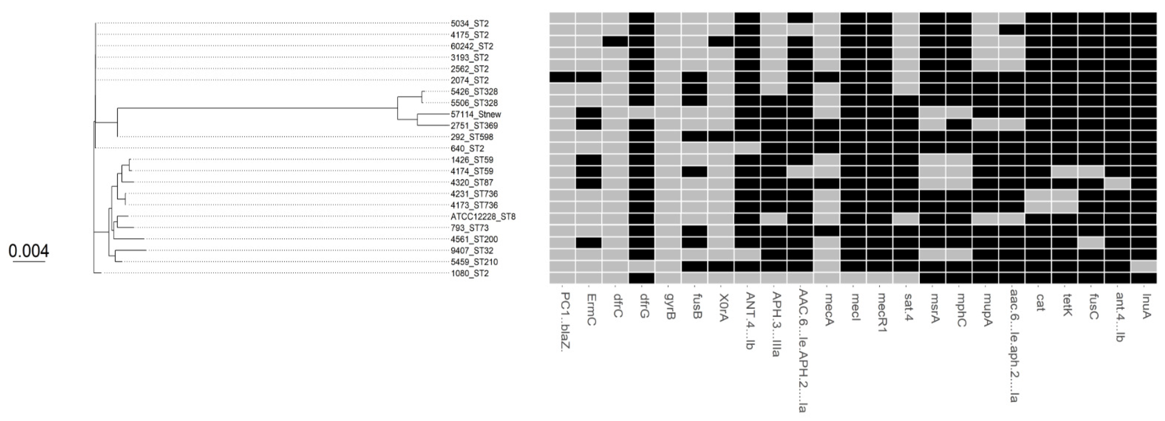
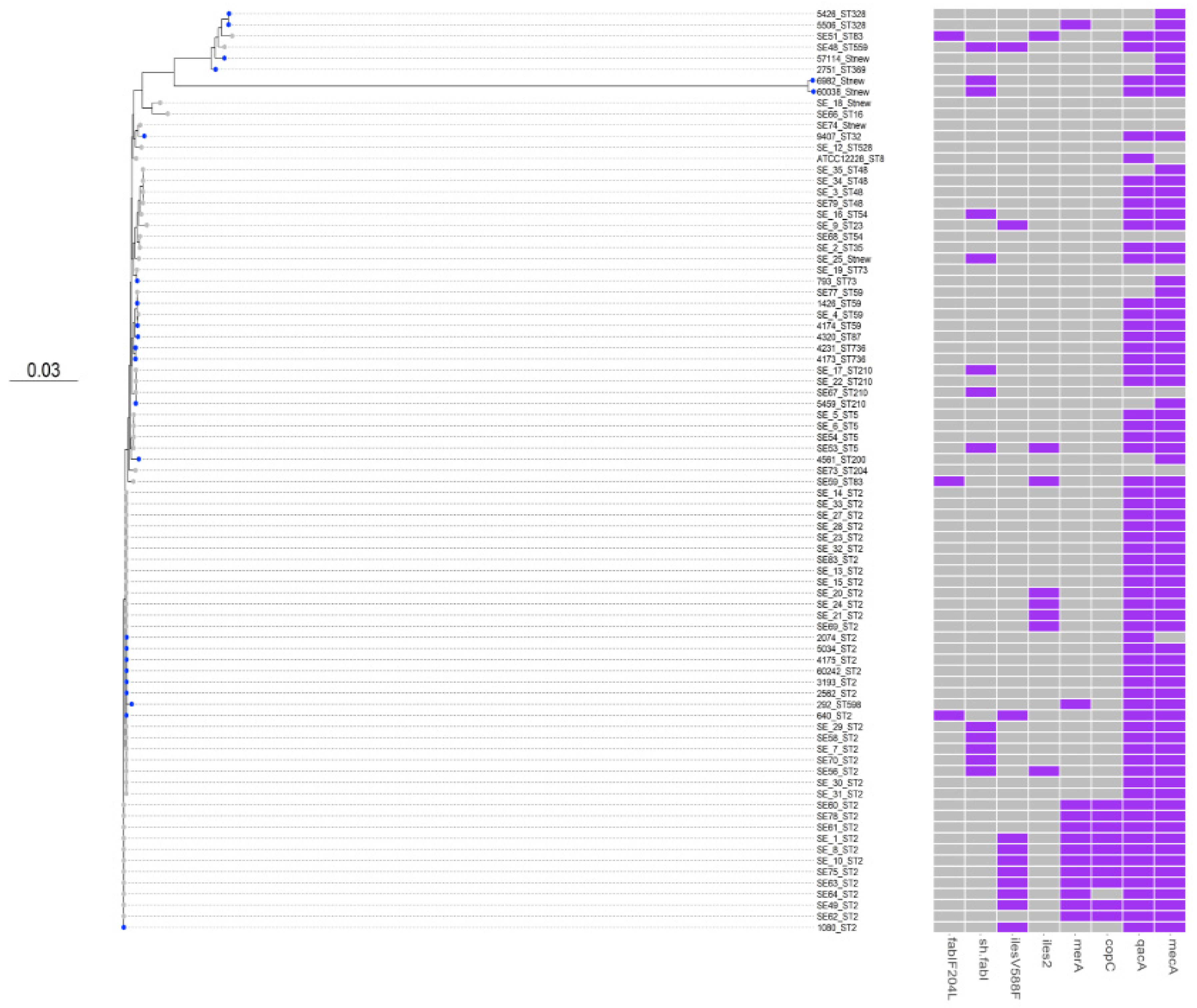
3.1. Comparative Phylogenetic Tree Analysis

Multilocus sequence typing (MLST) from whole genome data showed that 33.3% (n = 8) of our S. epidermidis isolates belong to ST-2 clustered in one branch of the tree (Table 1, Figure 1). ST-2 is the most predominant MDR sequence type worldwide. The remaining MLST were miscellaneous with random distribution throughout the tree (of which two were ST-59, two were ST-328, and two were ST-736) (Figure 1). Three isolates belong to a novel ST, of which two appear to be single locus variants. Out of the 24 S. epidermidis strains, 92% were methicillin-resistant S. epidermidis (MRSE) harboring the mecA gene. Most (81%) of the MRSE isolates carry mainly a type IV SCCmec (Table 1). The remaining two MSSE isolates lack mecA gene. With the exception of one isolate (2074), all isolates carry PC1 (blaZ) gene encoding penicillin resistance. COMER-like element in S. epidermidis seems to be larger than COMER in S. aureus with additional type I restriction modification system and arsenic resistance (ars) operon and exhibited a highly conserved structure in all isolates [3]. However, WGS showed that our S. epidermidis lack this composite COMER-like element downstream of SCCmec-IV, unlike the COMER element in USA300, with the exception of two isolates (5506 and 292) with merA only and one isolate (4526) with merB only (Figure 1).

3.2. SCCmec and ACME Types

Types of SCCmec are shown in Table 1. The content of mec gene complexes and multiple ccr gene complexes resulted sometimes to contradicting predictions of the SCCmec type in most isolates. All our isolates harbor the arginine catabolic mobile element (ACME), including both MRSE and MSSE strains. As previously described, ACME types are based on their composition of either one or more of the opp3, kdp, and arc operons as shown in Table 1 [8,10,45,46,47]. In our collection, the vast majority of S. epidermidis isolates (67%) belong to ACME type I with ST-2 (n = 8), ST-328 (n = 2), ST-59 (n = 1), ST-new (n = 2), ST-73 (n = 1), ST-200 (n = 1), and ST-598 (n = 1). ACME type-II was seen in three isolates (ST-369, ST-8, and ST-59), and ACME type-III in one isolate (ST-new). ACME type-V was seen in five isolates (ST-736 (n = 2), ST-87 (n = 1), ST-32 (n = 1), and ST-210 (n = 1)). Unlike COMER-like elements, ACME elements are highly variable in composition, as these are flanked by variable internal direct repeats allowing rearrangement of genes in modules [10]. Upon assembly of ACME in our isolates using the published DRs (A, B, and C) [8,48,49], four different composite islands were constructed (Figure S1, Supplementary Materials) with variable sizes. In addition, eight isolates carrying the ACME-I were located downstream of the SCCmec-IV, with cop and ars genes sandwiched in between and flanked by DR_A and B, respectively, at the 5′ end in orfX.
It was observed that partial deletion of SCCmec type IV occurred in 2 out of 24 isolates, 1426 and 9407, in which class B mec gene complex and composite ccrA4 were deleted. Deletion of ACME (type IIa) in 4174 (ST-59) was also observed. These rearrangements indicate the high genomic plasticity of S. epidermidis particularly in the SCCmec and ACME region compared to other regions. This observation has also been noted in in vitro studies where ACME have highly variable structures with 100 times higher excision frequency found for the SCCmec elements [3,19].
To examine the relation of our collection of S. epidermidis from Oman and the other published S. epidermidis strains, the core genome phylogenetic tree was then expanded to include a total of 58 isolates from the Aberdeen, the United Kingdom showed a large clustering of ST-2 lineage [3].

3.3. Antimicrobial Susceptibility and Virulence Conferring Genes

Twenty-nine percent of our S. epidermidis strains (n = 7/24) were mecA-negative (Figure 2). In staphylococcal spp., the cefoxitin 30-μg disk was used to predict the mecA status, and thus to predict methicillin resistance. Our data were interpreted according to the screening breakpoints published by the CLSI for coagulase-negative staphylococci (CoNS). Inhibition zone diameters for mecA-positive isolates were <28 mm for 80% of S. epidermidis, indicating methicillin resistance, whereas the remaining 20% mecA-positive strains were cefoxitin-sensitive (n = 4). Two out of 24 isolates showed oxacillin susceptibility, both of which are mecA positive and cefoxitin susceptible. This observation might suggest that mecA may be expressed variably in these isolates. Only one strain that was mecA-, mecI-, and mecR-negative showed cefoxitin resistance. Previous studies reported similar findings and suggested that the cefoxitin screening breakpoints for methicillin resistance in CoNS need to be adjusted [50].
In one isolate (2072-ST-2), the blaZ gene conferring resistance to penicillin was not detected. However, this isolate lacks the mecA gene (coding for altered penicillin-binding protein) and yet showed resistance to penicillin and other beta-lactams. In addition, this isolate is found within a cluster of blaZ-positive isolates of the same ST-2, suggesting a possible loss of bla-Z.
Out of 24 isolates, only one strain (60038), which has a mutation in the rpoB gene (D471G), showed rifampicin resistance (Table 2 and Supplementary Figure S1). Resistance to ciprofloxacin was found in 14/24 isolates. Mutations in the gyrA (S84L) and parC (S80Y) gene, which have been described for the USA300 lineage, were present in these resistant isolates. Overall, for the remaining antibiotics, we found a good phenotype-to-genotype correlation for antibiotic susceptibility data. For instance, the presence of macrolide efflux gene (msrA) and emrC correlates well with the susceptibility phenotype in our isolates as shown in Table 2 and Figure S2 in the Supplementary Materials.

3.4. Biocides Susceptibility

WGS sequencing results enabled the identification of all biocide resistance genes in the 24 S. epidermidis isolates (Figure 2). All of our S. epidermidis isolates carry a wild type fabI gene with no substitutions. The fabI gene encodes an enoyl-acyl carrier protein reductase (ENR), which is essential for fatty acid synthesis in bacteria. Isolate (640-ST-2), which carries F204L amino acid substitution resulting in reduced susceptibility to the antiseptic triclosan, is an exception. Moreover, two isolates (60038 and 6982, ST-new) carry an additional copy of sh-fabi allele, which is believed to be derived from Staphylococcus haemolyticus and results in increased resistance to triclosan [51].
Out of 24 isolates, six isolates carry the mupA gene coding for mupirocin resistance. Two isolates carry the isoleucyl-tRNA synthetase mutation V588F conferring mupirocin resistance. However, ileS2 (conferring high level of resistance to mupirocin) was absent in all of our S. epidermidis isolates. The carriage of qacA, qacB and qacC genes causing reduced susceptibility to quaternary ammonium compounds was 16.6%, 8.3%, and 12.5%, respectively.

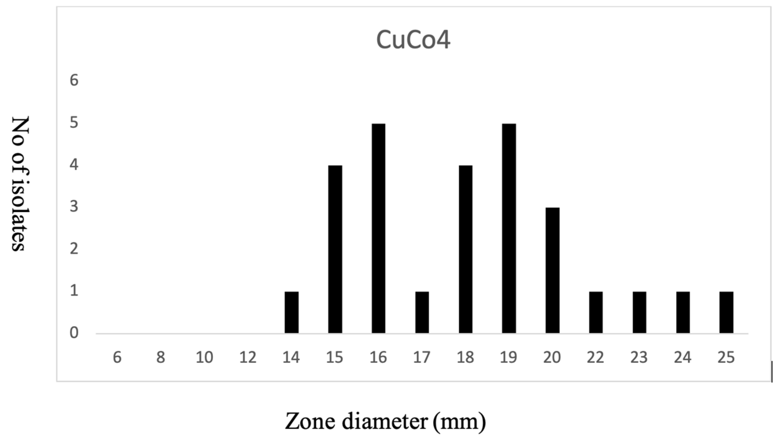
3.5. Copper Susceptibility

A phenotypic analysis of copper susceptibility was determined using disc diffusion test on our local S. epidermidis isolates (Figure 3). ECOFF was determined based on the normal distribution of the susceptibility, as breakpoints for metal susceptibility is not available [44]. copR was present in two strains (793 ST-73 and 5459 ST-210). Phenotypically, these two strains were copper-sensitive. copAZ with a second copy was present in all strains except five strains where copAZ2 was missing. All S. aureus species carry a putative copper-sensitive operon repressor (CsoR) -copA/copZ operon that is autoregulated by copper-sensitive transcritional repressor (CstR) and appears to encode at least two CsoR-like proteins [52]. Similarly in our collection, all S. epidermidis carry copper-resistance-encoding genes, even in the absence of COMER-like elements. This might provide a fitness cost to S. epidermidis under high selective pressure environment, as in the hospital settings.

4. Discussion

S. epidermidis have successfully emerged from a skin commensal to potentially pathogenic bacteria, owing to its plasticity and increasing capacity of acquiring a reservoir of resistance genes. Research in S. epidermidis genomics confirms the high likelihood of spread of the multidrug resistance ST-2 lineage worldwide as well as in our local isolates, where about one third of S. epidermis isolates belong to ST-2 [3,4]. The dissemination of MDR ST-2 lineage has been linked to SNPs in the rpoB gene, allowing its persistence [4], with possible co-occurrence of the highly stable COMER-like element in S. epidermidis clinical isolates [3]. In S. aureus, the COMER element enhances its survival in the host by amelioration of the survival capabilities and as a result the infection remains hidden from the immune system [12,13]. However, in our collection, the COMER-like element was not found in any of the isolates, despite its stability in other global lineages. This observation could suggest that the COMER-like element might not commonly occur in our region. All of our S. epidermidis isolates carry ACME of various types (I, II, III, and V), which was described initially in oral isolates of S. epidermidis and later in bloodstream infections [3,11]. This observation is similar to the epidemiology described in a global ST-2 lineage of Aberdeen, UK [3,53]. Therefore, reduced susceptibility to copper observed in some isolates cannot be contributed by either SCCmec alone or ACME or both. In addition, reduced susceptibility to copper was seen in eight isolates belonging to various ST types but predominantly ST-2 (n = 4), among others: ST-59 (n = 1), ST-328 (n = 1), ST-87 (n = 1), and ST-736 (n = 1). Five out of 8 S. epidermidis isolates with reduced copper susceptibility carry ACME-type I. ACME type-II was found in one isolate only. The remaining three belong to ACME type-V (n = 2), which is characterized by presence of all three gene clusters: arc, which encodes an arginine deaminase pathway; opp3, which encodes an oligopeptide permease ABC transporter; and kdp, which encodes a potassium ABC transporter [10].
ACME elements have been described previously to be more commonly associated with MSSE compared to MRSE [3,46,47,54]. To the contrary, in our collection, 29% of ACME harboring isolates were MSSE and the remaining 71% were MRSE. The relationship between these ACME types and copper susceptibility cannot be inferred at this stage as ST-2 and ACME type I are the predominant genotypes in our limited collection of isolates and a larger sample size will be needed to draw a conclusion. Furthermore, reduced copper susceptibility could well be due to the ubiquity of copper resistance gene clusters in the core chromosome, as it has a fitness advantage to the persistence and evolution of S. epidermidis species. Previous studies using infection models have demonstrated that several pathogens have evolved to control excess intracellular copper by either efflux pumps or sequestration as virulence mechanisms [17,18,55,56,57].
In this study, ST-2 S. epidermidis clinical isolates showed a lower antibiotics resistance profile compared with the global patterns of some hospital-adapted lineages with a pandrug resistance profile [4]. Apart from the intrinsic resistance to beta-lactams in MRSE, our collection of S. epidermidis spares a sensitivity phenotype to several antibiotic groups, including vancomycin, teicoplanin, rifampicin, chloramphenicol, and tigecycline, that have a high level of agreement with their respective genotypes (Figure S2, Supplementary Materials).
In addition to heavy metals, S. epidermidis have adapted to biocides commonly used in hospital settings such as quaternary ammonium compounds and triclosan, as well as decolonization antibiotics including mupirocin. Genetic determinants of resistance to mupirocin have been identified (ileS2, V588F IleS) and/or triclosan (sh-fabI, F204L in FabI) in 65% of qacA S. epidermidis isolates [53]. qacA, qacB, and qacC encode efflux pumps for a variety of lipophilic cations and are strongly associated with reduced microbial susceptibility to chlorhexidine, which is the most commonly used biocide in the community as well as hospital decontamination and infection control, with applications ranging from mouthwashes to impregnated catheters and skin/mucosal surface decontamination in intensive care units (ICUs). Notably, in a number of cases, mupirocin resistance genes were colocated with qacA on variants of known MRSA mobile elements, raising the possibility of horizontal transfer of multidrug resistance genes between S. epidermidis and S. aureus. However, previous studies have demonstrated the low fitness cost of ielS-V588F mutations, as it is consistent and is associated with low burden [58]. However, our study clearly showed high carriage of biocide resistance genes among ST-2 isolates along with MDR and metal resistance genes that are carried on MGEs. Our observation goes in line with the increasing concern from genomics and phylogenetic tree analyses of previous studies—that MDR hospital-adapted lineages consistently carry chlorhexidine, mupirocin, and triclosan-resistance-conferring genes, which is concerning and necessitates further action [59].
In conclusion, in this work, we identify a common prevalence of highly variable ACME elements in different hospital STs of S. epidermidis in Oman and high carriage of biocide resistance conferring genes, thus strongly suggesting the hypothesis that ACME types evolved from closely related STs.

Supplementary Materials

The following are available online at https://www.mdpi.com/article/10.3390/microorganisms9091824/s1, Figure S1: Schematic representation of ACME-I in S. epidermidis., Figure S2: Maximum likelihood phylogenetic tree constructed from WGS data and heat map for the distribution of antibiotic resistance genes of 24 S. epidermidis isolates. Table S1: WGS data of S. epidermidis: coverage, trimmed reads, taxonomic distribution, and assemblies. Table S2: Antibiotics, copper, and biocide genotype and phenotype results.

Author Contributions

Conceptualization, Z.A.-J. and Z.A.-S.; methodology, A.A.-B. and A.A.-H.; software, Z.A.-S.; validation, Z.A.-J., Z.A.-S. and A.A.-S.; formal analysis, Z.A.-S.; investigation, Z.A.-J.; resources, A.A.-B.; data curation, Z.A.-J. and Z.A.-S.; writing—original draft preparation, Z.A.-J.; writing—review and editing, Z.A.-S. and M.R.; visualization, M.R.; supervision, Z.A.-J.; project administration, A.A.-B.; funding acquisition, A.A.-B. All authors have read and agreed to the published version of the manuscript.

Funding

This research was funded by Deanship of Research, Sultan Qaboos University, Muscat, Oman, grant number RF/MED/MICR/19/02.

Institutional Review Board Statement

This study has been approved by the Medical Research Ethics Committee (MREC), College of Medicine and Health Sciences, Sultan Qaboos University, Oman. The MREC approval number (1832).

Informed Consent Statement

Not applicable.

Data Availability Statement

Whole genome sequences as draft contigs are available upon request from the corresponding author. The WGS data will be uploaded in GenBank after finishing the project.

Acknowledgments

We would like to express our sincere thanks to the sequencing company provided by MicrobesNG (Birmingham, UK) (https://microbesng.com (accessed on 4 July 2019)) for performing WGS and bioinformatics analysis. We would like to thank supervising staff, College of Agriculture, Department of Food Microbiology for providing instrumental support to conduct some experiments for cultivating the bacterial isolates.

Conflicts of Interest

The authors declare no conflict of interest.

References

  1. Brescó, M.S.; Harris, L.G.; Thompson, K.; Stanic, B.; Morgenstern, M.; O’Mahony, L.; Richards, R.G.; Moriarty, T.F. Pathogenic Mechanisms and Host Interactions in Staphylococcus Epidermidis Device-Related Infection. Front. Microbiol. 2017, 8, 1401. [Google Scholar] [CrossRef] [Green Version]
  2. Otto, M. Staphylococcus Epidermidis—The “accidental” Pathogen. Nat. Rev. Microbiol. 2009, 7, 555–567. [Google Scholar] [CrossRef] [Green Version]
  3. Almebairik, N.; Zamudio, R.; Ironside, C.; Joshi, C.; Ralph, J.D.; Roberts, A.P.; Gould, I.M.; Morrissey, J.A.; Hijazi, K.; Oggioni, M.R. Genomic Stability of Composite SCCmec ACME and COMER-Like Genetic Elements in Staphylococcus Epidermidis Correlates With Rate of Excision. Front. Microbiol. 2020, 11, 166. [Google Scholar] [CrossRef] [Green Version]
  4. Lee, J.Y.H.; Monk, I.R.; da Silva, A.G.; Seemann, T.; Chua, K.Y.L.; Kearns, A.; Hill, R.; Woodford, N.; Bartels, M.D.; Strommenger, B.; et al. Global Spread of Three Multidrug-Resistant Lineages of Staphylococcus Epidermidis. Nat. Microbiol. 2018, 3, 1175–1185. [Google Scholar] [CrossRef]
  5. Dhawan, B.; Rao, C.; Udo, E.E.; Gadepalli, R.; Vishnubhatla, S.; Kapil, A. Dissemination of Methicillin-Resistant Staphylococcus Aureus SCCmec Type IV and SCCmec Type v Epidemic Clones in a Tertiary Hospital: Challenge to Infection Control. Epidemiol. Infect. 2015, 143, 343–353. [Google Scholar] [CrossRef] [Green Version]
  6. Diep, B.A.; Stone, G.G.; Basuino, L.; Graber, C.J.; Miller, A.; des Etages, S.A.; Jones, A.; Palazzolo-Ballance, A.M.; Perdreau-Remington, F.; Sensabaugh, G.F.; et al. The Arginine Catabolic Mobile Element and Staphylococcal Chromosomal Cassette Mec Linkage: Convergence of Virulence and Resistance in the USA300 Clone of Methicillin-Resistant Staphylococcus Aureus. J. Infect. Dis. 2008, 197, 1523–1530. [Google Scholar] [CrossRef] [Green Version]
  7. Planet, P.J.; Diaz, L.; Kolokotronis, S.O.; Narechania, A.; Reyes, J.; Xing, G.; Rincon, S.; Smith, H.; Panesso, D.; Ryan, C.; et al. Parallel Epidemics of Community-Associated Methicillin-Resistant Staphylococcus Aureus USA300 Infection in North and South America. J. Infect. Dis. 2015, 212, 1874–1882. [Google Scholar] [CrossRef] [Green Version]
  8. O’Connor, A.M.; McManus, B.A.; Coleman, D.C. First Description of Novel Arginine Catabolic Mobile Elements (ACMEs) Types IV and V Harboring a Kdp Operon in Staphylococcus Epidermidis Characterized by Whole Genome Sequencing. Infect. Genet. Evol. 2018, 61, 60–66. [Google Scholar] [CrossRef] [PubMed]
  9. Planet, P.J.; Larussa, S.J.; Dana, A.; Smith, H.; Xu, A.; Ryan, C.; Uhlemann, A.C.; Boundy, S.; Goldberg, J.; Narechania, A.; et al. Emergence of the Epidemic Methicillin-Resistant Staphylococcus Aureus Strain USA300 Coincides with Horizontal Transfer of the Arginine Catabolic Mobile Element and SpeG-Mediated Adaptations for Survival on Skin. mBio 2013, 4, e00889-13. [Google Scholar] [CrossRef] [Green Version]
  10. McManus, B.A.; O’Connor, A.M.; Egan, S.A.; Flanagan, P.R.; Coleman, D.C. First Description of Arginine Catabolic Mobile Element (ACME) Type VI Harboring the Kdp Operon Only in Staphylococcus Epidermidis Using Short and Long Read Whole Genome Sequencing: Further Evidence of ACME Diversity. Infect. Genet. Evol. 2019, 71, 51–53. [Google Scholar] [CrossRef]
  11. O’Connor, A.M.; McManus, B.A.; Kinnevey, P.M.; Brennan, G.I.; Fleming, T.E.; Cashin, P.J.; O’Sullivan, M.; Polyzois, I.; Coleman, D.C. Significant Enrichment and Diversity of the Staphylococcal Arginine Catabolic Mobile Element ACME in Staphylococcus Epidermidis Isolates from Subgingival Peri-Implantitis Sites and Periodontal Pockets. Front. Microbiol. 2018, 9, 1588. [Google Scholar] [CrossRef]
  12. Zapotoczna, M.; Riboldi, G.P.; Moustafa, A.M.; Dickson, E.; Narechania, A.; Morrissey, J.A.; Planet, P.J.; Holden, M.T.G.; Waldron, K.J.; Geoghegan, J.A. Mobile-Genetic-Element-Encoded Hypertolerance to Copper Protects Staphylococcus Aureus from Killing by Host Phagocytes. mBio 2018, 9, e00550-18. [Google Scholar] [CrossRef] [Green Version]
  13. Purves, J.; Thomas, J.; Riboldi, G.P.; Zapotoczna, M.; Tarrant, E.; Andrew, P.W.; Londoño, A.; Planet, P.J.; Geoghegan, J.A.; Waldron, K.J.; et al. A Horizontally Gene Transferred Copper Resistance Locus Confers Hyper-Resistance to Antibacterial Copper Toxicity and Enables Survival of Community Acquired Methicillin Resistant Staphylococcus Aureus USA300 in Macrophages. Environ. Microbiol. 2018, 20, 1576–1589. [Google Scholar] [CrossRef] [Green Version]
  14. Fones, H.; Preston, G.M. The Impact of Transition Metals on Bacterial Plant Disease. FEMS Microbiol. Rev. 2013, 37, 495–519. [Google Scholar] [CrossRef] [Green Version]
  15. Chaturvedi, K.S.; Henderson, J.P. Pathogenic Adaptations to Host-Derived Antibacterial Copper. Front. Cell. Infect. Microbiol. 2014, 4, 3. [Google Scholar] [CrossRef] [Green Version]
  16. Fu, Y.; Chang, F.M.J.; Giedroc, D.P. Copper Transport and Trafficking at the Host-Bacterial Pathogen Interface. Acc. Chem. Res. 2014, 47, 3605–3613. [Google Scholar] [CrossRef]
  17. Johnson, M.D.L.; Kehl-Fie, T.E.; Klein, R.; Kelly, J.; Burnham, C.; Mann, B.; Rosch, J.W. Role of Copper Efflux in Pneumococcal Pathogenesis and Resistance to Macrophage-Mediated Immune Clearance. Infect. Immun. 2015, 83, 1684–1694. [Google Scholar] [CrossRef] [Green Version]
  18. Hyre, A.N.; Kavanagh, K.; Kock, N.D.; Donati, G.L.; Subashchandrabose, S. Copper Is a Host Effector Mobilized to Urine during Urinary Tract Infection to Impair Bacterial Colonization. Infect. Immun. 2017, 85, e01041-16. [Google Scholar] [CrossRef] [Green Version]
  19. Stojanov, M.; Moreillon, P.; Sakwinska, O. Excision of Staphylococcal Cassette Chromosome Mec in Methicillin-Resistant Staphylococcus Aureus Assessed by Quantitative PCR Microbiology. BMC Res. Notes 2015, 8, 1–8. [Google Scholar] [CrossRef] [Green Version]
  20. Wang, L.; Archer, G.L. Roles of CcrA and CcrB in Excision and Integration of Staphylococcal Cassette Chromosome Mec, a Staphylococcus Aureus Genomic Island. J. Bacteriol. 2010, 192, 3204–3212. [Google Scholar] [CrossRef] [PubMed] [Green Version]
  21. Rosario-Cruz, Z.; Eletsky, A.; Daigham, N.S.; Al-Tameemi, H.; Swapna, G.V.T.; Kahn, P.C.; Szyperski, T.; Montelione, G.T.; Boyd, J.M. The CopBL Operon Protects Staphylococcus Aureus from Copper Toxicity: CopL Is an Extracellular Membrane–Associated Copper-Binding Protein. J. Biol. Chem. 2019, 294, 4027–4044. [Google Scholar] [CrossRef] [Green Version]
  22. Bolger, A.M.; Lohse, M.; Usadel, B. Trimmomatic: A Flexible Trimmer for Illumina Sequence Data. Bioinformatics 2014, 30, 2114–2120. [Google Scholar] [CrossRef] [PubMed] [Green Version]
  23. Wood, D.E.; Salzberg, S.L. Kraken: Ultrafast Metagenomic Sequence Classification Using Exact Alignments. Genome Biol. 2014, 15, 1–12. [Google Scholar] [CrossRef] [Green Version]
  24. Li, H.; Durbin, R. Fast and Accurate Long-Read Alignment with Burrows-Wheeler Transform. Bioinformatics 2010, 26, 589–595. [Google Scholar] [CrossRef] [Green Version]
  25. Bankevich, A.; Nurk, S.; Antipov, D.; Gurevich, A.A.; Dvorkin, M.; Kulikov, A.S.; Lesin, V.M.; Nikolenko, S.I.; Pham, S.; Prjibelski, A.D.; et al. SPAdes: A New Genome Assembly Algorithm and Its Applications to Single-Cell Sequencing. J. Comput. Biol. 2012, 19, 455–477. [Google Scholar] [CrossRef] [PubMed] [Green Version]
  26. Koboldt, D.C.; Larson, D.E.; Wilson, R.K. Using Varscan 2 for Germline Variant Calling and Somatic Mutation Detection. Curr. Protoc. Bioinform. 2013, 44, 15.4.1–15.4.17. [Google Scholar] [CrossRef] [Green Version]
  27. Seemann, T. Prokka: Rapid Prokaryotic Genome Annotation. Bioinformatics 2014, 30, 2068–2069. [Google Scholar] [CrossRef]
  28. Larsen, J.; Enright, M.C.; Godoy, D.; Spratt, B.G.; Larsen, A.R.; Skov, R.L. Multilocus Sequence Typing Scheme for Staphylococcus Aureus: Revision of the Gmk Locus. J. Clin. Microbiol. 2012, 50, 2538–2539. [Google Scholar] [CrossRef] [Green Version]
  29. Seemann, T. Snippy: Fast Bacterial Variant Calling from NGS Reads [Internet]. Available online: www.github.com (accessed on 15 January 2020).
  30. Nguyen, L.T.; Schmidt, H.A.; von Haeseler, A.; Minh, B.Q. IQ-TREE: A Fast and Effective Stochastic Algorithm for Estimating Maximum-Likelihood Phylogenies. Mol. Biol. Evol. 2015, 32, 268–274. [Google Scholar] [CrossRef]
  31. Kalyaanamoorthy, S.; Minh, B.Q.; Wong, T.K.F.; von Haeseler, A.; Jermiin, L.S. ModelFinder: Fast Model Selection for Accurate Phylogenetic Estimates. Nat. Methods 2017, 14, 587–589. [Google Scholar] [CrossRef] [Green Version]
  32. Thi Hoang, D.; Chernomor, O.; von Haeseler, A.; Quang Minh, B.; Sy Vinh, L.; Rosenberg, M.S. UFBoot2: Improving the Ultrafast Bootstrap Approximation. Mol. Biol. Evol. 2017, 35, 518–522. [Google Scholar] [CrossRef] [PubMed]
  33. Yu, G.; Smith, D.K.; Zhu, H.; Guan, Y.; Lam, T.T.Y. Ggtree: An r Package for Visualization and Annotation of Phylogenetic Trees with Their Covariates and Other Associated Data. Methods Ecol. Evol. 2017, 8, 28–36. [Google Scholar] [CrossRef]
  34. Kaya, H.; Hasman, H.; Larsen, J.; Stegger, M.; Johannesen, T.B.; Allesøe, R.L.; Lemvigh, C.K.; Aarestrup, F.M.; Lund, O.; Larsen, A.R. SCC Mec Finder, a Web-Based Tool for Typing of Staphylococcal Cassette Chromosome Mec in Staphylococcus Aureus Using Whole-Genome Sequence Data. mSphere 2018, 3, e00612-17. [Google Scholar] [CrossRef] [Green Version]
  35. Joensen, K.G.; Scheutz, F.; Lund, O.; Hasman, H.; Kaas, R.S.; Nielsen, E.M.; Aarestrup, F.M. Real-Time Whole-Genome Sequencing for Routine Typing, Surveillance, and Outbreak Detection of Verotoxigenic Escherichia Coli. J. Clin. Microbiol. 2014, 52, 1501–1510. [Google Scholar] [CrossRef] [Green Version]
  36. Maria, A.; Tetzschner, M.; Johnson, J.R.; Johnston, B.D.; Lund, O.; Scheutz, F. In Silico Genotyping of Escherichia Coli Isolates for Extraintestinal Virulence Genes by Use of Whole-Genome Sequencing Data. J. Clin. Microbiol. 2020, 58, e01269-20. [Google Scholar]
  37. Carver, T.J.; Rutherford, K.M.; Berriman, M.; Rajandream, M.A.; Barrell, B.G.; Parkhill, J. ACT: The Artemis Comparison Tool. Bioinformatics 2005, 21, 3422–3423. [Google Scholar] [CrossRef] [Green Version]
  38. Altschup, S.F.; Gish, W.; Miller, W.; Myers, E.W.; Lipman, D.J. Basic Local Alignment Search Tool. J. Mol. Biol. 1990, 215, 403–410. [Google Scholar] [CrossRef]
  39. Siguier, P.; Perochon, J.; Lestrade, L.; Mahillon, J.; Chandler, M. ISfinder: The Reference Centre for Bacterial Insertion Sequences. Nucleic Acids Res. 2006, 34, W32–W37. [Google Scholar] [CrossRef] [Green Version]
  40. Russell Editor, D.J. Multiple Sequence Alignment; Humana Press: New work, NY, USA, 2014; Volume 15, pp. 75–101. [Google Scholar]
  41. Camacho, C.; Coulouris, G.; Avagyan, V.; Ma, N.; Papadopoulos, J.; Bealer, K.; Madden, T.L. BLAST+: Architecture and Applications. BMC Bioinform. 2009, 10, 421. [Google Scholar] [CrossRef] [Green Version]
  42. Clausen, P.T.L.C.; Aarestrup, F.M.; Lund, O. Rapid and Precise Alignment of Raw Reads against Redundant Databases with KMA. BMC Bioinform. 2018, 19, 1–8. [Google Scholar] [CrossRef]
  43. Alcock, B.P.; Raphenya, A.R.; Lau, T.T.Y.; Tsang, K.K.; Bouchard, M.; Edalatmand, A.; Huynh, W.; Nguyen, A.L.V.; Cheng, A.A.; Liu, S.; et al. CARD 2020: Antibiotic Resistome Surveillance with the Comprehensive Antibiotic Resistance Database. Nucleic Acids Res. 2020, 48, D517–D525. [Google Scholar] [CrossRef] [PubMed]
  44. Morrissey, I.; Oggioni, M.R.; Knight, D.; Curiao, T.; Coque, T.; Kalkanci, A.; Martinez, J.L.; Baldassarri, L.; Orefici, G.; Yetiş, Ü.; et al. Evaluation of Epidemiological Cut-off Values Indicates That Biocide Resistant Subpopulations Are Uncommon in Natural Isolates of Clinically-Relevant Microorganisms. PLoS ONE 2014, 9, e86669. [Google Scholar] [CrossRef] [PubMed] [Green Version]
  45. Weinstein, M.P.; Clinical and Laboratory Standards Institute. Performance Standards for Antimicrobial Susceptibility Testing. 20th Information Supplement CLSI Document M100-S20. Available online: https://kaldur.landspitali.is/focal/gaedahandbaekur/gnhsykla.nsf/5e27f2e5a88c898e00256500003c98c2/9c4f4955ccb9f8100025751a0046b075/$FILE/M100-S20%20Performance%20Standards%20for%20Antimicrobial%20Susceptibility%20Testing.pdf (accessed on 30 July 2021).
  46. Barbier, F.; Lebeaux, D.; Hernandez, D.; Delannoy, A.S.; Caro, V.; François, P.; Schrenzel, J.; Ruppé, E.; Gaillard, K.; Wolff, M.; et al. High Prevalence of the Arginine Catabolic Mobile Element in Carriage Isolates of Methicillin-Resistant Staphylococcus Epidermidis. J. Antimicrob. Chemother. 2011, 66, 29–36. [Google Scholar] [CrossRef] [Green Version]
  47. Onishi, M.; Urushibara, N.; Kawaguchiya, M.; Ghosh, S.; Shinagawa, M.; Watanabe, N.; Kobayashi, N. Prevalence and Genetic Diversity of Arginine Catabolic Mobile Element (ACME) in Clinical Isolates of Coagulase-Negative Staphylococci: Identification of ACME Type I Variants in Staphylococcus Epidermidis. Infect. Genet. Evol. 2013, 20, 381–388. [Google Scholar] [CrossRef] [PubMed]
  48. Soroush, S.; Jabalameli, F.; Taherikalani, M.; Amirmozafari, N.; Imani Fooladi, A.A.; Asadollahi, K.; Beigverdi, R.; Emaneini, M. Investigation of Biofilm Formation Ability, Antimicrobial Resistance and the Staphylococcal Cassette Chromosome Mec Patterns of Methicillin Resistant Staphylococcus Epidermidis with Different Sequence Types Isolated from Children. Microb. Pathog. 2016, 93, 126–130. [Google Scholar] [CrossRef]
  49. Ito, T.; Katayama, Y.; Hiramatsu, K. Cloning and Nucleotide Sequence Determination of the Entire Mec DNA of Pre-Methicillin-Resistant Staphylococcus Aureus N315. Antimicrob. Agents Chemother. 1999, 43, 1449–1458. [Google Scholar] [CrossRef] [Green Version]
  50. Ahman, J.; Matuschek, E.; Webster, C.; Kahlmeter, G. Methicillin Resistance in Coagulase-Negative Staphylococci-Can. We Use the Same Screening Breakpoints for All Species? ESCMID eLibrary, 2017 ECCMID Conference Vienna. Available online: https://www.escmid.org/escmid_publications/escmid_elibrary/?q=Methicillin+Resistance+in+Coagulase-Negative+Staphylococci-Can&id=2173&L=0&x=0&y=0&tx_solr%5Bfilter%5D%5B0%5D=main_category%253ABacterial%2BSusceptibility%2B%2526%2BResistance&tx_solr%5Bfilter%5D%5B1%5D=author%253AJenny%2BAhman (accessed on 30 July 2021).
  51. Ciusa, M.L.; Furi, L.; Knight, D.; Decorosi, F.; Fondi, M.; Raggi, C.; Coelho, J.R.; Aragones, L.; Moce, L.; Visa, P.; et al. A Novel Resistance Mechanism to Triclosan That Suggests Horizontal Gene Transfer and Demonstrates a Potential Selective Pressure for Reduced Biocide Susceptibility in Clinical Strains of Staphylococcus Aureus. Int. J. Antimicrob. Agents 2012, 40, 210–220. [Google Scholar] [CrossRef] [Green Version]
  52. Grossoehme, N.; Kehl-Fie, T.E.; Ma, Z.; Adams, K.W.; Cowart, D.M.; Scott, R.A.; Skaar, E.P.; Giedroc, D.P. Control of Copper Resistance and Inorganic Sulfur Metabolism by Paralogous Regulators in Staphylococcus Aureus. J. Biol. Chem. 2011, 286, 13522–13531. [Google Scholar] [CrossRef] [Green Version]
  53. Hijazi, K.; Mukhopadhya, I.; Abbott, F.; Milne, K.; Al-Jabri, Z.J.; Oggioni, M.R.; Gould, I.M. Susceptibility to Chlorhexidine amongst Multidrug-Resistant Clinical Isolates of Staphylococcus Epidermidis from Bloodstream Infections. Int. J. Antimicrob. Agents 2016, 48, 86–90. [Google Scholar] [CrossRef] [Green Version]
  54. Miragaia, M.; de Lencastre, H.; Perdreau-Remington, F.; Chambers, H.F.; Higashi, J.; Sullam, P.M.; Lin, J.; Wong, K.I.; King, K.A.; Otto, M.; et al. Genetic Diversity of Arginine Catabolic Mobile Element in Staphylococcus Epidermidis. PLoS ONE 2009, 4, e7722. [Google Scholar] [CrossRef] [Green Version]
  55. Schwan, W.R.; Warrener, P.; Keunz, E.; Kendall Stover, C.; Folger, K.R. Mutations in the CueA Gene Encoding a Copper Homeostasis P-Type ATPase Reduce the Pathogenicity of Pseudomonas Aeruginosa in Mice. Int. J. Med. Microbiol. 2005, 295, 237–242. [Google Scholar] [CrossRef]
  56. Shafeeq, S.; Yesilkaya, H.; Kloosterman, T.G.; Narayanan, G.; Wandel, M.; Andrew, P.W.; Kuipers, O.P.; Morrissey, J.A. The Cop Operon Is Required for Copper Homeostasis and Contributes to Virulence in Streptococcus Pneumoniae. Mol. Microbiol. 2011, 81, 1255–1270. [Google Scholar] [CrossRef] [Green Version]
  57. Ladomersky, E.; Khan, A.; Shanbhag, V.; Cavet, J.S.; Chan, J.; Weisman, G.A.; Petris, M.J. Host and Pathogen Copper-Transporting P-Type ATPases Function Antagonistically during Salmonella Infection. Infect. Immun. 2017, 85, e00351-17. [Google Scholar] [CrossRef] [Green Version]
  58. Hurdle, J.G.; O’Neill, A.J.; Chopra, I. The Isoleucyl-TRNA Synthetase Mutation V588F Conferring Mupirocin Resistance in Glycopeptide-Intermediate Staphylococcus Aureus Is Not Associated with a Significant Fitness Burden. J. Antimicrob. Chemother. 2004, 53, 102–104. [Google Scholar] [CrossRef]
  59. Furi, L.; Haigh, R.; al Jabri, Z.J.H.; Morrissey, I.; Ou, H.Y.; León-Sampedro, R.; Martinez, J.L.; Coque, T.M.; Oggioni, M.R. Dissemination of Novel Antimicrobial Resistance Mechanisms through the Insertion Sequence Mediated Spread of Metabolic Genes. Front. Microbiol. 2016, 7, 1008. [Google Scholar] [CrossRef] [Green Version]
Figure 1. Maximum likelihood phylogenetic tree constructed using core genome alignment from the 24 S. epidermidis clinical isolates. The phylogenetic tree is annotated with the isolate’s sequence type ST. S. epidermidis ATCC 12228 as a reference strain (GenBank accession number: AE015929).
Microorganisms 09 01824 g001
Figure 2. Core genome phylogenetic tree of S. epidermidis strains genomes from Aberdeen, UK. Maximum likelihood tree of the 24 isolates from Oman (Table 1) (isolates in marked in blue) and the data set from Aberdeen of 58 isolates (in gray) (NCBI BioProject number: PRJNA574294). As in Figure 1, the tree is annotated with the isolate’s sequence type (ST). Biocide’s tolerance genes (qacA/B, fabI) and an additional fabI allele derived from Staphylococcus haemolyticus (sh-fabI) are shown. ATCC 12228 is used as the reference strain in this phylogenetic tree (GenBank accession number: AE015929).
Microorganisms 09 01824 g002
Figure 3. Susceptibility profile of S. epidermidis to copper. Disc diffusion inhibition zones were defined for 24 S. epidermidis isolates when exposed to 10 µL of 1M stock (1 µg/µL) of copper sulfate (CuSo4).
Microorganisms 09 01824 g003
Table 1. Staphylococcus epidermidis isolates.
Isolate Date MLSTSCCmec Type ACME Operons & (Type)
5034November 20182IV (2B)arc & opp3 (I)
4175November 20182IV (2B)arc & opp3 (I)
60242August 20182IV (2B)arc & opp3 (I)
3193August 20182IV (2B)arc & opp3 (I)
2562August 20182IV (2B)arc & opp3 (I)
2074September 20182 -arc & opp3 (I)
5426December 2018328IV (2B&5)arc & opp3 (I)
5506November 2018328V (5C2)arc & opp3 (I)
57114July 2018newIva (2B)arc & opp3 (I)
2751October 2018369IVa (2B)arc (II)
292January 2019598IV (2B&5)arc & opp3 (I)
640October 20182 -arc & opp3 (I)
1426October 201859 -arc & opp3 (I)
4174November 201859Iva (2B)arc (II)
4320November 201887 kdp, arc & opp3 (V)
4231November 2018736Iva (2B)kdp, arc & opp3 (V)
4173October 2018736Iva (2B)kdp, arc & opp3 (V)
793December 201873 -arc & opp3 (I)
4561November 2018200 -arc & opp3 (I)
9407September 201832 -kdp, arc & opp3 (V)
5459January 2019210V (5C2)kdp, arc & opp3 (V)
1080August 20182II (2A)arc & opp3 (I)
60038August 2018newIva (2B)opp3 (III)
6982November 2018new-arc & opp3 (I)
Table 2. Minimum inhibitory concentrations (MIC) of S. epidermidis isolates were determined using the disc diffusion method. The number of isolates that are susceptible, intermediate, or resistant are shown with the percentages in brackets. Broth microdilution methods were used for vancomycin and teicoplanin MICs. Interpretive categories and zone diameter breakpoints were determined according to CLSI.
AntibioticNo of Isolates (%)
SusceptibleIntermediate Resistant
Penicillin G (P)0 (0%)-24 (100%)
Amoxycillin/clavulanic acid (AMC)14(58%)-10 (42%)
Oxacillin (OX)2(8%)-22 (92%)
Erythromycin (E)2 (8%)0 (0%)22 (92%)
Cefoxitin (FOX)4 (16%)-20 (83%)
Ciprofloxacin (CIP)9 (38%)1 (4%)14 (58%)
Gentamicin (CN)13 (54%)1 (4%)10 (42%)
Clindamycin (DA)13 (54%)0 (0%)11 (46%)
Rifampicin (RD)23 (96%)0 (0%)1 (4%)
Chloramphenicol (C)22 (92%)0 (0%)2 (8%)
Tigecycline (TGC)24 (100%)-0 (0%)
* Vancomycin (VA, MIC)24 (100%)-0 (0%)
** Teicoplanin (TEC MIC)24 (100%)-0 (0%)
* For vancomycin, DD does not differentiate between susceptible, intermediate, or resistant CONs all give similar zone of inhibition. ** Teicoplanin DD interpretive criteria were not re-evaluated concurrent with revaluation of vancomycin DD interpretive criteria. Therefore, the ability of these teicoplanin interpretive criteria to differentiate teicoplanin-intermediate and teicoplanin-susceptible strains is not known.
Publisher’s Note: MDPI stays neutral with regard to jurisdictional claims in published maps and institutional affiliations.

