Article

Loss and Gain of Gut Bacterial Phylotype Symbionts in Afrotropical Stingless Bee Species (Apidae: Meliponinae)

by
Yosef Hamba Tola
1,2,
Jacqueline Wahura Waweru
1,
Nelly N. Ndungu
1,
Kiatoko Nkoba
1,
Bernard Slippers
2 and
Juan C. Paredes
1,*
1
International Centre of Insect Physiology and Ecology, Nairobi 30772-00100, Kenya
2
Department of Biochemistry, Genetics and Microbiology, Forestry and Agricultural Biotechnology Institute (FABI), University of Pretoria, Pretoria 0002, South Africa
*
Author to whom correspondence should be addressed.
Microorganisms 2021, 9(12), 2420; https://doi.org/10.3390/microorganisms9122420
Submission received: 8 October 2021 / Revised: 2 November 2021 / Accepted: 15 November 2021 / Published: 24 November 2021
(This article belongs to the Section Gut Microbiota)

Abstract

:
Stingless bees (Apidae: Meliponini) are the most diverse group of corbiculate bees and are important managed and wild pollinators distributed in the tropical and subtropical regions of the globe. However, little is known about their associated beneficial microbes that play major roles in host nutrition, detoxification, growth, activation of immune responses, and protection against pathogens in their sister groups, honeybees and bumble bees. Here, we provide an initial characterization of the gut bacterial microbiota of eight stingless bee species from sub-Saharan Africa using 16S rRNA amplicon sequencing. Our findings revealed that Firmicutes, Actinobacteria, and Proteobacteria were the dominant and conserved phyla across the eight stingless bee species. Additionally, we found significant geographical and host intra-species-specific bacterial diversity. Notably, African strains showed significant phylogenetic clustering when compared with strains from other continents, and each stingless bee species has its own microbial composition with its own dominant bacterial genus. Our results suggest host selective mechanisms maintain distinct gut communities among sympatric species and thus constitute an important resource for future studies on bee health management and host-microbe co-evolution and adaptation.

1. Introduction

More than 90% of tropical plant flowers are pollinated by animals; among them, bees are the most important pollinators [1,2,3]. Stingless bees (Meliponinae) represent the most diverse group among corbiculate bees and are commonly distributed in the tropical and subtropical regions; 77% are found in the Neotropics (Central America, Caribbean, and South America), followed by 16% in the Indo Malay/Australasian region and 7% in the Afro tropics [4,5,6,7,8,9]. Worldwide, there are approximately 61 identified stingless bee genera and over 550 species [4,8,10]. In Africa, there are 10 genera and 36 species of stingless bees described, and they are commonly distributed in tropical forests, savannahs, and deserts [4,10,11]. In Kenya, 6 genera (Cleptotrigona, Dactylurina, Hypotrigona, Liotrigona, Meliponula, and Plebeina) have been reported and are composed of 12 species that inhabit the Kakamega forest, Mwingi, the Arabuko-Sokoke Forest, and the Taita hills [10,12,13,14]. The Meliponula genus has been further divided into three sub-genera, namely Meliponula (one species), Axestotrigona (two species), and Meliplebeia (five species) [6,10].
The current increasing demand for managed pollinators and the advantages that stingless bees present have further encouraged their domestication [15,16,17,18,19]. In contrast to honeybees, stingless bees do not sting, they can be used for crop pollination in greenhouses, and they produce honey, propolis, and wax with important medicinal properties (e.g., antioxidant capacity, antimicrobial activity) of high economical value [17,20,21,22,23].
Bacterial symbiosis is a profound mechanism that has shaped insect physiology and ecology throughout their evolution [24,25]. With the increased number of insect models used for beneficial gut bacterial studies, there is cumulative evidence that insect gut microbiota play an important role in insect physiology, organ development, protection against natural enemies and help in their nutrition and detoxification. Additionally, they are involved in immune response activation and communication [26,27,28,29,30,31]. Bee gut bacterial communities are not an exception [32,33,34,35,36,37,38,39].
The vast majority of bee gut bacterial microbiota research has been conducted on honeybees (Apini tribe), and to a lesser extent, in bumble bees (Bombini tribe), but very little is known regarding stingless bees [40,41,42,43,44,45]. The handful of studies investigating stingless bee gut bacterial microbiota have reported substantial community diversity that varies across bee species [40,42,43,44,45,46]. Lactobacillus and Bifidobacterium have been reported as the most abundant genera, and in contrast to Apini and Bombini tribes, in Meliponini, Gilliamella and Snodgrassella are not always present or abundant. Additionally, stingless bees harbor numerous and specific Acetobacter species [41,42,43,44,45,46,47].
Despite being the most diverse group of corbiculate bees, only a few stingless bee species have been studied for their gut bacterial microbiota. Thus, much greater diversity is expected since they occupy a variety of tropical and subtropical habitats, and they present a large range of behaviors, such as, specific plant foraging and the construction of perennial nests that have different locations and structures [4,10,11,18,48]. Therefore, a better characterization of stingless bee gut bacterial microbiota will contribute to understanding host–symbiont interactions which likely impact stingless bee physiology. This will lay the foundation for different strategies to ensure beneficial microbe persistence in bees for agricultural sustainability and biodiversity conservation.
Here, we characterized the gut bacterial microbiota of eight different Afrotropical stingless bee species belonging to Meliponula, Dactylurina, Hypotrigona, and Liotrigona genera and evaluated their phylogenetic diversity and their relationship with other corbiculate bees from other continents.

2. Materials and Methods

2.1. Sample Collection and Preparation

Foragers from eight African stingless bee species belonging to four genera (Meliponula, Dactylurina, Hypotrigona, Liotrigona) were collected from colonies located at the International Centre of Insect Physiology and Ecology (icipe) meliponary, Duduville campus, Nairobi, Kenya, from October 2019 to February 2020. The following bees were originally collected from the Kakamega forest (Western Kenya): Meliponula bocandei (collected in February 2019), Meliponula togoensis and Meliponula ferruginea (collected in September 2018), and Meliponula lendliana and Liotrigona spp. (collected in September 2019). From the Mwingi (Eastern Kenya), the following bees were collected: Hypotrigona spp. 1 (collected in February 2017) and Hypotrigona spp. 2 (collected in August 2015). From the Taita Hills and Kilifi (Coastal Kenya), Dactylurina schmidti were collected (collected in September 2018). At the icipe meliponary, all colonies had access to the same floral resources and faced the same ecological conditions (e.g., temperature, humidity). Gardens in their foraging range included Acacia, Eucalyptus, Psidium, Cordia Africana, croton megalocarpus, Leucaena glauca, and mixed crops.
Fifty forager worker bees per hive from two hives per species were collected at the hive entrance and washed in 4% sodium hypochlorite, followed by a wash in 70% ethanol and finally 1× PBS for 2 min to eliminate any external microorganisms or contaminant DNA attached to the cuticle [49,50]. The entire gut, with its contents, was dissected aseptically from each bee using forceps, and the guts were placed in a 2 mL microcentrifuge-tube containing 500 µL PBS. Samples were stored at −80 °C before DNA extraction.

2.2. DNA Extraction

DNA extraction from the entire gut was done using the CTAB-Phenol–Chloroform extraction method described in [50]. DNA was resuspended in 200 µL sterile water, and DNA concentration and quality per sample was confirmed using NanoDrop 2000 Spectrophotometer (Thermo Scientific, Wilmington, NC, USA).

2.3. 16S rRNA Gene Amplification, Sequencing, and Gut Community Analysis

All 16S rRNA gene amplification and sequencing was done at Macrogen Europe BV (Meibergdreef 31, 1105 AZ Amsterdam, The Netherlands, https://dna.macrogen-europe.com/eng/, accessed on 17 April 2020). We analyzed 8,563,992 paired-end sequences spanning the V3-V4 region of the 16S rRNA with an average of 155,709 reads per sample (10 pooled guts per sample, and the number of reads ranged from 106,894 to 198,324). We used the DADA2 pipeline embedded in R (version 4.0.2) to analyze our sequence reads [51]. Reads were checked for quality and primer sequences trimmed using Cutadapt (version 2.10). Taxonomic classification was performed against the SILVA138 database using a pre-trained Naive Bayes classifier [52]. Taxa and ASVs with cumulative abundance below five were discarded from analysis and from our negative control (a “blank” sample was included in the run, and we recovered only 2 reads), as well as those classified as unwanted sequences of animal and fungal origin (Chloroplast, Chloroflexi, Mitochondria, Archaea, and Eukaryota). Data were rarefied for the downstream alpha and beta diversity analysis (Figure S1). Alpha diversity was determined using Evenness, Faith’s phylogenetic diversity, and the Shannon index, and the statistical differences of gut bacterial microbiota diversity across the species were tested using Kruskal–Wallis H test. Average diversity values were plotted for each sample at each even sampling depth and samples were grouped based on sample metadata. To visualize the clustering of the microbial communities per species, principle coordinate analysis (PCoA) was done using Bray–Curtis distances and UniFrac distances (Weighted and Unweighted UniFrac). A Pearson correlation test was additionally performed to identify the impact of bee size on the bacterial richness and evenness.

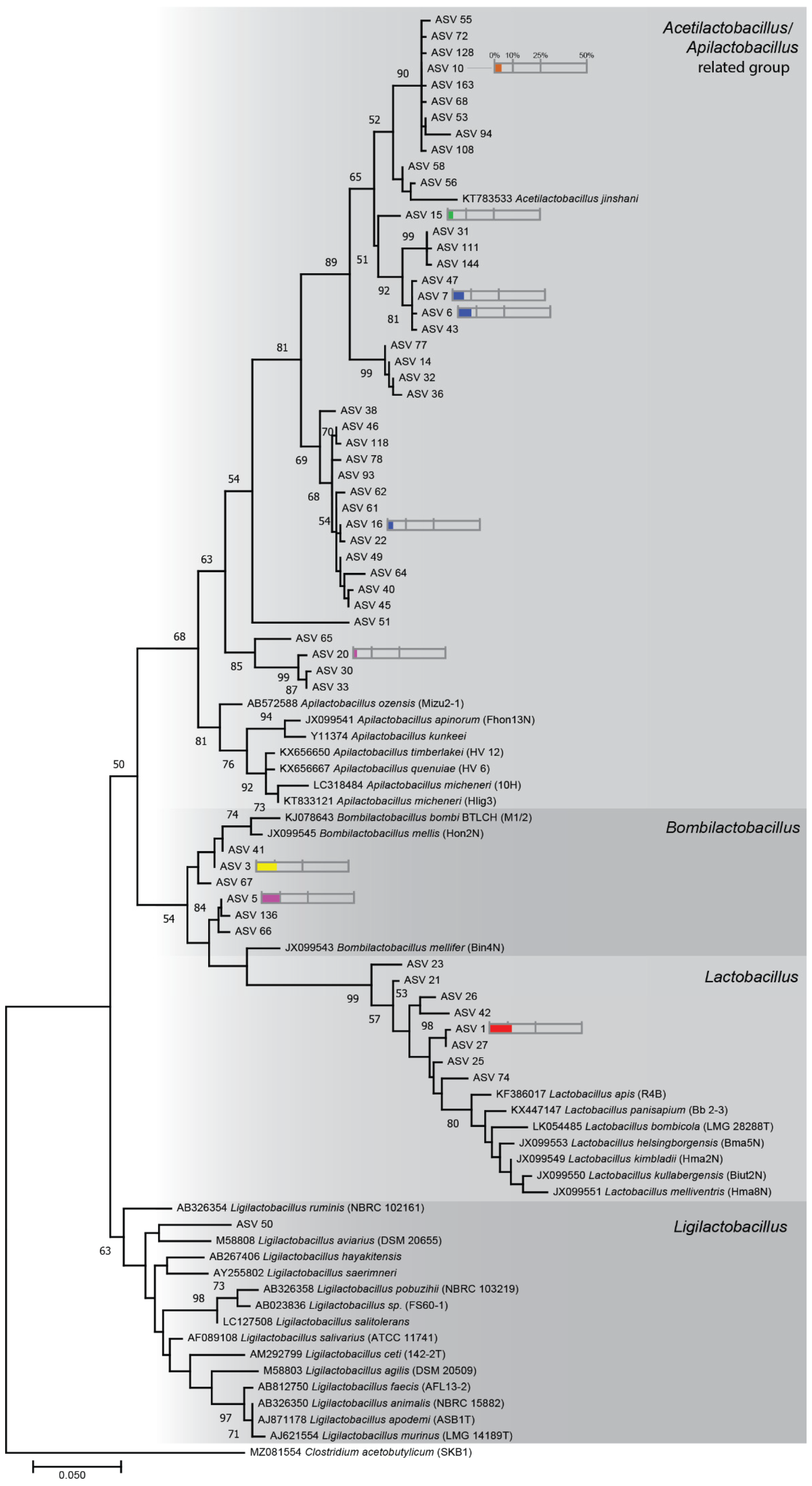
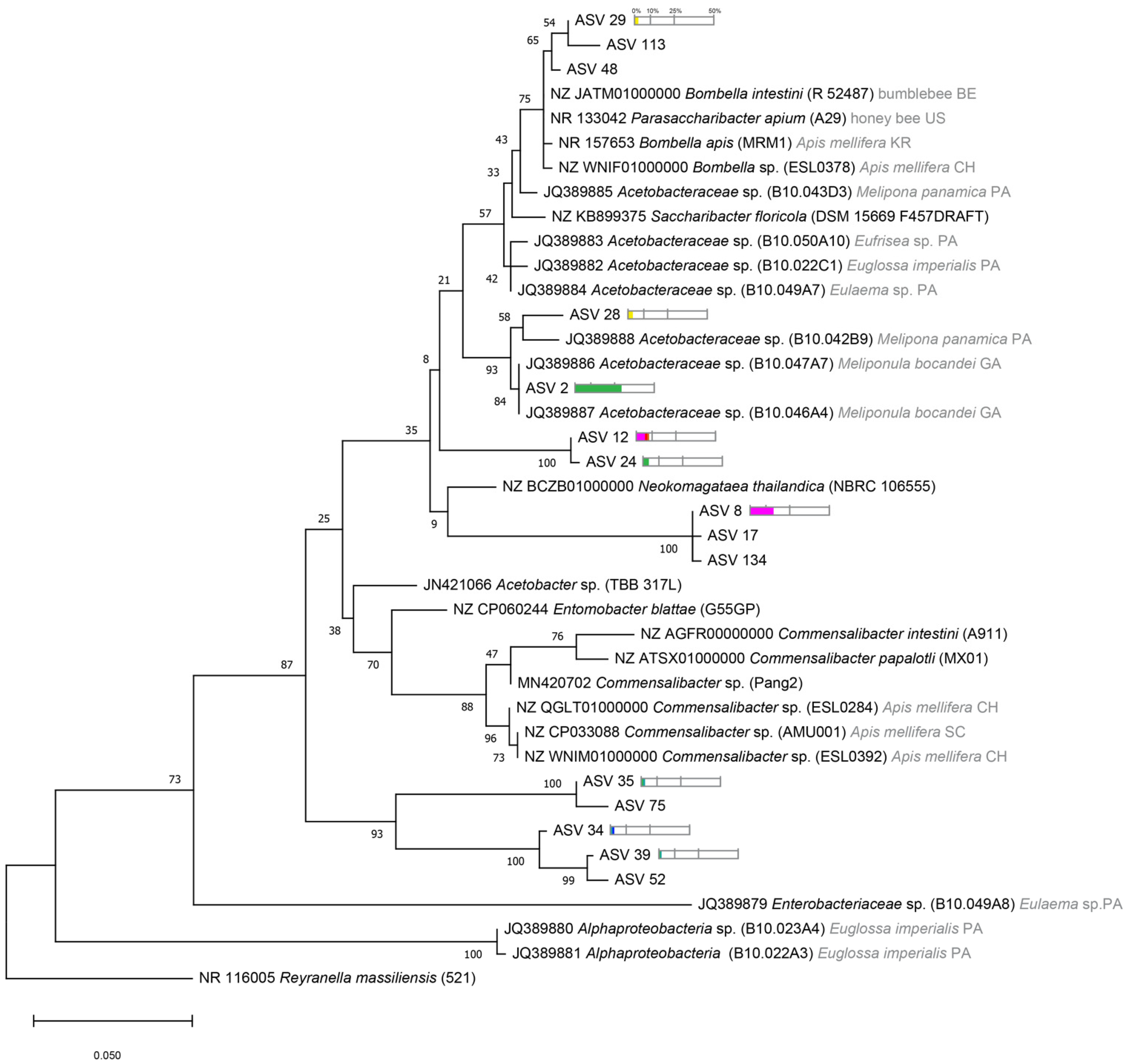
2.4. Phylogenetic Tree Analysis

Phylogenetic trees were constructed for Lactobacillaceae, Bifidobacteriaceae, and Acetobacteraceae members using MEGA X [53]. Sequences were aligned using Unweighted Pair Group Method with Arithmetic Mean (UPGMA). Phylogenetic trees were computed with the Kimura 2-parameter model using the Maximum Likelihood method [54]. The model was selected based on the Bayesian Information Criterion (BIC) values. The bootstrap consensus tree was inferred from 1000 replicates, and less than 50% bootstrap replicates collapsed.

3. Results

3.1. Bacterial Communities Associated with Stingless Bee Guts

We sampled eight different stingless bee species in Kenya: Meliponula bocandei, M. togoensis, M. ferruginea, M. lendliana, Dactylurina schmidti, Liotrigona spp., and Hypotrigona sp. 1 and sp. 2. We obtained 2038 amplicon sequence variants (ASVs). We found 409 genera of which 13 genera represented more than 99% of total reads (Figure 1A and Figure S2 and Table S1).
We found that Firmicutes, Proteobacteria, and Actinobacteria were the dominant phyla across the eight stingless bee species (Table S1). The most abundant was Firmicutes with about 60% of total reads encompassing Lactobacillus, Bombilactobacillus, Acetilactobacillus, and Apilactobacillus, among others. Lactobacillus accounted for approximately 25% of total reads (Figure 1 and Figure S2, Table S1). The second highest phylum was Proteobacteria with approximately 18% of total reads including Saccharibacter, Neokomagataea, Wolbachia, Bombella, Gluconacetobacter, Nguyenibacter, Acinetobacter, Zymobacter, Klebsiella, and Acetobacter. Saccharibacter accounted for about 8% of total sample reads (Figure 1 and Figure S2, Table S1). The actinobacteria phylum including Bifidobacterium accounted for approximately 10% of total reads (Figure 1 and Figure S2, Table S1). Interestingly, whereas almost all stingless bee species were dominated by a member of the Lactobacillaceae family, the dominant genera varied across species. Acetilactobacillus and Apilactobacillus related genera dominated in Hypotrigona sp. 2, M. lendliana, and M. ferruginea, Lactobacillus dominated in Hypotrigona sp. 1 and M. togoensis, and Bombilactobacillus in D. schmidti. Bifidobacterium was present in all species except Hypotrigona sp. 2 and was the most abundant in Hypotrigona sp. 1. Among the Acetobacteraceae family, Bombella was the most abundant in M. bocandei, and Saccharibacter was the most abundant in D. schmidti (Figure 1 and Figure S2, Table S1). Interestingly, we also uncovered the endosymbiont, Wolbachia, which was highly abundant in all Liotrigona sp. (59% of Liotrigona sp. total reads) and in one sample of M. lendliana (0.5% of M. lendliana total reads) (Table S1).

3.2. Bacterial Communities Varied across Stingless Bee Species

To measure the bacterial community variation across species, we analyzed the overall diversity of the gut bacteria in the eight stingless bee species (Figure 2). As seen in Figure 1, alpha diversity varied significantly when richness (Shannon: p = 0.000038, H = 29.672, (Figure 2A)), evenness (Evenness: p = 0.00005, H = 29.12, (Figure 2B)), and phylogenetic distances (Faith’s phylogenetic diversity, p = 0.00018, H = 24.888, (Figure 2C)) were tested. These results are consistent with the PCoA using Bray–Curtis and from the weighted and unweighted UniFrac distances (Figure 3).
Even though M. lendliana, Hypotrigona sp. 2, and Liotrigona sp. clustered together in Bray–Curtis, they did not cluster in Unifrac distances. It is also noteworthy that these are the species where we obtained a lower number of samples (2 to 4 compared to 8 to 10 for others (Figure 3)).

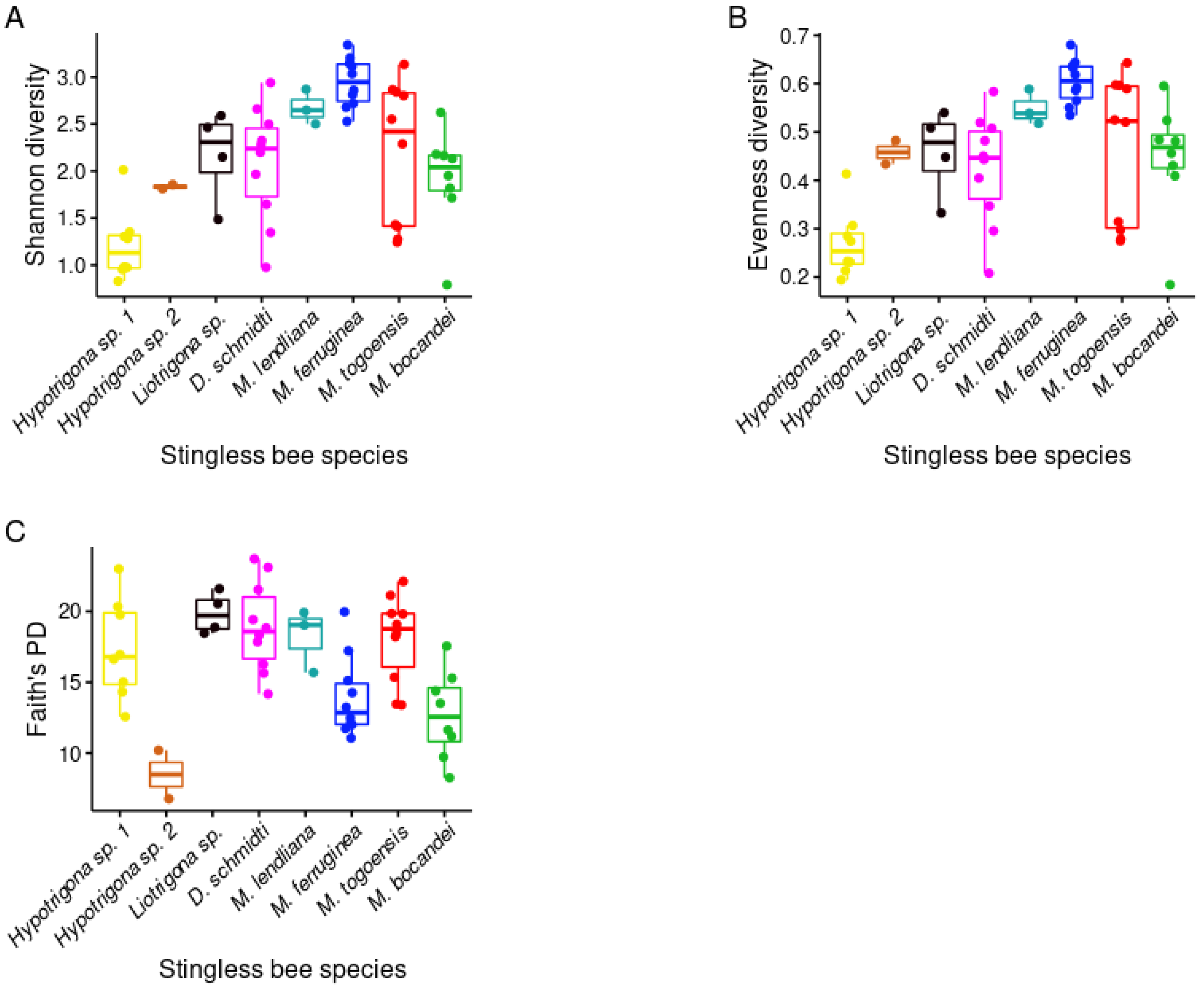
3.3. Phylogenetic Diversity of Stingless Bee Gut Bacterial Microbiota

To evaluate the relationship between the bacteria generated in this study to other bacterial sequences isolated in other corbiculate bees, we constructed phylogenetic trees with the three most abundant families, Lactobacillaceae, Bifidobacteriaceae, and Acetobacteraceae (Figure 4).
ASVs related to Lactobacillaceae were found overall distributed with other reported sequences from corbiculate bees. The most abundant ASVs were closely related to strains from Lactobacillus (Firmicutes 5 group) and Bombilactobacillus (Firmicutes 4 group), known members of honeybee and bumble bee core gut bacterial microbiota. Interestingly, we also uncovered multiple ASVs related to environmental or hive-associated Lactobacillaceae such as Acetilactobacillus and Apilactobacillus with an important abundance and prevalence across samples. One ASVs was related to Ligilactobacillus but at a very low abundance (Figure 1 and Figure 4).
Bifidobacteriaceae associated ASVs were found to cluster in two subgroups (Figure 5). The most numerous and abundant were highly related to Bifidobacterium commune, Bifidobacterium bohemicum, and Bifidobacterium bombi, all of which are found in bumble bee guts. Two other ASV clustered with Bifidobacterium coryneforme strains found in Apis mellifera and M. bocandei, from previous studies. Interestingly, none of the represented ASVs in Figure 4 were related to Bifidobacterium asteroids found in honeybees or bumble bees, but were found in the stingless bee M. bocandei from Kenya [44].
The Acetobacteraceae-associated ASVs clustered with other reported strains and in specific groups (Figure 6). The most abundant cluster was found to be highly related to other Acetobacter species isolated in other stingless bees, Melipona sp. and Meliponula sp., from Panama and Gabon [41,63,64]. We found one group also related to Bombella apis and Bombella intestini reported in honey bees and bumble bees, and another group with very long branches related to Neokomagataea thailandica isolated from flowers [41,65]. Interestingly, we found that some ASVs form a group on their own, with no closest known related strain (ASV 34, 35, 39, 52, and 75, Figure 6). In contrast to Apis mellifera bacterial microbiota, we did not uncover any Commensalibacter related strains in our study [66,67].

4. Discussion

We reported the first characterization of the gut bacterial microbiota of eight endemic African stingless bee species (Apidae: Meliponini). We found that Firmicutes, Actinobacteria, and Proteobacteria were the dominant and conserved phyla across the stingless bee species. Interestingly, there were significant variations amongst the genera that dominated the gut of each stingless bee species. Furthermore, phylogenetic analysis showed substantial bacterial strain diversity, which is shared with other corbiculate bees, but also with several African specific bacterial clusters. As has been reported for South American stingless bee species, the East African stingless bee species we examined lost the honeybee and bumble bee specific symbionts Snodgrassella, Gilliamella, and Frischella [42,46,69,70]. Together, these results are an important contribution towards the study of the common origin of corbiculate bee gut bacterial microbiota and their co-evolution with their host.
Among the African stingless bee populations that were sampled, we identified Lactobacillaceae (including Lactobacillus, Paucilactobacillus, Bombilactobacillus, and Lentilactobacillus), Bifidobacterium, and Acetobacteraceae (including Saccharibacter and Neokomagataea) as the most abundant families, which account for more than 73% of the total gut bacterial microbiota. Interestingly, phylogenetic analyses revealed novel diversity of strains from these groups that has not been reported in other studies on stingless bees [41,42]. Additionally, despite sharing the same habitat for at least six months, each stingless bee species was dominated by a certain number of specific bacterial strains. Evaluating bacterial gut composition across longer time scales—months or years—from feral colonies or after a change of location would be important to understand the causes of this diversity [40]. These results demonstrate that there is a vast diversity of stingless bee gut bacterial microbiota members that are yet to be characterized and that conducting research on multiple strains, as done in this study, is needed to have a comprehensive and comparative picture of stingless and corbiculate bee gut bacterial microbiota and their co-evolutionary patterns.
We found that gut bacterial microbiota richness and evenness varied across the stingless bee species. To get a better understanding of the drivers of bacterial diversity, we investigated potential correlations between bacterial diversity and bee size; however, we did not find any correlation (Figure S3). A recent study reported that larger bees have higher bacterial diversity, and that this correlation is higher when colony size was considered [42]. Since African stingless bee research is in its early stages, little is known about bee colony and hive size. This enables us to better calculate a potential correlation between bee habitat size and the gut bacterial microbiota diversity in their guts.
Whereas library sequencing of the 16S rRNA V3-V4 region has proved to be informative and very useful for bee bacterial gut microbiota characterization, it has important limitations in resolution and absolute bacterial quantification. Complementary studies evaluating whole bacterial genome sequences (DNA metagenomics) and bacteria metabolically active pathways (RNA metagenomics) will provide better information about bacterial classification and metabolic capabilities [36]. Additionally, such approaches will help to discriminate between metabolically active resident bacteria and transient inactive food-associated microbes.
Interestingly, we found the endosymbiotic bacteria Wolbachia in Liotrigona sp. with 100% prevalence that accounted for 59% of total reads, and in M. lendliana with 33% prevalence. The two Wolbachia ASVs found in this study were closely related to the Wolbachia strain WCAAL1 (MT590312, 97% similarity) which is found in the Aedes albopictus mosquito. Studies have shown that Wolbachia protects its host against viruses [71,72]. Since bees harbor a substantial diversity of viruses, it might be relevant to investigate the potential Wolbachia antiviral protection phenotype in these two stingless bee species [73]. Additionally, Wolbachia has also been shown to manipulate host reproduction by inducing parthenogenesis [74]. Most of stingless bee workers contribute to the production of haploid eggs, which generate male offspring or trophic eggs that can be eaten by the queen as a food source [75,76]. It would be interesting to investigate if there is any involvement of Wolbachia in these processes in Liotrigona spp. or M. lendliana.
Tropical regions are profitable environments to conduct gut bacterial microbiota studies due to their number of sympatric honeybee and stingless bee species and their diversity, which enables research on the evolutionary history of their bacterial microbiota [4,10,11]. This study contributes to the understanding of general concepts in microbial ecology and evolution in these bee species that could only be found in tropical latitudes and microbe rich environments.

Supplementary Materials

The following are available online at https://www.mdpi.com/article/10.3390/microorganisms9122420/s1, Figure S1: Alpha rarefaction curve. Figure S2: Gut bacterial genera associated with each of the eight stingless bee species in Kenya. Figure S3: Bacterial alpha diversity did not correlate with the stingless bee size. Table S1: 16S rRNA sequencing data analysis (Excel file).

Author Contributions

Conceptualization, J.C.P. and B.S.; investigation, Y.H.T.; data curation, Y.H.T. and J.W.W.; formal analysis, Y.H.T., J.W.W. and J.C.P.; funding acquisition, J.C.P.; methodology, Y.H.T. and J.C.P.; visualization, Y.H.T., J.W.W. and J.C.P.; writing—original draft preparation, Y.H.T. and J.C.P.; writing—review and editing, Y.H.T., J.W.W., N.N.N., K.N., B.S. and J.C.P.; project administration: J.C.P.; All authors have read and agreed to the published version of the manuscript.

Funding

This research was funded by the Swiss National Science Foundation Postdoc Mobility (grants P300PA_174329 and P2ELP3_165168), the German Academic Exchange Service (DAAD), grant 91671945, Norwegian Agency for Development Cooperation, the Section for Research, Innovation, and Higher Education grant number RAF-3058 KEN-18/0005 (CAP-Africa), and by icipe core donors (see Acknowledgments).

Institutional Review Board Statement

Not applicable for insects.

Informed Consent Statement

Not applicable.

Data Availability Statement

The datasets presented in this study can be found in the following online repository PRJNA776526 and in Supplementary Material.

Acknowledgments

The authors gratefully acknowledge the financial support for this research by the following organizations and agencies as core donors of icipe; UK’s Foreign, Commonwealth & Development Office (FCDO); the Swedish International Development Cooperation Agency (Sida); the Swiss Agency for Development and Cooperation (SDC); the Federal Democratic Republic of Ethiopia; and the Government of the Republic of Kenya. The views expressed herein do not necessarily reflect the official opinion of the donors. We are additionally grateful to Jeremy Herren for manuscript review and to icipe stingless bee team Gilvian Onsomu and Sophia Kariuki, and icipe bee health gut bacterial microbiota laboratory team.

Conflicts of Interest

The authors declare no conflict of interest. The funders had no role in the design of the study; in the collection, analyses, or interpretation of data; in the writing of the manuscript; or in the decision to publish the results.

References

  1. Bawa, K.S. Plant-Pollinator Interactions in Tropical Rain Forests. Annu. Rev. Ecol. Syst. 1990, 21, 399–422. [Google Scholar] [CrossRef]
  2. Van Dulmen, A. Pollination and phenology of flowers in the canopy of two contrasting rain forest types in Amazonia, Colombia. Plant Ecol. 2001, 153, 73–85. [Google Scholar] [CrossRef]
  3. Garibaldi, L.A.; Carvalheiro, L.G.; Vaissière, B.E.; Gemmill-Herren, B.; Hipólito, J.; Freitas, B.M.; Ngo, H.T.; Azzu, N.; Sáez, A.; Åström, J.; et al. Mutually beneficial pollinator diversity and crop yield outcomes in small and large farms. Science 2016, 351, 388–391. [Google Scholar] [CrossRef] [Green Version]
  4. Grüter, C. Stingless Bees, Their Behaviour, Ecology and Evolution; Springer Nature Switzerland AG: Cham, Switzerland, 2020; ISBN 9783030600891. [Google Scholar]
  5. Jalil, A.H. Beescape for Meliponines: Conservation of Indo-Malayan Stingless Bees; Partridge Publishing Singapore: Singapore, 2014; ISBN 9781482823622. [Google Scholar]
  6. Michener, C.D. The Bees of the World; The Johns Hopkins University Press: Baltimore, MD, USA, 2000; Volume 58, ISBN 0-8018-6133-0. [Google Scholar]
  7. Quezada-Euán, J.J.G. Stingless Bees of Mexico; Springer: Berlin/Heidelberg, Germany, 2018; ISBN 9783319777849. [Google Scholar]
  8. Rasmussen, C.; Cameron, S.A. Global stingless bee phylogeny supports ancient divergence, vicariance, and long distance dispersal. Biol. J. Linn. Soc. 2010, 99, 206–232. [Google Scholar] [CrossRef]
  9. Roig-Alsina, A.; Alvarez, L.J. Southern distributional limits of Meliponini bees (Hymenoptera, Apidae) in the Neotropics: Taxonomic notes and distribution of Plebeia droryana and P. emerinoides in Argentina. Zootaxa 2017, 4244, 261–268. [Google Scholar] [CrossRef]
  10. Eardley, C.D. Taxonomic revision of the African stingless bees (Apoidea: Apidae: Apinae: Meliponini). Afr. Plant Prot. 2004, 10, 63–96. [Google Scholar]
  11. Fabre Anguilet, E.; Nguyen, B.K.; Bengone Ndong, T.; Haubruge, É.; Francis, F. Meliponini and Apini in Africa (Apidae: Apinae): A review on the challenges and stakes bound to their diversity and their distribution. Biotechnol. Agron. Soc. Environ. 2015, 19, 382–391. [Google Scholar]
  12. Ndungu, N.N.; Nkoba, K.; Sole, C.; Pirk, C.; Abdullahi, A.Y.; Raina, S.K.; Masiga, D.K. Resolving taxonomic ambiguity and cryptic speciation of Hypotrigona species through morphometrics and DNA barcoding. J. Apic. Res. 2018, 57, 354–363. [Google Scholar] [CrossRef]
  13. Ndungu, N.N.; Kiatoko, N.; Ciosi, M.; Salifu, D.; Nyansera, D.; Masiga, D.; Raina, S.K. Identification of stingless bees (Hymenoptera: Apidae) in Kenya using morphometrics and DNA barcoding. J. Apic. Res. 2017, 56, 341–353. [Google Scholar] [CrossRef] [Green Version]
  14. Nkoba, K.; Raina, S.K.; Muli, E.; Mithöfer, K.; Mueke, J. Species richness and nest dispersion of some tropical meliponine bees (Apidae: Meliponinae) in six habitat types in the Kakamega forest, western Kenya. Int. J. Trop. Insect Sci. 2012, 32, 194–202. [Google Scholar] [CrossRef]
  15. Azmi, W.A.; Sembok, W.Z.W.; Yusuf, N.; Hatta, M.F.M.; Salleh, A.F.; Hamzah, M.A.H.; Ramli, S.N. Effects of Pollination by the Indo-Malaya Stingless Bee (Hymenoptera: Apidae) on the Quality of Greenhouse-Produced Rockmelon. J. Econ. Entomol. 2019, 112, 20–24. [Google Scholar] [CrossRef] [PubMed]
  16. Potts, S.G.; Imperatriz-Fonseca, V.; Ngo, H.T.; Aizen, M.; Biesmeijer, J.C.; Breeze, T.; Dicks, L.; Garibaldi, L.A.; Hill, R.; Settele, J.; et al. Safeguarding pollinators and their values to human well-being. Nature 2016, 540, 220–229. [Google Scholar] [CrossRef] [PubMed]
  17. Kiatoko, N.; Raina, S.K.; Muli, E.; Mueke, J. Enhancement of fruit quality in Capsicum annum through pollination by Hypotrigona gribodoi in Kakamega, Western Kenya. Entomol. Sci. 2014, 17, 106–110. [Google Scholar] [CrossRef]
  18. Slaa, E.J.; Chaves, L.A.S.; Malagodi-Braga, K.S.; Hofstede, F.E. Stingless bees in applied pollination: Practice and perspectives. Apidologie 2006, 37, 293–315. [Google Scholar] [CrossRef] [Green Version]
  19. Nkoba, K.; Raina, S.K.; Langevelde, F. A vertical compartmented hive design for reducing post-harvest colony losses in three afrotropical stingless bee species (Apidae: Meliponinae). Int. J. Dev. Res. 2016, 6, 9026–9034. [Google Scholar]
  20. Kishan Tej, M.; Srinivasan, M.R.; Rajashree, V.; Thakur, R.K. Stingless bee Tetragonula iridipennis Smith for pollination of greenhouse cucumber. J. Entomol. Zool. Stud. 2017, 5, 1729–1733. [Google Scholar]
  21. Nishio, E.K.; Ribeiro, J.; de Oliveira, A.G.; Andrade, C.G.T.J.; Proni, E.A.; Kobayashi, R.K.T.; Nakazato, G. Antibacterial synergic effect of honey from two stingless bees: Scaptotrigona bipunctata Lepeletier, 1836, and S. postica Latreille, 1807. Sci. Rep. 2016, 6, 21641. [Google Scholar] [CrossRef]
  22. Dos Santos, S.B.; Roselino, A.; Hrncir, M.; Bego, L. Pollination of tomatoes by the stingless bee Melipona quadrifasciata and the honey bee Apis mellifera (Hymenoptera, Apidae). Genet. Mol. Res. 2009, 8, 751–757. [Google Scholar] [CrossRef]
  23. Mokaya, H.O.; Nkoba, K.; Ndunda, R.M.; Vereecken, N.J. Characterization of honeys produced by sympatric species of Afrotropical stingless bees (Hymenoptera, Meliponini). Food Chem. 2021, 366, 130597. [Google Scholar] [CrossRef]
  24. Moran, N.A.; Tran, P.; Gerardo, N.M.; Tannock, G.W.; Ghazally, S.; Walter, J.; Loach, D.; Brooks, H.; Cook, G.; Surette, M.; et al. Symbiosis and Insect Diversification: An Ancient Symbiont of Sap-Feeding Insects from the Bacterial Phylum Bacteroidetes. Appl. Environ. Microbiol. 2005, 71, 8419–8425. [Google Scholar] [CrossRef] [Green Version]
  25. Gerardo, N.; Hurst, G. Q&A: Friends (but sometimes foes) within: The complex evolutionary ecology of symbioses between host and microbes. BMC Biol. 2017, 15, 126. [Google Scholar] [CrossRef]
  26. Schmidt, K.; Engel, P. Mechanisms underlying gut microbiota–host interactions in insects. J. Exp. Biol. 2021, 224, jeb207696. [Google Scholar] [CrossRef]
  27. Salem, H.; Kaltenpoth, M. Beetle–Bacterial Symbioses: Endless Forms Most Functional. Annu. Rev. Entomol. 2022, 67, 201–219. [Google Scholar] [CrossRef]
  28. Weisskopf, L.; Schulz, S.; Garbeva, P. Microbial volatile organic compounds in intra-kingdom and inter-kingdom interactions. Nat. Rev. Microbiol. 2021, 19, 391–404. [Google Scholar] [CrossRef]
  29. Duplais, C.; Sarou-Kanian, V.; Massiot, D.; Hassan, A.; Perrone, B.; Estevez, Y.; Wertz, J.T.; Martineau, E.; Farjon, J.; Giraudeau, P.; et al. Gut bacteria are essential for normal cuticle development in herbivorous turtle ants. Nat. Commun. 2021, 12, 1–6. [Google Scholar] [CrossRef] [PubMed]
  30. Ma, Q.; Cui, Y.; Chu, X.; Li, G.; Yang, M.; Wang, R.; Liang, G.; Wu, S.; Tigabu, M.; Zhang, F.; et al. Gut Bacterial Communities of Lymantria xylina and Their Associations with Host Development and Diet. Microorganisms 2021, 9, 1860. [Google Scholar] [CrossRef]
  31. Chakraborty, A.; Roy, A. Microbial influence on plant–insect interaction. In Plant-Pest Interactions: From Molecular Mechanisms to Chemical Ecology; Springer: Singapore, 2021. [Google Scholar]
  32. Emery, O.; Schmidt, K.; Engel, P. Immune system stimulation by the gut symbiont Frischella perrarain the honey bee (Apis mellifera). Mol. Ecol. 2017, 26, 2576–2590. [Google Scholar] [CrossRef]
  33. Engel, P.; Bartlett, K.D.; Moran, N.A. The Bacterium Frischella perrara Causes Scab Formation in the Gut of its Honeybee Host. MBio 2015, 6, 1–8. [Google Scholar] [CrossRef] [PubMed] [Green Version]
  34. Koch, H.; Schmid-Hempel, P. Socially transmitted gut microbiota protect bumble bees against an intestinal parasite. Proc. Natl. Acad. Sci. USA 2011, 108, 19288–19292. [Google Scholar] [CrossRef] [PubMed] [Green Version]
  35. Vernier, C.L.; Chin, I.M.; Adu-Oppong, B.; Krupp, J.J.; Levine, J.; Dantas, G.; Ben-Shahar, Y. The gut microbiome defines social group membership in honey bee colonies. Sci. Adv. 2020, 6, 42. [Google Scholar] [CrossRef] [PubMed]
  36. Kešnerová, L.; Mars, R.A.T.; Ellegaard, K.M.; Troilo, M.; Sauer, U.; Engel, P. Disentangling metabolic functions of bacteria in the honey bee gut. PLoS Biol. 2017, 15, e2003467. [Google Scholar] [CrossRef] [Green Version]
  37. Zheng, H.; Nishida, A.; Kwong, W.K.; Koch, H.; Engel, P.; Steele, M.I.; Moran, N.A. Metabolism of Toxic Sugars by Strains of the Bee Gut Symbiont Gilliamella apicola. MBio 2016, 7, e01326-16. [Google Scholar] [CrossRef] [PubMed] [Green Version]
  38. Zheng, H.; Powell, J.E.; Steele, M.I.; Dietrich, C.; Moran, N.A. Honeybee gut microbiota promotes host weight gain via bacterial metabolism and hormonal signaling. Proc. Natl. Acad. Sci. USA 2017, 114, 4775–4780. [Google Scholar] [CrossRef] [PubMed] [Green Version]
  39. Zheng, H.; Perreau, J.; Powell, J.; Han, B.; Zhang, Z.; Kwong, W.K.; Tringe, S.G.; Moran, N.A. Division of labor in honey bee gut microbiota for plant polysaccharide digestion. Proc. Natl. Acad. Sci. USA 2019, 116, 25909–25916. [Google Scholar] [CrossRef] [PubMed]
  40. Hall, M.A.; Brettell, L.E.; Liu, H.; Nacko, S.; Spooner-Hart, R.; Riegler, M.; Cook, J.M. Temporal changes in the microbiome of stingless bee foragers following colony relocation. FEMS Microbiol. Ecol. 2020, 97, fiaa236. [Google Scholar] [CrossRef]
  41. Koch, H.; Abrol, D.P.; Li, J.; Schmid-Hempel, P. Diversity and evolutionary patterns of bacterial gut associates of corbiculate bees. Mol. Ecol. 2013, 22, 2028–2044. [Google Scholar] [CrossRef]
  42. Kwong, W.K.; Medina, L.A.; Koch, H.; Sing, K.-W.; Soh, E.J.Y.; Ascher, J.S.; Jaffé, R.; Moran, N.A. Dynamic microbiome evolution in social bees. Sci. Adv. 2017, 3, e1600513. [Google Scholar] [CrossRef] [Green Version]
  43. Leonhardt, S.D.; Kaltenpoth, M. Microbial Communities of Three Sympatric Australian Stingless Bee Species. PLoS ONE 2014, 9, e105718. [Google Scholar] [CrossRef]
  44. Vásquez, A.; Forsgren, E.; Fries, I.; Paxton, R.; Flaberg, E.; Szekely, L.; Olofsson, T.C. Symbionts as Major Modulators of Insect Health: Lactic Acid Bacteria and Honeybees. PLoS ONE 2012, 7, e33188. [Google Scholar] [CrossRef]
  45. Tang, Q.-H.; Miao, C.-H.; Chen, Y.-F.; Dong, Z.-X.; Cao, Z.; Liao, S.-Q.; Wang, J.-X.; Wang, Z.-W.; Guo, J. The composition of bacteria in gut and beebread of stingless bees (Apidae: Meliponini) from tropics Yunnan, China. Antonie Leeuwenhoek 2021, 114, 1293–1305. [Google Scholar] [CrossRef]
  46. Cerqueira, A.E.S.; Hammer, T.J.; Moran, N.A.; Santana, W.C.; Kasuya, M.C.M.; da Silva, C.C. Extinction of anciently associated gut bacterial symbionts in a clade of stingless bees. ISME J. 2021, 15, 2813–2816. [Google Scholar] [CrossRef] [PubMed]
  47. Díaz, S.; Urbano, S.D.S.; Caesar, L.; Blochtein, B.; Sattler, A.; Zuge, V.; Haag, K.L. Report on the microbiota of Melipona quadrifasciata affected by a recurrent disease. J. Invertebr. Pathol. 2017, 143, 35–39. [Google Scholar] [CrossRef] [PubMed]
  48. Vit, P.; Roubik, D.W.; Pedro, S.R.M. Taxonomy as a Tool for Conservation of African Stingless Bees and Their Honey. In Eardley, Connal Kwapong, Peter; Springer: Berlin/Heidelberg, Germany, 2013; pp. 1–654. ISBN 9781461449607. [Google Scholar]
  49. Engel, P.; James, R.R.; Koga, R.; Kwong, W.K.; Mcfrederick, Q.S.; Moran, N.A. Standard methods for research on Apis mellifera gut symbionts. J. Apic. Res. 2013, 524, 1–24. [Google Scholar] [CrossRef] [Green Version]
  50. Tola, Y.; Waweru, J.; Hurst, G.; Slippers, B.; Paredes, J. Characterization of the Kenyan Honey Bee (Apis mellifera) Gut Microbiota: A First Look at Tropical and Sub-Saharan African Bee Associated Microbiomes. Microorganisms 2020, 8, 1721. [Google Scholar] [CrossRef] [PubMed]
  51. Callahan, B.J.; McMurdie, P.J.; Rosen, M.J.; Han, A.W.; Johnson, A.J.A.; Holmes, S.P. DADA2: High-resolution sample inference from Illumina amplicon data. Nat. Methods 2016, 13, 581–583. [Google Scholar] [CrossRef] [Green Version]
  52. Wang, Q.; Garrity, G.M.; Tiedje, J.M.; Cole, J.R. Naïve Bayesian Classifier for Rapid Assignment of rRNA Sequences into the New Bacterial Taxonomy. Appl. Environ. Microbiol. 2007, 73, 5261–5267. [Google Scholar] [CrossRef] [Green Version]
  53. Kumar, S.; Stecher, G.; Li, M.; Knyaz, C.; Tamura, K. MEGA X: Molecular Evolutionary Genetics Analysis across Computing Platforms. Mol. Biol. Evol. 2018, 35, 1547–1549. [Google Scholar] [CrossRef]
  54. Kimura, M. A simple method for estimating evolutionary rates of base substitutions through comparative studies of nucleotide sequences. J. Mol. Evol. 1980, 16, 111–120. [Google Scholar] [CrossRef]
  55. Kapheim, K.M.; Rao, V.D.; Yeoman, C.J.; Wilson, B.A.; White, B.A.; Goldenfeld, N.; Robinson, G.E. Caste-Specific Differences in Hindgut Microbial Communities of Honey Bees (Apis mellifera). PLoS ONE 2015, 10, e0123911. [Google Scholar] [CrossRef]
  56. Killer, J.; Dubná, S.; Sedláček, I.; Švec, P. Lactobacillus apis sp. nov., from the stomach of honeybees (Apis mellifera), having an in vitro inhibitory effect on the causative agents of American and European foulbrood. Int. J. Syst. Evol. Microbiol. 2014, 64, 152–157. [Google Scholar] [CrossRef] [Green Version]
  57. Olofsson, T.C.; Vásquez, A. Detection and Identification of a Novel Lactic Acid Bacterial Flora within the Honey Stomach of the Honeybee Apis mellifera. Curr. Microbiol. 2008, 57, 356–363. [Google Scholar] [CrossRef] [PubMed]
  58. Praet, J.; Meeus, I.; Cnockaert, M.; Houf, K.; Smagghe, G.; Vandamme, P. Novel lactic acid bacteria isolated from the bumble bee gut: Convivina intestini gen. nov., sp. nov., Lactobacillus bombicola sp. nov., and Weissella bombi sp. nov. Antonie Leeuwenhoek 2015, 107, 1337–1349. [Google Scholar] [CrossRef]
  59. Anderson, K.E.; Johansson, A.; Sheehan, T.H.; Mott, B.M.; Corby-Harris, V.; Johnstone, L.; Sprissler, R.; Fitz, W. Draft genome sequences of two Bifidobacterium sp. from the honey bee (Apis mellifera). Gut Pathog. 2013, 5, 42. [Google Scholar] [CrossRef] [Green Version]
  60. Engel, P.; Martinson, V.G.; Moran, N.A. Functional diversity within the simple gut microbiota of the honey bee. Proc. Natl. Acad. Sci. USA 2012, 109, 11002–11007. [Google Scholar] [CrossRef] [Green Version]
  61. Kapheim, K.M.; Johnson, M.M.; Jolley, M. Composition and acquisition of the microbiome in solitary, ground-nesting alkali bees. Sci. Rep. 2021, 11, 1–11. [Google Scholar] [CrossRef] [PubMed]
  62. Killer, J.; Kopečný, J.; Mrazek, J.; Rada, V.; Benada, O.; Koppová, I.; Havlik, J.; Straka, J. Bifidobacterium bombi sp. nov., from the bumblebee digestive tract. Int. J. Syst. Evol. Microbiol. 2009, 59, 2020–2024. [Google Scholar] [CrossRef] [Green Version]
  63. Yun, J.-H.; Lee, J.-Y.; Hyun, D.-W.; Jung, M.-J.; Bae, J.-W. Bombella apis sp. nov., an acetic acid bacterium isolated from the midgut of a honey bee. Int. J. Syst. Evol. Microbiol. 2017, 67, 2184–2188. [Google Scholar] [CrossRef] [PubMed]
  64. Li, L.; Illeghems, K.; Van Kerrebroeck, S.; Borremans, W.; Cleenwerck, I.; Smagghe, G.; De Vuyst, L.; Vandamme, P. Whole-Genome Sequence Analysis of Bombella intestini LMG 28161T, a Novel Acetic Acid Bacterium Isolated from the Crop of a Red-Tailed Bumble Bee, Bombus lapidarius. PLoS ONE 2016, 11, e0165611. [Google Scholar] [CrossRef] [Green Version]
  65. Yukphan, P.; Malimas, T.; Muramatsu, Y.; Potacharoen, W.; Tanasupawat, S.; Nakagawa, Y.; Tanticharoen, M.; Yamada, Y. Neokomagataea gen. Nov., with descriptions of Neokomagataea thailandica sp. nov. and Neokomagataea tanensis sp. Nov., osmotolerant acetic acid bacteria of the α-proteobacteria. Biosci. Biotechnol. Biochem. 2011, 75, 419–426. [Google Scholar] [CrossRef] [Green Version]
  66. Ellegaard, K.; Engel, P. New Reference Genome Sequences for 17 Bacterial Strains of the Honey Bee Gut Microbiota. Microbiol. Resour. Announc. 2018, 7, 4–6. [Google Scholar] [CrossRef] [Green Version]
  67. Siozios, S.; Moran, J.; Chege, M.; Hurst, G.D.D.; Paredes, J.C. Complete Reference Genome Assembly for Commensalibacter sp. Strain AMU001, an Acetic Acid Bacterium Isolated from the Gut of Honey Bees. Microbiol. Resour. Announc. 2019, 8, 1–2. [Google Scholar] [CrossRef] [PubMed] [Green Version]
  68. Corby-Harris, V.; Maes, P.; Anderson, K.E. The Bacterial Communities Associated with Honey Bee (Apis mellifera) Foragers. PLoS ONE 2014, 9, e95056. [Google Scholar] [CrossRef] [PubMed] [Green Version]
  69. Rosso, G.B.; Engel, P. Functional roles and metabolic niches in the honey bee gut microbiota. Curr. Opin. Microbiol. 2018, 43, 69–76. [Google Scholar] [CrossRef] [Green Version]
  70. Raymann, K.; Moran, N.A. The role of the gut microbiome in health and disease of adult honey bee workers. Curr. Opin. Insect Sci. 2018, 26, 97–104. [Google Scholar] [CrossRef] [PubMed]
  71. Hedges, L.M.; Brownlie, J.C.; O’Neill, S.L.; Johnson, K.N. Wolbachia and Virus Protection in Insects. Science 2008, 322, 702. [Google Scholar] [CrossRef]
  72. Teixeira, L.; Ferreira, Á.; Ashburner, M. The Bacterial Symbiont Wolbachia Induces Resistance to RNA Viral Infections in Drosophila melanogaster. PLoS Biol. 2008, 6, 2753–2763. [Google Scholar] [CrossRef] [Green Version]
  73. Galbraith, D.A.; Fuller, Z.L.; Ray, A.M.; Brockmann, A.; Frazier, M.; Gikungu, M.W.; Martinez, J.F.I.; Kapheim, K.M.; Kerby, J.T.; Kocher, S.D.; et al. Investigating the viral ecology of global bee communities with high-throughput metagenomics. Sci. Rep. 2018, 8, 1–11. [Google Scholar] [CrossRef] [Green Version]
  74. Werren, J.H.; Baldo, L.; Clark, M.E. Wolbachia: Master manipulators of invertebrate biology. Nat. Rev. Genet. 2008, 6, 741–751. [Google Scholar] [CrossRef] [PubMed]
  75. Luna-Lucena, D.; Rabico, F.; Simoes, Z.L. Reproductive capacity and castes in eusocial stingless bees (Hymenoptera: Apidae). Curr. Opin. Insect Sci. 2019, 31, 20–28. [Google Scholar] [CrossRef]
  76. Vollet-Neto, A.; Koffler, S.; Dos Santos, C.F.; Menezes, C.; Nunes, F.; Hartfelder, K.; Imperatriz-Fonseca, V.L.; Alves, D.A. Recent advances in reproductive biology of stingless bees. Insectes Sociaux 2018, 65, 201–212. [Google Scholar] [CrossRef]
Figure 1. Gut bacterial genera associated with eight stingless bee species in Kenya. (A) A bar plot representation of all the stingless bee gut bacterial genera with an overall abundance higher than 1%. All genera with abundances below 1% were categorized as “Others”. The phylogeny of Afrotropical stingless bees was based on [8]. (B,C) heatmap comprising of the ASVs from the five most abundant genera across the eight stingless bee species. ASV ID numbers (Table S1) are indicated at the top. Darker squares correspond to higher mean relative abundances for a given ASV in each bee species.
Microorganisms 09 02420 g001
Figure 2. Alpha diversity estimates from the different stingless bee species. (A) Shannon diversity (p = 0.000038, H = 29.672), (B) Evenness diversity (p = 0.00005, H = 29.12), (C) Faith’s phylogenetic diversity (Faith’s PD) (p =0.00018, H = 24.888).
Microorganisms 09 02420 g002
Figure 3. PCoA clustering using (A) Bray–Curtis PCoA, (B) Weighted UniFrac PCoA, (C) Unweighted UniFrac PCoA. The ellipse cluster indicate the distribution of the gut bacterial microbiota with respect to the different species of stingless bees (standard errors at 95% confidence).
Microorganisms 09 02420 g003
Figure 4. Phylogenetic tree showing the relationships between Lactobacillaceae ASVs from this study and reference Lactobacillaceae sequences. Accession Number are indicated at the beginning of each name reported from [41,44,55,56,57,58], and bacterial strains are indicated in brackets. Only ASVs that represent more than 1% among Lactobacillaceae family per species were represented. Percentage greater than 1% of the relative abundance from total reads of each ASV are represented by color bars. Each color represents each stingless bee species using the same color code as in Figure 2 and Figure 3.
Microorganisms 09 02420 g004
Figure 5. Phylogenetic tree showing the relationships between Bifidobacterium from stingless bees from Africa compared to other continents. Accession Number is indicated at the beginning of each name reported in [42,44,57,58,59,60,61,62]. Species from which the bacterial strain was reported and the country of origin (two letter code) are indicated in grey. Only ASVs that represent more than 1% among Bifidobacteriaceae family per species were represented. Percentage greater than 1% of the relative abundance from total reads of each ASV are represented by color bars. Each color represents each stingless bee species using the same color code as in Figure 2 and Figure 3.
Microorganisms 09 02420 g005
Figure 6. Phylogenetic tree showing the relationships between Acetobacteraceae family from African stingless bees compared to other continents. Accession Number is indicated at the beginning of each name reported in [41,42,61,63,64,66,67,68]. Species from which the bacterial strain was reported and the country of origin (two letter code) are indicated in grey. Only ASVs that represent more than 1% among Acetobacteraceae family per species are represented. Percentage greater than 1% of the relative abundance from total reads of each ASV are represented by color bars. Each color represents each stingless bee species using the same color code as in Figure 2 and Figure 3.
Microorganisms 09 02420 g006
Publisher’s Note: MDPI stays neutral with regard to jurisdictional claims in published maps and institutional affiliations.

