Communication

Genetic Diversity of Staphylococcus aureus Strains from a Tertiary Care Hospital in Rawalpindi, Pakistan

1
Department of Microbiology, The University of Haripur, Haripur 22620, Pakistan
2
BJ Micro Lab (SMC Pvt) Limited, Islamabad 44000, Pakistan
3
Hygiene and Zoonoses Department, Faculty of Veterinary Medicine, Mansoura University, Mansoura 35516, Egypt
4
Bacterial Epidemiology and Antimicrobial Resistance Research Unit, USDA-ARS, US National Poultry Research Center, Athens, GA 30605, USA
5
One Health Research Group, Discipline of Zoology, Department of Wildlife and Ecology, University of Veterinary and Animal Sciences, Lahore 54000, Pakistan
*
Author to whom correspondence should be addressed.
Both authors contributed equally to this work.
Microorganisms 2021, 9(11), 2301; https://doi.org/10.3390/microorganisms9112301
Submission received: 1 October 2021 / Revised: 26 October 2021 / Accepted: 2 November 2021 / Published: 5 November 2021
(This article belongs to the Special Issue Staphylococcal Infections (Host and Pathogenic Factors) 2.0)

Abstract

:
Staphylococcus aureus is an important healthcare-associated bacterium that causes a multitude of infections in humans such as superficial skin and soft tissue infections, necrotizing pneumonia, foodborne illnesses and postsurgical infections. Treatment of S. aureus infections has become more complicated due to the emergence of Methicillin-Resistant Staphylococcus aureus (MRSA), some of which are multidrug resistant. The present study aimed to characterize S. aureus isolates from a tertiary care hospital in the Rawalpindi district of Pakistan. Staphylococci were isolated from 300 clinical samples collected from January 2018 to January 2019 and S. aureus isolates were tested for antimicrobial susceptibility and analyzed using Pulsed-Field Gel Electrophoresis (PFGE), Multi-Locus Sequence Typing (MLST), staphylococcal cassette chromosome mec (SCCmec) and spa typing. Approximately 25.3% (76/300) of the clinical samples were positive for S. aureus; of those, 88.2% (67/76) were mecA+ (MRSA). In addition to the β-lactam antibiotics, high levels of resistance were also found to the fluoroquinolones (ciprofloxacin, gatifloxacin and levofloxacin (73.7% each)). Of the 23 different spa types identified, the majority of isolates belonged to spa type t632 and t657 (9/66; 13.6% each spa type). ST772-t657 (Bengal Bay clone) was the most commonly identified clone in this study although other clones circulating around different regions of the world were also found indicating the diversity in MRSA isolates from this area of Pakistan. This study emphasizes the need to monitor MRSA in the clinical setting for improved infection control and treatment options.

1. Introduction

Staphylococcus aureus is a well-known commensal found on different parts of the human body such as skin, skin glands, nose and gut mucus membranes [1]. However, S. aureus is a major cause of skin diseases and invasive infections, such as endocarditis, pneumonia and osteomyelitis, in both healthcare and community settings. It has been estimated that 20% of humans are persistent nasal carriers for S. aureus, while 30% are intermittent and 50% are non-carriers for S. aureus [2]. Persistent and intermittent colonization increases the opportunity for infection, which is primarily caused by the same strains present as a commensal on an individuals’ body [3,4,5]. Different infections, skin lesions and injuries, catheters, implants and chronic diseases (diabetes, AIDS, innate and acquired deficiencies of immune system) favor S. aureus infections [6,7].
Emergence of resistant phenotypes is usually linked with injudicious use of antimicrobial agents. A study by Schentag et al. (1998) reported changes in normal biota of a patient within 24–48 h under antibiotic pressure [8]. Some studies have supported causal relationships between antibiotic administration and emergence of Methicillin-Resistant S. aureus (MRSA) [9,10]. MRSA emergence was first observed in 1961, soon after the clinical application of penicillinase-resistant penicillin [1]. Patients infected with these resistant bacteria take longer to recover as compared to those infected with other staphylococcal species, especially those that are susceptible to antibiotics [9]. As such, MRSA isolates have been recognized as a source of infections with resistance to antibiotics in the β-lactam antibiotic class as a main characteristic contributing to its disease-causing ability along with other virulence factors in the bacterium [9].
The presence of mobile genetic elements also plays a major role in conferring resistance to antibiotics in MRSA such as resistance to vancomycin [7]. Mobile genetic elements include plasmids, transposons, bacteriophage and pathogenicity islands [5]. MRSA isolates also contain a mobile genetic element, Staphylococcal cassette chromosome (SCCmec), that may be horizontally disseminated among S. aureus isolates resulting in spread of antimicrobial resistance genes among the isolates [11]. SCCmec is composed of two parts, namely the mec gene complex and cassette chromosome recombinase (ccr) gene complex both of which contribute to production of different variants of MRSA.
Studies conducted in Pakistan in the last decade have reported high prevalence of MRSA [12,13,14,15,16]. Recent studies in our laboratory conducted on MRSA isolates from Peshawar and Malakand cities using microarray technology showed epidemiological links to the Middle Eastern/Arabian Gulf region [17,18]. However, available data for this region is still limited and there is a need for continued surveillance of S. aureus and characterization of isolates from local hospitals for control and better treatment options. The aim of the present study was to characterize clinical S. aureus isolates from a tertiary care hospital in Rawalpindi city of Pakistan. S. aureus from clinical samples were isolated and analyzed using antimicrobial susceptibility testing, presence of antimicrobial resistance genes, Pulsed-field Gel Electrophoresis (PFGE), Multi-Locus Sequence Typing (MLST), SCCmec and spa typing.

2. Materials and Methods

2.1. Sample Collection and Bacterial Identification

Three hundred clinical samples (urine, pus, tracheal tubes, vaginal swabs, body fluids, blood and cannula) were collected from January 2018 to January 2019 from Fauji Foundation hospital, Rawalpindi, Punjab, Pakistan. Samples were collected from patients for routine procedures to more urgent conditions including infected wounds, abscesses, burns and serious or life-threatening medical conditions. The samples were streaked onto Mannitol Salt Agar (MSA) using sterile cotton swabs and incubated at 37 °C for 24 h. For liquid samples (such as urine, blood, etc.), 50 μL of the sample was placed onto the surface of the MSA plate and then spread on the media surface using a sterile swab. Initial bacterial identification was performed by microscopy and biochemical tests (catalase test, tube coagulase test, mannitol fermentation and DNase). Confirmation of the isolates was performed by PCR using previously reported species-specific primers for S. aureus [19]. Pure cultures were preserved in glycerol stock at −80 °C. The work was approved by the Ethics Review Committee of Department of Microbiology of The University of Haripur.

2.2. Antibiotic Susceptibility Testing

Minimum inhibitory concentration (MIC, µg/mL) for S. aureus was determined by broth microdilution with the SensititreTM semi-automated antimicrobial susceptibility system (Trek Diagnostic Systems, Inc., Cleveland, OH, USA) and the SensititreTM Gram-Positive Plate GPN3F. Antimicrobials and breakpoints were: ampicillin (≥5 µg/mL), ceftriaxone (≥64 µg/mL), ciprofloxacin (≥4 µg/mL), clindamycin (≥4 µg/mL), daptomycin (>1 µg/mL), erythromycin (≥8 µg/mL), gatifloxacin (≥2 µg/mL), gentamicin (≥16 µg/mL), levofloxacin (≥4 µg/mL), linezolid (≥8 µg/mL), oxacillin (≥4 µg/mL), penicillin G (≥0.25 µg/mL), rifampin (≥4 µg/mL), streptomycin (≥1000 µg/mL), Synercid (Quinupristin/Dalfopristin (Q/D)) (≥4 µg/mL), tetracycline (≥16 µg/mL), trimethoprim/sulfamethoxazole (≥4/76 µg/mL) and vancomycin (≥16 µg/mL). MIC values were manually recorded using the Sensitouch system. Clinical and Laboratory Standards Institute (CLSI) standards were used to determine resistance [20,21]. Only susceptible breakpoints for daptomycin (<1 µg/mL) have been established by CLSI; resistance for this drug was defined as MICs greater than that value. S. aureus ATCC 29213 was used as a quality control strain [20,21].

2.3. Molecular Characterization

Multiplex PCR was used to test for the presence of resistance genes to aminoglycosides (aacA-aphD), macrolides (erm(A), erm(C)), oxacillin (mecA), streptogramins (vat(A), vat(B), vat(C)) and tetracycline (tet(K), tet(M)) [22]. PFGE was used to generate macro-restriction patterns using 30 U of SmaI (Roche, Indianapolis, IN, USA) as previously described [23]. Cluster analysis was performed with BioNumerics software v6 (Applied Maths, Sint-Martens-Latem, Belgium) using Dice coefficient and the unweighted pair group method (UPGMA). Optimization settings for dendrograms were 2% with a band tolerance of 2%. SCCmec type [24], spa type [25,26], MLST/clonal complexes (CC) [27] were performed as previously described.

3. Results

3.1. Bacterial Isolation and Identification

Of the 300 samples collected, 76 (25.3%) were positive for S. aureus. Although the number of samples per source varied, the majority of positive samples were from pus (41.1%; 51/121), tracheal tubes (19.4%; 6/31), vaginal swabs (18.2; 2/11) and urine (16.7%; 1/6). Samples from blood (14.3%; 5/35), body fluids (12.7%; 7/55) and cannula (9.8%; 4/41) were also positive for S. aureus.

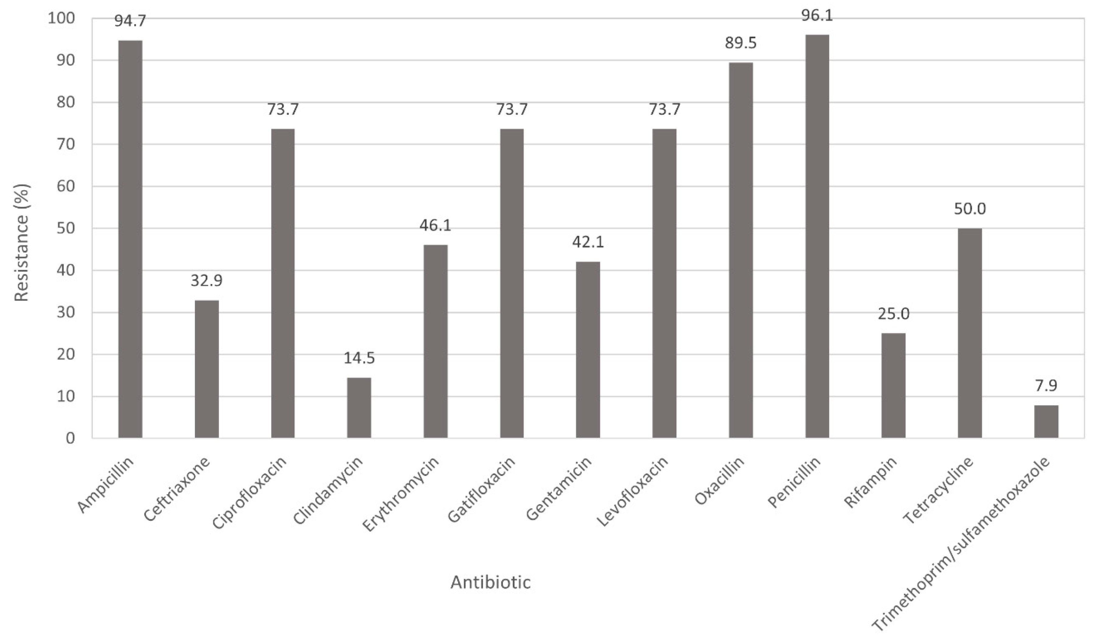
3.2. Antimicrobial Susceptibility Testing

Percent resistance of S. aureus isolates (n = 76) to the tested antibiotics is shown in Figure 1. Higher frequencies of antibiotic resistance were observed to ampicillin (94.7%; 72/76), oxacillin (89.5%; 68/76), ciprofloxacin (73.7%; 56/76), gatifloxacin (73.7%; 56/76) and levofloxacin (73.7%; 56/76). Moderate frequencies of antibiotic resistance ranging from 30% to 60% were found against tetracycline (50%), erythromycin (46.1%), gentamicin (42.1%) and ceftriaxone (32.9%). The lowest resistance observed was identified to rifampin (19; 25%), clindamycin (11; 14.5%) and trimethoprim/sulfamethoxazole (6; 7.9%). All isolates were susceptible to daptomycin, linezolid, high-level streptomycin, Q/D and vancomycin. Multidrug resistance (MDR; resistance to two or more antimicrobial classes) was observed with most common combinations of aminoglycoside/β-lactams/fluoroquinolones/macrolide-lincosamide-streptogramin/rifampin/tetracycline (Table 1). MDR patterns AmpCefCipCliEryGatGenLevOxaPenRifTet and AmpCefCipEryGatGenOxaPenRifTet contained eight MRSA isolates and one methicillin-susceptible S. aureus (MSSA) isolate for each pattern. One MRSA isolate was resistant to seven antibiotic classes (AmpCefCipCliEryGatGenLevOxaPenRifTetTri).

3.3. Frequency of Antibiotic Resistance Genes

Of the S. aureus isolates, 88.2% (67/76) were mecA+ (MRSA), while nine (11.8%) were negative for mecA and classified as MSSA. Isolates were also positive for aacA-aphD (34.2%), erm(A) (22.3%), erm(C) (15.8%), tet(K) (13.2%) and tet(M) (23.7%). No PCR positive results were found for the streptogramin resistance genes, vat(A), vat(B) and vat(C).

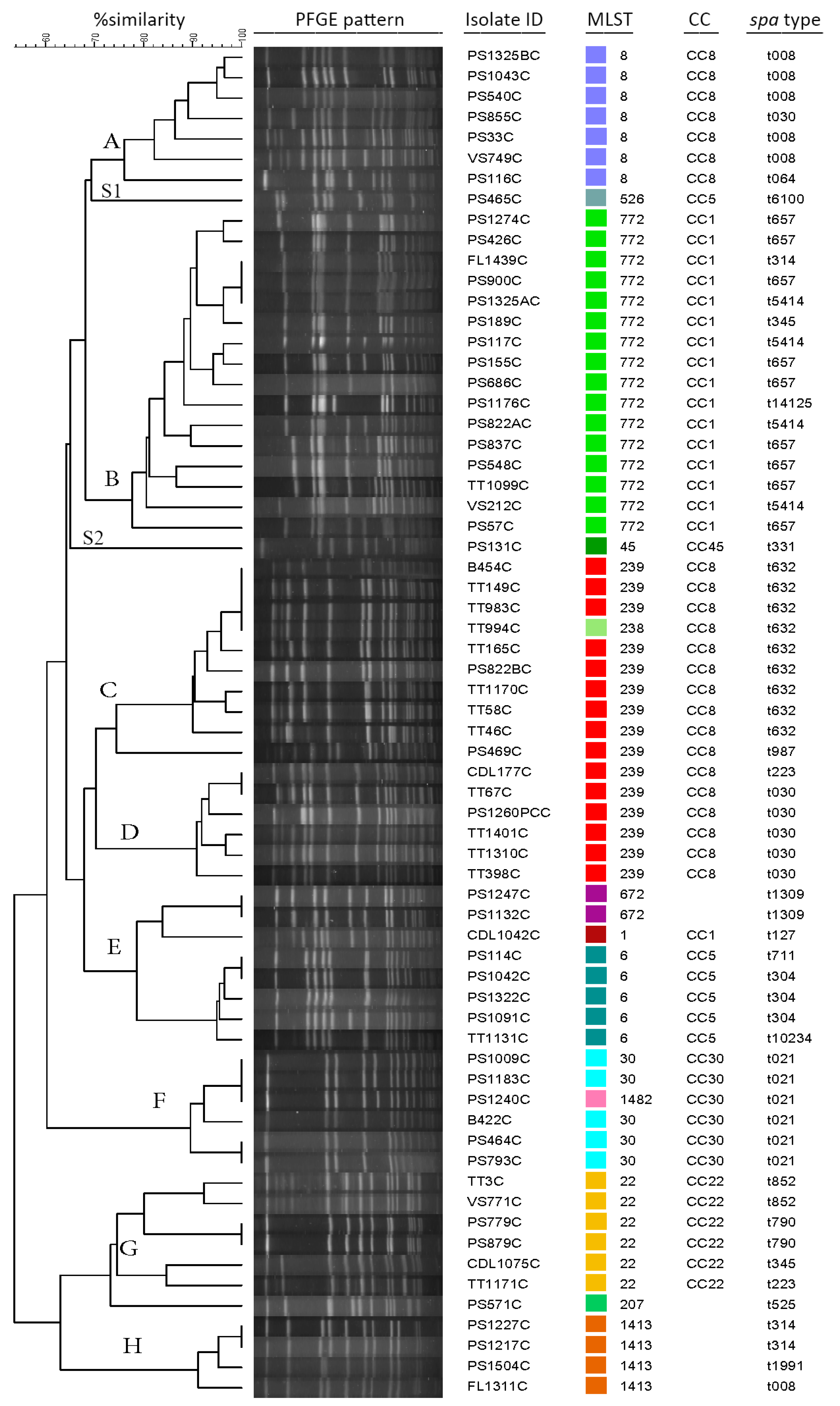
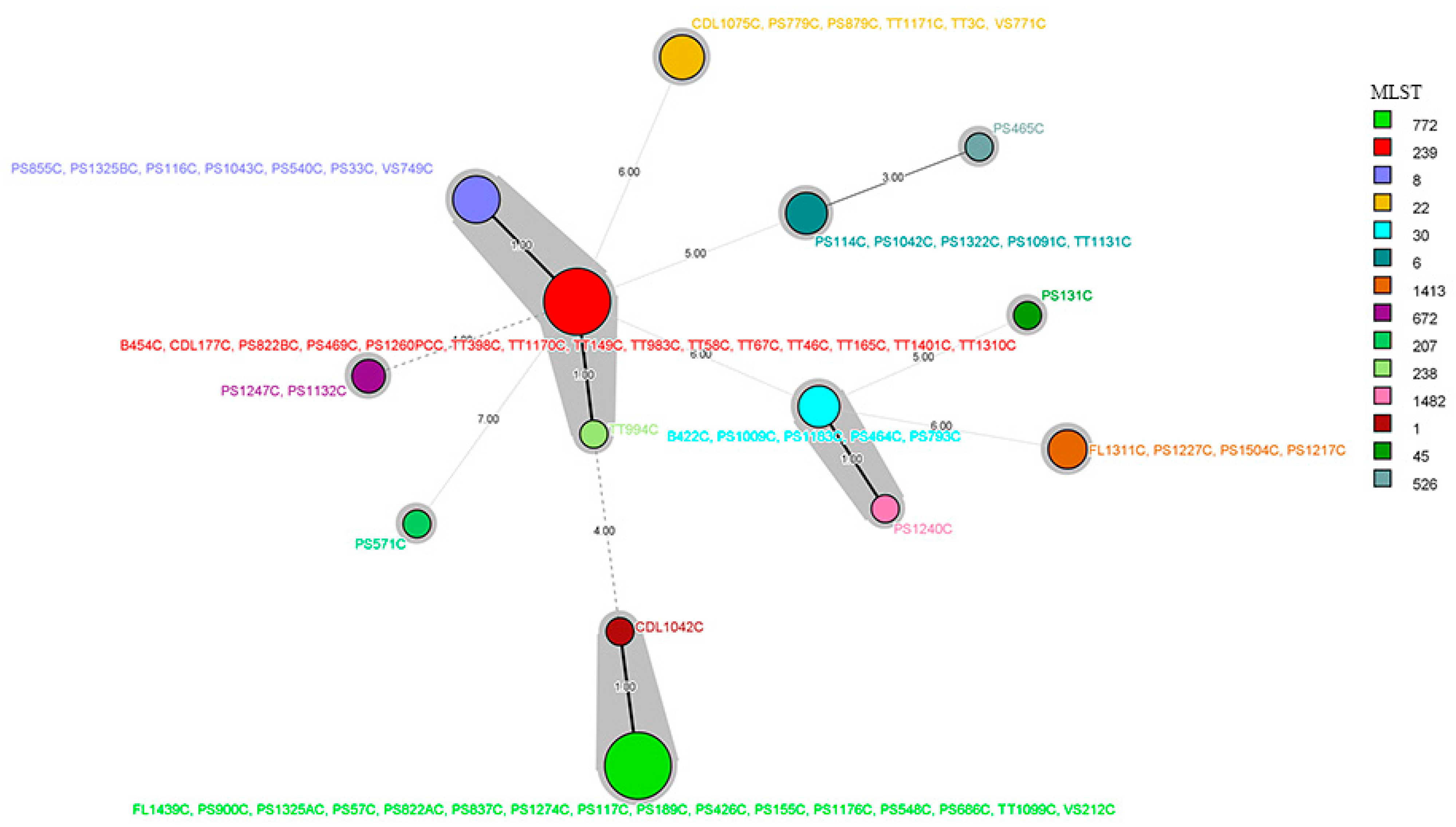
3.4. Molecular Analysis

PFGE was conducted on all but one MRSA isolate as the isolate did not produce a useable PFGE pattern. Results showed the clustering of MRSA isolates (n = 66) based on 75% similarity into eight clusters (A, B, C, D, E, F, G, H) and two singletons (S1, isolate PS465C and S2, isolate PS131C) (Figure 2). Two PFGE clusters encompassed approximately 40% (26/66) of the examined isolates; clusters B and C contained 16 and 10 isolates, respectively. Other isolates were distributed into six clusters as follows: eight isolates in cluster E, seven isolates each in clusters A and G, six isolates each in clusters D and F and four isolates in cluster H. spa types among the MRSA isolates were highly diverse as isolates were distributed among 23 different spa types (Figure 2). The majority of isolates belonged to spa type t632 and t657 (9/66; 13.6% each spa type). Over half of the spa types were represented by 1–2 isolates only. Using MLST, isolates were assigned into 14 different sequence types (ST772, ST239, ST8, ST22, ST6, ST30, ST1413, ST672, ST1, ST526, ST45, ST238, ST1482, ST207) (Figure 2 and Figure 3). Of these STs, ST772 and ST239 were the prevalent STSs that constituted 24.2% (16/66) and 22.7% (15/66) of isolates, respectively. ST1, ST45, ST526, ST238, ST1482 and ST207 were each represented by 1.5% (1/66) of isolates. Comparing the MLST to PFGE results, isolates grouped in the same PFGE cluster were assigned to the same ST. For example, all isolates in PFGE clusters A, B, D, and H were assigned to ST8, ST772, ST239, and ST1413, respectively. However, two different STs were identified in clusters C (ST239 and ST238), F (ST30 and ST1482), and G (ST22 and ST207) and three different STs in cluster E (ST6, ST672 and ST1). All MRSA isolates were distributed within six different CC types, CC8 (23; 34.8%), CC1 (17; 25.7%), CC5 (6; 9%), CC22 (6; 9.0%), CC30 (6; 9%) and CC45 (1; 1.5%) (Figure 2). CC8 was the most frequent clonal type followed by CC1. CC8 consisted of six different spa types including t632, one of the most prevalent spa types. CC8 also contained isolates belonging to sequence type ST239 (15; 22.7%), considered a pandemic strain, followed by CC1 with sixdifferent spa types. The most common clonal types were ST772-CC1-t657 (n = 9) followed by ST239-CC-8-t632 (n = 8), ST239-CC8-t030 (n = 5), ST8-CC8-t008 (n = 5) and ST30-CC30-t021 (n = 5). Clonal types of sequence type ST672, ST207 and ST1413 were unknown.

4. Discussion

Like other geographical regions around the globe, MRSA is a serious health concern in Pakistan [28]. MRSA emergence was reported for the first time in the 1960s in the United Kingdom and since then its prevalence has been increasing throughout the world [8]. In the present study, high prevalence of MRSA (88%) among the collected S. aureus was observed while only 12% isolates were MSSA. Although high prevalence of mecA in clinical S. aureus isolates has also been reported in Pakistan previously, this work is one of those reporting the highest frequency of MRSA [15,17,28]. For example, a study conducted by Siddiqui et al. on clinical isolates in Karachi reported 52% prevalence of MRSA among S. aureus isolates, while 68% of the isolates were MDR S. aureus [29]. A similar study conducted in Peshawar city of Pakistan on clinical isolates by Ullah et al. reported prevalence of 36% [28]. Furthermore, a recent study conducted at a tertiary care hospital of Islamabad city of Pakistan reported 65% prevalence of MRSA isolates; 83% of those MRSA isolates were MDR S. aureus [13]. However, it is important to note that MRSA prevalence data may differ from town to town and even between hospitals of the same city [16]. Therefore, additional studies will provide needed data to inform decisions in specific clinical settings and to increase positive outcomes with infections caused by MRSA. The results of this study are alarming, since the majority of the isolates were resistant to drugs commonly used to treat S. aureus infections. Inappropriate use of antibiotics in Pakistan is widespread as physicians prescribe the antibiotics without performing sensitivity tests [30]. Self-medication along with inappropriate dispensing of antibiotics among patients are some of the other weaknesses in the clinical health system in Pakistan that further enhance the prevalence of antibiotic resistance [31,32]. In contrast to the previous reported results of different studies [12,33,34], no vancomycin resistant S. aureus were identified in the present study. Vancomycin intermediate resistant and vancomycin resistant S. aureus (VISA and VRSA, respectively) are a serious public health concern. In Pakistan, VISA was reported for the first time in 2004 while VRSA was reported for the first time in 2009 [14,35]. Concern for emergence of VISA increased after the study conducted by Hakim et al. (2007) which reported an incidence rate of 13% of VISA isolates [33]. According to some of the studies conducted on usage of antibiotics in Pakistan, the most commonly used antibiotics are penicillin, cephalosporins, fluoroquinolones, aminoglycosides, ceftriaxone, cefradine, ciprofloxacin and tetracycline [32,36,37,38], which would account for the resistance patterns among MRSA observed in this study.
The hospital sampled in this study receives patient samples from different neighboring cities, which was reflected in the genetic diversity of the population structure of the isolated strains. In the present study, MRSA isolates exhibited 23 different spa types. Although a paucity of data is available on spa typing in Pakistan, approximately 10 spa types from this study have been previously reported in other studies. For example, Syed et al. (2018) reported spa type t657 in Pakistan from table egg samples [39] while spa types t632, t030, t021, t008, t5414, t314, t345, t127 and t064 detected in the present study have also been reported in Pakistan, primarily from clinical samples [13,39,40]. To the best of our knowledge, no previous record is available for Pakistan on most spa types identified for the first time in this study (t657, t304, t1309, t223, t852, t345, t790, t525, t331, t10234, t14125, t711, t1991 and t6100).
ST772-CC1-t657 or the Bengal Bay clone was one of the most identified clones in this study. This clone was first reported in Bangladesh and has spread to other locations due to travel to India or contact with travelers to that region [41,42]. It is known to be endemic in regions around the Bay of Bengal and resulted in the discovery of its first clinical case which was associated with surrounding regions [18,43]. This clone was first classified as HA-MRSA and caused health care issues in multiple countries as defined in other studies [12,28,41,44,45]. It is now also associated with community infections and has linked Pakistan with the Arabian Peninsula epidemiologically as described previously [18]. The diversity of MRSA clones in this study may also be due to travel or the expansion of clones within the hospital or community. Two other prevalent clones in this study, ST239-CC8-t632 and ST239-CC8-t030, have also been recently identified as dominant epidemic MRSA clones from clinical specimens from hospitals in China [46]. According to that study, ST239-t030 has been a dominant epidemic clone in China for the last 20 years, displacing the previous dominant clone, ST239-t037. ST239 was also once considered to be hospital-associated MRSA (HA-MRSA), but it is no longer confined to hospitals only and has been reported as a commonly circulating strain in Middle East and Gulf countries [17,18].
Like many of the MRSA clones detected in this study, ST30-CC30-t021 has been described in different regions of the world and from various sources. For example, it was one of four major MRSA clades detected in Argentina between 2004 and 2015 while also circulating among a veterinarian and a dog from the same veterinary clinic in Japan [47,48]. Recently, the USA300 pulsotype (ST8-CC8-t008) has been detected in studies of clinical MRSA isolates from Pakistan [17,18,49]. Some of these same clones from Pakistan show a close epidemiological association of Pakistan with Arab Gulf countries and the Middle East, as approximately 3.6 million Pakistanis are employed there [17]. The proportion of CC5 and CC22 in isolates from the present study was 9.0% each while CC30 was the fourth most abundant clonal complex found. This clonal complex was reported for the first time in the UK but has now disseminated to Australia, Belgium, Canada, Greece, Finland, Denmark, Ireland, Spain, Sweden and the US [50]. The CC30/ST30 clonal type was also previously reported in Pakistan [51]; six isolates in the present study were found to belong to this clonal type. However, other MRSA clones reported previously in studies conducted in Pakistan, such as CC509 and CC6, were not found in this study [17,18].

5. Conclusions

Antibiotic resistance remains a serious health concern globally as well as in local hospitals. The results of the current study present an alarming situation as frequency of MRSA was very high among the S. aureus isolated. The genetic fingerprinting results indicate that the MRSA population of the present study is diverse and comprised of several global and regional clones. Monitoring evolution and circulation of antimicrobial resistant bacterial clones in the clinical setting allows for: (1) making informed decisions in prescribing the most effective antimicrobial treatment therapy for patients and (2) forming strategies to develop effective control measures to limit dissemination of these clones.

Author Contributions

Conceptualization, M.A.S., B.J. and C.R.J.; methodology, M.A.S., B.J., C.R.J., M.R., S.A. and S.A.A.; validation, M.R., S.A., S.A.A., H.R. and T.A.W.; formal analysis, M.R., S.A., S.A.A., H.R. and T.A.W.; investigation, H.R. and T.A.W.; resources, M.A.S., B.J. and C.R.J.; data curation, H.R. and T.A.W.; writing—original draft preparation, M.A.S., B.J. and M.R.; writing—review and editing, H.R. and C.R.J.; visualization, M.A.S. and B.J.; supervision, M.A.S., B.J. and C.R.J.; project administration, M.A.S., B.J. and C.R.J.; funding acquisition, M.A.S., B.J. and C.R.J. All authors have read and agreed to the published version of the manuscript.

Funding

This research was partially funded by the US Department of Agriculture-Agricultural Research Service (USDA-ARS) Project No. 6040-32000-009-00D.

Institutional Review Board Statement

The study was approved by Advance Study and Research Board of the University of Haripur. Ethical review and approval were waived for this study, as the study did not require IRB approval since it did not involve experimentation on humans and the identity of the subjects whose samples were collected was kept confidential.

Informed Consent Statement

Informed consent was obtained from all subjects involved in the study. Each subject had the right to withdraw from participation at any time during the study. However, the identity of subjects whose clinical samples were used in the study was kept confidential.

Data Availability Statement

Data is contained within the article.

Acknowledgments

The authors are grateful to all participating institutions for providing infrastructure to conduct this research. The mention of trade names or commercial products in this article is solely for the purpose of providing specific information and does not imply recommendation or endorsement by the US Department of Agriculture. The US Department of Agriculture is an equal opportunity provider and employer.

Conflicts of Interest

The authors declare no conflict of interest.

References

  1. Gould, D.; Chamberlaine, A. Staphylococcus aureus: A review of the literature. J. Clin. Nurs. 1995, 4, 5–12. [Google Scholar] [CrossRef] [PubMed]
  2. Wertheim, H.F.; Melles, D.C.; Vos, M.C.; van Leeuwen, W.; van Belkum, A.; Verbrugh, H.A.; Nouwen, J.L. The role of nasal carriage in Staphylococcus aureus infections. Lancet Infect. Dis. 2005, 5, 751–762. [Google Scholar] [CrossRef]
  3. Williams, R.E.; Jevons, M.P.; Shooter, R.A.; Hunter, C.J.; Girling, J.A.; Griffiths, J.D.; Taylor, G.W. Nasal staphylococci and sepsis in hospital patients. Br. Med. J. 1959, 2, 658–662. [Google Scholar] [CrossRef] [PubMed] [Green Version]
  4. Omuse, G.; Van Zyl, K.N.; Hoek, K.; Abdulgader, S.; Kariuki, S.; Whitelaw, A.; Revathi, G. Molecular characterization of Staphylococcus aureus isolates from various healthcare institutions in Nairobi, Kenya: A cross sectional study. Ann. Clin. Microbiol. Antimicrob. 2016, 15, 51. [Google Scholar] [CrossRef] [Green Version]
  5. Choudhury, R.; Panda, S.; Singh, D.V. Emergence and dissemination of antibiotic resistance: A global problem. Indian J. Med. Microbiol. 2012, 30, 384–390. [Google Scholar] [CrossRef]
  6. Sergelidis, D.; Angelidis, A.S. Methicillin-resistant Staphylococcus aureus: A controversial food-borne pathogen. Lett. Appl. Microbiol. 2017, 64, 409–418. [Google Scholar] [CrossRef] [PubMed] [Green Version]
  7. McGuinness, W.A.; Malachowa, N.; DeLeo, F.R. Vancomycin resistance in Staphylococcus aureus. Yale J. Biol. Med. 2017, 90, 269–281. [Google Scholar]
  8. Schentag, J.J.; Hyatt, J.M.; Carr, J.R.; Paladino, J.A.; Birmingham, M.C.; Zimmer, G.S.; Cumbo, T.J. Genesis of methicillin-resistant Staphylococcus aureus (MRSA), how treatment of MRSA infections has selected for vancomycin-resistant Enterococcus faecium, and the importance of antibiotic management and infection control. Clin. Infect. Dis. 1998, 26, 1204–1214. [Google Scholar] [CrossRef] [Green Version]
  9. Cosgrove, S.E.; Qi, Y.; Kaye, K.S.; Harbarth, S.; Karchmer, A.W.; Carmeli, Y. The impact of methicillin resistance in Staphylococcus aureus bacteremia on patient outcomes: Mortality, length of stay, and hospital charges. Infect. Control. Hosp. Epidemiol. 2005, 26, 166–174. [Google Scholar] [CrossRef]
  10. DeLeo, F.R.; Otto, M.; Kreiswirth, B.N.; Chambers, H.F. Community-associated meticillin-resistant Staphylococcus aureus. Lancet 2010, 375, 1557–1568. [Google Scholar] [CrossRef] [Green Version]
  11. Stapleton, P.D.; Taylor, P.W. Methicillin resistance in Staphylococcus aureus: Mechanisms and modulation. Sci. Prog. 2002, 85 Pt 1, 57–72. [Google Scholar] [CrossRef]
  12. Rukan, M.; Jamil, H.; Bokhari, H.A.; Khattak, A.A.; Khan, A.N.; Ullah, Z.; Hussain, S.; Syed, M.A. Nasal carriage of highly resistant methicillin resistant Staphylococcus aureus (MRSA) strains by hospital staff in Hazara region of Pakistan. J. Pak. Med. Assoc. 2021, 71, 47–50. [Google Scholar] [PubMed]
  13. Khan, A.A.; Ali, A.; Tharmalingam, N.; Mylonakis, E.; Zahra, R. First report of mecC gene in clinical methicillin resistant S. aureus (MRSA) from tertiary care hospital Islamabad, Pakistan. J. Infect. Public Health 2020, 13, 1501–1507. [Google Scholar] [CrossRef]
  14. Taj, Y.; Abdullah, F.E.; Kazmi, S.U. Current pattern of antibiotic resistance in Staphylococcus aureus clinical isolates and the emergence of vancomycin resistance. J. Coll. Physicians Surg. Pak. 2010, 20, 728–732. [Google Scholar]
  15. Monecke, S.; Coombs, G.; Shore, A.C.; Coleman, D.C.; Akpaka, P.; Borg, M.; Chow, H.; Ip, M.; Jatzwauk, L.; Jonas, D.; et al. A field guide to pandemic, epidemic and sporadic clones of methicillin-resistant Staphylococcus aureus. PLoS ONE 2011, 6, e17936. [Google Scholar] [CrossRef] [PubMed]
  16. Bukhari, S.Z.; Ahmed, S. Prevalence of meticillin resistance among Staphylococcus aureus isolates in Pakistan and its clinical outcome. J. Hosp. Infect. 2007, 67, 101–102. [Google Scholar] [CrossRef]
  17. Jamil, B.; Gawlik, D.; Syed, M.A.; Shah, A.A.; Abbasi, S.A.; Muller, E.; Reissig, A.; Ehricht, R.; Monecke, S. Hospital-acquired methicillin-resistant Staphylococcus aureus (MRSA) from Pakistan: Molecular characterisation by microarray technology. Eur. J. Clin. Microbiol. Infect. Dis. 2018, 37, 691–700. [Google Scholar] [CrossRef] [PubMed]
  18. Madzgalla, S.; Syed, M.A.; Khan, M.A.; Rehman, S.S.; Muller, E.; Reissig, A.; Ehricht, R.; Monecke, S. Molecular characterization of Staphylococcus aureus isolates causing skin and soft tissue infections in patients from Malakand, Pakistan. Eur. J. Clin. Microbiol. Infect. Dis. 2016, 35, 1541–1547. [Google Scholar] [CrossRef]
  19. Poulsen, A.B.; Skov, R.; Pallesen, L.V. Detection of methicillin resistance in coagulase-negative staphylococci and in staphylococci directly from simulated blood cultures using the EVIGENE MRSA Detection Kit. J. Antimicrob. Chemother. 2003, 51, 419–421. [Google Scholar] [CrossRef] [Green Version]
  20. CLSI. Performance Standards for Antimicrobial Susceptibility Testing, 30th ed.; Volume CLSI Supplement M100; Clinical and Laboratory Standards Institute: Wayne, PA, USA, 2020. [Google Scholar]
  21. CLSI. Performance Standards for Antimicrobial Susceptibility Testing: Seventeenth Informational Supplement, 28th ed.; Volume CLSI Supplement M100-S17 No. 1; Clinical and Laboratory Standards Institute: Wayne, PA, USA, 2007. [Google Scholar]
  22. Strommenger, B.; Kettlitz, C.; Werner, G.; Witte, W. Multiplex PCR assay for simultaneous detection of nine clinically relevant antibiotic resistance genes in Staphylococcus aureus. J. Clin. Microbiol. 2003, 41, 4089–4094. [Google Scholar] [CrossRef] [Green Version]
  23. McDougal, L.K.; Steward, C.D.; Killgore, G.E.; Chaitram, J.M.; McAllister, S.K.; Tenover, F.C. Pulsed-field gel electrophoresis typing of oxacillin-resistant Staphylococcus aureus isolates from the United States: Establishing a national database. J. Clin. Microbiol. 2003, 41, 5113–5120. [Google Scholar] [CrossRef] [Green Version]
  24. Oliveira, D.C.; de Lencastre, H. Multiplex PCR strategy for rapid identification of structural types and variants of the mec element in methicillin-resistant Staphylococcus aureus. Antimicrob. Agents. Chemother. 2002, 46, 2155–2161. [Google Scholar] [CrossRef] [PubMed] [Green Version]
  25. Strommenger, B.; Braulke, C.; Heuck, D.; Schmidt, C.; Pasemann, B.; Nubel, U.; Witte, W. spa Typing of Staphylococcus aureus as a frontline tool in epidemiological typing. J. Clin. Microbiol. 2008, 46, 574–581. [Google Scholar] [CrossRef] [PubMed] [Green Version]
  26. Harmsen, D.; Claus, H.; Witte, W.; Rothganger, J.; Claus, H.; Turnwald, D.; Vogel, U. Typing of methicillin-resistant Staphylococcus aureus in a university hospital setting by using novel software for spa repeat determination and database management. J. Clin. Microbiol. 2003, 41, 5442–5448. [Google Scholar] [CrossRef] [Green Version]
  27. Enright, M.C.; Day, N.P.; Davies, C.E.; Peacock, S.J.; Spratt, B.G. Multilocus sequence typing for characterization of methicillin-resistant and methicillin-susceptible clones of Staphylococcus aureus. J. Clin. Microbiol. 2000, 38, 1008–1015. [Google Scholar] [CrossRef] [PubMed] [Green Version]
  28. Ullah, A.; Qasim, M.; Rahman, H.; Khan, J.; Haroon, M.; Muhammad, N.; Khan, A.; Muhammad, N. High frequency of methicillin-resistant Staphylococcus aureus in Peshawar Region of Pakistan. Springerplus 2016, 5, 600. [Google Scholar] [CrossRef] [PubMed] [Green Version]
  29. Siddiqui, T.; Muhammad, I.N.; Khan, M.N.; Naz, S.; Bashir, L.; Sarosh, N.; Masood, R.; Ali, A.; Fatima, S.; Naqvi, T. MRSA: Prevalence and susceptibility pattern in health care setups of Karachi. Pak. J. Pharm. Sci. 2017, 30 (Suppl. S6), 2417–2421. [Google Scholar]
  30. Atif, M.; Azeem, M.; Saqib, A.; Scahill, S. Investigation of antimicrobial use at a tertiary care hospital in Southern Punjab, Pakistan using WHO methodology. Antimicrob. Resist. Infect. Control. 2017, 6, 41. [Google Scholar] [CrossRef] [Green Version]
  31. Hanif, A.; Ashar, S.M.; Rabnawaz, R.; Yasmeen, S. Self-medication of antibiotics among the students of Hamdard University, Pakistan. J. Public Health Dev. Ctries 2016, 2, 145–148. [Google Scholar]
  32. Asghar, S.; Atif, M.; Mushtaq, I.; Malik, I.; Hayat, K.; Babar, Z.U. Factors associated with inappropriate dispensing of antibiotics among non-pharmacist pharmacy workers. Res. Social Adm Pharm 2020, 16, 805–811. [Google Scholar] [CrossRef] [PubMed]
  33. Hakim, S.T.; Arshed, S.; Iqbal, M.; Javaid, S. Vancomycin sensitivity of Staphylococcus aureus isolates from hospital patients in Karachi, Pakistan. Libyan J. Med. 2007, 2, 176–179. [Google Scholar] [CrossRef]
  34. Brohi, N.A.; Noor, A.A. Frequency of the occurrence of Methicillin-resistant Staphylococcus aureus (MRSA) infections in Hyderabad, Pakistan. Pak. J. Anal. Environ. Chem. 2017, 18, 84–90. [Google Scholar] [CrossRef]
  35. Bukhari, S.Z.; Ahmed, S.; Zia, N. Antimicrobial susceptibility pattern of Staphylococcus aureus on clinical isolates and efficacy of laboratory tests to diagnose MRSA: A multi-centre study. J. Ayub Med. Coll. Abbottabad 2011, 23, 139–142. [Google Scholar]
  36. Khan, Z.; Ahmed, N.; Rehman, A.; Khan, F.U.; Karatas, Y. Utilization pattern of antibiotics and patient care indicators in the teaching hospitals, Islamabad, Pakistan. SN Comp. Clin. Med. 2019, 1, 812–816. [Google Scholar] [CrossRef] [Green Version]
  37. Atif, M.; Azeem, M.; Sarwar, M.R.; Shahid, S.; Javaid, S.; Ikram, H.; Baig, U.; Scahill, S. WHO/INRUD prescribing indicators and prescribing trends of antibiotics in the Accident and Emergency Department of Bahawal Victoria Hospital, Pakistan. Springerplus 2016, 5, 1928. [Google Scholar] [CrossRef] [PubMed] [Green Version]
  38. Khan, S.; Shehzad, A.; Shehzad, O.; Al-Suhaimi, E.A. Inpatient antibiotics pharmacology and physiological use in Hayatabad medical complex, Pakistan. Int. J. Physiol. Pathophysiol. Pharmacol. 2013, 5, 120–127. [Google Scholar] [PubMed]
  39. Syed, M.A.; Shah, S.H.H.; Sherafzal, Y.; Shafi-Ur-Rehman, S.; Khan, M.A.; Barrett, J.B.; Woodley, T.A.; Jamil, B.; Abbasi, S.A.; Jackson, C.R. Detection and molecular characterization of methicillin-resistant Staphylococcus aureus from table eggs in Haripur, Pakistan. Foodborne Pathog. Dis. 2018, 15, 86–93. [Google Scholar] [CrossRef] [PubMed]
  40. Arfat, Y.; Johnson, M.; Malik, S.A.; Morrisey, J.A.; Bayliss, C.D. Epidemiology of methicillin-resistant Staphylococcus aureus (MRSA) isolates from Pakistan. African J. Microbiol. Res. 2013, 7, 568–576. [Google Scholar]
  41. Brennan, G.I.; Shore, A.C.; Corcoran, S.; Tecklenborg, S.; Coleman, D.C.; O’Connell, B. Emergence of hospital- and community-associated panton-valentine leukocidin-positive methicillin-resistant Staphylococcus aureus genotype ST772-MRSA-V in Ireland and detailed investigation of an ST772-MRSA-V cluster in a neonatal intensive care unit. J. Clin. Microbiol. 2012, 50, 841–847. [Google Scholar] [CrossRef] [Green Version]
  42. Rajan, V.; Schoenfelder, S.M.; Ziebuhr, W.; Gopal, S. Genotyping of community-associated methicillin resistant Staphylococcus aureus (CA-MRSA) in a tertiary care centre in Mysore, South India: ST2371-SCCmec IV emerges as the major clone. Infect. Genet. Evol. 2015, 34, 230–235. [Google Scholar] [CrossRef]
  43. Prabhakara, S.; Khedkar, S.; Loganathan, R.M.; Chandana, S.; Gowda, M.; Arakere, G.; Seshasayee, A.S. Draft genome sequence of Staphylococcus aureus 118 (ST772), a major disease clone from India. J. Bacteriol. 2012, 194, 3727–3728. [Google Scholar] [CrossRef] [Green Version]
  44. D’Souza, N.; Rodrigues, C.; Mehta, A. Molecular characterization of methicillin-resistant Staphylococcus aureus with emergence of epidemic clones of sequence type (ST) 22 and ST 772 in Mumbai, India. J. Clin. Microbiol. 2010, 48, 1806–1811. [Google Scholar] [CrossRef] [Green Version]
  45. Ho, W.Y.; Choo, Q.C.; Chew, C.H. Predominance of three closely related methicillin-resistant Staphylococcus aureus clones carrying a unique ccrC-positive SCCmec type III and the emergence of spa t304 and t690 SCCmec type IV pvl(+) MRSA isolates in Kinta Valley, Malaysia. Microb. Drug. Resist. 2017, 23, 215–223. [Google Scholar] [CrossRef]
  46. Tang, Y.T.; Cao, R.; Xiao, N.; Li, Z.S.; Wang, R.; Zou, J.M.; Pei, J. Molecular epidemiology and antimicrobial susceptibility of methicillin-resistant Staphylococcus aureus isolates in Xiangyang, China. J. Glob. Antimicrob. Resist. 2018, 12, 31–36. [Google Scholar] [CrossRef] [PubMed]
  47. Di Gregorio, S.; Haim, M.S.; Vielma Vallenilla, J.; Cohen, V.; Rago, L.; Gulone, L.; Aanensen, D.M.; Argimon, S.; Mollerach, M. Genomic epidemiology of CC30 methicillin-resistant Staphylococcus aureus strains from Argentina reveals four major clades with distinctive genetic features. mSphere 2021, 6, e01297-20. [Google Scholar] [CrossRef]
  48. Ishihara, K.; Saito, M.; Shimokubo, N.; Muramatsu, Y.; Maetani, S.; Tamura, Y. Methicillin-resistant Staphylococcus aureus carriage among veterinary staff and dogs in private veterinary clinics in Hokkaido, Japan. Microbiol. Immunol. 2014, 58, 149–154. [Google Scholar] [CrossRef]
  49. Monecke, S.; Syed, M.A.; Khan, M.A.; Ahmed, S.; Tabassum, S.; Gawlik, D.; Muller, E.; Reissig, A.; Braun, S.D.; Ehricht, R. Genotyping of methicillin-resistant Staphylococcus aureus from sepsis patients in Pakistan and detection of antibodies against staphylococcal virulence factors. Eur. J. Clin. Microbiol. Infect. Dis. 2020, 39, 85–92. [Google Scholar] [CrossRef] [PubMed]
  50. Deurenberg, R.H.; Vink, C.; Kalenic, S.; Friedrich, A.W.; Bruggeman, C.A.; Stobberingh, E.E. The molecular evolution of methicillin-resistant Staphylococcus aureus. Clin. Microbiol. Infect. 2007, 13, 222–235. [Google Scholar] [CrossRef] [PubMed] [Green Version]
  51. Shabir, S.; Hardy, K.J.; Abbasi, W.S.; McMurray, C.L.; Malik, S.A.; Wattal, C.; Hawkey, P.M. Epidemiological typing of meticillin-resistant Staphylococcus aureus isolates from Pakistan and India. J. Med. Microbiol. 2010, 59 Pt 3, 330–337. [Google Scholar] [CrossRef] [Green Version]
Figure 1. Percent resistance of Staphylococcus aureus (n = 76) to tested antimicrobials using broth microdilution. No resistance to daptomycin, linezolid, high-level streptomycin, Quinupristin/Dalfopristin, tetracycline, or vancomycin was detected.
Microorganisms 09 02301 g001
Figure 2. Pulsed-field Gel Electrophoresis (PFGE) analysis and genetic profiles (Multilocus Sequence Typing (MLST), clonal complex (CC), and spa types) of MRSA from clinical samples. DNA for PFGE was digested with SmaI. Levels of similarity were determined using Dice coefficient and the unweighted pair group method (UPGMA). PFGE clusters were determined using a 75% similarity cutoff and labeled A–H. Sample source Isolate ID: B = blood, CDL = cannula double lumen, FL = fluid. TT = tracheal tube, VS = vaginal swab.
Microorganisms 09 02301 g002
Figure 3. Minimum spanning tree (MST) of Multilocus Sequence Typing (MLST) of the examined clinical Staphylococcus aureus from humans. Each circle corresponds to an individual MLST type; the circle size is proportional to the number of isolates belonging to the same MLST type. The color of each circle denotes the MLST type. Connecting lines (solid and dashed) between circles represents allelic variations between MLST types; the grey shadowing indicates no more than one different loci between MLST types.
Microorganisms 09 02301 g003
Table 1. Multidrug resistance patterns among clinical Staphylococcus aureus.
No. IsolatesNo. Antibiotic Classes
Resistance Pattern aMRSAMSSA
AmpCefCipCliEryGatGenLevOxaPenRifTetTri107
AmpCefCipCliEryGatGenLevOxaPenRifTet816
AmpCefCipEryGatGenLevOxaPenTetTri016
AmpCefCipEryGatGenOxaPenRifTet816
AmpCefCipEryGatLevOxaPenTet104
AmpCefCipEryGatLevOxaPen103
AmpCefCipGatGenLevOxaPen203
AmpCefCipGatLevOxaPen102
AmpCipCliEryGatGenLevOxaPen104
AmpCipEryGatGenLevOxaPen014
AmpCipEryGatLevOxaPen503
AmpCipEryGatLevOxaPenTet304
AmpCipEryGatLevPenTet024
AmpCipGatGenLevOxaPen603
AmpCipGatGenLevOxaPenTet104
AmpCipGatGenLevOxaPenTri104
AmpCipGatLevOxaPenTet203
AmpCipGatLevOxaPen702
AmpEryOxaPen102
AmpOxaPen801
AmpOxaPenTetTri103
AmpOxaPenTet402
AmpPenTet212
AmpPen101
CipGatLevOxaPen102
CipGatLevOxa012
Pan-susceptible110
a Amp, ampicillin; Cef, ceftriaxone; Cip, ciprofloxacin; Cli, clindamycin; Ery, erythromycin; Gat, gatifloxacin; Gen, gentamicin; Lev, levofloxacin; Oxa, oxacillin; Pen, penicillin; Rif, rifampin; Tet, tetracycline; Tri. Trimethoprim/sulfamethoxazole.
Publisher’s Note: MDPI stays neutral with regard to jurisdictional claims in published maps and institutional affiliations.

