Article

Flexibility of Microcystis Overwintering Strategy in Response to Winter Temperatures

1
Key Laboratory of Algal Biology, Institute of Hydrobiology, Chinese Academy of Sciences, Wuhan 430072, China
2
University of Chinese Academy of Sciences, Beijing 100049, China
3
Research Academy of Plateau Lake Dianchi, Kunming 671500, China
*
Author to whom correspondence should be addressed.
Microorganisms 2021, 9(11), 2278; https://doi.org/10.3390/microorganisms9112278
Submission received: 27 September 2021 / Revised: 28 October 2021 / Accepted: 28 October 2021 / Published: 1 November 2021
(This article belongs to the Section Environmental Microbiology)

Abstract

:
Microcystis is one of the most common bloom-forming cyanobacteria in freshwater ecosystems throughout the world. However, the underlying life history mechanism and distinct temporal dynamics (inter- and intra-annual) of Microcystis populations in different geographical locations and lakes remain unclear but is critical information needed for the development of robust prediction, prevention, and management strategies. Perennial observations indicate that temperature may be the key factor driving differences in the overwintering strategy. This study quantitatively compared the overwintering abilities of Microcystis aeruginosa (Ma) in both the water column and sediments under a gradient of overwintering water temperatures (i.e., 4, 8, and 12 °C) using the death and proliferation rates of Ma. The results show that the dynamics of the Microcystis overwintering strategy were significantly affected by water temperatures. At 4 and 8 °C, Ma mainly overwintered in sediments and disappeared from the water column after exposure to low temperatures for a long duration, although some Microcystis cells can overwinter in the water column for short durations at low temperatures. At 12 °C, most Ma can overwinter in the water column. Rising temperatures promoted the proliferation of pelagic Ma but accelerated the death of benthic Ma. With warmer winter temperatures, pelagic Microcystis might become the primary inoculum sources in the spring. Our study highlights the overwintering strategy flexibility in explaining temporal dynamics differences of Microcystis among in geographical locations and should be considered in the context of global warming.

1. Introduction

Cyanobacterial blooms appear to be increasing in frequency, intensity, and duration due to global warming and anthropogenic activities, and they threaten human and ecosystem health, as well as ecosystem services [1]. Microcystis is the most frequently reported bloom-forming cyanobacteria in freshwater ecosystems, and certain Microcystis species can produce potent toxins [2,3]. Microcystis is ecologically characterized by an annual life cycle comprised of four phenological stages: overwintering, reinvasion, pelagic growth, and sinking out [4,5,6]. In eutrophic lakes, large amounts of Microcystis cyanobacterium may sink to the sediment layer during mid-summer and autumn and subsequently survive on the bottom of the lake [7]. The accumulated biomass of Microcystis colonies in the sediment could be considered a “seed-bank,” potentially serving as an inoculum source for pelagic blooms after reinvading the water column, thereby contributing to the ecological success and bloom formation of this cyanobacterium in freshwater ecosystems [8,9]. Several bloom mitigation methods have been proposed to eliminate overwintering Microcystis populations that are deemed cost-effective [10]. However, the typical four stage life cycle may not in actuality be in accordance with the inter- and intra-annual bloom dynamics of Microcystis populations in Chinese lakes.
In China, three large shallow lakes—Lake Taihu, Lake Chaohu, and Lake Dianchi—have suffered from severe Microcystis blooms for decades. Previous studies found that the low winter temperatures of Lake Taihu (4.8–7.3 °C) [11] and Lake Chaohu (3.3–6.3 °C) [12] lead to the senescence and cell death of pelagic Microcystis but provide optimal conditions for Microcystis to overwinter in the sediment [8,13]. However, winter bloom events of Microcystis spp. were also observed in Lake Taihu during winters when water temperatures were below 10 °C [14]. Microcystis blooms in Lake Dianchi have become a year-round occurrence, with a minimum abundance of 1.3 × 10⁷ cells/L in the winter [15]. It has been clearly demonstrated that Microcystis could overwinter, and even bloom, in the water column of Chinese lakes, suggesting that the four-stage life cycle theory does not accurately explain the bloom process of Microcystis in these subtropical lakes. In the context of global warming and the expansion of Microcystis blooms, overwintering in sediment may not be necessary for Microcystis perennial succession, while overwintering, and even blooming, in the water column may be more common. If so, Microcystis blooms could become more plaguing and pose an even greater threat to ecosystem health. Therefore, more systematic, and quantitative research on the overwintering patterns of Microcystis populations in Chinese lakes and changes in these patterns in response to global climate change are needed.
When overwintering in sediments, the proliferation rate of Microcystis can decrease to nearly zero, in which case the death rate is used to determine if the Microcystis population density could remain stable. Many researchers have reported that Microcystis can survive in sediment for long time periods (>6 years) under low temperatures [16]. Our previous research also revealed that most Microcystis spp. remained viable after overwintering at 5 °C in the sediment for over 4 months and could grow after reinvasion of the water column [17]. However, quantitative research concerning the proliferation and death rates of Microcystis in the water column at low temperatures are relatively sparse. Yang et al. [18] found that the viability of Microcystis aeruginosa showed little decrease under chill-light stress (5 °C, 100 μmol photons m−2 s−1) within 10 days after preconditioning at 15 °C for 2 days. However, their research mainly focused on the molecular mechanisms involved in the adaptation of Microcystis to chill-light conditions. As such, more research is needed to quantify the proliferation and death rates of Microcystis overwintering in the water column.
In this study, the growth and survival dynamics of Ma were determined under different culture conditions and overwintering growth capabilities were evaluated. Results yielded possible explanations for the overwintering strategies employed by benthic and pelagic Microcystis spp. in three shallow lakes in China and potential ramifications in relation to global warming. In this study, fluorescence staining followed by flow cytometry (FCM) was used to directly quantify the proliferation and death rates of Microcystis cells. To quantify the proliferation capacity of Microcystis cells, the intracellular protein dye carboxin fluorescein diacetate succinimide ester (CFSE) was used in conjunction with flow cytometry [19,20]. The fluorescent nucleic acid dye, SYTOX Green, was used to quantify damaged cells and indirectly determine the death rate of Microcystis cells [21].

2. Materials and Methods

2.1. Cyanobacteria Cultures

Microcystis aeruginosa NIES-843 (Ma) was obtained from the Freshwater Algae Culture Collection at the Institute of Hydrobiology, Chinese Academy of Sciences (FACHB-Collection; Wuhan, China). Cells were cultivated in BG11 medium at 25 ± 1 °C and illuminated by white cool fluorescent light lamps (40 μmol photons m−2 s−1) with a 12:12 h light/dark cycle. The cultures were manually shaken 3 or 4 times each day during incubation. Log-phase cells were centrifuged and resuspended in BG11 medium, and the initial cell density of the experimental cultures was adjusted to 4 × 10⁶ cells/mL.

2.2. Experimental Design

Microcystis cultures were divided in two, one which was incubated with the fluorochrome CFSE for the detection of cell division, and the other was used for evaluating cell viability and photosynthetic efficiency. The cultures, with or without CFSE, were inoculated into 12-mL sterilized culture tubes containing 10 mL of microalgae cultures. The culture tubes were placed in incubators kept at 4, 8, and 12 °C, respectively. To simulate the luminous environment of the water column and sediment, half of the microalgal cultures were illuminated with 40 μmol photons m−2 s−1 under a 12:12 h light/dark cycle, serving as the light group, and the other half were covered with tinfoil to serve as the dark group. Microalgal cultures incubated at 25 ± 1 °C and illuminated by white cool fluorescent light lamps (40 μmol photons m−2 s−1) were used as controls in all experiments. Three tubes of microalgae cultures were randomly sampled weekly from each treatment group. About 1 mL of microalgae cultures with CFSE stain were used for FCM analysis, and 9 mL was returned to the standard growth conditions (25 ± 1 °C, 40 μmol photons m−2 s−1, under a 12:12 h light/dark cycle) for one week to resume growth.

2.3. Flow Cytometric Analysis of Microalgal Cells

Flow cytometric analyses of Microcystis cells were performed on a CytoFLEX flow cytometer (Beckman Coulter Inc., Fullerton, CA, USA) equipped with an argon-ion excitation laser (488 nm), forward (FS) and side (SS) light scatter detectors, and four fluorescence detectors corresponding to four different wavelength intervals: 505–550, 550–600, 600–645, and >645 nm. From the start of the experiments, samples were taken weekly from each culture for the different cytometric analyses. Fluorescence of chlorophyll a (>645 nm) was used as a FCM gate to exclude non-microalgal particles. At least 30,000 cells per culture were analyzed.

2.3.1. Cell Division

When incubated with cells, CFSE crosses the cell membrane and binds to the abundant amine groups present in the cytoplasm, after which CFSE acquires identical spectral characteristics to fluorescein after removal of the acetate groups by endogenous intracellular esterases. After each cell division, the cytoplasmic CFSE fluorescence of the mother cell will be distributed equally between daughter cells, allowing analysis of population proliferation at the cellular level [22,23]. The log-phase Microcystis culture was stained with fluorochrome CFSE (Sigma-Aldrich, Louis, MO, USA) according the to the methods of Rioboo et al. [20] and Zhou et al. [24]. A CFSE stock solution was prepared by dissolving CFSE powder in DMSO (Sigma-Aldrich) to a final concentration of 10 mM. Next, 130 mL of log-phase Microcystis culture (9 × 10⁶ cells/mL) was incubated with 65 μL of CFSE (10 mM) at a final concentration of 5 μM per 6.8 × 10⁶ cells ml−1, in BG11 medium for 1 h at 20 °C in the dark. The CFSE-labeled culture was diluted five times with BG11 medium and kept in a culture room for 1 day to allow the cells to recover from the treatments. Cultures without CFSE that cultured under the above conditions were used as controls. Fluorescence of fluorescein was analyzed by the FITC detector (505–550 nm) of the flow cytometer.

2.3.2. Growth Measurement

The cell density of Microcystis cultures was measured by FCM. Due to the presence of chlorophyll and phycobiliproteins, Microcystis cells showed very strong natural fluorescence [25]. Autofluorescence corresponds to the signal gathered in the PerCP-A detector (660–700 nm), emitted by chlorophyll and related pigments after being excited at 488 nm. This autofluorescence was used to distinguish non-microalgal particles, thus, fluorescence of chlorophyll a (PerCP; 660–700 nm) was used as a gate to select Microcystis cells, which showed red fluorescence [26,27].

2.3.3. Cell Viability

The nucleic acid stain SYTOX-Green (Life Technologies, Carlsbad, CA, USA) has been used to distinguish dead and living cells in many studies [28]. Two microliters of SYTOX-Green were added to 1 mL of sample, which was then incubated for 15 min at room temperature in the dark. Fluorescence was analyzed by flow cytometry with excitation and emission wavelengths of 488 nm and 523 nm, respectively. When a cell dies, the integrity of the cell membrane declines, allowing SYTOX Green to easily penetrate cells and bind with DNA to produce a bright green, fluorescent complex which can be analyzed by the FITC detector (505–550 nm) of the flow cytometer [29]. The membrane-damaged cells and integrated membrane cells can be distinguished by differences in fluorescence.

2.3.4. Cell Size, Complexity, and Autofluorescence

The FCM is also equipped with forward (FS) and side (SS) light scatter detectors. Both have been widely applied to measure the cell morphology of phytoplankton [30]. As forward light scatter increases linearly with the square of the cell diameter or cross section, cell size is usually measured by forward scatter [31]. Relating to the refractive index of the cellular content, side light scatter is used to measure intracellular complexity, especially starch content [32]. The samples were analyzed by flow cytometer to identify potential alterations in the cell size or intracellular complexity of Microcystis cultures. In the same way, the autofluorescence of Microcystis cells (PerCP detector; 660–700 nm) was analyzed by FCM to identify potential changes in the chlorophyll a content of microalgae. Only viable cells were analyzed.

2.4. Analyzing the Photochemical Efficiency of Microcystis

The maximum quantum yield for primary photochemistry (Fv/Fm) was measured using a pulse-amplitude-modulated fluorescence monitoring system (Phyto-PAM, Walz, Germany). After a 5-min dark exposure for adaptation, the photosynthetic parameters can be obtained using the PHYTO-PAM analyzer [33].

2.5. Calculations and Statistics

The growth rates (week−1) of Microcystis cultures were calculated using the following formula:
μ   =   ln ( N t / N 0 ) t t 0
where Nt is the cell density (cells/mL) at time t (weeks) and N0 is the cell density at time t0.
All treatments contained three replicates. All data are presented as mean ± standard errors (SE). Significant differences between treated and control samples were determined by comparing the means. Repeated-measures of multi-factor analysis of variance (MANOVA), followed by least-significant difference (LSD) tests, were applied to determine statistical differences between culture treatments under different temperature and light conditions. Statistical analysis was carried out with the SPSS 26.0 software. Differences were considered to be significant when p < 0.05.

3. Results

3.1. Microcystis Growth under Different Temperature and Light Conditions

The cell density of the Ma cultures kept under control conditions suitable to their proliferation increased sharply from 4.4 × 10⁶ cell/mL to 1.4 × 10⁷ cell/mL in the first two weeks, then gradually decreased to 8.9 × 10⁶ cell/mL over the following three weeks (Figure 1). The light and dark conditions and low temperature used serve as a simplified simulation of the overwintering conditions experienced by Ma cells in the water column and sediment. The cell density of Ma cultures grown at 12 °C in light decreased by about 10% the first week, then experienced little change (p > 0.05) afterwards, while the cell density of Ma cultures grown at 4 and 8 °C continued to decline over time during the experiment. After 5 weeks of incubation in light, the cell density of the Ma cultures grown at 4 and 8 °C decreased by 86.5% and 49.3%, respectively, compared with the initial value but only decreased by 10.3% when cultured at 12 °C. Under dark conditions, the cell density of the Ma cultures grown at 4 and 8 °C remained stable during the first three weeks, then significantly declined in the fourth and fifth week, respectively; at 12 °C, the cell density began to decline by the third week. After culturing for 5 weeks under dark conditions at 4, 8, and 12 °C, the survival rates of Ma were 87.3%, 74.2%, and 8.9%, respectively. It can be inferred that increased temperatures were favorable to Microcystis overwintering in the water column but unfavorable to Microcystis overwintering in sediment.
As shown in Figure 2, the net growth rate of Ma cultures under different temperatures and light conditions were calculated based on the dynamics of Ma cell density at the 5-week incubation period. Low temperature stress had significantly negative effects on the growth rate of Ma. Under light conditions, the net growth rate of Ma cells at 12 °C was close to 0 and significantly higher than that at 4 and 8 °C (p < 0.05), indicating the decline in Ma slowed with increasing temperature. On the contrary, increasing temperature accelerated the decline in Ma under dark conditions. The net growth rate of Ma was lower than −0.4 week−1 when cultured at both 12 °C in the dark and 4 °C in the light for 5 weeks.

3.2. Re-Growth of Microcystis Cells after an Overwintering for a Period

When Ma cultures were transferred from low temperature conditions to control conditions (25 °C with illumination) for one week, the re-growth rate was calculated based on changes in Ma cell density during this week (Figure 3). Comparing the re-growth rate of Ma cultures overwintering at 4 °C under light and dark conditions, no significant differences were found (p = 0.505). The re-growth rate of Ma cells decreased significantly after overwintering at 4 °C for more than 2 weeks. The length of overwintering time under 8 °C and light conditions had no effect on the re-growth ability (p = 0.293). The re-growth rate of Ma overwintering under dark conditions was lower than that under light conditions at 8 °C (p = 0.004) or 12 °C (p = 0.006). The re-growth rate of Ma cells decreased significantly after overwintering at 12 °C in light for 2 weeks, which did not change with the length of overwintering time. The re-growth rate of Ma was still high after 2 weeks at 12 °C in the dark; however, when the overwintering time exceeded 2 weeks, the re-growth rate was negative, indicating that the ability to re-grow was lost.

3.3. Cell Division of Microcystis under Different Temperature and Light Conditions

Through fluorescence staining and flow cytometry detection, the densities of undivided (mother) and divided (daughter) cells were quantified (Figure 4). Along with the cell division of Ma cells, the proportion of mother cells decreased. Thus, we use the proportion of mother cells to reveal the proliferation rate of Ma cells.
In the control treatment (25 °C and light), the proportion of mother cells decreased to 10% in the second week and continued to decrease to near zero across the following three weeks. Under low temperature and light conditions, the percentage of mother cells decreased with time during the experiment. At 4 °C in the light, the proportion of mother cells decreased to 30% in the first three weeks and continued to gradually decrease to 21% over the following two weeks. At 8 °C in the light, the proportion of mother cells decreased to 20% over the first three weeks and continued to gradually decrease to 3% over the following two weeks. At 12 °C in the light, the proportion of mother cells decreased to 10% over the first three weeks and continued to gradually decrease to 2% over the following two weeks. Contrary to the results of the light condition treatments, the proportion of mother cells remained stable at 4–12 °C when grown in the dark. These results indicate that even though higher temperatures promoted the proliferation of Ma cells, the cells could continue proliferating even at 4 °C in the light; however, under dark conditions the cells could not proliferate.

3.4. Ratio of Undamaged Microcystis Cells under Different Temperature and Light Conditions

Damaged and undamaged cells are distinguished by the fluorescence staining with SYTO and enumerated with flow cytometry. The ratio of undamaged cells to the sum of damaged and undamaged cells was used to represent the variability of overwintering Ma cells. Under light conditions, there was no significant difference in the ratio of undamaged cells grown at 8 and 12 °C (p > 0.05). The ratios of undamaged cells grown at 8 and 12 °C both decreased at first, then gradually rose, with the final values being close to the initial value (Figure 5). However, the ratio of undamaged Ma cells in cultures grown at 4 °C decreased significantly after the third week (p < 0.05), and 20% of the total population was comprised of damaged cells after 5 weeks. Under dark conditions, Ma exposed to 4 and 8 °C did not lose cell viability, and the undamaged cells accounted for over 97% of the total population after 5 weeks incubation. The ratio of undamaged cells in Ma cultures grown at 12 °C decreased to 84% in the fifth week.

3.5. Cell Size and Complexity of Microcystis Cultured under Different Temperature and Light Conditions

Forward scatter is correlated with the size or volume of a cell or particle. As a unicellular organism, the life cycle of Ma is the same as the cell cycle, which includes three phases: (1) the growth of mother cells, (2) cell division, and (3) the liberation of daughter cells. To maintain cell size, cell volume usually increases during the division process. A down and up trend in the values of forward scatter of Ma cells was observed in both control cultures and those exposed to light conditions, corresponding with the liberation of daughter cells and growth of mother cells in the cell cycle (Figure 6). A significantly higher value of forward scatter in control cultures indicated that more cells were dividing than in cultures exposed to low temperatures. When exposed to darkness, the forward scatter value of Ma cells decreased gradually with time. Under light conditions, the forward scatter value increased with temperature (p < 0.05, LSD), while under dark conditions, the temperature had no significant effect on the forward scatter value (p = 0.105).
Side scatter (SS) is correlated with intracellular complexity. The side scatter values of the Ma cells recorded in control cultures was significantly higher than in the treated cultures (p < 0.05). Under light conditions, the value of the side scatter initially declined in the first 2 weeks, then increased gradually (Figure 7). The value of the side scatter of the Ma cells cultured at 12 °C was significantly higher than those cultured at 4 and 8 °C (p < 0.05, LSD). A significant decrease in cell side scatter was also observed in the Ma cells exposed to darkness in the first 2 weeks. Subsequently, the side scatter changed little when cultured at 12 °C but continued to decrease slowly at 4 and 8 °C. The side scatter of Ma cells cultured at 12 °C was significantly higher than those at 4 and 8 °C. Since cell division is often characterized by an increase in cell complexity, these results indicate that Microcystis cell division likely occurred under light conditions, while the Ma cells did not divide under dark conditions.

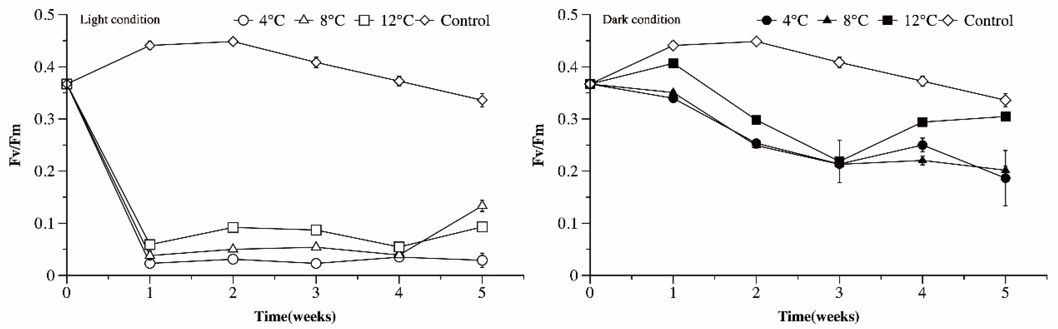
3.6. Photosynthetic Efficiency of Microcystis Cultures under Different Temperature and Light Conditions

Under light conditions, the Fv/Fm values of the Ma cultures exposed to low temperatures declined dramatically from 0.36 to under 0.1 in the first week (p < 0.05). Afterwards, the Fv/Fm values of the Ma cells cultured at 4 °C changed little, while the Fv/Fm values of the Ma cells grown at 8 and 12 °C increased slightly (Figure 8). As the culture temperature increased, so too did the Fv/Fm values of the Ma cultures (p < 0.05, LSD). The Fv/Fm values of the Ma cultured in darkness declined over time and were much higher than those cultured in light conditions after 5 weeks of incubation.

4. Discussion

4.1. Response of Microcystis to Different Temperatures and Light Conditions

By analyzing the net growth rate, proliferation rate (represented by the ratio of divided cells to the sum of divided and undivided cells), and death rate (represented by the ratio of damaged cells to the sum of damaged and undamaged cells), the response of Ma cultures to low temperatures and light/dark conditions was comprehensively evaluated. The net growth rates under all tested conditions (except for the control) were negative, indicating that the biomass of Ma would not increase when temperatures were lower than 12 °C. However, the response of Ma to increasing temperature was different under light and dark conditions. At temperatures ranging from 4 °C to 12 °C in light conditions, rising temperatures increased the proliferation rate and slowed the death rate of Ma cells, thereby increasing the net growth rate of Ma cultures. At 12 °C, the net growth rate was close to zero, showing that the proliferation rate was approximately equal to the death rate. In contrast, under dark conditions, increasing temperature increased the death rate of Ma cells. As Ma cells did not proliferate under dark conditions, the death rate was equal to the net growth rate. This and the results of our previous study [17] indicate that the death rate of Microcystis under dark conditions is extremely low, which is beneficial to its long-term survival in sediment during the winter. Field investigations also determined that Microcystis can overwinter in sediments under cold and dark conditions and could survive for a long period of time [4,8].
Temperature is one of the dominant environmental factors affecting photosynthetic ability. The Fv/Fm values of Ma cells exposed to low temperature and light conditions fall sharply below 0.1 in the first week, then changed little thereafter. The Fv/Fm values of Ma cells exposed to low temperature and dark conditions, however, decreased slowly and were higher than 0.2 after 5 weeks incubation. This suggests that the photosystem of Microcystis under light conditions might be impaired more severely than under dark conditions. Interestingly, when transplanted to normal conditions for re-growth, the re-growth rate of Ma cells overwintering in light conditions were significantly higher than those overwintering in dark conditions. Moreover, the re-growth rate of Ma cells decreased, or ceased entirely, as the length of time of overwintering in dark conditions was extended. These results could suggest that the first decrease in photosynthetic efficiency may be a protective mechanism against photoinhibition occurring under chill and light conditions, rather than permanent photodamage to PSII [34,35], and long exposure to darkness might result in a slow but irreversible deterioration of the physiological state of Microcystis.

4.2. Overwintering Strategies of Microcystis Populations in Lakes Taihu, Chaohu, and Dianchi

The temporal dynamics of Microcystis blooms in Lake Taihu and Chaohu were significantly different from those in Lake Dianchi. In Lake Taihu and Lake Chaohu, Microcystis in the water column gradually become dominant when water temperatures increase in spring and summer and gradually disappear when water temperatures decrease in autumn and winter. In contrast, Microcystis biomass persisted at high levels throughout the year in Lake Dianchi [15,36,37,38]. Previous studies found that temperature has the most significant effect on the annual duration of blooms [39]. It was also hypothesized that temperature was a key factor in the growth and cessation of planktonic cyanobacteria blooms [40,41].
In this study, we investigated the effects of temperature on the proliferation and death rates of Microcystis in the water column and sediment. Our study showed that Microcystis biomass decreased significantly over time when cultured at 4 and 8 °C under light conditions, resulting in a higher cell death rate. By fitting the cell density with linear function, the survival times of Ma cells cultured at 4 and 8 °C can be estimated. The results showed that it takes only 5.65 and 10.28 weeks for the cell density of Ma to decrease to zero at 4 and 8 °C, respectively, while the density of Ma remained stable at 12 °C. These results indicate that Microcystis can survive in the water column for a short period at low temperatures, and the cell density of Microcystis in the water column depends on the duration of exposure to low wintertime temperatures. If the duration of low temperature (4 and 8 °C) exposure exceeded 5.65 and 10.28 weeks, respectively, then Microcystis may completely vanish from the water column. Otherwise, there should be a certain number of Microcystis that survived. Contrary to that observed in the water column, the overwintering ability of Microcystis in sediments is significantly higher at lower temperatures.
Accordingly, in Lake Dianchi, where wintertime water temperatures are higher than 10 °C most of the time, the cell density of pelagic Microcystis could remain stable, but the death rate of benthic Microcystis would be higher. Moreover, the re-growth ability of benthic Microcystis cells after overwintering for several weeks was significantly lower than that of pelagic populations. Thus, most Microcystis can overwinter in the water column and become the primary inoculum source in Lake Dianchi. It could be speculated that the high proliferation rate and low death rate of Microcystis populations under warmer winter temperatures are important factors driving the persistent winter blooms in Lake Dianchi.
As for Lake Taihu and Lake Chaohu, where winter temperatures are usually below 8 °C, the biomass of pelagic Microcystis might gradually decrease because of the high death rates and low proliferation rates. When the duration of low temperatures is short, some Microcystis can still survive in the water column and may be able to grow rapidly as spring brings warmer temperatures, thereby contributing to blooms in the summer. When low temperatures are prolonged, the pelagic Microcystis cells will disappear from the water column, in which case the death rate of the benthic Microcystis populations was very low, creating inoculum in the sediment that feeds subsequent blooms. Regardless of whether winters were warm or cold, surviving Microcystis cells may be sufficient to seed the following blooms. This also showed the flexibility of the Microcystis overwintering strategy response to different winter temperature conditions.

4.3. Response of Winter Microcystis Blooms to Global Warming

Winter temperatures are predicted to be warmer in the future, providing greater opportunities for overwintering pelagic Microcystis populations to grow [42]. As winter temperatures increased, the proliferation rate of pelagic Microcystis populations also increased while the death rate decreased. In Lakes Chaohu and Taihu, there will likely be more Microcystis overwintering in the water column during warmer winters, which might make it easier for pelagic Microcystis populations to accumulate to high enough densities to cause more frequent winter Microcystis blooms in these lakes. Additionally, with winter temperatures increasing, the overwintering and re-growth abilities of pelagic Microcystis are significantly higher than those of benthic Microcystis, implying that pelagic Microcystis will be the primary sources of inoculum for growth and recruitment in the spring.

5. Conclusions

The overwintering strategy of Microcystis is related to winter temperature. In shallow lakes with cold winters (e.g., Lake Taihu and Lake Chaohu), Microcystis primarily overwinter in sediments. In lakes having warmer water in the winter (e.g., Lake Dianchi), the water column is more suitable for Microcystis overwintering. Regardless of whether winter temperatures are warm or cold, the flexibility of the overwintering strategy makes it possible for Microcystis to conserve a large enough quantity of cells to seed future blooms. Warmer winter temperatures appear to promote the proliferation of Microcystis in the water column, while accelerating its death in the sediment.

Author Contributions

Conceptualization, P.C. and C.W.; methodology, P.C. and Q.C.; software, F.H.; validation, P.C., Q.C., and F.H.; formal analysis, Y.H.; investigation, P.C.; resources, Y.H.; data curation, C.T.; writing—original draft preparation, P.C.; writing—review and editing, C.W.; visualization, P.C.; supervision, B.X.; project administration, C.T.; funding acquisition, X.W. and C.W. All authors have read and agreed to the published version of the manuscript.

Funding

This research was funded by the National Natural Science Foundation of China, grant number 31800390 and 42061134013, and the Liu Yongding Academician Workstation of Yunnan (Kunming, China).

Institutional Review Board Statement

Not applicable.

Informed Consent Statement

Not applicable.

Data Availability Statement

Not applicable.

Acknowledgments

The authors thank Yan Wang (the Center for Instrumental Analysis and Metrology, Institute of Hydrobiology, Chinese Academy of Science) for technical assistance in flow cytometry.

Conflicts of Interest

The authors declare no conflict of interest.

References

  1. Ortiz, D.; Palmer, J.; Wilkinson, G. Detecting changes in statistical indicators of resilience prior to algal blooms in shallow eutrophic lakes. Ecosphere 2020, 11, e03200. [Google Scholar] [CrossRef]
  2. Harke, M.J.; Steffen, M.M.; Gobler, C.J.; Otten, T.G.; Wilhelm, S.W.; Wood, S.A.; Paerl, H.W. A review of the global ecology, genomics, and biogeography of the toxic cyanobacterium Microcystis spp. Harmful Algae 2016, 54, 4–20. [Google Scholar] [CrossRef] [Green Version]
  3. Dittmann, E.; Wiegand, C. Cyanobacterial toxins—Occurrence, biosynthesis and impact on human affairs. Mol. Nutr. Food Res. 2006, 50, 7–17. [Google Scholar] [CrossRef] [PubMed]
  4. Reynolds, C.S.; Jaworski, G.H.M.; Cmiech, H.A.; Leedale, G.F. On the annual cycle of the blue-green alga Microcystis Aeruginosa Kütz. Emend. Elenkin. Philos. Trans. R. Soc. B Biol. Sci. 1981, 293, 419–477. [Google Scholar] [CrossRef]
  5. Ihle, T.; Jähnichen, S.; Benndorf, J. Wax and wane of Microcystis (Cyanophyceae) and microcystins in Lake sediments: A case study in Quitzdorf reservoir (Germany). J. Phycol. 2005, 41, 479–488. [Google Scholar] [CrossRef]
  6. Kong, F.X.; Gao, G. Hypothesis on cyanobacteria bloom-forming mechanism in large shallow eutrophic lakes. Acta Ecol. Sin. 2005, 25, 589–595. [Google Scholar]
  7. Tsujimura, S.; Tsukada, H.; Nakahara, H.; Nakajima, T.; Nishino, M. Seasonal variations of Microcystis populations in sediments of Lake Biwa, Japan. Hydrobiologia 2000, 434, 183–192. [Google Scholar] [CrossRef]
  8. Brunberg, A.K.; Blomqvist, P. Benthic overwintering of Microcystis colonies under different environmental conditions. J. Plankton Res. 2002, 24, 1247–1252. [Google Scholar] [CrossRef]
  9. Sabart, M.; Misson, B.; Jobard, M.; Bronner, G.; Donnadieu-Bernard, F.; Duffaud, E.; Salençon, M.-J.; Amblard, C.; Latour, D. Genetic diversity along the life cycle of the cyanobacterium Microcystis: Highlight on the complexity of benthic and planktonic interactions. Environ. Microbiol. 2014, 17, 901–911. [Google Scholar] [CrossRef] [PubMed]
  10. Shi, K.; Zhang, Y.; Zhang, Y.; Li, N.; Qin, B.; Zhu, G.; Zhou, Y. Phenology of Phytoplankton Blooms in a Trophic Lake Observed from Long-Term MODIS Data. Environ. Sci. Technol. 2019, 53, 2324–2331. [Google Scholar] [CrossRef]
  11. Chen, S.L.; Lu, J.W.; Shen, J.Q. Study of the spatio-temporal change of water temperature in Lake Taihu. Jiangshu Water Res. 2009, 3, 38–39. (In Chinese) [Google Scholar]
  12. Hu, M.Q.; Zhang, Y.C.; Ma, R.H.; Zhang, Y.X. Spatial and Temporal Dynamics of Floating Algal Blooms in Lake Chaohu in 2016 and Their Environmental Drivers. Environ. Sci. 2018, 39, 87–99. (In Chinese) [Google Scholar]
  13. Sigee, D.C.; Selwyn, A.; Gallois, P.; Dean, A.P. Patterns of cell death in fresh- water colonial cyanobacteria during the late summer bloom. Phycologia 2007, 46, 284–292. [Google Scholar] [CrossRef]
  14. Ma, J.; Qin, B.; Paerl, H.W.; Brookes, J.D.; Hall, N.S.; Shi, K.; Zhou, Y.; Guo, J.; Li, Z.; Xu, H.; et al. The persistence of cyanobacterial (Microcystis spp.) blooms throughout winter in Lake Taihu, China. Limnol. Oceanogr. 2016, 61, 711–722. [Google Scholar] [CrossRef] [Green Version]
  15. Wu, Y.; Li, L.; Gan, N.; Zheng, L.; Ma, H.; Shan, K.; Liu, J.; Xiao, B.; Song, L. Seasonal dynamics of water bloom-forming Microcystis morphospecies and the associated extracellular microcystin concentrations in large, shallow, eutrophic Dianchi Lake. J. Environ. Sci. 2014, 26, 1921–1929. [Google Scholar] [CrossRef]
  16. Latour, D.; Salençon, M.J.; Reyss, J.L.; Giraudet, H. Sedimentary imprint of Microcystis aeruginosa (Cyanobacteria) blooms in Grangent reservoir (Loire, France). J. Phycol. 2007, 43, 417–425. [Google Scholar] [CrossRef]
  17. Wang, C.; Feng, B.; Tian, C.; Tian, Y.; Chen, D.; Wu, X.; Li, G.; Xiao, B. Quantitative study on the survivability of Microcystis colonies in lake sediments. Environ. Boil. Fishes 2017, 30, 495–506. [Google Scholar] [CrossRef]
  18. Yang, Y.; Yin, C.T.; Li, W.Z.; Xu, X.D. α-Tocopherol is essential for acquired chill-light tolerance in the cyanobacterium Synechocystis sp. Strain PCC 6803. J. Bacteriol. 2008, 190, 1554–1560. [Google Scholar] [CrossRef] [PubMed] [Green Version]
  19. Kamau, S.W.; Nunez, R.; Grimm, F. Flow cytometry analysis of the effect of allopurinol and the dinitroaniline compound (Chloralin) on the viability and proliferation of Leishmania infantum promastigotes. BMC Pharmacol. 2001, 1, 1471–1481. [Google Scholar] [CrossRef] [PubMed]
  20. Rioboo, C.; O’Connor, J.E.; Prado, R.; Herrero, C.; Cid, A. Cell proliferation alterations in Chlorella cells under stress conditions. Aquat. Toxicol. 2009, 94, 229–237. [Google Scholar] [CrossRef] [PubMed] [Green Version]
  21. Li, X.; Chen, S.; Zeng, J.; Song, W.; Yu, X. Comparing the effects of chlorination on membrane integrity and toxin fate of high- and low-viability cyanobacteria. Water Res. 2020, 177, 115769. [Google Scholar] [CrossRef] [PubMed]
  22. Parish, C.R. Fluorescent dyes for lymphocyte migration and proliferation studies. Immunol. Cell Biol. 1999, 77, 499–508. [Google Scholar] [CrossRef] [Green Version]
  23. Lyons, A.B.; Hasbold, J.; Hodgkin, P.D. Flow cytometric analysis of cell division history using dilution of carboxyfluorescein diacetate succinimidyl ester, a stably integrated fluorescent probe. In Methods in Cell Biology: Cytometry, 3rd ed.; Darzynkiewicz, Z., Crissman, H.A., Robinson, J.P., Eds.; Academic Press: London, UK, 2001; Volume 63, pp. 375–398. [Google Scholar]
  24. Zhou, C.; Place, A.R.; Yan, X.; Xu, J.; Luo, Q.; William, E.; Jiang, Y. Interactions between Karlodinium veneficum and Prorocentrum donghaiense from the East China Sea. Harmful Algae 2015, 49, 50–57. [Google Scholar] [CrossRef]
  25. Cunningham, A. Analysis of microalgae and cyanobacteria by flow cytometry. In Flow Cytometry in Microbiology; Lloyd, D., Ed.; Springer: London, UK, 1993; pp. 131–142. [Google Scholar]
  26. Trask, B.J.; Engh, G.J.V.D.; Elgershuizen, J.H.B.W. Analysis of phytoplankton by flow cytometry. Cytometry 1982, 2, 258–264. [Google Scholar] [CrossRef]
  27. Olson, R.J.; Frankel, S.L.; Chisholm, S.W.; Shapiro, H.M. An inexpensive flow cytometer for the analysis of fluorescence signals in phytoplankton: Chlorophyll and DNA distributions. J. Exp. Mar. Biol. Ecol. 1983, 68, 129–144. [Google Scholar] [CrossRef]
  28. Brussaard, C.; Marie, D.; Thyrhaug, R.; Bratbak, G. Flow cytometric analysis of phytoplankton viability following viral infection. Aquat. Microb. Ecol. 2001, 26, 157–166. [Google Scholar] [CrossRef] [Green Version]
  29. Roth, B.L.; Poot, M.; Yue, S.T.; Millard, P.J. Bacterial viability and antibiotic susceptibility testing with SYTOX green nucleic acid stain. Appl. Environ. Microbiol. 1997, 63, 2421–2431. [Google Scholar] [CrossRef] [PubMed] [Green Version]
  30. Cermeño, P.; Marañón, E.; Rodríguez, J.; Fernandez, E. Large-sized phytoplankton sustain higher carbon-specific photosynthesis than smaller cells in a coastal eutrophic ecosystem. Mar. Ecol. Prog. Ser. 2005, 297, 51–60. [Google Scholar] [CrossRef]
  31. Toepel, J.; Wilhelm, C.; Meister, A.; Beckert, A.; Martinez-Ballesta, M.D. Cytometry of freshwater phytoplankton. Methods Cell Biol. 2004, 75, 375–407. [Google Scholar] [PubMed]
  32. Yang, Y.-P.; Juang, Y.-S.; Hsu, B.-D. A quick method for assessing chloroplastic starch granules by flow cytometry. J. Plant Physiol. 2002, 159, 103–106. [Google Scholar] [CrossRef]
  33. Ou, H.; Gao, N.; Deng, Y.; Qiao, J.; Wang, H. Immediate and long-term impacts of UV-C irradiation on photosynthetic capacity, survival and microcystin-LR release risk of Microcystis aeruginosa. Water Res. 2012, 46, 1241–1250. [Google Scholar] [CrossRef]
  34. Allakhverdiev, S.I.; Kreslavski, V.D.; Klimov, V.V.; Los, D.; Carpentier, R.; Mohanty, P. Heat stress: An overview of molecular responses in photosynthesis. Photosynth. Res. 2008, 98, 541–550. [Google Scholar] [CrossRef] [PubMed]
  35. Ding, Y.; Gan, N.; Liu, J.; Zheng, L.; Li, L.; Song, L. Survival, recovery and microcystin release of Microcystis aeruginosa in cold or dark condition. Chin. J. Oceanol. Limnol. 2016, 35, 313–323. [Google Scholar] [CrossRef]
  36. Shan, K.; Song, L.; Chen, W.; Li, L.; Liu, L.; Wu, Y.; Jia, Y.; Zhou, Q.; Peng, L. Analysis of environmental drivers influencing interspecific variations and associations among bloom-forming cyanobacteria in large, shallow eutrophic lakes. Harmful Algae 2019, 84, 84–94. [Google Scholar] [CrossRef]
  37. Yang, Z.; Zhang, M.; Yu, Y.; Shi, X. Temperature triggers the annual cycle of Microcystis, comparable results from the laboratory and a large shallow lake. Chemosphere 2020, 260, 127543. [Google Scholar] [CrossRef]
  38. Zhang, M.; Zhang, Y.; Yang, Z.; Wei, L.; Yang, W.; Chen, C.; Kong, F. Spatial and seasonal shifts in bloom-forming cyanobacteria in Lake Chaohu: Patterns and driving factors. Phycol. Res. 2015, 64, 44–55. [Google Scholar] [CrossRef]
  39. Zhang, Y.; Ma, R.; Zhang, M.; Duan, H.; Loiselle, S.; Xu, J. Fourteen-Year Record (2000–2013) of the Spatial and Temporal Dynamics of Floating Algae Blooms in Lake Chaohu, Observed from Time Series of MODIS Images. Remote Sens. 2016, 7, 10523–10542. [Google Scholar] [CrossRef] [Green Version]
  40. Sellner, K.G. Physiology, ecology, and toxic properties of marine cyanobacteria blooms. Limnol. Oceanogr. 1997, 42, 1089–1104. [Google Scholar] [CrossRef] [Green Version]
  41. Paerl, H.W.; Huisman, J. Blooms like it hot. Science 2008, 320, 57–58. [Google Scholar] [CrossRef] [PubMed] [Green Version]
  42. Qin, B.; Zhu, G.; Gao, G.; Zhang, Y.; Li, W.; Paerl, H.W.; Carmichael, W.W. A drinking water crisis in Lake Taihu, China: Linkage to climatic variability and lake management. Environ. Manag. 2010, 45, 105–112. [Google Scholar] [CrossRef] [PubMed]
Figure 1. The cell density dynamics of Ma under different temperature and light conditions, as a function of time: Inserts—the control group: The cell density dynamics of Ma under 25 °C and light condition.
Microorganisms 09 02278 g001
Figure 2. Net Growth rate of Ma cells at different temperature and light conditions.
Microorganisms 09 02278 g002
Figure 3. After overwintering for 1–4 weeks under different conditions ((A) 4 °C; (B) 8 °C; (C) 12 °C), Ma cells were cultured at 25 °C in the light. The net growth rate was used to represent the re-growth ability of Ma cells. * Represents a negative growth rate.
Microorganisms 09 02278 g003
Figure 4. The ratio of mother cells to the sum of mother cells and daughter cells under different temperature and light conditions, as a function of time.
Microorganisms 09 02278 g004
Figure 5. The ratio of undamaged cells to the sum of damaged and undamaged cells at different temperature and light conditions.
Microorganisms 09 02278 g005
Figure 6. Forward scatter, analyzed by FCM, of Ma cells exposed to different temperature and light conditions as a function of time.
Microorganisms 09 02278 g006
Figure 7. Side scatter, analyzed by FCM, of Ma cells exposed to different temperature and light conditions as a function of time.
Microorganisms 09 02278 g007
Figure 8. The maximum quantum yield for primary photochemistry (Fv/Fm) of Microcystis cultures exposed to different temperature and light conditions as a function of time.
Microorganisms 09 02278 g008
Publisher’s Note: MDPI stays neutral with regard to jurisdictional claims in published maps and institutional affiliations.

