Article

Genome-Wide Analysis of Sugar Transporters Identifies the gtsA Gene for Glucose Transportation in Pseudomonas stutzeri A1501

1
School of Life and Pharmaceutical Sciences, Hainan University, Haikou 570100, China
2
Biotechnology Research Institute, Chinese Academy of Agricultural Sciences, Beijing 100081, China
*
Authors to whom correspondence should be addressed.
Microorganisms 2020, 8(4), 592; https://doi.org/10.3390/microorganisms8040592
Submission received: 23 March 2020 / Revised: 15 April 2020 / Accepted: 18 April 2020 / Published: 19 April 2020
(This article belongs to the Section Environmental Microbiology)

Abstract

:
Pseudomonas stutzeri A1501 possesses an extraordinary number of transporters which confer this rhizosphere bacterium with the sophisticated ability to metabolize various carbon sources. However, sugars are not a preferred carbon source for P. stutzeri A1501. The P. stutzeri A1501 genome has been sequenced, allowing for the homology-based in silico identification of genes potentially encoding sugar-transport systems by using established microbial sugar transporters as a template sequence. Genomic analysis revealed that there were 10 sugar transporters in P. stutzeri A1501, most of which belong to the ATP-binding cassette (ABC) family (5/10); the others belong to the phosphotransferase system (PTS), major intrinsic protein (MIP) family, major facilitator superfamily (MFS) and the sodium solute superfamily (SSS). These systems might serve for the import of glucose, galactose, fructose and other types of sugar. Growth analysis showed that the only effective medium was glucose and its corresponding metabolic system was relatively complete. Notably, the loci of glucose metabolism regulatory systems HexR, GltR/GtrS, and GntR were adjacent to the transporters ABCMalEFGK, ABCGtsABCD, and ABCMtlEFGK, respectively. Only the ABCGtsABCD expression was significantly upregulated under both glucose-sufficient and -limited conditions. The predicted structure and mutant phenotype data of the key protein GtsA provided biochemical evidence that P. stutzeri A1501 predominantly utilized the ABCGtsABCD transporter for glucose uptake. We speculate that gene absence and gene diversity in P. stutzeri A1501 was caused by sugar-deficient environmental factors and hope that this report can provide guidance for further analysis of similar bacterial lifestyles.

1. Introduction

Sugar is a stable and widely distributed carbon source in nature and has traditionally been considered an important source of carbon skeleton material and energy supply in bacteria. Sugar is not the dominant carbon source for Pseudomonas, which prefers amino acids or organic acids [1]. The basic sugar biosynthesis and degradation pathways have been established for some time and have more recently become a subject of great interest and enthusiastic study [2,3]. It is perhaps somewhat natural to focus on intracellular activities and forget the preceding step whereby sugar enters the cell via membrane transport. Sugar usually enters the cell through various specific transporters, such as the glucose phosphotransferase transport system (PTSGlu) in Escherichia coli [4]. Saier summarized about 20 families of secondary carriers, including porins and various transport systems, and constructed the transporter classification database (TCDB) [5]. Due to the limited data in TCDB, we extended our search to include other databases (UniProt and the National Center for Biotechnology Information (NCBI)) and referred to the well-studied model strains of E. coli and Sulfolobus solfataricus to provide more comprehensive and important information for our research [6].
Pseudomonas stutzeri A1501 (China General Microbiological Culture Collection Center Accession No. 0351) was originally isolated from rice rhizosphere soil in South China and was previously named Alcaligenes faecalis A15 [7]. It possesses a strong nitrogen fixation ability and can effectively colonize the root surface to promote plant growth [8]. The ability of P. stutzeri A1501 to utilize nitrogen has been studied extensively, but little is known about its utilization of carbon sources, especially regarding sugars. We explored the reasons for the growth status of P. stutzeri A1501 by considering the sugar-transport system.
The genome of P. stutzeri A1501 has been sequenced, enabling the rapid identification and screening of the potential sugar-transport system [9]. In this study, we compiled the sequence data for all sugar transporters present in P. stutzeri A1501 using similarity searches and evaluated the potential substrate specificity by analyzing its adjacent metabolism and regulatory genes. Glucose was assessed as the available sugar source by combining growth analysis and metabolic system gene characterization. Hence, the function of the putative glucose transport system was evaluated through the characterization of candidate gene expression under the conditions of sufficient and limited glucose, and their contribution to glucose use was examined using a mutation analysis and consumption experiments.

2. Materials and Methods

2.1. Bacterial Strains and Growth Conditions

The bacterial strains and plasmids used in this study are shown in Table 1. P. stutzeri A1501 was grown in a minimal lactate medium or in a Luria-Bertani (LB) medium at 30 °C as previously described [10]. E. coli was grown at 37 °C in the LB medium. Chloramphenicol (Cm), tetracycline (Tc), and kanamycin (Km) were added to the media at concentrations of 20, 10, and 50 μg/mL, respectively.

2.2. Computer Analyses and Screening Strategies

All data were based upon sequence comparisons conducted using the freely accessible genome data of P. stutzeri A1501. Functional annotations of the protein sequences were carried out by a BLASTP search of the translations versus GenBank’s non-redundant protein database (NR) [9]. To more comprehensively identify the sugar transporter, protein sequences of known sugar-transport systems from the transporter classification database (http://www.tcdb.org/search/index.php) were also used to screen the corresponding sequence of P. stutzeri A1501 at the BLAST server of the NCBI (http://ncbi.nlm.nih.gov) with an E-value cutoff of 10−5. The identified candidate proteins were then cross-checked against the original list of annotated proteins. To find the possible substrates, the predicted sugar transporters in P. stutzeri A1501 were similarity searched by BLASTP alignment in the UniProt database with an E-value cutoff of 10−5. Finally, the protein sequence alignments were conducted with the DNAMAN software (Lynnon BioSoft, Vaudreuil, QC, Canada). Candidate sugar-transport protein was speculated to have the same substrate if the protein identity of the P. stutzeri A1501 protein was more than a 60% match with the corresponding protein of Pseudomonas, or more than a 30% match with other bacteria. Moreover, the coverage should be more than 60% of the protein sequence full-length.

2.3. Monitoring Growth

The bacteria were cultured overnight in an LB medium. After centrifugation and washing, they were diluted in the experimental medium to an initial optical density (OD600) of 0.1. The OD600 was measured every two hours until the cells reached a steady growth. Three replicates of each strain were performed.

2.4. Reverse Transcription PCR (RT-PCR) and Quantitative Real-Time PCR (qPCR)

The total RNA was isolated using the innuPREP RNA Mini Kit (Analytik Jena, Jena, Germany) and reverse transcription was performed using the Prime Script™ RT Reagent Kit with gDNA Eraser (Takara, Dalian, China). The produced cDNA was used to perform qPCR using the ChamQ™ Universal SYBR qPCR Master Mix (Vazyme, Nanjing, China). All primer pairs are shown in Table S1, and the procedures followed the manufacturer’s instructions. The relative gene expression levels were calculated using the 2−ΔΔCt method and the 16S rRNA was used as a reference gene.

2.5. Phylogenetic Analysis

For the phylogenetic analysis, the amino acid sequences of glucose-binding proteins from different organisms were obtained from the NCBI. Multiple sequence alignments of full-length proteins were performed using ClustalX [14]. The pairwise deletion option was used to circumvent the gaps and missing data. We used the neighbor-joining tree generated by the MEGA (Molecular Evolutionary Genetics Analysis) program [15] with 1000 replicates of bootstrap analysis.

2.6. Construction of the gtsA Deletion Mutant and Complementation

To verify the role of gtsA gene in P. stutzeri A1501, the gtsA region was deleted by homologous recombination according to a PCR-based fusion strategy. The upstream and downstream homologous arm fragments of gtsA and the chloramphenicol resistance gene cat with its own promoter in pKatCAT5 were fused. The fusion PCR product was then cloned into the multiple cloning site of pK18mobsacB and the resulting plasmid was named pK18gtsA. The obtained recombinant plasmid was transferred into the wild type P. stutzeri A1501 by triparental mating, producing a mutant strain. Candidate clones resulting from a double-crossover event were isolated on the LB agar with chloramphenicol (Figure S1A). The correct recombination was confirmed by PCR analysis (Figure S1B). To complement the gtsA gene, a PCR fragment containing gtsA was cloned into the plasmid pLAFR3. The resulting vector (pLgtsA) was then used to transform the ΔgtsA mutant (Figure S2A). The correct recombination was confirmed by PCR analysis (Figure S2B). The test result was validated by gene sequencing. To compare the phenotype of the complemented strain and the wild type, pLAFR3 was also introduced into P. stutzeri A1501.

2.7. Measurement of Glucose Uptake

The samples were harvested during the exponential growth of P. stutzeri A1501 by filtering the medium. The determination and quantification of glucose were performed using an ion chromatograph (Thermo Scientific™ Dionex™ ICS-5000, Waltham, MA, USA). The gradients of sodium hydroxide, generated by an eluent generator (Thermo Scientific Dionex ICS 5000EG, Waltham, MA, USA), were as follows: 200 mM (0–10 min, equilibration), 200 mM (10–40 min, column cleaning), and 10 mM (40–52 min, sample). Quantitative analyses were performed by measuring the peak area using the external standard method.

3. Results

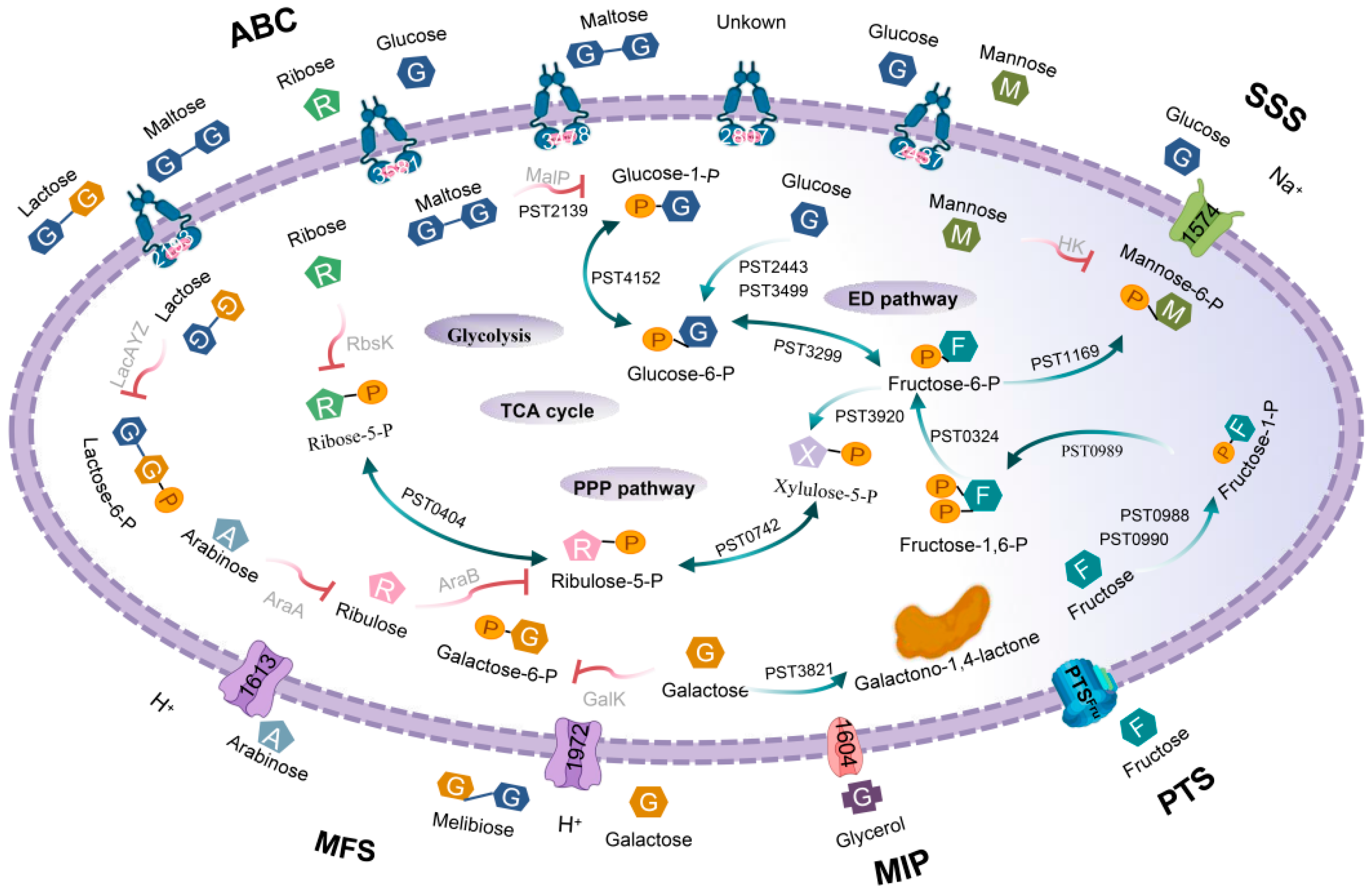
3.1. Prediction of Sugar-Transport Systems

The bioinformatics analysis predicted 10 sugar-transport systems in P. stutzeri A1501, including the ATP-binding cassette family (ABC), phosphotransferase system (PTS), major facilitator superfamily (MFS), major intrinsic protein family (MIP) and the sodium solute superfamily (SSS) (Figure S3).
The five ABC-type transport systems consisted of one required ATP-binding protein (ABP) and two permeases. With the exception of the transporters encoded by PST2907-2909, the others also contained one substrate-binding protein (SBP). Although this system had an amino acid sequence identity of 66–83% with the monosaccharide ABC transport system (PA0136-0138) in Pseudomonas aeruginosa [16], the substrate specificity could not be determined due to the absence of SBP. It may be linked to the E. coli galactose/methylgalactoside MglABC transport system [17], but the sequence similarity was very low. The proteins encoded by PST3478-3486 and PST3579-3583 share an identity with the maltose transport system malEFGK [18] and the ribose transport system rbsABC [19] in E. coli, respectively. The latter was also similar to the Haloferax volcanii DS2 glucose ABC transport system (HVOB3014-3018). The second potential maltose transporters were encoded by PST2190-2193, and shared 77–83% similarity with P. aeruginosa MtlEFGK [20]. Notably, this system was also similar to the Burkholderia multivorans multiple sugar transporter encoded by BMULJ02554-02558 (55–61% identity) and Aquimixticola soesokkakensis lactose transporter subunit LacEF encoded by AQS8620_00608-00609 (55–60% identity). The last ABC transport system encoded by PST2437-2440 was similar to the Pseudomonas putida glucose transport system GtsABCD2 [21], but no corresponding protein was found in E. coli.
Only one fructose-specific PTS transport system was discovered. The gene order of fruBfruKfruA was the same as in E. coli, but the locus of fruR was closely linked to this cluster, whereas E. coli fruR was expressed independently [4]. The protein encoded by PST0988 was only 15% identical to E. coli FruB, but more than 30% identical to the Pseudomonas fluorescens glucose PTS transport protein PtsP (PFL_4931) and the Aquitalea magnusonii mannose PTS transport protein (VI06_09940).
In addition to the aforementioned ABC and PTS sugar-transport systems, the two genes PST1613 and PST1972 may be involved in the MFS transport system. The protein encoded by PST1613 shared 58% identity with the sugar efflux transporter YdeA (also known as SotB) in E. coli [22] and 63% with the Acinetobacter marinus arabinose export protein. The protein encoded by PST1972 shared 46% similarity with Acidovorax sp. RAC01 MFS/sugar transporter (A0A1B3PGF1_9BURK). It also had many functions beyond MFS, such as SSS and glycoside-pentoside-hexuronide (GPH) cation symporter transporter, because it was highly similar not only to melibiose and galactose SSS transporters in Chitinimonas taiwanensis and Janthinobacterium sp. B9-8, but also to GPH in Jannaschia helgolandensis. Another sodium/glucose co-transporter also belongs to the SSS family and was encoded by PST1574. Finally, an MIP family protein encoded by PST1604 had 67% similarity with E. coli GlpF [23].
We noticed that the neighborhood of the candidate transport system included sugar-related metabolic genes. The adjacent genes of ABCGtsABCD, ABCMtlEFGK, ABCMalEFGK and PTSFru were mainly involved in glucose metabolism, namely edd-2/glk2/hexR/zwf/pgl/eda (PST3495PST3550), edd-1/glk-1/gltR/gltS (PST2441PST2444), gntR/gntK (PST2198, PST2221) and gcd (PST0991), respectively. However, the genes around the other systems were not related to sugar metabolism.
Two sugar porins were located upstream of ABCGtsABCD and downstream of ABCMalEFGK, which were highly similar to LamB in E. coli (>30% identity), so we named them LamB1 and LamB2, respectively [24]. A sugar porin OprB was also present upstream of ABCGtsABCD in P. aeruginosa and P. putida and was reported to transport glucose into the cell [25]. However, the sequence similarity between LamB1, LamB2, and OprB was very low, and no OprB homologues were found in the P. stutzeri A1501 genome.

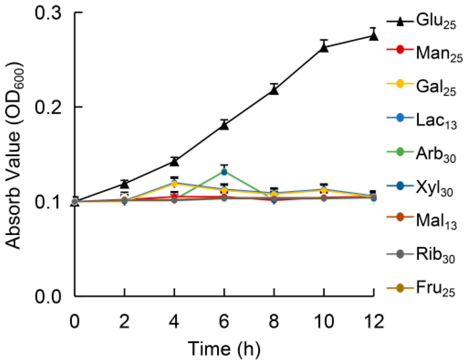
3.2. Growth in Different Sugars

The whole-genome BLAST analysis revealed that P. stutzeri A1501 might possess an uptake system for glucose, maltose, mannose, ribose, fructose, xylose, melibiose, lactose, arabinose and galactose. Therefore, growth experiments were performed in a minimal medium (containing all the essential nutrients and trace elements) with sugar as the only carbon source. Surprisingly, with the exception of glucose, P. stutzeri A1501 hardly grew on other sugars (Figure 1). We further analyzed the related metabolic pathways of the sugars in cytosol to explore the reasons.
Firstly, glucose was converted to glucose-6-phosphate and fructose-6-phosphate by glucokinase Glk and glucose-6-phosphate isomerase Pgi. Fructose was phosphorylated into fructose-1,6-diphosphate by fructokinase MtlZ and 1-phosphofructokinase FruK [26]. These three products eventually entered the Entner–Doudoroff (ED) pathway, pentose phosphate (PPP) pathway and the tricarboxylic acid (TCA) cycle. The pattern diagram and regulatory genes were published elsewhere and will not be repeated here [9].
The PPP pathway involved the conversion between xylulose-5-phosphate, ribose-5-phosphate, and ribulose-5-phosphate. However, xylose, arabinose and ribose could not be metabolized through the PPP pathway because xylose isomerase XlyA [27], arabinose isomerase AraA [28] and ribokinase Rbsk [29] were not found in P. stutzeri A1501. Similarly, even though mannose-6-phosphate could be converted from fructose-6-phosphate by mannose-6-phosphate isomerase AlgA, there was no hexokinase to phosphorylate mannose [30]. Maltodextrin phosphorylase (MalP) and amylomaltase (MalQ) were essential enzymes for maltose and maltodextrin metabolism [18], galactose dehydrogenase (GalDH) and 2-dehydro-3-deoxy-6-phosphate galactate aldolase (DgoA) were equally important for galactose [31] and only MalQ and GalDH were present in this organism. We did not find any proteins related to lactose and melibiose metabolism. In short, only glucose and fructose metabolic systems were relatively complete metabolic systems, whereas others were incomplete or non-existent (Figure 2).

3.3. Effect of Glucose on the Expression of Sugar Transporters

We discussed the influence of P. stutzeri A1501 when the substrate was glucose for the following four reasons: one system can interact with multiple substrates; the candidate transporters neighborhood genes are related to glucose metabolism; this bacterium can only grow in glucose and has a more complete glucose metabolic system.
The expression level of related genes may vary with glucose concentration to maintain glucose homeostasis. Therefore, we analyzed the expressions of the genes of these candidate sugar-transport systems under glucose-sufficient (25 mmol/L) and -limited (3 mmol/L) conditions to confirm their hypothetical role. RNA was isolated rapidly after shaking the culture for 1 h because the growth of P. stutzeri A1501 was limited in the absence of a carbon source or low concentration of glucose and long-term culture would affect RNA quality.
Table 2 shows that both concentrations of glucose could significantly induce the mRNA expression of PST2437, PST2438, PST2439, and PST2440, especially at 25 mmol/L. The PST3484, PST1972, PST3079, PST3685, and PST1221 mRNA levels were upregulated only with 25 mmol/L glucose, but not with 3 mmol/L. The other sugar transporter genes were not affected regardless of the concentration.

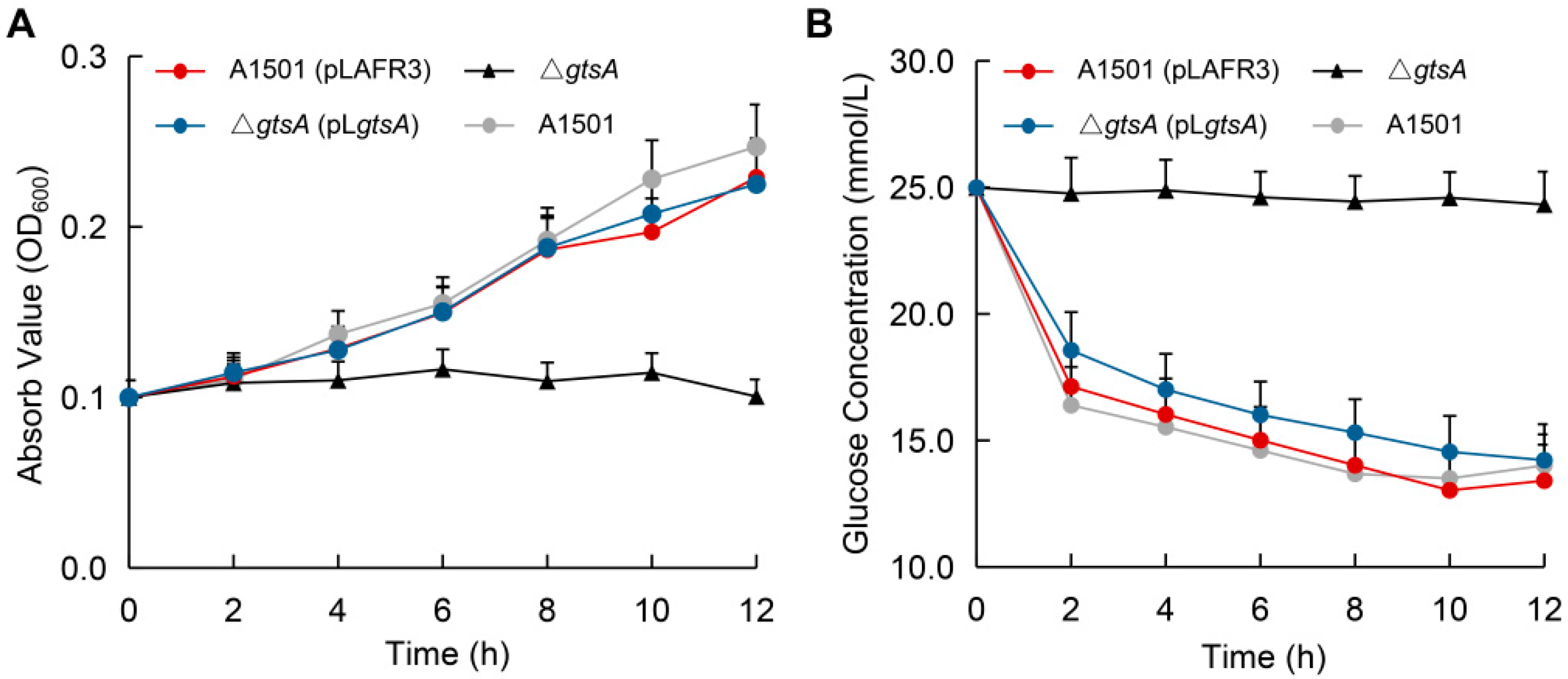
3.4. Functional Analysis of Glucose-Binding Protein GtsA

A striking result was the strong induction of the ABCGtsABCD transport system by different concentrations of glucose, so we focused on the gtsA gene which encodes the periplasmic binding protein, a key component of this system. The genomic sequence analysis showed that gtsA (PST2440) was located upstream from gtsBCD/lamB1 (PST2436PST2439, encoding permease, ABP, and porin) and transcribed in the same orientation, suggesting that they may be co-transcribed. This possibility was determined using the indicated primer pairs to amplify cDNA and genomic DNA (gDNA). The results showed that the PCR products of gtsBgtsC were obtained using both cDNA and gDNA as templates. However, gtsAgtsB produced a PCR product only with gDNA (Figure 3A). This result indicated that gtsA was independently transcribed in P. stutzeri A1501.
Th phylogenetic analysis revealed that the product of gtsA was highly conserved in Pseudomonas species and was most closely related to Ochrobactrum anthropi glucose-binding protein (oaGBP) [32]. Compared with E. coli GBP, GtsA was more closely related to the corresponding protein from extreme environmental microorganisms such as Thermus thermophilus ttGBP [33], Saccharolobus solfataricus GlcS [6], and Thermotoga maritima GBP [34] (Figure 3B). Although X-ray crystallographic analysis was not performed, the tertiary structure of GtsA was inferred by homology-modeling based on the crystal structure of P. putida CSV86 glucose-binding protein (ppGBP; Protein Data Bank code 5DVI) [32] (Figure 3C; Figure S4).
To study the biological function of gtsA gene, a knockout mutant was constructed by replacing the gtsA gene with a chloramphenicol resistance cassette. The growth and consumption curve of the strains in the medium containing glucose as the sole carbon source showed that the mutant strain ΔgtsA could hardly grow (Figure 4A) and the glucose content in the medium did not change (Figure 4B), which meant that the strain could not utilize glucose. The results also showed that the complemented strain ΔgtsA (pLgtsA) could restore the above phenotypes while the complementary plasmid pLAFR3 had no effect on the strain.

4. Discussion

All free-living bacteria in nature can rapidly and flexibly adapt their behavior to the changing environment, thereby effectively utilizing the available carbon source. Sugar, the most common nutrient, plays a vital role in microbial metabolism. For example, it is the main carbon source for E. coli, which is rich in sugar-transport systems and metabolic pathways. Although Pseudomonas are ubiquitous bacteria, unlike E. coli, sugar is not their preferred carbon source [35]. P. aeruginosa prefers succinic acid rather than glucose and fructose [36]. The poor sugar use may be due to catabolite repression control [37] or related genes loss, such as the well-known deficiency of the glycolysis key enzyme phosphofructokinase [38].
In this study, a total of 10 limited and incomplete potential sugar-transport systems in P. stutzeri A1501 were identified by bioinformatics and genome analysis. This was consistent with a previous report that only fructose enters into Pseudomonas through the PTS system, whereas other sugars are independent of PTS [35]. The sugar metabolism system of P. stutzeri A1501 was also deficient. There was only one relatively complete glucose metabolism system, and other sugar degrading enzymes that participated in the central carbon metabolism were lacking. L-rhamnose isomerase RHI (Q75WH8_PSEST) can metabolize rhamnose, mannose, ribose, and many other sugars in Pseudomonas sp. LL172 [39,40]; it is strange that its homologues are found in Rhizobium and not Pseudomonas. Fewer genes for sugar metabolism were present in P. stutzeri A1501 compared with other Pseudomonas species, such as ribokinase RbsK (PFLU_4156) which was present in P. fluorescens but not in P. stutzeri A1501. This phenomenon may be influenced by the living environment; P. fluorescens was isolated from the phyllosphere [41], whereas P. stutzeri A1501 was isolated from the rhizosphere, which has a lower sugar content [42,43]. Previous studies suggested that endophytic Pseudomonas could utilize the sugars in xylem fluid in contrast to the rhizosphere Pseudomonas [44]. Based on their distinct material conditions, microorganisms will selectively discard some “useless” genes in their long-term adaptive evolution process and prioritize genes that are conducive to competitive survival [45]. Studies generally have reported that environmentally induced gene loss is an adaptive evolutionary ability of bacteria to avoid invalid gene replication, transcription and translation, to save energy and to improve space efficiency [46].
Of all the assayed sugars, P. stutzeri A1501 only grew on glucose, which may be due to it having a more complete system for glucose transport and metabolism. Different concentrations of glucose had varying degrees of influence on the transport system; what attracted our attention is that ABCGtsABCD could be significantly induced regardless of the concentration, indicating that it participated in glucose transport. This system has been deeply studied in P. putida and P. aeruginosa [21]. A gtsA deletion mutant was constructed to further clarify the function, and P. stutzeri A1501 almost completely lost the ability to utilize glucose after inferring with the function of the ABCGtsABCD transport system. Glucose transport and chemotaxis were also defective in the absence of glucose-binding proteins in P. aeruginosa [47].
Glucose was internalized into E. coli by the ABCMglABC, the PTSGlu system, and GalP [48]; its homologues GalP (PST4041) and ABCMglAC (PST2907PST2908, <30% identity) were found in P. stutzeri A1501, but the substrate-binding protein MglB and the PTSGlu transport system were absent. The same situation exists in Archaea isolated from extreme environments, such as Sulfolobus solfataricus [49], Thermophilic archaea [50] and Thermotoga maritima [51]. They lack the PTSGlu system and rely on the ABC transport system [52], which is more similar to ABCGtsABCD than ABCMglABC. We speculate that this genetic diversity is guided by different survival circumstances, and the microorganism shows the optimum phenotype to cater to its surroundings, which is the basis for a higher-level evolution of species [53]. For E. coli, glucose was sufficient, and its transport system was more comprehensive and powerful, whereas the ABC transport system was more suitable for sugar-deficient bacterium such as Pseudomonas and Archaea. The ABC transporter is considered essential for thermophiles to survive in harsh habitats because it has a stronger binding capacity than other systems and can bind to very low concentrations of substrates [54].
At present, the sugar-transport systems of Pseudomonas have not been systematically analyzed. We hope that the summary provided in this article can guide the further development of related research, which needs to be improved and supplemented continuously, even though the genome of P. stutzeri A1501 has been comprehensively analyzed. P. stutzeri A1501 lacks many sugar-related genes, but retains some genes related to glucose metabolism whose functional roles deserve attention.

Supplementary Materials

Supplementary materials can be found at https://www.mdpi.com/2076-2607/8/4/592/s1.

Author Contributions

Conceptualization, Y.L., Z.L., M.L., and Y.Y.; methodology, Y.L. and Y.Y.; validation, Y.L.; formal analysis, Y.L., L.S., and Y.Z.; investigation, Y.L.; writing—original draft preparation, Y.L.; writing—review and editing, Y.L., Z.L., and Y.Y.; supervision, Z.L. and Y.Y. All authors have read and agreed to the published version of the manuscript.

Funding

This work was funded by National Key R&D Program of China (No. 2019YFA0904700), the National Natural Science Foundation of China (Nos. 31770067 and 31930004), the Agricultural Science and Technology Innovation Program of CAAS (No. CAAS-ZDRW202009) and the Central Public Interest Scientific Institution Basal Research Fund (No. Y2020GH04-3).

Conflicts of Interest

The authors declare no conflicts of interest.

References

  1. Daniels, C.; Godoy, P.; Duque, E.; Molina-Henares, M.A.; De La Torre, J.; Del Arco, J.M.; Herrera, C.; Segura, A.; Guazzaroni, M.E.; Ferrer, M.; et al. Global regulation of food supply by Pseudomonas putida DOT-T1E. J. Bacteriol. 2010, 192, 2169–2181. [Google Scholar] [CrossRef] [Green Version]
  2. Antonovsky, N.; Gleizer, S.; Noor, E.; Zohar, Y.; Herz, E.; Barenholz, U.; Zelcbuch, L.; Amram, S.; Wides, A.; Tepper, N.; et al. Sugar synthesis from CO2 in Escherichia coli. Cell 2016, 166, 115–125. [Google Scholar] [CrossRef] [Green Version]
  3. Thattai, M.; Shraiman, B.I. Metabolic switching in the sugar phosphotransferase system of Escherichia coli. Biophys. J. 2003, 85, 744–754. [Google Scholar] [CrossRef] [Green Version]
  4. Tchieu, J.H.; Norris, V.; Edwards, J.S.; Saier, J. The complete phosphotransferase system in Escherichia coli. J. Mol. Microbiol. Biotechnol. 2001, 3, 329–346. [Google Scholar] [PubMed]
  5. Saier, M.H. Families of transmembrane sugar transport proteins. Mol. Microbiol. 2000, 35, 699–710. [Google Scholar] [CrossRef] [PubMed]
  6. Elferink, M.G.L.; Albers, S.V.; Konings, W.N.; Driessen, A.J.M. Sugar transport in Sulfolobus solfataricus is mediated by two families of binding protein-dependent ABC transporters. Mol. Microbiol. 2001, 39, 1494–1503. [Google Scholar] [CrossRef] [PubMed] [Green Version]
  7. Verimeiren, H.; Willems, A.; Schoofs, G.; de Mot, R.; Keijers, V.; Hai, W.L.; Vanderleyden, J. The rice inoculant strain Alcaligenes faecalis A15 is a nitrogen-fixing Pseudomonas stutzeri. Syst. Appl. Microbiol. 1999, 22, 215–224. [Google Scholar] [CrossRef]
  8. Han, Y.; Wang, R.; Yang, Z.; Zhan, Y.; Ma, Y.; Ping, S.; Zhang, L.; Lin, M.; Yan, Y. 1-aminocyclopropane-1-carboxylate deaminase from Pseudomonas stutzeri A1501 facilitates the growth of rice in the presence of salt or heavy metals. J. Microbiol. Biotechnol. 2015, 25, 1119–1128. [Google Scholar] [CrossRef]
  9. Yan, Y.; Yang, J.; Dou, Y.; Chen, M.; Ping, S.; Peng, J.; Lu, W.; Zhang, W.; Yao, Z.; Li, H.; et al. Nitrogen fixation island and rhizosphere competence traits in the genome of root-associated Pseudomonas stutzeri A1501. Proc. Natl. Acad. Sci. USA 2008, 105, 7564–7569. [Google Scholar] [CrossRef] [Green Version]
  10. Desnoues, N.; Lin, M.; Guo, X.; Ma, L.; Carreño-Lopez, R.; Elmerich, C. Nitrogen fixation genetics and regulation in a Pseudomonas stutzeri strain associated with rice. Microbiology 2003, 149, 2251–2262. [Google Scholar] [CrossRef] [Green Version]
  11. Staskawicz, B.; Dahlbeck, D.; Keen, N.; Napoli, C. Molecular characterization of cloned avirulence genes from race 0 and race 1 of Pseudomonas syringae pv. glycinea. J. Bacteriol. 1987, 169, 5789–5794. [Google Scholar] [CrossRef] [PubMed] [Green Version]
  12. Schäfer, A.; Tauch, A.; Jäger, W.; Kalinowski, J.; Thierbachb, G.; Pühler, A. Small mobilizable multi-purpose cloning vectors derived from the Escherichia coli plasmids pK18 and pK19: Selection of defined deletions in the chromosome of Corynebacterium glutamicum. Gene 1994, 145, 69–73. [Google Scholar] [CrossRef]
  13. Figurski, D.H.; Helinski, D.R. Replication of an origin-containing derivative of plasmid RK2 dependent on a plasmid function provided in trans. Proc. Natl. Acad. Sci. USA 1979, 76, 1648–1652. [Google Scholar] [CrossRef] [PubMed] [Green Version]
  14. Thompson, J.D.; Gibson, T.J.; Higgins, D.G. Multiple sequence alignment using ClustalW and ClustalX. Curr. Protoc. Bioinform. 2003, 2–3. [Google Scholar] [CrossRef] [PubMed]
  15. Tamura, K.; Stecher, G.; Peterson, D.; Filipski, A.; Kumar, S. MEGA6: Molecular evolutionary genetics analysis version 6.0. Mol. Biol. Evol. 2013, 30, 2725–2729. [Google Scholar] [CrossRef] [Green Version]
  16. Mortimer, M.; Devarajan, N.; Li, D. Multiwall carbon nanotubes induce more pronounced transcriptomic responses in Pseudomonas aeruginosa PG201 than graphene, exfoliated boron nitride, or carbon black. ACS Nano 2018, 12, 2728–2740. [Google Scholar] [CrossRef] [Green Version]
  17. Yaagoubi, A.E.I.; Kohiyama, M.; Richarme, G. Defect in export and synthesis of the periplasmic galactose receptor MglB in dnaK mutants of Escherichia coli, and decreased stability of the mglB mRN. Microbiology 1996, 142, 2595–2602. [Google Scholar] [CrossRef] [Green Version]
  18. Boos, W.; Shuman, H. Maltose/maltodextrin system of Escherichia coli: Transport, metabolism, and regulation. Microbiol. Mol. Biol. Rev. 1998, 62, 204–229. [Google Scholar] [CrossRef] [Green Version]
  19. Bell, A.W.; Buckel, S.D.; Groarkes, M.; Hope, J.N.; David, H.; Hermodson, M.A. The nucleotide sequences of the rbsD, rbsA, and rbsC genes of Escherichia coli K12. J. Biol. Chem. 1986, 261, 7652–7658. [Google Scholar]
  20. Small, D.A.; Chang, W.; Toghrol, F.; Bentley, W.E. Comparative global transcription analysis of sodium hypochlorite, peracetic acid, and hydrogen peroxide on Pseudomonas aeruginosa. Appl. Microbiol. Biotechnol. 2007, 76, 1093–1105. [Google Scholar] [CrossRef]
  21. Del Castillo, T.; Duque, E.; Ramos, J.L. A set of activators and repressors control peripheral glucose pathways in Pseudomonas putida to yield a common central intermediate. J. Bacteriol. 2008, 190, 2331–2339. [Google Scholar] [CrossRef] [PubMed] [Green Version]
  22. Carolé, S.; Pichoff, S.; Bouché, J.-P. Escherichia coli gene ydeA encodes a major facilitator pump which exports l-arabinose and isopropyl-β-d-thiogalactopyranoside. J. Bacteriol. 1999, 181, 5123–5125. [Google Scholar] [CrossRef] [Green Version]
  23. Sweet, G.; Gandor, C.; Voegele, R.; Wittekindt, N.; Beuerle, J.; Truniger, V.; Lin, E.C.C.; Boos, W. Glycerol facilitator of Escherichia coli: Cloning of glpF and identification of the glpF product. J. Bacteriol. 1990, 172, 424–430. [Google Scholar] [CrossRef] [Green Version]
  24. Schulein, K.; Benz, R. LamB (maltoporin) of Salmonella typhimurium: Isolation, purification and comparison of sugar binding with LamB of Escherichia Coli. Mol. Microbiol. 1990, 4, 625–632. [Google Scholar] [CrossRef] [PubMed]
  25. Wylie, J.L.; Worobec, E.A. Cloning and nucleotide sequence of the Pseudomonas aeruginosa glucose-selective OprB porin gene and distribution of OprB within the family Pseudomonadaceae. Eur. J. Biochem. 1994, 220, 505–512. [Google Scholar] [CrossRef] [PubMed]
  26. Kornberg, H.L. Routes for fructose utilization by Escherichia coli. J. Mol. Microbiol. Biotechnol. 2001, 3, 355–359. [Google Scholar]
  27. Maleszka, R.; Wang, P.Y.; Schneider, H. A Col E1 hybrid plasmid containing Escherichia coli genes complementing d-xylose negative mutants of Escherichia coli and Salmonella typhimurium. Can. J. Biochem. 1982, 60, 144–151. [Google Scholar] [CrossRef]
  28. Helling, R.B.; Weinberg, R. Complementation studies of arabinose genes in Escherichia coli. Genetics 1963, 48, 1397–1410. [Google Scholar]
  29. Hope, J.N.; Bell, A.W.; Hermodson, M.A.; Groarke, J.M. Ribokinase from Escherichia coli K12. Nucleotide sequence and overexpression of the rbsK gene and purification of ribokinase. J. Biol. Chem. 1986, 261, 7663–7668. [Google Scholar]
  30. Herman, R.H. Mannose metabolism. I. Am. J. Clin. Nutr. 1971, 24, 488–498. [Google Scholar] [CrossRef]
  31. Cooper, R.A. The utilisation of d-galactonate and d-2-oxo-3-deoxygalactonate by Escherichia coli K-12. Arch. Microbiol. 1978, 118, 199–206. [Google Scholar] [CrossRef] [PubMed]
  32. Pandey, S.; Modak, A.; Phale, P.S.; Bhaumik, P. High resolution structures of periplasmic glucose-binding protein of Pseudomonas putida CSV86 reveal structural basis of its substrate specificity. J. Biol. Chem. 2016, 291, 7844–7857. [Google Scholar] [CrossRef] [PubMed] [Green Version]
  33. Cuneo, M.J.; Changela, A.; Warren, J.J.; Beese, L.S.; Hellinga, H.W. The crystal structure of a thermophilic glucose binding protein reveals adaptations that interconvert mono and di-saccharide binding sites. J. Mol. Biol. 2006, 362, 259–270. [Google Scholar] [CrossRef]
  34. Tian, Y.; Cuneo, M.J.; Changela, A.; Höcker, B.; Beese, L.S.; Hellinga, H.W. Structure-based design of robust glucose biosensors using a Thermotoga maritima periplasmic glucose-binding protein. Protein Sci. 2007, 16, 2240–2250. [Google Scholar] [CrossRef] [PubMed] [Green Version]
  35. Rojo, F. Carbon catabolite repression in Pseudomonas: Optimizing metabolic versatility and interactions with the environment. Fems Microbiol. Rev. 2010, 34, 658–684. [Google Scholar] [CrossRef] [PubMed] [Green Version]
  36. Hylemon, P.B.; Phibbs, P.V., Jr. Independant regulation of hexose catabolizing enzymes and glucose transport activity in Pseudomonas aeruginosa. Biochem. Biophys. Res. Commun. 1972, 48, 1041–1048. [Google Scholar] [CrossRef]
  37. Collier, D.N.; Hager, P.W.; Phibbs, P.V. Catabolite repression control in the Pseudomonads. Res. Microbiol. 1996, 147, 551–561. [Google Scholar] [CrossRef]
  38. Tiwari, N.P.; Campbell, J.J.R. Enzymatic control of the metabolic activity of Pseudomonas aeruginosa grown in glucose or succinate media. BBA Gen. Subj. 1969, 192, 395–401. [Google Scholar] [CrossRef]
  39. Bhuiyan, S.H.; Ami, Y.I.T.; Izumori, K. Isolation of an L-rhamnose isomerase-constitutive mutant of Pseudomonas sp. Strain LL172: Purification and characterization of the enzyme. J. Ferment. Bioeng. 1997, 84, 319–323. [Google Scholar] [CrossRef]
  40. Leang, K.; Takada, G.; Fukai, Y.; Morimoto, K.; Granström, T.B.; Izumori, K. Novel reactions of L-rhamnose isomerase from Pseudomonas stutzeri and its relation with D-xylose isomerase via substrate specificity. Biochim. Biophys. Acta Gen. Subj. 2004, 1674, 68–77. [Google Scholar] [CrossRef]
  41. Liu, Y.; Rainey, P.B.; Zhang, X. Molecular mechanisms of xylose utilization by Pseudomonas fluorescens: Overlapping genetic responses to xylose, xylulose, ribose and mannitol. Mol. Microbiol. 2015, 98, 553–570. [Google Scholar] [CrossRef] [PubMed] [Green Version]
  42. Lin, M.; You, C.B. Root exudates of rice (Oryza Sativa L.) and its interaction with Alcaligenes faecalis. Sci. Agric. Sin. 1989, 6. [Google Scholar]
  43. Kamilova, F.; Kravchenko, L.V.; Shaposhnikov, A.I.; Azarova, T.; Makarova, N.; Lugtenberg, B. Organic acids, sugars, and L-tryptophane in exudates of vegetables growing on stonewool and their effects on activities of rhizosphere bacteria. Mol. Plant Microbe Interact. 2006, 19, 250–256. [Google Scholar] [CrossRef] [PubMed] [Green Version]
  44. Malfanova, N.; Kamilova, F.; Validov, S.; Chebotar, V.; Lugtenberg, B. Is L-arabinose important for the endophytic lifestyle of Pseudomonas spp.? Arch. Microbiol. 2013, 195, 9–17. [Google Scholar] [CrossRef]
  45. Albalat, R.; Cañestro, C. Evolution by gene loss. Nat. Rev. Genet. 2016, 17, 379–391. [Google Scholar] [CrossRef]
  46. Cavalier-Smith, T. Economy, speed and size matter: Evolutionary forces driving nuclear genome miniaturization and expansion. Ann. Bot. 2005, 95, 147–175. [Google Scholar] [CrossRef] [Green Version]
  47. Sly, L.M.; Worobec, E.A.; Perkins, R.E.; Phibbs, P.V., Jr. Reconstitution of glucose uptake and chemotaxis in Pseudomonas aeruginosa glucose transport defective mutants. Can. J. Microbiol. 1993, 39, 1079–1083. [Google Scholar] [CrossRef]
  48. Fuentes, L.G.; Lara, A.R.; Martínez, L.M.; Ramírez, O.T.; Martínez, A.; Bolívar, F.; Gosset, G. Modification of glucose import capacity in Escherichia coli: Physiologic consequences and utility for improving DNA vaccine production. Microb. Cell Fact. 2013, 12, 1–11. [Google Scholar] [CrossRef] [Green Version]
  49. Sensen, C.W.; Charlebois, R.L.; Chow, C.; Clausen, I.G.; Curtis, B.; Doolittle, W.F.; Duguet, M.; Erauso, G.; Gaasterland, T.; Garrett, R.A. Completing the sequence of the Sulfolobus solfataricus P2 genome. Extremophiles 1998, 2, 305–312. [Google Scholar] [CrossRef]
  50. Koning, S.M.; Albers, S.V.; Konings, W.N.; Driessen, A.J.M. Sugar transport in (hyper)thermophilic archaea. Res. Microbiol. 2002, 153, 61–67. [Google Scholar] [CrossRef] [Green Version]
  51. Nelson, K.E.; Clayton, R.A.; Gill, S.R.; Gwinn, M.L.; Dodson, R.J.; Haft, D.H.; Hickey, E.K.; Peterson, J.D.; Nelson, W.C.; Ketchum, K.A. Evidence for lateral gene transfer between Archaea and bacteria from genome sequence of Thermotoga maritima. Nature 1999, 399, 323–329. [Google Scholar] [CrossRef] [PubMed]
  52. Gosset, G. Improvement of Escherichia coli production strains by modification of the phosphoenolpyruvate:sugar phosphotransferase system. Microb. Cell Fact. 2005, 4, 1–11. [Google Scholar] [CrossRef] [PubMed] [Green Version]
  53. Badyaev, A.V. Stress-induced variation in evolution: From behavioural plasticity to genetic assimilation. Proc. R. Soc. B Biol. Sci. 2005, 272, 877–886. [Google Scholar] [CrossRef] [PubMed] [Green Version]
  54. Konings, W.N.; Albers, S.-V.; Koning, S.; Driessen, A.J.M. The cell membrane plays a crucial role in survival of bacteria and archaea in extreme environments. Antonie Van Leeuwenhoek 2002, 81, 61–72. [Google Scholar] [CrossRef] [Green Version]
Figure 1. Growth of Pseudomonas stutzeri A1501 in a minimal medium containing different sugars with the same number of carbon atoms (glucose, 25 mmol/L; mannose, 25 mmol/L; galactose, 25 mmol/L; lactose, 13 mmol/L; arabinose, 30 mmol/L; xylose, 30 mmol/L; maltose, 13 mmol/L; ribose, 30 mmol/L; and fructose, 25 mmol/L). The values are the means of three independent experiments. Error bars represent the standard deviation (SD) of the three biological replicates from a single experiment. For some data points, the SDs were smaller than the symbol size, so the error bars are indiscernible.
Microorganisms 08 00592 g001
Figure 2. The substrate and metabolic pathways of the inferred sugar-transport system in P. stutzeri A1501. Detailed gene annotation is provided in Figure S3.
Microorganisms 08 00592 g002
Figure 3. Gene organization of gtsABC operon, phylogenetic analysis and proposed structure of GtsA. (A) Independent transcription of the gtsA locus genes determined with RT-PCR. The gtsA–gtsB (a, lanes 1 and 2) and gtsB–gtsC (b, lanes 3 and 4) junctions were amplified using genomic DNA (gDNA, lanes 2 and 4) and cDNA (lanes 1 and 3) as the template. Lane M, 2 kb plus DNA ladder and the sizes of the molecular markers are indicated at the side in bp. (B) Unrooted neighbor-joining phylogenetic tree of the P. stutzeri A1501 GtsA was constructed after the multiple alignment of data by ClustalX [14]. Bootstrap values based on 1000 replications are listed as percentages at branching points. (C) Homology model of the GtsA involved in the regulation of glucose transport pathways. The N- and C-terminal domains are shown, and glucose is represented by a black stick. The figure was generated using Swiss-Model (https://swissmodel.expasy.org/) and PyMOL (http://www.pymol.org).
Microorganisms 08 00592 g003
Figure 4. (A) Growth and (B) substrate consumption curves of the ΔgtsA mutant strain in the glucose-sufficient medium (25 mmol/L).
Microorganisms 08 00592 g004
Table 1. Strains and plasmids used in this study.
Strains or PlasmidsRelevant CharacteristicsSource or Reference
Strains
P. stutzeri A1501Wild type, Chinese Culture Collection: CGMCC (China General Microbiological Culture Collection Center) 0351[10]
ΔgtsAgtsA deleted mutant strain, CmrThis study
ΔgtsA (pLgtsA)ΔgtsA complemented with pLgtsA, Kmr, TcrThis study
A1501 (pLgtsA)A1501 complemented with pLgtsA, KmrThis study
Plasmids
pLAFR3Broad host range cloning vector, Tcr[11]
pK18mobsacBSuicide plasmid for gene knockout, Kmr[12]
pRK2013Used as mobilizing plasmid in triparental crosses, Kmr[13]
pK18gtsADeleted gtsA fragment cloned into pK18mobsacB, Kmr, CmrThis study
pLgtsApLAFR3 derivative carried a fragment encoding the gtsA gene, used to complement, TcrThis study
Table 2. Sugar-transport system of P. stutzeri A1501 and the effect of glucose on related genes.
Family/SubstrateLocus Tag(s)Fold Change aHomologue/References *
Glucose25Glucose3
ATP-binding Cassette Family
Maltose/Mannitol/LactosePST2190-2193NSS bNSS bmtlEFGK, P. aeruginosa [20]
Glucose/MannosePST2437-2440+727.96+24.21gtsABCD, P. putida [21]
+181.73+25.23
+241.87+81.08
+222.30+42.64
UnknownPST2907-2909NSS bNSS b
Maltose/Maltodextrin/MaltooligosaccharidePST3478+2.33NSS bmalEFGK, E. coli [18]
PST3484-3486NSS b
Ribose/GlucosePST3579-3583NSS bNSS brbsABC, E. coli [19]
tsgABCD13, H. volcanii DS2
Phosphotransferase System
FructosePST0987-0990NSS bNSS bfruAB, E. coli [4]
Major Facilitator Superfamily
Sugar/ArabinosePST1613NSS bNSS bydeA, E. coli [22]
Melibiose/GalactosePST1972+2.81NSS b
Major Intrinsic Protein Family
GlycerolPST1604NSS bNSS bglpF, E. coli [23]
Sodium Solute Superfamily
GlucosePST1574NSS bNSS b
* The selected strains and references are representative, and homologues of these transporters have been found in other microorganisms that might interact with other substrates (Figure S3). a Relative mRNA expression levels of the sugar-transport system genes under different glucose concentrations (glucose25: 25 mmol/L; glucose3: 3 mmol/L). Data in the ABC transport system are sorted by gene order. b Not statistically significant.

