Article

SpoVG Is Necessary for Sporulation in Bacillus anthracis

State Key Laboratory of Pathogens and Biosecurity, Beijing Institute of Biotechnology, 20 Dongdajie Street, Fengtai District, Beijng 100071, China
*
Authors to whom correspondence should be addressed.
These authors contributed equally to this work.
Microorganisms 2020, 8(4), 548; https://doi.org/10.3390/microorganisms8040548
Submission received: 3 March 2020 / Revised: 1 April 2020 / Accepted: 8 April 2020 / Published: 10 April 2020
(This article belongs to the Special Issue Bacillus: Molecular Considerations)

Abstract

:
The Bacillus anthracis spore constitutes the infectious form of the bacterium, and sporulation is an important process in the organism’s life cycle. Herein, we show that disruption of SpoVG resulted in defective B. anthracis sporulation. Confocal microscopy demonstrated that a ΔspoVG mutant could not form an asymmetric septum, the first morphological change observed during sporulation. Moreover, levels of spoIIE mRNA were reduced in the spoVG mutant, as demonstrated using β-galactosidase activity assays. The effects on sporulation of the ΔspoVG mutation differed in B. anthracis from those in B. subtilis because of the redundant functions of SpoVG and SpoIIB in B. subtilis. SpoVG is highly conserved between B. anthracis and B. subtilis. Conversely, BA4688 (the protein tentatively assigned as SpoIIB in B. anthracis) and B. subtilis SpoIIB (SpoIIBBs) share only 27.9% sequence identity. On complementation of the B. anthracis ΔspoVG strain with spoIIBBs, the resulting strain pBspoIIBBsspoVG could not form resistant spores, but partially completed the prespore engulfment stage. In agreement with this finding, mRNA levels of the prespore engulfment gene spoIIM were significantly increased in strain pBspoIIBBsspoVG compared with the ΔspoVG strain. Transcription of the coat development gene cotE was similar in the pBspoIIBBsspoVG and ΔspoVG strains. Thus, unlike in B. subtilis, SpoVG appears to be required for sporulation in B. anthracis, which provides further insight into the sporulation mechanisms of this pathogen.

1. Introduction

Bacillus anthracis, the causative agent of anthrax, mostly exists in nature in the form of spores, which play an important role in anthrax infection. Sporulation is initiated in harsh conditions, such as nutritional deficiency [1]. The process of B. anthracis sporulation is still poorly understood, which has hindered progress towards understanding its impacts on physiology and pathology.
Bacillus subtilis has been studied in detail as a model organism for sporulation. The regulation of B. anthracis sporulation is generally assumed to be broadly like that of B. subtilis. Sporulation in B. subtilis has been divided into so-called “stages” 0 to VII using electron microscopy [2,3]—vegetative cells (stage 0), axial filamentation (stage I), asymmetric division (stage II), engulfment (stage III), formation of the cortex and coat (stages IV and V), and spore maturation and mother cell lysis (stages VI and VII) [2,4,5,6]. Asymmetric division is the earliest morphological change that distinguishes a sporulating cell from a nonsporulating stationary-phase cell [7,8]. SpoIIE, an integral membrane protein [9], is involved in asymmetric septum formation in B. subtilis and plays a crucial role in sporulation in B. anthracis. The spoIIM and spoIIQ genes play important roles in the early and late stages of engulfment, respectively [10,11]. The cotE gene is involved in assembly of the outer coat structure in stage V of sporulation [12,13].
Spore formation in the B. cereus group (which includes B. anthracis) is similar to that in B. subtilis; however, there are some differences. For example, coat assembly begins at the mother-cell-proximal pole of the forespore in B. subtilis, whereas coat material first appears on the long axis of the forespore in B. anthracis and B. cereus [13]. Spo0B of B. anthracis has autophosphorylation and ATPase activities, while its ortholog in B. subtilis acts only as a phosphotransferase [14].
SpoVG is a pleiotropic regulatory factor in sporulation. In B. subtilis, SpoVG negatively regulates asymmetric septum formation and positively regulates cortex formation [7,15,16]. Moreover, SpoVG is involved in the production of hemolysin in B. subtilis [15,17]. B. subtilis SpoIIB plays a role in engulfment [15,18]. Strikingly, B. subtilis single mutants of spoIIB or spoVGspoVG or ΔspoIIB) showed only minor effects on sporulation, while the ΔspoVGspoIIB double mutant had severe defects in spore formation at the engulfment stage, with little or no thinning of the septal peptidoglycan [7,11]. However, the function of the spoVG gene in B. anthracis is almost completely unknown.
In this study, we investigated the function of SpoVG in B. anthracis vaccine strain A16R (pXO1+, pXO2) [19] by constructing a ΔspoVG mutant. We found that in the ΔspoVG mutant, sporulation was blocked before asymmetric septum formation. Thus, the function of SpoVG in B. anthracis differs from that in B. subtilis; in the latter organism, the absence of spoVG caused no significant changes in sporulation efficiency because of the redundancy of spoVG and spoIIB [5,7,20].

2. Materials and Methods

2.1. Strain Construction and Growth Conditions

B. anthracis A16R strains were routinely cultured in LB medium. Sporulation was induced by nutrient exhaustion in Difco sporulation medium (DSM), with the start of sporulation (T0) defined as the end of exponential growth; Tn indicates n hours after T0 [5]. All strains and plasmids used in this study are listed in Table 1. All primers used for strain construction are listed in Supplementary Table S1.
The spoVG mutant strain was constructed as described previously [24] (Figure 1a). Briefly, vectors up-T, spc-T, and down-T and the acceptor vector pKMBKI were digested with BsaI and then ligated. Subsequently, pKMUSD was introduced into strain A16R for homologous recombination, followed by introduction of plasmid pSS4332 and spoVG deletion screening.
A plasmid containing an amylase promoter, pBE2A, was used to construct the RΔspoVG complementation strain; replication of the promoter region of spoVG on multicopy plasmids inhibits sporulation [25]. A fragment containing the complete spoVG open reading frame (ORF) was PCR-amplified from B. anthracis A16R genomic DNA. This fragment was then inserted into vector pBE2A to yield pBE2AspoVG. Escherichia coli JM110 cells that were transformed with pBE2AspoVG to demethylate the plasmid. Subsequently, ΔspoVG-competent cells were transformed with demethylated pBE2AspoVG using electroporation (500 U, 25 mF, 0.6 kV) [26], which yielded the strain RΔspoVG (Figure 1b).
In a similar manner, a fragment containing the spoIIB promoter region and ORF was PCR-amplified from B. subtilis strain 168. This fragment was inserted into pBE2, to yield pBE2spoIIBBs. This vector was used to transform B. anthracis ΔspoVG-competent cells using the method described above, to give strain pBspoIIBBsspoVG (Figure 1b).

2.2. Assay of Spore Formation Rate

Sporulation efficiency was determined as described previously [21,27]. In this method, cells cultured in DSM at 37 °C for 24 or 120 h were heat-inactivated. The number of spores was measured by measuring heat-resistant (70 °C for 30 min) colony-forming units (CFU) on LB-agar plates, while viable cells were measured as total CFU on LB-agar plates. Spore % = (spores/mL)/(viable cells/mL) × 100%.

2.3. Analysis of Sporulation Using Microscopy

Strains were stained with malachite green (a dormant spore-specific stain) and safranin O (a vegetative cell-specific stain) and examined using optical microscopy. Cells cultured in DSM for 24 or 120 h were collected and washed once with ddH2O, then resuspended in a small volume of ddH2O. Ten microliters of cell suspension were evenly smeared onto a glass slide and allowed to air dry. The air-dried cells were covered with a piece of filter paper, and a sufficient amount of 5% malachite green was added to the filter paper, which was baked under an alcohol lamp for 5 min. During this period, malachite green was periodically added to the slide to ensure that the filter paper did not dry. The back of the slide was washed with water for 1 min. The slide was stained with safranin O for 1 min, then washed again. After drying, slides were observed using a 100× objective lens.

2.4. β-Galactosidase Activity Assay

B. anthracis strains encoding lacZ under the transcriptional control of the spoIIE promoter were cultured in DSM at 37 °C with shaking at 220 rpm. From T1 to T17 (where T0 is the end of the exponential phase and Tn is n hours after T0), 2 mL aliquots of cells were collected every 2 h, centrifuged at 12,000× g at 4 °C (3–30 k, Sigma, St. Louis, MO, USA), washed with low-salt phosphate-buffered saline, and stored at −80 °C. β-Galactosidase activity was determined as previously described [28,29]. The averages from at least three independent assays are reported. LacZ activity was calculated in Miller units [30].

2.5. RNA Isolation and Reverse Transcription Real-Time Quantitative PCR (RT-qPCR)

Total RNA was isolated from B. anthracis strains at the indicated time points using TRIzol Up reagent (Trans, Beijing, China). RNA concentrations were measured using a NanoDrop2000 instrument (Thermo, Wilmington, DE, USA). Total RNA was treated with DNase (Thermo, Vilnius, Lithuania) to digest genomic DNA. RNA was reverse transcribed into cDNA using EasyQuick RT MasterMix (CWBIO, Beijing, China) according to the manufacturer’s instructions. qPCR was performed using SYBR Green real-time master mix (CWBIO, Beijing, China) according to the manufacturer’s instructions.

2.6. Ultrastructural Studies of Sporulation Using Transmission Electron Microscopy (TEM)

A 1 mL sample of bacterial cells cultured in DSM for 24 h was collected and centrifuged at 12,000× g for 3 min at 4 °C. The supernatant was discarded and the pellet was washed twice with ddH2O. The pellet was resuspended in ddH2O, centrifuged at 4000× g for 5 min, and the supernatant was discarded. Two hundred microliters of 2.5% glutaraldehyde were added to the edge of the microcentrifuge tube and slowly mixed with the bacterial cells, then the mixture was incubated at 4 °C overnight. Electron microscopy was conducted as described previously [11].

2.7. Evaluation of Heat Resistance on LB-Agar Medium

B. anthracis strains A16R, ΔspoVG, RΔspoVG, and pBspoIIBBsspoVG were cultured in DSM for 24, 72, and 120 h. The cultures were serially diluted 10-fold (10−1 through 10−5) as described previously [31], and 10 μL aliquots from each dilution were plated on LB-agar plates. Cultures were heated to 70 °C for 30 min to inactive vegetative cells, and spores were plated using the method described above. Plates were incubated at 37 °C overnight and then photographed.

2.8. Phylogenetic Analysis

A phylogenetic tree was constructed based on alignments of the SpoVG and SpoIIB amino acid sequences from 10 strains using data obtained from NCBI (the National Center for Biotechnology Information; https://www.ncbi.nlm.nih.gov/). Phylogenetic trees were constructed using the unweighted pair group method with arithmetic mean (UPGMA) [32]. Trees were drawn to scale, with branch lengths in the same units as those of the evolutionary distances used to infer the phylogenetic tree. Evolutionary distances were computed using the Poisson correction method. Phylogenetic analyses were conducted using Mega X software [33].

2.9. Confocal Laser-Scanning Microscopy

Confocal laser-scanning microscopy was performed as previously described [23]. Briefly, cells were stained with the membrane dye FM4-64 (final concentration 100 µM; Molecular Probes, Inc., Eugene, OR, USA). Cells collected at the designated time points (T1 and T3 indicate the early stationary phase during spore development, and T17 indicates the late stationary phase) were observed under a confocal laser-scanning microscope (Carl Zeiss, Germany). Each strain was viewed in at least three fields.

3. Results

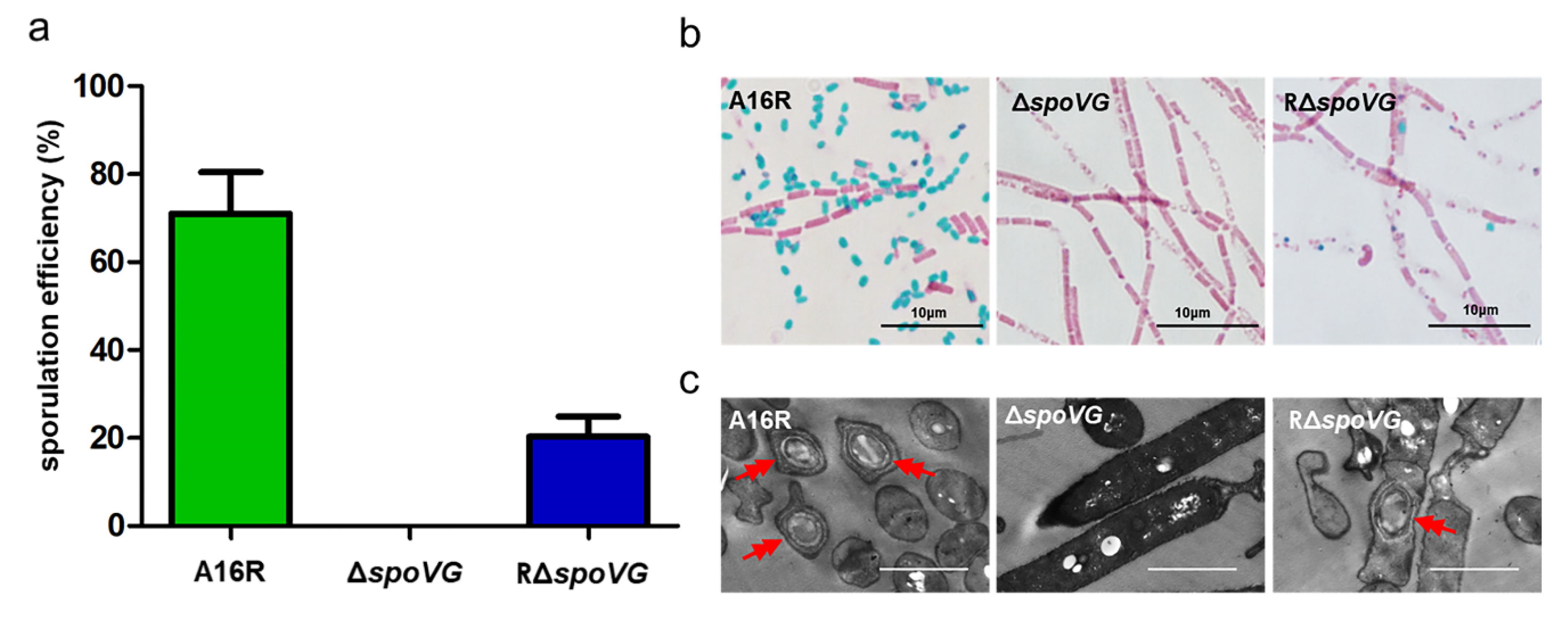
3.1. Deletion of spoVG Results in a Spore Formation Defect in B. anthracis

Although the absence of spoVG causes little impairment in sporulation in the model organism B. subtilis, it caused aberrations in the cortex [7,16] and amplified the effects of a spoIIB mutation [5,15]. To investigate whether SpoVG influenced sporulation in B. anthracis A16R, a spoVG mutant (ΔspoVG) and a plasmid-based complementation strain (RΔspoVG) were constructed. The sporulation efficiency of the ΔspoVG strain was assessed via its heat-resistance (70 °C for 30 min) after cells were cultured in DSM for 24 h. The ΔspoVG mutant had a spore formation defect, with no heat-resistant spore forms observed (Figure 2a). The complemented strain RΔspoVG showed partly restored sporulation, although the sporulation efficiency of RΔspoVG was lower than that of the wild-type strain A16R.
To further verify this mutant phenotype, cultures of the three strains were collected at 24 h and stained with malachite green and safranin O, followed by optical microscopy examination. Green spores were observed in cultures of both the wild-type strain A16R and the complemented strain RΔspoVG, but not in the ΔspoVG culture (Figure 2b). Additionally, cultures of these three strains collected after 24 h were examined using TEM. TEM images of A16R and the complemented strain RΔspoVG revealed that both formed intact spores (Figure 2c). However, TEM images of the ΔspoVG strain showed only rod-shaped, vegetative cells with no spores. These results indicated that deletion of spoVG results in a spore formation defect in B. anthracis.

3.2. Deletion of spoVG Resulted in a Complete Blockage Prior to Asymmetric Division in B. anthracis

As shown in Figure 2, no spores were formed by the ∆spoVG strain. To identify the sporulation stage at which blockage occurred, the membranes of live cells collected at T1 (8 h), T3 (10 h) and T17 (24 h) were stained with membrane-impermeable FM4-64 dye. Cells were observed using confocal microscopy, with the red fluorescent signal indicating the bacterial cell membrane. As indicated by arrows (Figure 3a), the wild-type strain A16R and the complemented strain RΔspoVG showed complete asymmetric septum formation (yellow arrow) and the engulfment process (white arrow) at T1 and T3, respectively. Bright-field imaging showed that both strains produced many mature spores (red arrows) at T17, although the number of RΔspoVG spores was lower than the number of A16R spores. However, the ΔspoVG strain did not form an asymmetric septum and showed no morphological changes. Therefore, deletion of spoVG prevented formation of an asymmetric septum.
Asymmetric division is the first obvious morphological feature of sporulation. The A16R cells underwent normal asymmetric division by T1, while ∆spoVG cells did not. To understand the physiological consequences of spoVG deletion at the proteomic level, total protein was collected from A16R and ∆spoVG cells at T1 and iTRAQ-based proteomic analysis (iTRAQ: isobaric tag for relative and absolute quantitation) was performed (Supplementary Table S2, Figure S1). The expression level of the stage II sporulation protein SpoIIE was significantly decreased in the ΔspoVG mutant (0.23-fold downregulation) (Supplementary Table S3). SpoIIE not only plays an important role in the formation of an asymmetric septum in B. subtilis, but is required for spore formation in B. anthracis [34,35]. To determine whether the spoIIE gene was responsible for the B. anthracis ΔspoVG mutant phenotype, the transcription levels and promoter activities of spoIIE were assayed. The mRNA expression level of spoIIE was significantly downregulated (50.63 ± 1.82-fold) in the ΔspoVG strain as measured using RT-qPCR (Figure 3b). Additionally, a PspoIIE–lacZ fusion was constructed. Following transformation into the wild-type strain A16R and the mutant strain ΔspoVG, this construct was used to assess transcription and regulation of the spoIIE promoter. β-Galactosidase assays showed that the transcriptional activity of PspoIIE in the ΔspoVG strain was much lower than that in the wild-type (Figure 3c). These results suggest that SpoVG positively regulates expression and transcription of spoIIE.

3.3. SpoIIB is Poorly Conserved Between B. anthracis and B. subtilis

In this study, we found that, unlike in B. subtilis, SpoVG was required for sporulation in B. anthracis. SpoVG, a pleiotropic regulatory factor, has little effect on spore formation in the model organism B. subtilis because of functional redundancy of SpoVG and SpoIIB [5]. To investigate whether this phenotypic difference is related to SpoIIB, similarity searching of SpoIIB from B. subtilis (SpoIIBBs) was performed against the NCBI and Kyoto Encyclopedia of Genes and Genomes (KEGG) databases. We identified protein BA4688 and tentatively assigned this as SpoIIB in B. anthracis; among all B. anthracis proteins, BA4688 had the highest similarity to SpoIIBBs, but the sequence similarity was low (27.9%). We aligned the amino acid sequences of SpoIIB and SpoVG from Bacillus spp. The amino acid sequence of SpoVG was highly conserved between B. anthracis, B. cereus, B. subtilis, B. thuringiensis, and B. amyloliquefaciens (Figure 4a). However, the sequences of SpoIIB proteins from the B. cereus group species [36] were distinct from those of B. subtilis and B. amyloliquefaciens (Figure 4a). I.e., the amino acid sequences of SpoIIBBs and BA4688 are quite different.
In the genome of B. anthracis, the tentative spoIIB ORF (BA4688) was located between folC and maF. However, an additional gene, comC, was located between folC and maF adjacent to spoIIB in the genome of B. subtilis (Figure 4b). In B. anthracis, the comC gene (BA1318, NCBI Reference Sequence: NC_003997.3, region 1265988 to 1266710) is located between BA1317 and BA1319. The amino acid sequence of the ComC proteins of B. anthracis and B. subtilis share only 38.91% sequence identity (92% sequence coverage) (Supplemental Table S4). These results revealed that the amino acid sequence of SpoIIB was poorly conserved between B. subtilis and B. anthracis, and that the location of the spoIIB gene differed in B. anthracis and B. subtilis.

3.4. SpoIIBBs Could Not Restore the ΔspoVG Strain to Mature Resistant Spores

A single spoVG mutation had almost no effect on sporulation in B. subtilis; this phenotype was very different from that of the ΔspoVG mutant in B. anthracis. In B. subtilis, the ΔspoVGspoIIB double mutation prevented sporulation at the engulfment stage [20]. spoIIBBs (the spoIIB gene of B. subtilis) was transferred into the B. anthracisspoVG strain derived from strain A16R to explore whether SpoIIBBs could complement the sporulation deficiency of the B. anthracisspoVG mutant. Spore formation efficiency was evaluated by culturing strains for 5 days in DSM (to ensure all spores developed to maturity). The pBspoIIBBsspoVG strain, as well as the ΔspoVG mutant, showed no spore formation (Table 2). Additionally, the heat resistance of the pBspoIIBBsspoVG strain was assayed at 24, 72, and 120 h, respectively. The pBspoIIBBsspoVG strain did not form heat-resistant spores (Figure 5a). Spore staining showed that the pBspoIIBBsspoVG strain did not form spores (green; Figure 5b). Thus, SpoIIBBs could not restore the ability of the B. anthracis ΔspoVG strain to form mature resistant spores.

3.5. SpoIIBBs Partially Restored Sporulation of ΔspoVG at the Engulfment Stage

Although the pBspoIIBBsspoVG strain could not form mature resistant spores, a normal asymmetric septum (yellow arrow) was formed at T1. The pBspoIIBBsspoVG strain subsequently underwent the prespore engulfment stage (white arrow) at T3 (Figure 6a). These results revealed that SpoIIBBs could partially restore the ability of the B. anthracis ΔspoVG strain to reach the prespore engulfment stage of sporulation, although the numbers of cells undergoing asymmetric division and engulfment were low.
To confirm this result, RNA was extracted from the A16R (wild-type), ΔspoVG, RΔspoVG, and pBspoIIBBsspoVG strains at T17. Levels of spoIIM, spoIIQ, and cotE mRNA were quantitated using RT-qPCR. Levels of spoIIM mRNA were significantly downregulated in ΔspoVG cells compared with the wild-type strain A16R (Figure 6b–d). Levels of spoIIM mRNA in the pBspoIIBBsspoVG strain were almost identical to those in strain A16R. However, levels of spoIIQ and cotE mRNA had no significant change in the pBspoIIBBsspoVG strain compared with those in strain ∆spoVG. These results indicated that SpoIIBBs could partially restore sporulation of the B. anthracis ΔspoVG strain at the engulfment stage.

4. Discussion

In this study, SpoVG was found to play a critical role in spore formation in B. anthracis. Sporulation was abolished, with blockage occurring before asymmetric septum formation, in the absence of spoVG (Figure 2 and Figure 3a). To investigate the impact of spoVG deletion on regulation of sporulation, total protein was collected from A16R (wild-type) and ΔspoVG cells at T1. iTRAQ-based proteomic analysis detected many differentially expressed proteins relating to sporulation (Supplemental Tables S2 and S3). SpoIIE, an integral membrane protein, participates in asymmetric septum formation in B. subtilis [37,38], and spoIIE mutants cannot form spores in B. anthracis [35]. Expression of SpoIIE was significantly downregulated in the ΔspoVG mutant compared with the wild-type strain, as determined using iTRAQ analysis, and RT-qPCR showed that mRNA levels of spoIIE in the ΔspoVG strain were significantly downregulated compared with those in the wild-type (Figure 3b). PspoIIE showed almost no activity in the ΔspoVG strain in a β-galactosidase activity assay (Figure 3c). These data indicate that SpoVG has a positive regulatory effect on the spoIIE gene in B. anthracis; SpoVG affects sporulation by positively regulating spoIIE (Figure 7). This finding may provide a foundation for the investigation of a potential interaction between SpoVG and SpoIIE in B. subtilis.
It is interesting that the phenotype of the ΔspoVG strain of B. anthracis was completely different from that of the ΔspoVG B. subtilis mutant, even though the spoVG gene is highly conserved between these species (Figure 4a). In B. subtilis, the absence of spoVG from sporulating cells causes aberrations in the cortex, and defects were observed at stage V of sporulation [16]. Deletion of spoVG had little effect on spore formation in B. subtilis. Only double-mutation of spoIIB and spoVG strongly blocked sporulation, at the engulfment stage [11,39] (Figure 7a). Protein BA4688 is the most similar protein encoded in the B. anthracis genome to SpoIIBBs, but the similarity is low, and the location of the putative spoIIB gene differed from that in the B. subtilis genome (Figure 4). Thus, we suspect that the low sequence similarity of SpoIIB results in the B. subtilis and B. anthracis spoVG mutants having different phenotypes.
To test this hypothesis, the spoIIBBs gene was transferred into the B. anthracis ΔspoVG strain, producing strain pBspoIIBBsspoVG. No resistant spores of this strain formed (Figure 5). In the pBspoIIBBsspoVG strain, mRNA expression of the spoIIM gene was similar to that in the wild-type strain A16R (Figure 6b), which indicates that transcription of spoIIM was not impaired in the pBspoIIBBsspoVG strain. However, the pBspoIIBBsspoVG mutant showed low-level transcription of spoIIQ and cotE (Figure 6c,d). SpoIIM is required during the early stage of engulfment, and spoIIM mutants showed blocked sporulation prior to the completion of engulfment in B. subtilis [40]. Sporulation is blocked at the late stage of engulfment in spoIIQ mutants of B. subtilis [11]. CotE plays an essential role in assembly of both the coat and exosporium in B. anthracis and B. cereus spores [13,21,41,42]. Thus, SpoIIBBs may be able to stimulate sporulation of the B. anthracis pBspoIIBBsspoVG strain to the engulfment stage (Figure 7b). This result provides evidence supporting our hypothesis—the poor sequence conservation of SpoIIB between B. anthracis and B. subtilis may be related to functional differences in SpoVG. The phenotypic differences of spoVG mutants of B. anthracis and B. subtilis may be one of the factors underlying B. anthracis pathogenicity. These data may help us further understand sporulation and provide new perspectives on B. anthracis pathogenesis.
Additionally, the B. anthracis spoVG mutant and the wild-type A16R strain showed similar growth rates during the exponential phase. However, cell densities of the ΔspoVG strain during the stationary phase were much lower than those of the wild-type (Supplementary Figure S2), consistent with the results of heat-resistance assays (Figure 5a). Thus, we speculate that SpoVG might influence cell lysis rather than growth rate.

5. Conclusions

SpoVG was found to play an indispensable role in spore formation in B. anthracis. Our study serves to deepen understanding of the function of SpoVG in B. anthracis, and of the regulatory relationships of SpoVG (Figure 7). As illustrated in Figure 7b, SpoVG is involved in spore formation by modulating the expression of SpoIIE. Because of the low sequence similarity between B. subtilis SpoIIB (SpoIIBBs) and BA4688 (the putative spoIIB of B. anthracis), we believe that SpoVG alone plays a crucial role in spore formation in B. anthracis, which is supported by our finding that sporulation was blocked before asymmetric division in the ΔspoVG strain derived from B. anthracis A16R. Our findings on the function of SpoVG in this study not only explain the functional mechanism of SpoVG in B. anthracis, but also increase our understanding of the phenotypic differences between B. anthracis and B. subtilis.

Supplementary Materials

The following are available online at https://www.mdpi.com/2076-2607/8/4/548/s1: Figure S1: Verification of differentially expressed genes using RT-qPCR; Figure S2: Effect of SpoVG on growth. Table S1: Primer sequences; Table S2: List of all differentially-expressed proteins identified via iTRAQ analysis; Table S3: List of differentially-expressed proteins related to growth and spore formation; Table S4: Conservation of comC between B. anthracis and B. subtilis.

Author Contributions

Conceptualization, H.W., D.W., Y.L. and M.C.; methodology, M.C. and Y.L.; data analysis, H.W., D.W., Y.L., L.Z. and M.C.; investigation, M.C.; resources, E.F. and H.W.; data curation, X.L., C.P.; writing—original draft preparation, M.C.; writing—review and editing, D.W., C.P. and Y.L.; supervision, funding acquisition, and project administration, D.W., X.L. and H.W. All authors have read and agreed to the published version of the manuscript.

Funding

This research was funded by the National Natural Science Foundation of China [grant numbers 81871619, 81671979, 81571958, and 81601739] and the National Science and Technology Major Project of China [grant number 2018ZX10714002].

Acknowledgments

We are very grateful to Fuping Song from the Institute of Plant Protection of China for plasmid pHT304-lacZ and his generous help.

Conflicts of Interest

The authors declare no conflict of interest. The funders of this research had no role in the design of the study; in the collection, analyses, or interpretation of the data; in the writing of the manuscript; or in the decision to publish the results.

References

  1. Hagan, A.K.; Plotnick, Y.M.; Dingle, R.E.; Mendel, Z.I.; Cendrowski, S.R.; Sherman, D.H.; Tripathi, A.; Hanna, P.C. Petrobactin Protects against Oxidative Stress and Enhances Sporulation Efficiency in Bacillus anthracis Sterne. mBio 2018, 9. [Google Scholar] [CrossRef] [PubMed] [Green Version]
  2. Tan, I.S.; Ramamurthi, K.S. Spore formation in Bacillus subtilis. Environ. Microbiol. Rep. 2014, 6, 212–225. [Google Scholar] [CrossRef] [PubMed] [Green Version]
  3. Piggot, P.J.; Coote, J.G. Genetic aspects of bacterial endospore formation. Bacteriol. Rev. 1976, 40, 908–962. [Google Scholar] [CrossRef] [PubMed] [Green Version]
  4. Stragier, P.; Losick, R. Molecular genetics of sporulation in Bacillus subtilis. Annu. Rev. Genet. 1996, 30, 297–341. [Google Scholar] [CrossRef]
  5. Margolis, P.S.; Driks, A.; Losick, R. Sporulation gene spoIIB from Bacillus subtilis. J. Bacteriol. 1993, 175, 528–540. [Google Scholar] [CrossRef] [Green Version]
  6. Wilkinson, B.J.; Deans, J.A.; Ellar, D.J. Biochemical evidence for the reversed polarity of the outer membrane of the bacterial forespore. Biochem. J. 1975, 152, 561–569. [Google Scholar] [CrossRef] [Green Version]
  7. Matsuno, K.; Sonenshein, A.L. Role of SpoVG in asymmetric septation in Bacillus subtilis. J. Bacteriol. 1999, 181, 3392–3401. [Google Scholar] [CrossRef] [Green Version]
  8. Barak, I.; Muchova, K.; Labajova, N. Asymmetric cell division during Bacillus subtilis sporulation. Future Microbiol. 2019, 14, 353–363. [Google Scholar] [CrossRef]
  9. Feucht, A.; Abbotts, L.; Errington, J. The cell differentiation protein SpoIIE contains a regulatory site that controls its phosphatase activity in response to asymmetric septation. Mol. Microbiol. 2002, 45, 1119–1130. [Google Scholar] [CrossRef]
  10. Smith, K.; Youngman, P. Evidence that the spoIIM gene of Bacillus subtilis is transcribed by RNA polymerase associated with sigma E. J. Bacteriol. 1993, 175, 3618–3627. [Google Scholar] [CrossRef] [PubMed] [Green Version]
  11. Londono-Vallejo, J.A.; Frehel, C.; Stragier, P. SpoIIQ, a forespore-expressed gene required for engulfment in Bacillus subtilis. Mol. Microbiol. 1997, 24, 29–39. [Google Scholar] [CrossRef] [PubMed]
  12. Costa, T.; Serrano, M.; Steil, L.; Volker, U.; Moran, C.P., Jr.; Henriques, A.O. The timing of cotE expression affects Bacillus subtilis spore coat morphology but not lysozyme resistance. J. Bacteriol. 2007, 189, 2401–2410. [Google Scholar] [CrossRef] [PubMed] [Green Version]
  13. Boone, T.J.; Mallozzi, M.; Nelson, A.; Thompson, B.; Khemmani, M.; Lehmann, D.; Dunkle, A.; Hoeprich, P.; Rasley, A.; Stewart, G.; et al. Coordinated Assembly of the Bacillus anthracis Coat and Exosporium during Bacterial Spore Outer Layer Formation. mBio 2018, 9. [Google Scholar] [CrossRef] [PubMed] [Green Version]
  14. Mattoo, A.R.; Saif Zaman, M.; Dubey, G.P.; Arora, A.; Narayan, A.; Jailkhani, N.; Rathore, K.; Maiti, S.; Singh, Y. Spo0B of Bacillus anthracis—A protein with pleiotropic functions. FEBS J. 2008, 275, 739–752. [Google Scholar] [CrossRef]
  15. Perez, A.R.; Abanes-De Mello, A.; Pogliano, K. SpoIIB localizes to active sites of septal biogenesis and spatially regulates septal thinning during engulfment in bacillus subtilis. J. Bacteriol. 2000, 182, 1096–1108. [Google Scholar] [CrossRef] [Green Version]
  16. Rosenbluh, A.; Banner, C.D.; Losick, R.; Fitz-James, P.C. Identification of a new developmental locus in Bacillus subtilis by construction of a deletion mutation in a cloned gene under sporulation control. J. Bacteriol. 1981, 148, 341–351. [Google Scholar] [CrossRef] [Green Version]
  17. Pan, X.; Chen, X.; Su, X.; Feng, Y.; Tao, Y.; Dong, Z. Involvement of SpoVG in hemolysis caused by Bacillus subtilis. Biochem. Biophys. Res. Commun. 2014, 443, 899–904. [Google Scholar] [CrossRef]
  18. Aung, S.; Shum, J.; Abanes-De Mello, A.; Broder, D.H.; Fredlund-Gutierrez, J.; Chiba, S.; Pogliano, K. Dual localization pathways for the engulfment proteins during Bacillus subtilis sporulation. Mol. Microbiol. 2007, 65, 1534–1546. [Google Scholar] [CrossRef] [Green Version]
  19. Liu, X.; Wang, D.; Ren, J.; Tong, C.; Feng, E.; Wang, X.; Zhu, L.; Wang, H. Identification of the immunogenic spore and vegetative proteins of Bacillus anthracis vaccine strain A16R. PLoS ONE 2013, 8, e57959. [Google Scholar] [CrossRef]
  20. Chastanet, A.; Losick, R. Engulfment during sporulation in Bacillus subtilis is governed by a multi-protein complex containing tandemly acting autolysins. Mol. Microbiol. 2007, 64, 139–152. [Google Scholar] [CrossRef]
  21. Lyu, Y.; Gu, M.; Chen, M.; Feng, E.; Zhu, L.; Pan, C.; Wang, D.; Liu, X.; Wang, H. Disruption of SpoIIID decreases sporulation, increases extracellular proteolytic activity and virulence in Bacillus anthracis. Biochem. Biophys. Res. Commun. 2019, 513, 651–656. [Google Scholar] [CrossRef] [PubMed]
  22. Arantes, O.; Lereclus, D. Construction of cloning vectors for Bacillus thuringiensis. Gene 1991, 108, 115–119. [Google Scholar] [CrossRef]
  23. Peng, Q.; Wu, J.; Chen, X.; Qiu, L.; Zhang, J.; Tian, H.; Song, F. Disruption of Two-component System LytSR Affects Forespore Engulfment in Bacillus thuringiensis. Front. Cell. Infect. Microbiol. 2017, 7, 468. [Google Scholar] [CrossRef] [PubMed] [Green Version]
  24. Wang, T.; Wang, D.; Lyu, Y.; Feng, E.; Zhu, L.; Liu, C.; Wang, Y.; Liu, X.; Wang, H. Construction of a high-efficiency cloning system using the Golden Gate method and I-SceI endonuclease for targeted gene replacement in Bacillus anthracis. J. Biotechnol. 2018, 271, 8–16. [Google Scholar] [CrossRef]
  25. Zuber, P.; Healy, J.M.; Losick, R. Effects of plasmid propagation of a sporulation promoter on promoter utilization and sporulation in Bacillus subtilis. J. Bacteriol. 1987, 169, 461–469. [Google Scholar] [CrossRef] [Green Version]
  26. Shatalin, K.Y.; Neyfakh, A.A. Efficient gene inactivation in Bacillus anthracis. FEMS Microbiol. Lett. 2005, 245, 315–319. [Google Scholar] [CrossRef] [PubMed]
  27. Grossman, A.D.; Losick, R. Extracellular control of spore formation in Bacillus subtilis. Proc. Natl. Acad. Sci. USA 1988, 85, 4369–4373. [Google Scholar] [CrossRef] [PubMed] [Green Version]
  28. Perchat, S.; Dubois, T.; Zouhir, S.; Gominet, M.; Poncet, S.; Lemy, C.; Aumont-Nicaise, M.; Deutscher, J.; Gohar, M.; Nessler, S.; et al. A cell-cell communication system regulates protease production during sporulation in bacteria of the Bacillus cereus group. Mol. Microbiol. 2011, 82, 619–633. [Google Scholar] [CrossRef] [PubMed]
  29. Peng, Q.; Kao, G.; Qu, N.; Zhang, J.; Li, J.; Song, F. The Regulation of Exosporium-Related Genes in Bacillus thuringiensis. Sci. Rep. 2016, 6, 19005. [Google Scholar] [CrossRef] [Green Version]
  30. Ter Beek, A.; Keijser, B.J.; Boorsma, A.; Zakrzewska, A.; Orij, R.; Smits, G.J.; Brul, S. Transcriptome analysis of sorbic acid-stressed Bacillus subtilis reveals a nutrient limitation response and indicates plasma membrane remodeling. J. Bacteriol. 2008, 190, 1751–1761. [Google Scholar] [CrossRef] [Green Version]
  31. Liszewski Zilla, M.; Lunderberg, J.M.; Schneewind, O.; Missiakas, D. Bacillus anthracis lcp Genes Support Vegetative Growth, Envelope Assembly, and Spore Formation. J. Bacteriol. 2015, 197, 3731–3741. [Google Scholar] [CrossRef] [PubMed] [Green Version]
  32. Jones, J.H.; Lennard-Jones, J.E.; Morson, B.C.; Chapman, M.; Sackin, M.J.; Sneath, P.H.; Spicer, C.C.; Card, W.I. Numerical taxonomy and discriminant analysis applied to non-specific colitis. Q. J. Med. 1973, 42, 715–732. [Google Scholar] [PubMed]
  33. Kumar, S.; Stecher, G.; Li, M.; Knyaz, C.; Tamura, K. MEGA X: Molecular Evolutionary Genetics Analysis across Computing Platforms. Mol. Biol. Evol. 2018, 35, 1547–1549. [Google Scholar] [CrossRef] [PubMed]
  34. Bradshaw, N.; Losick, R. Asymmetric division triggers cell-specific gene expression through coupled capture and stabilization of a phosphatase. eLife 2015, 4, e08145. [Google Scholar] [CrossRef] [PubMed]
  35. Skoble, J.; Beaber, J.W.; Gao, Y.; Lovchik, J.A.; Sower, L.E.; Liu, W.; Luckett, W.; Peterson, J.W.; Calendar, R.; Portnoy, D.A.; et al. Killed but metabolically active Bacillus anthracis vaccines induce broad and protective immunity against anthrax. Infect. Immun. 2009, 77, 1649–1663. [Google Scholar] [CrossRef] [Green Version]
  36. Cardoso, P.F.; Perchat, S.; Vilas-Boas, L.A.; Lereclus, D.; Vilas-Boas, G.T. Diversity of the Rap-Phr quorum-sensing systems in the Bacillus cereus group. Curr. Genet. 2019, 65, 1367–1381. [Google Scholar] [CrossRef]
  37. Bi, C.; Jones, S.W.; Hess, D.R.; Tracy, B.P.; Papoutsakis, E.T. SpoIIE is necessary for asymmetric division, sporulation, and expression of sigmaF, sigmaE, and sigmaG but does not control solvent production in Clostridium acetobutylicum ATCC 824. J. Bacteriol. 2011, 193, 5130–5137. [Google Scholar] [CrossRef] [Green Version]
  38. Levdikov, V.M.; Blagova, E.V.; Rawlings, A.E.; Jameson, K.; Tunaley, J.; Hart, D.J.; Barak, I.; Wilkinson, A.J. Structure of the phosphatase domain of the cell fate determinant SpoIIE from Bacillus subtilis. J. Mol. Biol. 2012, 415, 343–358. [Google Scholar] [CrossRef] [Green Version]
  39. Resnekov, O.; Driks, A.; Losick, R. Identification and characterization of sporulation gene spoVS from Bacillus subtilis. J. Bacteriol. 1995, 177, 5628–5635. [Google Scholar] [CrossRef] [Green Version]
  40. Smith, K.; Bayer, M.E.; Youngman, P. Physical and functional characterization of the Bacillus subtilis spoIIM gene. J. Bacteriol. 1993, 175, 3607–3617. [Google Scholar] [CrossRef] [Green Version]
  41. Bressuire-Isoard, C.; Bornard, I.; Henriques, A.O.; Carlin, F.; Broussolle, V. Sporulation Temperature Reveals a Requirement for CotE in the Assembly of both the Coat and Exosporium Layers of Bacillus cereus Spores. Appl. Environ. Microbiol. 2016, 82, 232–243. [Google Scholar] [CrossRef] [PubMed] [Green Version]
  42. Giorno, R.; Bozue, J.; Cote, C.; Wenzel, T.; Moody, K.S.; Mallozzi, M.; Ryan, M.; Wang, R.; Zielke, R.; Maddock, J.R.; et al. Morphogenesis of the Bacillus anthracis spore. J. Bacteriol. 2007, 189, 691–705. [Google Scholar] [CrossRef] [PubMed] [Green Version]
Figure 1. Schematic of construction of the B. anthracis ΔspoVG mutant and complemented strains. (a) Construction of the ΔspoVG strain. The construction method has been described in detail previously [24]. Briefly, the recombinant allelic exchange vector pKMUSD containing an I-SceI site was constructed. Then, pKMUSD was introduced into B. anthracis strain A16R, followed by introduction of pSS4332. Expression of the endonuclease I-SceI from pSS4332 promotes homologous recombination. When homologous recombination is completed, pSS4332 is driven out, producing a ∆spoVG mutant. (b) The complementation plasmids pBE2AspoVG and pBE2spoIIBBs were constructed as described in Materials and Methods. These plasmids were introduced into B. anthracis A16RΔspoVG competent cells, to yield strains RΔspoVG and pBspoIIBBsspoVG, respectively.
Microorganisms 08 00548 g001
Figure 2. Role of SpoVG in sporulation. (a) Sporulation efficiency of B. anthracis strains A16R (wild-type), ΔspoVG and RΔspoVG (complemented strain) cultured in DSM for 24 h. Values shown are means ± standard deviations (SDs) of triplicate experiments. (b) A16R, ΔspoVG and RΔspoVG strain cultures (24 h) were stained with malachite green and safranin O. Spores and vegetative cells are stained green and red, respectively. Scale bar, 10 μm. (c) Ultrastructural observations of the three strains using transmission electron microscopy. Red double arrows indicate spores. Scale bar, 2 μm.
Microorganisms 08 00548 g002
Figure 3. Defects in the formation of asymmetric septum in the B. anthracis ΔspoVG strain are associated with low transcriptional levels of spoIIE. (a) Confocal laser-scanning micrograph (scale bar, 10 µm) of the A16R (wild-type), ΔspoVG, and RΔspoVG strains at T1, T3, and T17 (37 °C), Tn being n hours after T0 (the end of the exponential growth phase). The cell membrane is visible as red fluorescence. Yellow, white and red arrows indicate asymmetric septum, engulfed cells (prespores), and mature spores, respectively. (b) Relative mRNA expression of spoIIE determined in the ΔspoVG and RΔspoVG strains compared with A16R (wild-type) cells at T1 using RT-qPCR. Values represent means ± SDs of triplicate experiments. (c) Transcription of PspoIIElacZ in A16R (wild-type) cells (green line) and ΔspoVG-mutant cells (red line) grown in DSM. Values represent the means of at least three independent replicates; error bars represent SDs.
Microorganisms 08 00548 g003
Figure 4. Amino acid sequence similarity of SpoVG and SpoIIB from species in the B. cereus group compared with those in B. subtilis. (a) Phylogenetic trees based on amino acid sequence alignment of SpoVG and SpoIIB, respectively. The unweighted pair group method with mean averages (UPGMA) tree was based on alignment of 10 amino acid sequences of SpoVG and SpoIIB proteins from strains belonging to the B. cereus group (seven sequences, including sequences from B. anthracis), B. subtilis (two sequences), and B. amyloliquefaciens (one sequence) available in the NCBI database (https://www.ncbi.nlm.nih.gov/). Multiple sequence alignment was conducted using ClustalX, and the tree was generated using Mega X software. The schematic shows regions of similarity rather than the complete sequence because of the length of the SpoIIB protein. SpoVG is highly conserved between B. anthracis and B. subtilis, while BA4688 (from B. anthracis) and SpoIIBBs (from B. subtilis) share a low amino acid sequence similarity. (b) Comparison of the spoIIB locus across Bacillus species. Arrows indicate the orientations of open reading frames. Original genome annotations are listed, and the names of organisms are abbreviated to: BA, B. anthracis Ames, and BS, B. subtilis 168. BA4688 represents the putative protein in B. anthracis that has the highest similarity to B. subtilis SpoIIB, but it still shares only a small region of similarity with the latter protein. There is some difference in the genetic location of BA4688 and spoIIBBs between B. anthracis and B. subtilis.
Microorganisms 08 00548 g004
Figure 5. Sporulation of the B. anthracis pBspoIIBBsspoVG strain. (a) Strains were cultured in DSM for 24, 72, and 120 h. The cultures were serially diluted and 10 μL aliquots of each dilution (10−1 through 10−5) were plated on LB-agar. After heat inactivation, spores were diluted and plated in the same manner. Images of plates after overnight incubation at 37 °C are shown. The ΔspoVG and pBspoIIBBsspoVG strains did not form heat-resistant spores. (b) Cultures of four strains were stained with malachite green and safranin O at 120 h. Spores and vegetative cells were stained green and red, respectively. Scale bar, 10 μm.
Microorganisms 08 00548 g005
Figure 6. Morphological and molecular characteristics of the B. anthracis pBspoIIBBsspoVG strain during sporulation. (a) Confocal laser-scanning micrographs (scale bar, 10 µm) of strain pBspoIIBBsspoVG at T1, T3, and T17. The cell membrane is visible as red fluorescence. Yellow and white arrows indicate the asymmetric septum and engulfed cells (prespores), respectively. (b–d) Relative mRNA expression of genes spoIIM, spoIIQ, and cotE determined in the ΔspoVG, RΔspoVG, and pBspoIIBBsspoVG strains compared with strain A16R (wild-type) at T17 using RT-qPCR. Values represent means ± SDs of at least two experiments.
Microorganisms 08 00548 g006
Figure 7. Schematic representation of regulatory pathways and their effectors in B. anthracis and B. subtilis based on the results of this study. (a) In B. subtilis, SpoVG is involved in spore formation at multiple stages (asymmetric division, engulfment, and cortex formation). Combined mutation of spoIIB and spoVG prevents spore formation at the engulfment stage. (b) In B. anthracis, the ΔspoVG mutant shows no asymmetric septum formation. SpoVG positively modulates spore formation through SpoIIE. B. subtilis SpoIIB (SpoIIBBs) partly restored spore formation in the B. anthracis ΔspoVG strain at the engulfment stage of sporulation.
Microorganisms 08 00548 g007
Table 1. Strains and plasmids used in this study.
Plasmid or StrainGenotype or DescriptionSource
Plasmid
pBE2AShuttle vector containing amylase promoter, Kanr in B. anthracis and Ampr in Escherichia coliOur lab
pBE2Shuttle vector, Kanr in B. anthracis and Ampr in E. coliOur lab [21]
pBE2AspoVGpBE2A carrying spoVG complete ORF, spoVG complementation plasmid, Ampr in E. coli, Kanr in B. anthracisThis study
pBE2spoIIBBspBE2 carrying spoIIBBs, spoIIB complementation plasmid, Ampr in E. coli, Kanr in B. anthracisThis study
pHT304Shuttle vectors, Ermr, AmprAgaisse and Lereclus [22]
pHT304-lacZPromoterless lacZ vector, Ermr, Ampr, 9.7 kbFuping Song [23]
pHT304-PspoIIEpHT304-lacZ carrying PspoIIE, Ampr in E. coli, Ermr in B. anthracisThis study
E. coli
DH5αF2, Q80d/lacZDM15, D(lacZYA-argF)U169, deoR, recA1, endA1, hsdR17(rk 2,mk + ), phoA, supE44l2, thi-1, gyrA96, relA1Transgen, Beijing, China
JM110rpsL(StrR), thr, leu, endA, thi-1, lacy, galK, galT, ara, tonA, tsx, dam-, dcm-, supE44(lac-proAB), F- [traD36, proAB, lacIqlacZΔM15]Transgen, Beijing, China
B. anthracis strain
A16RHuman vaccine strain in China; derived from A16; pXO1+, pXO2Our lab [19]
ΔspoVGA16R spoVG mutant, A16RΔspoVG: spcThis study
spoVGΔspoVG genetic complementation strain containing pBE2AspoVG plasmid; KanrThis study
pBspoIIBBsspoVG ΔspoVG genetic complementation strain containing pBE2spoIIBBs plasmid; KanrThis study
pHT304-PspoIIE/A16RA16R strain containing plasmid pHT304-PspoIIE, Ermr in B. anthracisThis study
pHT304-PspoIIE/ΔspoVGΔspoVG mutant strain containing plasmid pHT304-PspoIIE, Ermr in B. anthracisThis study
Table 2. Effect of SpoIIBBs complementation on sporulation of the B. anthracis ΔspoVG strain.
StrainViable Cells a
(CFU mL−1)
Spores a
(CFU mL−1)
Spores/Viable Cells
×100(%)
A16R1.34 × 1071.22 × 10791.04
ΔspoVG2.39 × 10400
spoVG2.73 × 1052.32 × 10584.98
pBspoIIBBsspoVG4.67 × 10400
a The values in each column represent the average of three independent experiments.

