Article

The First Step of Neurospora crassa Molybdenum Cofactor Biosynthesis: Regulatory Aspects under N-Derepressing and Nitrate-Inducing Conditions

1
TU Braunschweig, Institute of Plant Biology, 38106 Braunschweig, Germany
2
TU Braunschweig, Institute of Physical and Theoretical Chemistry, Antibody Facility, 38106 Braunschweig, Germany
3
Center of Molecular Ecophysiology (CMEP), College of Resources and Environment, Southwest University No. 2, Tiansheng Road, Beibei District, Chongqing 400715, China
*
Author to whom correspondence should be addressed.
Microorganisms 2020, 8(4), 534; https://doi.org/10.3390/microorganisms8040534
Submission received: 10 March 2020 / Revised: 30 March 2020 / Accepted: 3 April 2020 / Published: 7 April 2020
(This article belongs to the Special Issue The Filamentous Fungus Neurospora crassa)

Abstract

:
Molybdenum cofactor (Moco) is the active site prosthetic group found in all Moco dependent enzymes, except for nitrogenase. Mo-enzymes are crucial for viability throughout all kingdoms of life as they catalyze a diverse set of two electron transfer reactions. The highly conserved Moco biosynthesis pathway consists of four different steps in which guanosine triphosphate is converted into cyclic pyranopterin monophosphate, molybdopterin (MPT), and subsequently adenylated MPT and Moco. Although the enzymes and mechanisms involved in these steps are well characterized, the regulation of eukaryotic Moco biosynthesis is not. Within this work, we described the regulation of Moco biosynthesis in the filamentous fungus Neurospora crassa, which revealed the first step of the multi-step pathway to be under transcriptional control. We found, that upon the induction of high cellular Moco demand a single transcript variant of the nit-7 gene is increasingly formed pointing towards, that essentially the encoded enzyme NIT7-A is the key player for Moco biosynthesis activity in Neurospora.

1. Introduction

In order to gain biological activity, molybdenum (Mo) needs to be coordinated by molybdopterin (MPT), resulting in the formation of the molybdenum cofactor (Moco). Moco is an essential component of vitally important enzymes found in all kingdoms of life [1]. These enzymes contribute to the biogenic nitrogen, carbon, and sulfur cycles [2]. Moco biosynthesis is an evolutionary old and highly conserved multi-step pathway which can be divided into four main steps [3]. In eukaryotes, the first step of Moco biosynthesis is generally assumed to be localized in the mitochondria of the cell [3,4]. While in plants, this step is encoded by two gene loci (cnx2 and cnx3), in humans and the filamentous fungus Neurospora crassa (N. crassa), a single gene locus (MOCS1 and nit-7, respectively) encodes both domains required for catalytic activity [5,6,7]. nit-7 consists of two exons (exon 1 and exon 2) flanking the single intron 1 [7]. In the recent model, translation of the non-spliced nit-7 transcript leads to the protein NIT-7A, which is encoded by nit-7 exon 1 and to a small part by intron 1. Splicing of the nit-7 mRNA results in an inactive NIT-7A domain fused to the NIT-7B domain. This variant is referred to as NIT-7AB [7]. As shown for bacterial orthologues [8,9], NIT-7A is supposed to catalyze the conversion of guanosine triphosphate (GTP) to 3′,8-cH2GTP, which is subsequently processed by the NIT-7B domain to cyclic pyranopterin monophosphate (cPMP). In the following, cPMP is exported into the cytoplasm of the cell [1,4] where all subsequent Moco biosynthesis steps take place [1]. In the second step of Moco biosynthesis, the dithiolene motif is introduced into cPMP, thus forming the Mo chelating scaffold molybdopterin (MPT) [10,11,12]. This reaction is performed by the heterotetrameric MPT synthase complex, which consists of two sulfur carrier subunits (N. crassa NIT-8) and two catalytic subunits (N. crassa NIT-12) [1,3,7]. As the MPT synthase catalyzes the stoichiometric conversion of cPMP to MPT, after each reaction cycle, it needs to be regenerated by the MPT synthase sulfurase (N. crassa NIT-1) [1,3,7]. Within steps three and four of the pathway, MPT is initially adenylated yielding adenylated MPT (MPT-AMP) [13,14], which is subsequently hydrolyzed to form Moco [15]. In N. crassa, these steps are catalyzed by a two domain protein encoded by nit-9 [7]. Here, the NIT-9G domain adenylates MPT and the NIT-9E domain accepts the synthesized MPT-AMP as substrate for the subsequent molybdate insertion reaction, resulting in the formation of physiologically active Moco [1,3,7] (Figure 1).
While Moco biosynthesis has been studied extensively in prokaryotes [17,18,19] and eukaryotes [1], little is known about its regulation in eukaryotes. In this study, we took advantage of the model organism N. crassa in which one of the main users of Moco, i.e. nitrate reductase (NR; gene locus nit-3) [20] is under strong transcriptional control [21], which guarantees, that nit-3 is only expressed in the presence of nitrate and under nitrogen-derepressing conditions [22]. In the present work, we characterized the transcriptional regulation of the N. crassa Moco biosynthesis pathway, revealing the expression of nit-7 to be under nitrogen dependent control. Within this work, we revealed NIT-7A to be localized in the mitochondria of the cell and found that the mitochondrial import of NIT-7B requires the fusion of NIT-7A to NIT-7B. However, contrary to the present model of NIT-7 functionality [7], the NIT-7AB fusion protein was found to be degraded upon mitochondrial import, resulting in the release of NIT-7B from the fusion protein. Our new model for NIT-7 functionality was confirmed by functional complementation studies, which solidified the requirement of separate NIT-7A and a NIT-7AB derived, separate NIT-7B domain for cPMP synthesis in N. crassa.

2. Material and Methods

2.1. Neurospora crassa Strains and Growth Conditions

N. crassa strains used in this study are listed in Table 1.
Strains were grown on Vogel’s minimal medium (MM) [23] supplemented with 1.5% (w/v) sucrose as carbon source and at 30 °C. N. crassa liquid cultures were cultivated at 130 rpm. If required, 1.5% (w/v) agar were added to solidify media. Appropriate supplements and antibiotics were added for N. crassa knock out (ko) strains or strains carrying resistance markers for selection. For growth under nitrogen repressing (N-repressing) or N-derepressing and nitrate-inducing conditions [22], 75 mM ammonium chloride (NH4Cl) or 80 mM potassium nitrate (KNO3, nitrate medium) were added as sole nitrogen sources to the culture medium, respectively. Then, 300 mM sodium chlorate (NaClO3) were added to MM (chlorate medium) to test for catalytically active NR [24]. For crosses, synthetic cross medium (SC) was used [25]. Crossing plates were incubated at room temperature (RT) until ejected ascospores were seen. After incubating the ascospores for ~7 d in ripening buffer (0.1 M Tris-HCl pH 8.0, 2 mM EDTA) in the dark at RT, they were subsequently heat activated at 60 °C for 10 min and grown on BDES plates [26] at 30 °C overnight. Mating type determination of generated knock outs was carried out as described [27]. For race tube experiments, 3 mL solid nitrate or chlorate medium were filled in 10 mL sterile plastic serological pipets [28]. After the inoculation of 104 conidia, race tubes were incubated for 72 h at 30 °C and in the dark.

2.2. Transformation of Neurospora crassa

N. crassa strains were transformed by electroporation of macroconidia as described by [29]. Homokaryotic strains were obtained either by single spore isolation or by crossing with an appropriate partner.

2.3. PCR-Based Cloning and Mutagenesis

PCR-based cloning was carried out with gene-specific primers (Table S1) and Q5® High-Fidelity DNA Polymerase or Phusion® High-Fidelity DNA Polymerase (New England Biolabs, Ipswich, MA, USA). Obtained PCR products were subcloned using the CloneJET Cloning Kit (Thermo Fisher Scientific, Waltham, MA, USA). If indicated, nucleotide exchanges were introduced with QuikChange II (Agilent Technologies, Santa Clara, CA, USA) modified for the use of Phusion® High-Fidelity DNA Polymerase [30]. Primers used for site directed mutagenesis are listed in Table S1. Plasmids were generated by restriction digest based cloning, overlap extinction PCR based cloning, or the NEBuilder® HiFi DNA Assembly Cloning Kit (New England Biolabs, Ipswich, MA, USA). As shuttle vector for multiplying plasmids in E. coli and genomic integration of DNA fragments into the N. crassa his-3 locus, a pCCG-C-eGFP construct or derivatives were used (Table 1) [31].

2.4. RNA Extraction and cDNA Synthesis

Total RNA was extracted with the Quick-RNATM Fungal/Bacterial Kit (Zymo, Irvine, CA, USA) followed by DNA removal using the TURBO DNA-freeTM Kit (Thermo Fisher Scientific, Waltham, MA, USA) according to the manufacturer’s instructions. RNA concentrations of the same measurement series were subsequently adjusted to identical concentrations. For cDNA synthesis, SuperScript IV reverse transcriptase (Thermo Fisher Scientific, Waltham, MA, USA) was used according to the manufacturer’s instructions.

2.5. Transcript Analysis

After cDNA synthesis, Moco biosynthesis gene expression was analyzed PCR-based using gene-specific primers (Table S1) and the GoTaq® DNA Polymerase (Promega, Flitchburg, WI, USA). Upon DNA gel-electrophoresis, the PCR products were documented using a ChemiDoc XRS+ imager (Bio-Rad, Hercules, CA, USA) and quantified with the Image Lab 6.0 software (Bio-Rad, Hercules, CA, USA). Quantification of act-2 (NCU04173) cDNA served as control and reference here.

2.6. 5′ rapid amplification of cDNA ends

For 5′ rapid amplification of cDNA ends (5′ RACE), the SMARTer® RACE 5′/3′ Kit (Takara, Kusatsu, Shiga, Japan) was used. RNA extraction and TURBOTM DNase digestion was performed as described, followed by an additional RNA clean up step with the RNA Clean and ConcentratorTM-5 Kit (Zymo, Irvine, CA, USA). Then, 1 µg total RNA was used for first strand synthesis with a nit-7 gene-specific primer (Table S1). Touchdown PCR was carried out for the amplification of nit-7 transcripts according to the manufacturer’s instructions. Obtained PCR products were isolated and cloned into the pRACE (Takara, Kusatsu, Shiga, Japan) or pJET vector (Thermo Fisher Scientific, Waltham, MA, USA), respectively.

2.7. Protein and Molybdenum Cofactor Metabolites Extraction from N. crassa

After overnight growth of N. crassa in liquid medium, mycelium was harvested and ground in liquid nitrogen. Subsequently, 250 µL of ice cold extraction buffer (50 mM sodium phosphate buffer, 200 mM NaCl, 5 mM EDTA, 2 % (v/v) glycerol, 5 mM glutathione, protease inhibitor (Roche cOmpleteTM, EDTA-free), pH 7.2) were added to 100 mg ground mycelium and then incubated for 30 min on ice. Samples were mixed occasionally, followed by a centrifugation step at 21,000× g for 10 min at 4 °C. The obtained supernatants were transferred into fresh reaction tubes. Protein concentration was determined by Bradford protein assay (ROTI®-Quant, Carl Roth, Karlsruhe, BW, Germany) with bovine serum albumin serving as a concentration standard.

2.8. Neurospora crassa Nitrate Reductase Activity Assay

N. crassa NR activity was determined as described previously [32,33].

2.9. Generation of Monoclonal Antibodies for NIT-7A and Nitrate Reductase

The monoclonal antibodies 4C10 (anti-NIT-7A) and 5G7 (anti-NR) were generated by immunizing mice with recombinant NIT-7A or with the recombinant cytochrome c reducing fragment of N. crassa NR [33] following a standard immunization protocol. After hybridization and cloning, antibody producing hybridoma cells were screened for their binding to recombinant NIT-7A or the recombinant NR cytochrome c reducing fragment, respectively, by use of ELISA and Western blot analysis. Isotype analysis of the clones 4C10 and 5G7 revealed them to be an IgG1 subtype. Supernatants were grown either according to standard protocols or in serum-free medium. Both antibodies were used as culture supernatant.

2.10. Detection of Enhanced Green Fluorescent Protein

Recombinant eGFP fusion proteins in N. crassa were detected immune blot-based using an anti-green fluorescent protein mouse IgG1 κ monoclonal antibody (Roche, Basel, BS, Switzerland). To recognize the primary antibodies used in this study, we used Peroxidase-conjugated AffiniPure Goat Anti-Mouse IgG + IgM (H+L) (Dianova, Hamburg, Germany), which was diluted 1: 10,000 in 20 mL TBS-T and was incubated 1 h with the membrane at RT and under shaking.

2.11. Quantitative Detection of Molybdenum Cofactor and Its Metabolites

Oxidation and quantification of cPMP and Moco/MPT was carried out essentially as described previously [16,34,35,36,37].

2.12. Bright Field Microscopy

Hyphae of strains grown on MM, nitrate, and chlorate medium were documented with the Nikon Eclipse Ni upright microscope equipped with the DS-Qi2 camera (Nikon, Shinagawa, Tokyo, Japan). The 60× Plan Apo VC (numerical aperture 1.4, oil immersion) was used as objective. Images were taken with the NIS Elements software (Nikon, Shinagawa, Tokyo, Japan) and processed using ImageJ v.1.52e (NIH, Bethesda, MD, USA).

2.13. Confocal Microscopy

To determine the subcellular localization of NIT-7 variants, heterokaryons of strains expressing nit-7-egfp variants (Table 1) and an N. crassa mitochondrial marker strain expressing the atp-1 presequence fused to the mCherry coding sequence [31] were prepared as described previously [31]. Spatial localization was analyzed with the cLSM-510META scan head connected to the Axiovert 200M (Carl Zeiss, Oberkochen, BW, Germany) in a single-tracking mode. An argon laser with 488 nm (LGK 7812 ML4, Lasos) was used for eGFP excitation and fluorescence was detected by using the main dichroic beam splitter HFT UV/488/543/633 and the bandpass filter (505–530 nm) in the channel mode in front of the detector. For mCherry excitation, a helium-neon laser with 543 nm (LGK 7786P, Lasos, Jena, TH, Germany) was employed. mCherry fluorescence was detected by using the main dichroic beam splitter HFT UV/488/543/633 and a longpass filter (560 nm). When appropriate, bright field images of samples were taken with the transmitted light photomultiplier. Unidirectional sequential scanning was done with a line average of 4, 8, or 16. As objective, the 10x Plan-Neofluar (numerical aperture 0.3) and the 40× C-Apochromat (numerical aperture 1.2, water immersion) were used. Images were taken using the ZEN software (Carl Zeiss, Oberkochen, BW, Germany) and processed using ImageJ v.1.52e (NIH, Bethesda, MD, USA).

3. Results

Nitrate reductase expression is connected to Moco biosynthesis regulation—As nitrate reductase activity depends on active site bound Moco, biosynthesis of Moco is a requirement for nitrate assimilation. However, whereas the regulation of NR is well characterized in the fungi Aspergillus nidulans (A. nidulans) and N. crassa [22], regulation of Moco biosynthesis is not, and notably, this also holds true for any other eukaryote. To provide first insights into the regulatory aspects behind eukaryotic Moco biosynthesis, we characterized the effect(s) of induced NR gene expression on the N. crassa Mo-metabolism. To do so, N. crassa was initially cultivated under nitrogen repressing (N-repressing) conditions and subsequently transferred to nitrate-containing media, which was depleted of any reduced N-source to establish N-derepressing and nitrate-inducing conditions [22]. Here, upon defined periods of growth, NR formation was documented Western blot based by use of a highly specific, monoclonal NR antibody, derived against the NR cytochrome c reducing fragment (Figure 2A).
No NR is formed in mycelia grown under N-repressing conditions (Figure 2A, 0 min), while under N-derepressing and nitrate-inducing conditions, NR becomes detectable Western blot-based at t40 (Figure 2A). Consistently, NR activity is measurable here and reaches its maximum at time point t200 (Figure 2B). The amount of NR, that is detectable by Western blot (Figure 2A), nearly correlates with the quantified NR activities (Figure 2B). Therefore, growth under N-derepressing and nitrate-inducing conditions results in the formation of enzymatically active NR. NR is a Moco dependent enzyme, and consistently the total amount of Moco/MPT (t0 = 3.7 ± 0.7 pmol per mg crude extract, t200 = 6.01 ± 0.66 pmol per mg crude extract) detectable upon induction of NR gene expression was found to correlate with the detectable NR enzymatic activity (Figure 2B). Next to Moco/MPT (Moco biosynthesis steps 2 and 3–4, Figure 1), also the Moco biosynthesis first step product cPMP can be quantified by HPLC. The quantified cPMP amounts (t0 = 32.2 ± 16.9 pmol per mg crude extract, t200 = 59.31 ± 26.44 pmol per mg crude extract) were found to correlate with the detectable Moco/MPT amounts, whereas the absolute cPMP amounts exceed the absolute Moco/MPT amounts ~ 10 fold (Figure 2B). To shed light on the underlying reason(s) for enhanced cPMP and Moco/MPT amounts, we next quantified the Moco biosynthesis gene expression strengths.
Transcriptional regulation of Neurospora crassa Moco biosynthesis—In the following reverse transcription, PCR was employed to trace back changes of the Moco biosynthesis gene expression levels under N-derepressing and nitrate-inducing conditions. Notably, for the first step of N. crassa Moco biosynthesis, two functional nit-7 transcript variants were reported [7]. To distinguish between the intron-free (nit-7 Δ1460–1696) and the non-spliced nit-7 variant, the primers for nit-7 amplification were chosen to include the single nit-7 intron (I1) which encodes for the functional relevant double glycine motif of NIT-7A. NIT-7A is encoded by exon 1 (E1), while a linker-region and NIT-7B are encoded by exon 2 (E2). The positions of the primers used for transcript quantification are indicated by arrows above the schematic representation of the nit-7 splice variants in Figure 3A.
Amongst all transcripts analyzed (Figure S1), exclusively the amount of the non-spliced nit-7 mRNA (encoding NIT-7A) was found to be significantly enhanced under N-derepressing and nitrate-inducing conditions (Figure 3B). Therefore, enhanced Moco demand results in an increase of the non-spliced nit-7 transcript and hence the regulation of nit-7 splicing.
nit-7 splice variants—The human nit-7 homolog MOCS1 encodes for multiple splice variants with differences in the 5′- and 3′- ends [6,38,39,40]. To test whether or not also yet unidentified nit-7 splice variants contribute to fungal cPMP synthesis and are potentially subject to transcriptional regulation, we carried out a 5′ RACE (5′ rapid amplification of cDNA ends) experiment using mycelium grown under N-derepressing and nitrate-inducing conditions (t40, see also Figure 2). 5′ RACE yielded products sized ~2.5 kb (Figure 4). These were sequenced, revealing that the two known nit-7 splice variants (non-spliced nit-7 and nit-7 Δ1460–1696, see Figure 3A for comparison) were not separated by gel electrophoresis but were detected as one band. Next to these, a novel yet unknown splice variant nit-7 Δ1469–1696 was identified by sequencing (Figure 4), whose relevancy for Moco biosynthesis has been studied by us in the following.
The Neurospora crassa nit-phenotype—In the available literature, e.g., [7,22,41] N. crassa nit-strains are described to lack the nitrate utilization capability (nit is the abbreviation for nitrate non utilizer) and next to this rather qualitative description, no quantitative data describing the nit-phenotype is available so far. To document any impact of the novel splice variant nit-7 Δ1469–1696 on N. crassa Moco biosynthesis, we established quantitative methods which aimed to provide the experimental basis for any further phenotypic characterization of the N. crassa Moco biosynthesis. Therefore, we initially quantified the hyphal growth of the nit-7 knock out (ko) strain (Table 1, Figure 5) [7] and unexpectedly we found this strain to possess residual growth under N-derepressing and nitrate-inducing conditions (nitrate-containing medium) (wildtype (wt) = 21.7 ± 2.7 cm, nit-7 ko strain = 5.5 ± 1.0 cm), although significant growth on chlorate-containing medium was detected (wt = 2.4 ± 0.4 cm, nit-7 ko strain = 19.6 ± 0.6 cm), (Figure 5C).
As growth of any nit-strain on chlorate-containing medium naturally excludes growth on nitrate-containing medium [24], we next went on to identify the underlying reason behind this observation. We found that the nit-7 ko strain possesses a reduced lateral spreading as compared to the wt strain when grown on nitrate-containing medium. Furthermore, the diameter of primary hyphae of both, the wt and the nit-7 ko strain grown on nitrate and on chlorate-containing medium was quantified (Figure 5B), respectively. When grown on nitrate-containing medium, we identified nit-7 ko strain hyphae to possess a significantly reduced diameter and an enhanced number of vacuoles as compared to the wt strain (Figure 5B). However, when the N. crassa nit-7 ko strain was grown on chlorate-containing medium, the hyphae diameter and vacuole number was comparable to that documented for the N. crassa wt strain cultivated on nitrate-containing medium.
We conclude, that when cultivated on nitrate-containing medium, the nit-7 ko strain metabolizes endogenously available N-sources which—combined with (i) reduced lateral spreading, (ii) a reduction of the hyphae diameter, and (iii) an increase of the vacuole number—best explains the detected linear growth here.
Relevancy of splice variant nit-7 Δ1469–1696 for Moco biosynthesis—Upon establishing quantitative methods to characterize the N. crassa nit-phenotype, we were able to characterize the relevancy of nit-7 splice variant Δ1469–1696 for N. crassa Moco biosynthesis. For this, N. crassa strain #557 (Table 1), expressing the nit-7 variant incompetent of producing nit-7 Δ1469–1696 ectopically in a nit-7 ko background [7] (Table 1, Figure 5A) was created. For comparison, N. crassa strain #555 (Table 1) was generated, ectopically expressing the nit-7 variant incompetent of producing nit-7 Δ1460–1696 again in the nit-7 ko background [7].
Using the above described quantitative methods, we assayed strains #557 and #555, whereby we also established a control strain expressing wt nit-7 ectopically in the nit-7 ko background [7]. As expected, strain #554 displays significant growth on nitrate-containing medium (17.6 ± 1.1 cm), comparable to that of the wt strain (21.7 ± 2.7 cm) and also hyphal diameters were found to be wt like (wt strain = 4.6 ± 0.9 µm, #554 = 5.2 ± 0.7 µm). On the contrary, strain #555 (expressing the nit-7 variant incompetent of producing nit-7 Δ1460–1696) does not possess significant growth. Strain #557 (expressing the nit-7 variant incompetent of producing nit-7 Δ1469–1696) shows wt like growth behavior except for reduced hyphae diameter on nitrate-containing medium (Figure 5B). When grown on chlorate-containing medium, all strains showed the complementing phenotype expected from growth on nitrate-containing medium. Hence, we conclude that the novel nit-7 splice variant Δ1469–1696 is not essential for Moco biosynthesis.
The NIT-7 functional domains—As cPMP synthesis depends on both NIT-7 domains, the contribution of domains A, B, and the AB domain fusion to N. crassa cPMP synthesis was deciphered. The A. nidulans nit-7 homolog cnxABC was discussed to be expressed as bicistronic transcript, translating both catalytic domains separately [42]. In line with this, the nit-7 exon 2 harbors a potential start codon (http://atgpr.dbcls.jp/) [43], which encodes for the first residue of NIT-7B (M623). To test for a possible function of a separate NIT-7B domain starting with M623, three novel N. crassa strains within the nit-7 ko background [7] were created, each constitutively expressing either NIT-7A (N. crassa strain #552), NIT-7B (#558), or NIT-7AB (#553), respectively. The functional interplay of the respective NIT-7 domains was subsequently assayed by heterokaryon growth (Figure 6), which has been quantified as described above.
This setup revealed that growth on nitrate-containing medium depends on the presence of both, NIT-7A and NIT-7AB in the heterokaryon. Notably, wt-like growth of the heterokaryons formed by strains expressing NIT-7A plus NIT-7B or NIT-7AB plus NIT-7B was not detectable. As plant and mammalian cPMP synthesis was reported to be localized in the mitochondria of the cell [4,44], we determined the subcellular localization of the fungal NIT-7A, NIT-7AB, and NIT-7B domain.
Localization of NIT-7 domains—To determine the subcellular localization of NIT-7A, NIT-7B, and NIT-7AB, transgenic N. crassa strains expressing the respective C-terminal eGFP fusions were created (Table 1). NIT-7A- and NIT-7AB-eGFP fusions localized to the mitochondria (Figure 7A,B), while in contrast, NIT-7B did not (Figure 7C).
Recent work carried out in the human system reported that cytosolic cleavage of the NIT-7AB homologous protein MOCS1AB exposes an internal mitochondrial targeting signal, affecting the mitochondrial import of thus liberated MOCS1B [44]. To test for the putative cytosolic cleavage and subsequent mitochondrial import of NIT-7B, we created an N. crassa strain expressing a nit-7ab-egfp variant, lacking the mitochondrial targeting signal encoding sequence. This truncated NIT-7AB-eGFP fusion protein was shown to reside in the cytosol of the cell, while no mitochondrial import of NIT-7B eGFP was detectable (Figure S2). Therefore, we conclude that the mammalian import model for the first step Moco biosynthesis proteins [44] does not hold true for N. crassa NIT-7.
While in N. crassa, no cytosolic release of NIT-7B from the AB domain fusion was detectable, we identified the NIT-7AB-eGFP fusion (possessing the N-terminal mitochondrial targeting signal) to lack the A-domain after import into the mitochondria (Figure 7B). This finding was documented by the fact that the mitochondrial localized NIT-7B-eGFP fusion was detectable immunoblot-based while the A-domain was not (Figure 7B). We conclude that upon mitochondrial import, NIT-7B is released from NIT-7AB. Therefore, the current model of NIT-7 functionality [7] requires revision.

4. Discussion

Synthesis of Moco involves the stepwise conversion of GTP into the known Moco metabolites 3′,8-cH2GTP [8,9], cPMP [16] (Figure 8A), MPT [10,11,12], MPT-AMP [13,14], and finally Moco [1,45,46,47]. Numerous Moco biosynthesis enzymes are required for its synthesis [1,3], which demands the precise coordination of the expression strengths of all encoding genes. In our work, we identified the tight regulation of the (Moco-dependent) NR in N. crassa to be interlaced with the Moco biosynthesis pathway on the transcriptional level. Under nitrogen derepressing (N-derepressing) and nitrate-inducing conditions, expression of nitrate reductase (nit-3) [20], nitrite reductase (nit-6) [48], and the nitrate importer (nit-10) [49] is up-regulated (Figure 8B). In our work, we identified the amounts of the non-spliced NIT-7A encoding nit-7 gene transcript (Figure 8D) to be likewise enhanced under these conditions, while the expression strengths of the remaining Moco-biosynthesis genes were not up-regulated. This finding underlines the results of [50], who suggest that in Escherichia coli, the NIT-7A homolog MoaA catalyzes the rate-determining step in cPMP synthesis (Figure 8A), as documented by the finding that the kcat for the conversion of 3´,8-cH2GTP into cPMP is approximately 4-fold higher than the kcat determined for the conversion of GTP to 3´,8-cH2GTP [8,9].
However, to our surprise, the absolute HPLC-based pterin quantification revealed that in N. crassa, cPMP amounts are highly elevated as against Moco/MPT amounts at any time point in the experimental series (Figure 2B, Figure 8C). We conclude, that cPMP is not quantitatively converted into MPT— neither under N-repressing nor N-derepressing and nitrate-inducing conditions. Nonetheless, the cPMP to Moco/MPT ratio increased in a time-dependent fashion; evidently, nit-7 expression ultimately leads to enhanced cPMP production. As enhanced amounts of cellular cPMP goes hand in hand with the formation of enhanced Moco/MPT amounts and enhanced NR enzymatic activity, we conclude that steps two and three–four of the pathway provide a constant cPMP conversion capacity, which is only fully used when the cellular cPMP amount is maximal (Figure 8C). Consistently, our transcript quantification approach identified the amount of the remaining Moco-biosynthesis transcripts to be not significantly enhanced under N-derepressing and nitrate-inducing conditions (Figure 8B). Transcriptional regulation results in the enhanced formation of non-spliced nit-7 transcript (Figure 8D), which encodes the mitochondrial localized NIT-7A domain. However, for cPMP synthesis, the NIT-7B domain likewise requires mitochondrial import.
Most interestingly here, the NIT-7AB fusion was found to undergo proteolytic cleavage after mitochondrial import, which results in the degeneration of the A-domain and the liberation of NIT-7B. This finding is contrary to that reported for the mammalian system where native MOCS1AB was found to undergo proteolytic cleavage, thus exposing an internal mitochondrial import signal required for the mitochondrial import of the B-domain [44]. Therefore, in both—mammals and fungi—other than postulated earlier [7,51], functionality of the first Moco biosynthesis step essentially depends on the presence of separated A- and B-domains in the mitochondria. The existence of different pathways in mammals and fungi, which both result in the B-domain release from the AB precursor, allows it to conclude that these have evolved after the fungal and animal lineage diverged from their last common ancestor [52].

Supplementary Materials

Supplementary materials can be found at https://www.mdpi.com/2076-2607/8/4/534/s1.

Author Contributions

S.W.: Acquisition of data, data analysis and interpretation. T.W.H.: Adaption of the race tube system to nitrate and chlorate-containing media. S.B.: Generation of monoclonal antibodies. R.H.: LSM Microscopy. R.R.M.: Interpretation of data, final approval of the version to be published. T.K.: Conception and design, analysis and interpretation of data, drafting the article, final approval of the version to be published. All authors have read and approved the final version of the manuscript.

Funding

This work was financed by a grant of the Deutsche Forschungsgemeinschaft to R.R.M. and T.K. (GRK 2223/1), by a grant of the Deutsche Forschungsgemeinschaft to R.R.M. (Me1266/35-1) and by a grant of the Deutsche Forschungsgemeinschaft to R.R.M. (FOR12220, TP1).

Acknowledgments

We thank Brigitte M. Jockusch (TU Braunschweig, Cell Biology, Zool. Institute) for her expert support with generating the monoclonal antibodies used in this study. We thank Kevin D. Oliphant (TU Braunschweig, Institute of Plant Biology) for his excellent technical support. We thank Phillip Ringel (TU-Braunschweig, Institute of Plant biology) for his help with generating the monoclonal NR antibody used in this study. We acknowledge the FGSC (Kansas City, Missouri USA) for continuous support.

Conflicts of Interest

The authors declare no conflict of interest. The funders had no role in the design of the study; in the collection, analyses, or interpretation of data; in the writing of the manuscript, or in the decision to publish the results.

References

  1. Mendel, R.R.; Kruse, T. Cell biology of molybdenum in plants and humans. Biochim. Biophys. Acta 2012, 1823, 1568–1579. [Google Scholar] [CrossRef] [PubMed] [Green Version]
  2. Stiefel, E.I. The biogeochemistry of molybdenum and tungsten. Met. Ions Biol. Syst. 2002, 39, 1–29. [Google Scholar] [PubMed]
  3. Mendel, R.R. The molybdenum cofactor. J. Biol. Chem. 2013, 288, 13165–13172. [Google Scholar] [CrossRef] [PubMed] [Green Version]
  4. Teschner, J.; Lachmann, N.; Schulze, J.; Geisler, M.; Selbach, K.; Santamaria-Araujo, J.; Balk, J.; Mendel, R.R.; Bittner, F. A novel role for Arabidopsis mitochondrial ABC transporter ATM3 in molybdenum cofactor biosynthesis. Plant Cell 2010, 22, 468–480. [Google Scholar] [CrossRef] [Green Version]
  5. Reiss, J.; Christensen, E.; Kurlemann, G.; Zabot, M.T.; Dorche, C. Genomic structure and mutational spectrum of the bicistronic MOCS1 gene defective in molybdenum cofactor deficiency type A. Hum. Genet. 1998, 103, 639–644. [Google Scholar] [CrossRef]
  6. Reiss, J.; Cohen, N.; Dorche, C.; Mandel, H.; Mendel, R.R.; Stallmeyer, B.; Zabot, M.T.; Dierks, T. Mutations in a polycistronic nuclear gene associated with molybdenum cofactor deficiency. Nat. Genet. 1998, 20, 51–53. [Google Scholar] [CrossRef]
  7. Probst, C.; Ringel, P.; Boysen, V.; Wirsing, L.; Alexander, M.M.; Mendel, R.R.; Kruse, T. Genetic characterization of the Neurospora crassa molybdenum cofactor biosynthesis. Fungal Genet. Biol. 2014, 66, 69–78. [Google Scholar] [CrossRef]
  8. Hover, B.M.; Loksztejn, A.; Ribeiro, A.A.; Yokoyama, K. Identification of a cyclic nucleotide as a cryptic intermediate in molybdenum cofactor biosynthesis. J. Am. Chem. Soc. 2013, 135, 7019–7032. [Google Scholar] [CrossRef] [Green Version]
  9. Hover, B.M.; Tonthat, N.K.; Schumacher, M.A.; Yokoyama, K. Mechanism of pyranopterin ring formation in molybdenum cofactor biosynthesis. Proc. Natl. Acad. Sci. USA 2015, 112, 6347–6352. [Google Scholar] [CrossRef] [Green Version]
  10. Pitterle, D.M.; Johnson, J.L.; Rajagopalan, K.V. In vitro synthesis of molybdopterin from precursor Z using purified converting factor. Role of protein-bound sulfur in formation of the dithiolene. J. Biol. Chem. 1993, 268, 13506–13509. [Google Scholar]
  11. Johnson, M.E.; Rajagopalan, K.V. Involvement of chlA, E, M, and N loci in Escherichia coli molybdopterin biosynthesis. J. Bacteriol. 1987, 169, 117–125. [Google Scholar] [CrossRef] [PubMed] [Green Version]
  12. Johnson, M.E.; Rajagopalan, K.V. In vitro system for molybdopterin biosynthesis. J. Bacteriol. 1987, 169, 110–116. [Google Scholar] [CrossRef] [PubMed] [Green Version]
  13. Llamas, A.; Mendel, R.R.; Schwarz, G. Synthesis of adenylated molybdopterin: An essential step for molybdenum insertion. J. Biol. Chem. 2004, 279, 55241–55246. [Google Scholar] [CrossRef] [PubMed] [Green Version]
  14. Kuper, J.; Llamas, A.; Hecht, H.-J.; Mendel, R.R.; Schwarz, G. Structure of the molybdopterin-bound Cnx1G domain links molybdenum and copper metabolism. Nature 2004, 430, 803–806. [Google Scholar] [CrossRef]
  15. Llamas, A.; Otte, T.; Multhaup, G.; Mendel, R.R.; Schwarz, G. The Mechanism of nucleotide-assisted molybdenum insertion into molybdopterin. A novel route toward metal cofactor assembly. J. Biol. Chem. 2006, 281, 18343–18350. [Google Scholar] [CrossRef] [Green Version]
  16. Wuebbens, M.M.; Rajagopalan, K.V. Structural characterization of a molybdopterin precursor. J. Biol. Chem. 1993, 268, 13493–13498. [Google Scholar]
  17. Leimkühler, S.; Wuebbens, M.M.; Rajagopalan, K.V. The history of the discovery of the molybdenum cofactor and novel aspects of its biosynthesis in bacteria. Coord. Chem. Rev. 2011, 255, 1129–1144. [Google Scholar] [CrossRef] [Green Version]
  18. Mendel, R.R.; Leimkühler, S. The biosynthesis of the molybdenum cofactors. JBIC J. Biol. Inorg. Chem. 2015, 20, 337–347. [Google Scholar] [CrossRef]
  19. Zupok, A.; Iobbi-Nivol, C.; Mejean, V.; Leimkuhler, S. The regulation of Moco biosynthesis and molybdoenzyme gene expression by molybdenum and iron in bacteria. Metallomics 2019, 11, 1602–1624. [Google Scholar] [CrossRef]
  20. Nason, A.; Evans, H.J. Triphosphopyridine nucleotide-nitrate reductase in Neurospora. J. Biol. Chem. 1953, 202, 655–673. [Google Scholar]
  21. Marzluf, G.A. Metabolic Regulation in Fungi. Appl. Mycol. Biotechnol. 2001, 1, 55–72. [Google Scholar]
  22. Marzluf, G.A. Genetic regulation of nitrogen metabolism in the fungi. Microbiol. Mol. Biol. Rev. 1997, 61, 17–32. [Google Scholar] [CrossRef] [PubMed]
  23. Vogel, H.J. A convenient growth medium for Neurospora (Medium N). Microb. Genet. Bull. 1956, 13, 42–43. [Google Scholar]
  24. Tomsett, A.B.; Garrett, R.H. The isolation and characterization of mutants defective in nitrate assimilation in Neurospora crassa. Genetics 1980, 95, 649–660. [Google Scholar]
  25. Westergaard, M.; Mitchell, H.K.; Neurospora, V. A Synthetic Medium Favoring Sexual Reproduction. Am. J. Bot. 1947, 34, 573–577. [Google Scholar] [CrossRef]
  26. Davis, R.H.; de Serres, F.J. Genetic and microbiological research techniques for Neurospora crassa. Methods Enzymol. 1970, 27, 79–143. [Google Scholar]
  27. Perkins, D.D.; Turner, B.C.; Pollard, V.C.; Fairfield, A. Neurospora strains incorporating fluffy, and their use as testers. Fungal Genet. Rep. 1989, 36, 64. [Google Scholar] [CrossRef] [Green Version]
  28. White, B.; Woodward, D. A simple method for making disposable race tubes. Fungal Genet. Rep. 1995, 42, 79. [Google Scholar] [CrossRef] [Green Version]
  29. Chakraborty, B.N.; Kapoor, M. Transformation of filamentous fungi by electroporation. Nucleic Acids Res. 1990, 22, 6737. [Google Scholar] [CrossRef] [Green Version]
  30. Xia, Y.; Chu, W.; Qi, Q.; Xun, L. New insights into the QuikChangeTM process guide the use of Phusion DNA polymerase for site-directed mutagenesis. Nucleic Acids Res. 2015, 43, e12. [Google Scholar] [CrossRef] [Green Version]
  31. Wirsing, L.; Klawonn, F.; Sassen, W.A.; Lünsdorf, H.; Probst, C.; Hust, M.; Mendel, R.R.; Kruse, T.; Jänsch, L. Linear discriminant analysis identifies mitochondrially localized proteins in Neurospora crassa. J. Proteome Res. 2015, 14, 3900–3911. [Google Scholar] [CrossRef] [PubMed]
  32. Evans, H.J.; Nason, A. The effect of reduced triphosphopyridine nucleotide on nitrate reduction by purified nitrate reductase. Arch. Biochem. Biophys. 1952, 39, 234–235. [Google Scholar] [CrossRef]
  33. Ringel, P.; Krausze, J.; Van Heuvel, J.D.; Curth, U.; Pierik, A.J.; Herzog, S.; Mendel, R.R.; Kruse, T. Biochemical characterization of molybdenum cofactor-free nitrate reductase from Neurospora crassa. J. Biol. Chem. 2013, 288, 14657–14671. [Google Scholar] [CrossRef] [PubMed] [Green Version]
  34. Zupok, A.; Gorka, M.; Siemiatkowska, B.; Skirycz, A.; Leimkühler, S. Iron-dependent regulation of molybdenum cofactor biosynthesis genes in Escherichia coli. J. Bacteriol. 2019, 201, e00382-19. [Google Scholar] [CrossRef] [PubMed] [Green Version]
  35. Rieder, C.; Eisenreich, W.; O’Brien, J.; Richter, G.; Gotze, E.; Boyle, P.; Blanchard, S.; Bacher, A.; Simon, H. Rearrangement reactions in the biosynthesis of molybdopterin an NMR study with multiply 13C/15N labelled precursors. Eur. J. Biochem. 1998, 255, 24–36. [Google Scholar] [CrossRef] [PubMed]
  36. Johnson, J.L.; Hainline, B.E.; Rajagopalan, K.V.; Arison, B.H. The Pterin Component of the Molybdenum Cofactor—Structural Characterization of two Fluorescent Derivatives. J. Biol. Chem. 1984, 259, 5414–5422. [Google Scholar]
  37. Hercher, T.W.; Krausze, J.; Hoffmeister, S.; Zwerschke, D.; Lindel, T.; Blankenfeldt, W.; Mendel, R.-R.; Kruse, T. Insights into the Cnx1E catalyzed MPT-AMP hydrolysis. Biosci. Rep. 2020, 40. [Google Scholar] [CrossRef] [Green Version]
  38. Arenas, M.; Fairbanks, L.D.; Vijayakumar, K.; Carr, L.; Escuredo, E.; Marinaki, A.M. An unusual genetic variant in the MOCS1 gene leads to complete missplicing of an alternatively spliced exon in a patient with molybdenum cofactor deficiency. J. Inherit. Metab. Dis. 2009, 32, 560–569. [Google Scholar] [CrossRef]
  39. Gross-Hardt, S.; Reiss, J. The bicistronic MOCS1 gene has alternative start codons on two mutually exclusive exons. Mol. Genet. Metab. 2002, 76, 340–343. [Google Scholar] [CrossRef]
  40. Gray, T.A.; Nicholls, R.D. Diverse splicing mechanisms fuse the evolutionarily conserved bicistronic MOCS1A and MOCS1B open reading frames. RNA 2000, 6, 928–936. [Google Scholar] [CrossRef] [Green Version]
  41. Sorger, G.J.; Giles, N.H. Genetic control of nitrate reductase in Neurospora crassa. Genetics 1965, 52, 777–788. [Google Scholar] [PubMed]
  42. Reiss, J. Genetics of molybdenum cofactor deficiency. Hum. Genet. 2000, 2, 157–163. [Google Scholar] [CrossRef]
  43. Salamov, A.A.; Nishikawa, T.; Swindells, M.B. Assessing protein coding region integrity in cDNA sequencing projects. Bioinformatics 1998, 14, 384–390. [Google Scholar] [CrossRef] [PubMed] [Green Version]
  44. Mayr, S.J.; Röper, J.; Schwarz, G. Alternative splicing of the bicistronic gene molybdenum cofactor synthesis 1 (MOCS1) uncovers a novel mitochondrial protein maturation mechanism. J. Biol. Chem. 2020, 1. [Google Scholar] [CrossRef] [PubMed]
  45. Rajagopalan, K.V. Novel aspects of the biochemistry of the molybdenum cofactor. Adv. Enzymol. Relat. Areas Mol. Biol. 1991, 64, 215–290. [Google Scholar]
  46. Kramer, S.P.; Johnson, J.L.; Ribeiro, A.A.; Millington, D.S.; Rajagopalan, K.V. The structure of the molybdenum cofactor. Characterization of di-(carboxamidomethyl) molybdopterin from sulfite oxidase and xanthine oxidase. J. Biol. Chem. 1987, 262, 16357–16363. [Google Scholar]
  47. Romao, M.J.; Archer, M.; Moura, I.; Moura, J.J.; LeGall, J.; Engh, R.; Schneider, M.; Hof, P.; Huber, R. Crystal structure of the xanthine oxidase-related aldehyde oxido- reductase from D. gigas. Science 1995, 270, 1170–1176. [Google Scholar] [CrossRef]
  48. Exley, G.E.; Colandene, J.D.; Garrett, R.H. Molecular cloning, characterization, and nucleotide sequence of nit-6, the structural gene for nitrite reductase in Neurospora crassa. J. Bacteriol. 1993, 175, 2379–2392. [Google Scholar] [CrossRef] [Green Version]
  49. Gao-Rubinelli, F.; Marzluf, G.A. Identification and characterization of a nitrate transporter gene in Neurospora crassa. Biochem. Genet. 2004, 42, 21–34. [Google Scholar] [CrossRef]
  50. Pang, H.; Yokoyama, K. Lessons from the Studies of a C–C Bond Forming Radical SAM Enzyme in Molybdenum Cofactor Biosynthesis. Methods Enzymol. 2018, 606, 485–522. [Google Scholar]
  51. Reiss, J.; Hahnewald, R. Molybdenum cofactor deficiency: Mutations in GPHN, MOCS1, and MOCS2. Hum. Mutat. 2011, 32, 10–18. [Google Scholar] [CrossRef] [PubMed] [Green Version]
  52. Wainright, P.O.; Hinkle, G.; Sogin, M.L.; Stickel, S.K. Monophyletic origins of the metazoa: An evolutionary link with fungi. Science 1993, 260, 340–342. [Google Scholar] [CrossRef] [PubMed]
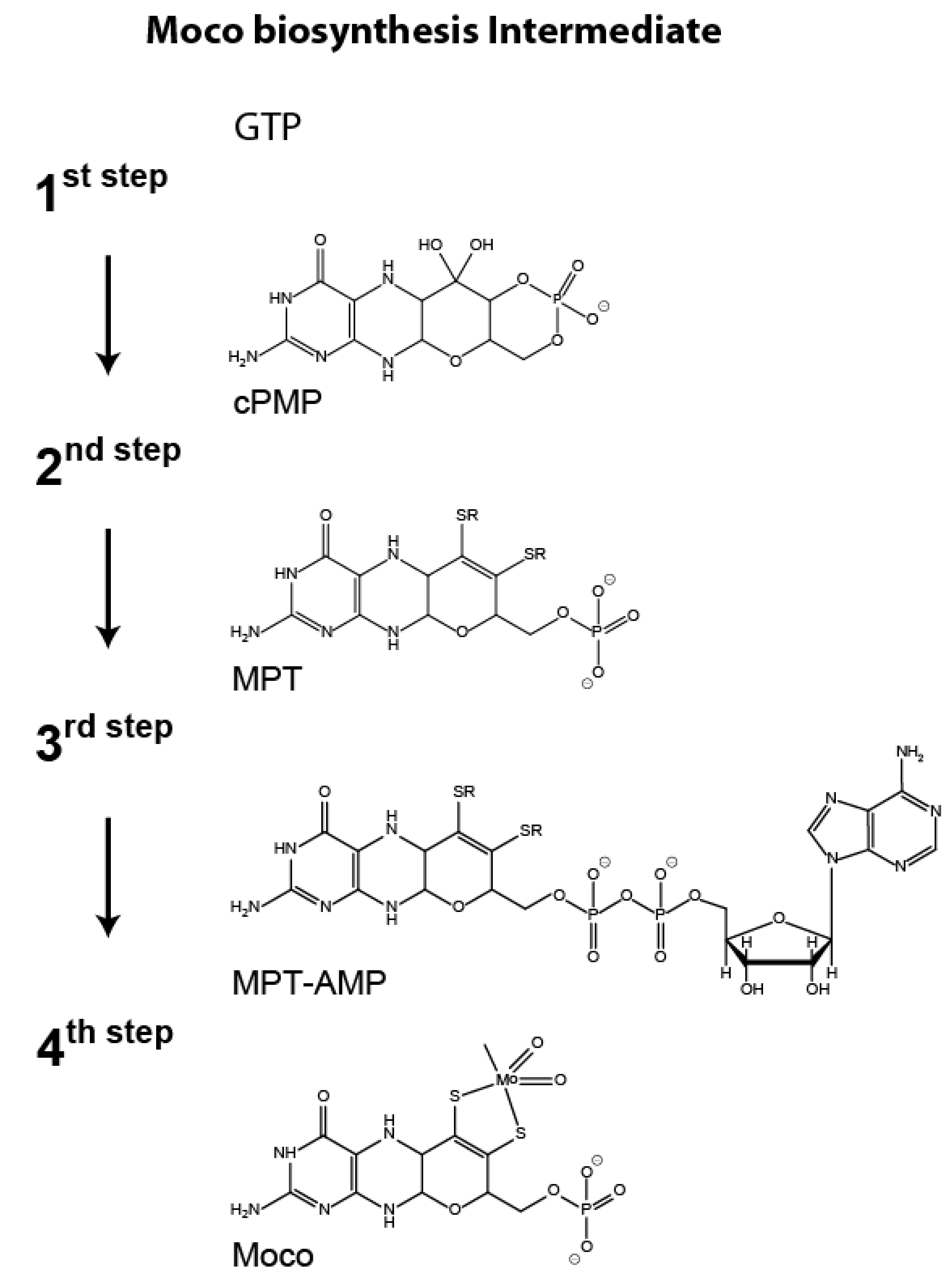
Figure 1. Scheme of the molybdenum cofactor biosynthesis pathway. Biosynthesis of the molybdenum cofactor (Moco) involves the stepwise conversion of guanosine triphosphate (GTP) to cyclic pyranopterin monophosphate (cPMP [8,9,16], step 1) which is converted to molybdopterin (MPT, [10,11,12]) in the following pathway step 2. In the third step, MPT is initially adenylated yielding adenylated MPT (MPT-AMP, [13,14]), which is subsequently converted to Moco [1,15] in the fourth pathway step.
Microorganisms 08 00534 g001
Figure 2. Connection of the Neurospora crassa Mo-metabolism with the nitrate assimilation pathway. (A) Western blot-based detection of nitrate reductase (NR) in Neurospora crassa. (N. crassa) crude extracts obtained at the indicated time points upon growth under nitrogen derepressing (N-derepressing) and nitrate-inducing conditions. (B) Quantitative detection of cyclic pyranopterin monophosphate (cPMP), molybdopterin (MPT), molybdenum cofactor (Moco), and NR activity in N. crassa crude extracts obtained at the indicated time points under N-derepressing and nitrate-inducing conditions. Four biological replicates were analyzed. Error bars indicate the standard deviation.
Microorganisms 08 00534 g002
Figure 3. Molecular characterization of nit-7 splice variants. (A) Schematic representation of the two known Neurospora crassa nit-7 splice variants and the respective, encoded proteins [7]. Numbers indicate the nucleotide or amino acid positions, respectively. (B) Relative expression levels of nit-7 non-spliced and nit-7 Δ1460–1696 (see also Figure S1). Three biological replicates were analyzed. Error bars indicate the standard deviation and asterisks indicate significant differences compared to N-repressing conditions with p ≤ 0.05 according to Student’s t-Test.
Microorganisms 08 00534 g003
Figure 4. Identification of the novel nit-7 splice variant nit-7 Δ1469–1696. (A) Results from 5′ RACE (5′ rapid amplification of cDNA ends). (B) Schematic representation of the nit-7 exon (E) / intron (I) structure of non-spliced nit-7, splice variants nit-7 Δ1460–1696 and nit-7 Δ1469–1696, respectively. Numbers below indicate the nucleotide position of the exon / intron borders. When existent in the respective nit-7 mRNA, the stop codon within intron 1 is indicated above. (C) Schematic representation of the encoded NIT-7 domains. The first and last amino acids of NIT-7A and NIT-7B are given below. The functional relevant double glycine motif encoded by I1 and the linker region encoded by exon 2 (white bar) are indicated. Numbers refer to the amino acid position within NIT-7.
Microorganisms 08 00534 g004
Figure 5. Quantification of the Neurospora crassa nit-phenotype. (A) Nitrate-dependent growth (i.e., growth under nitrogen-derepressing and nitrate-inducing conditions = nitrate medium) of indicated N. crassa strains, see Table 1, wt = wild type. In the complementary experimental setup, growth was quantified on medium containing chlorate. (B) The hyphal diameter of the strains analyzed in (A) has been determined and is shown as Tukey´s box blots. Multiple comparisons analysis was carried out using ordinary one-way ANOVA (Tukey´s method) with **** p ≤ 0.001. (C) Representative images of hyphae from strains assayed in (A+B) on different media.
Microorganisms 08 00534 g005
Figure 6. Functional complementation of the Neurospora crassa nit-7 mutant strain. (A) Neurospora crassa nit-7 knock out strains ectopically expressing either nit-7a, nit-7b, or nit-7ab (Table 1) were generated and grown as heterokaryons under nitrogen derepressing and nitrate-inducing conditions (nitrate-containing medium) and on chlorate-containing medium, respectively. (B) The hyphal diameter of the strains analyzed in (A) has been determined and is shown as Tukey´s box plots. Multiple comparisons analysis was carried out using ordinary one-way ANOVA (Tukey´s method). Differences, which are not significant, are indicated (ns).
Microorganisms 08 00534 g006
Figure 7. Localization of NIT-7. (A I) Subcellular localization of the NIT-7A-eGFP fusion protein, (A II) subcellular localization of ATP-1-Presequence-mCherryNC [31], (A III) overlay of the signals obtained for I and II. (B I) Subcellular localization of the NIT-7AB-eGFP fusion protein, (B II) subcellular localization of ATP-1-Presequence-mCherryNC, (B III) overlay of the signals obtained for I and II. (C I) Subcellular localization of the NIT-7B-eGFP fusion protein, (C II) subcellular localization of ATP-1-Presequence-mCherryNC, (C III) overlay of the signals obtained for I and II. (A) Scale bar = 10 µm. Scale bars for (B) and (C) were not shown but are identical to that shown in (A). The corresponding immuno-blots are shown next to the results from the live cell imaging approach, with the used antibodies indicated. For immune blotting, 20 µg of crude extracts prepared from the analyzed strains were used. The primary and secondary antibodies used are described in detail in the Materials and Methods section.
Microorganisms 08 00534 g007
Figure 8. Co-regulation of Neurospora crassa molybdenum cofactor biosynthesis and nitrate assimilation. (A) Schematic reaction detail of the first step of molybdenum cofactor (Moco) biosynthesis, where GTP is sequentially processed yielding 3´,8-cH2GTP as intermediate and cyclic pyranopterin monophosphate (cPMP) as reaction product. Here, NIT-7A is suggested to act as 3´,8-cH2GTP cyclase, whereby NIT-7B is assumed to function as cPMP synthase [8,9]. (B) Schematic representation of the interlaced regulation of the Moco biosynthesis and nitrate assimilation pathway in Neurospora crassa. + = nitrogen derepressing (N-derepressing) and nitrate-inducing conditions. Triangles indicate the promotors, black boxes the terminators of the genes indicated. (C) Quantitative data for cellular Moco/molybdopterin (MPT) and cPMP amounts at t40 and t200 (single biological experiment, see Figure 2). The dashed lines indicate the Moco/MPT synthesis implemented at t40 and t200, respectively. The regulatory range is defined as t200t40. (D) Detail of the regulatory scheme shown in (B). Under N-derepressing and nitrate-inducing conditions (red arrow), the non-splice nit-7 transcript (encoding NIT-7A, see A) is increasingly expressed.
Microorganisms 08 00534 g008
Table 1. Neurospora crassa strains used in this work. Next to the strain number, for Pccg-1 driven genes, the encoded protein product is given in parenthesis. mts: mitochondrial targeting sequence. eGFP: enhanced green fluorescent protein.
Strain NumberGenotypeOrigin
FGSC 248974-OR23-1V; mat AFGSC
FGSC 4317fl; mat AFGSC
FGSC 4347fl; mat aFGSC
FGSC 9720his-3-; Δmus-52::bar+; mat AFGSC
G200 (ATP-1-Preseq-mCherryNC)his-3::Pccg1-atp1.1_120-mcherry; Δmus-52::bar+; mat AWirsing et al., 2015
G253Δmus-52::bar+; Δnit-7::hph+; mat aProbst et al., 2014
G546his-3-; Δmus-52::bar+; Δnit-7::hph+; mat AThis study
G554his-3::nit-7+(EC); Δmus52::bar+; Δnit-7::hph+; mat AThis study
G555his-3::nit-7.1461T>C(EC); Δmus52::bar+; Δnit-7::hph+; mat AThis study
G557his-3::nit-7.1470T>C(EC); Δmus52::bar+; Δnit-7::hph+; mat AThis study
G552 (NIT-7A)his-3::Pccg1-nit-7.1_1467; Δnit-7::hph+; Δmus-52::bar+; mat AThis study
G553 (NIT-7AB)his-3::Pccg1-nit-7.1_1459+1697_2664; Δnit-7::hph+; Δmus-52::bar+; mat AThis study
G558 (NIT-7B)his-3::Pccg1-nit-7.2104_2664; Δnit-7::hph+; Δmus-52::bar+; mat AThis study
G542 (NIT-7A-eGFP)his-3::Pccg1-nit-7.1_1464-egfp; Δmus-52::bar+; mat AThis study
G544 (NIT-7AB-eGFP)his-3::Pccg1-nit-7.1_1459+1697_2661-egfp; Δmus-52::bar+; mat AThis study
G550 (NIT-7B-eGFP)his-3::Pccg1-nit-7.2104_2661-egfp; Δmus-52::bar+; mat AThis study
G575 (NIT-7A without mts-eGFP)his-3::Pccg1-nit-7.163_1464-egfp; Δmus-52::bar+; mat AThis study
G576 (NIT-7AB without mts-eGFP)his-3::Pccg1-nit-7.163_1459+1697_2661-egfp; Δmus-52::bar+; mat AThis study
G577 (Linker-NIT-7B-eGFP)his-3::Pccg1-nit-7.1459+1697_2661-egfp; Δmus-52::bar+; mat AThis study