Article

The Role of Petrimonas mucosa ING2-E5AT in Mesophilic Biogas Reactor Systems as Deduced from Multiomics Analyses

1
Center for Biotechnology (CeBiTec), Genome Research of Industrial Microorganisms, Bielefeld University, Universitätsstr. 27, 33615 Bielefeld, Germany
2
Faculty of Technology, Bielefeld University, Universitätsstr. 25, 33615 Bielefeld, Germany
3
Bioprocess Engineering, Otto von Guericke University Magdeburg, Universitätspl. 2, 39106 Magdeburg, Germany
4
Database and Software Engineering Group, Department of Computer Science, Institute for Technical and Business Information Systems, Otto von Guericke University Magdeburg, Universitätspl. 2, 39106 Magdeburg, Germany
5
Department of Bioengineering, Leibniz Institute for Agricultural Engineering and Bioeconomy, Max-Eyth-Allee 100, 14469 Potsdam, Germany
6
Biological and Chemical Engineering Section (BCE), Department of Engineering, Aarhus University, 8000 Aarhus, Denmark
7
Department Bioinformatics and Databases, Leibniz Institute DSMZ-German Collection of Microorganisms and Cell Cultures GmbH, Inhoffenstraße 7B, 38124 Braunschweig, Germany
8
Bioprocess Engineering, Max Planck Institute for Dynamics of Complex Technical Systems, Sandtorstr. 1, 39106 Magdeburg, Germany
9
Microbiology, Anhalt University of Applied Sciences, Bernburger Straße 55, 06354 Köthen, Germany
10
Chair of Microbiology, Technical University of Munich, Emil-Ramann-Str. 4, 85354 Freising, Germany
11
Institute of Molecular Genetics, National Research Centre «Kurchatov Institute», Kurchatov Sq. 2, 123128 Moscow, Russia
12
Institute of Agricultural and Urban Ecological Projects Affiliated to Berlin Humboldt University (IASP), Philippstraße 13, 10115 Berlin, Germany
*
Author to whom correspondence should be addressed.
Microorganisms 2020, 8(12), 2024; https://doi.org/10.3390/microorganisms8122024
Submission received: 12 November 2020 / Revised: 11 December 2020 / Accepted: 14 December 2020 / Published: 17 December 2020
(This article belongs to the Section Environmental Microbiology)

Abstract

:
Members of the genera Proteiniphilum and Petrimonas were speculated to represent indicators reflecting process instability within anaerobic digestion (AD) microbiomes. Therefore, Petrimonas mucosa ING2-E5AT was isolated from a biogas reactor sample and sequenced on the PacBio RSII and Illumina MiSeq sequencers. Phylogenetic classification positioned the strain ING2-E5AT in close proximity to Fermentimonas and Proteiniphilum species (family Dysgonomonadaceae). ING2-E5AT encodes a number of genes for glycosyl-hydrolyses (GH) which are organized in Polysaccharide Utilization Loci (PUL) comprising tandem susCD-like genes for a TonB-dependent outer-membrane transporter and a cell surface glycan-binding protein. Different GHs encoded in PUL are involved in pectin degradation, reflecting a pronounced specialization of the ING2-E5AT PUL systems regarding the decomposition of this polysaccharide. Genes encoding enzymes participating in amino acids fermentation were also identified. Fragment recruitments with the ING2-E5AT genome as a template and publicly available metagenomes of AD microbiomes revealed that Petrimonas species are present in 146 out of 257 datasets supporting their importance in AD microbiomes. Metatranscriptome analyses of AD microbiomes uncovered active sugar and amino acid fermentation pathways for Petrimonas species. Likewise, screening of metaproteome datasets demonstrated expression of the Petrimonas PUL-specific component SusC providing further evidence that PUL play a central role for the lifestyle of Petrimonas species.

1. Introduction

Anaerobic digestion (AD) is commonly applied for treatment of organic wastes in order to achieve reduction of waste combined with parallel recovery of bioenergy [1,2]. The degradation of organic matter is a complex microbial process featuring interaction of several groups of microorganisms with different interacting or even interfering metabolic capabilities and growth requirements [3]. The microbial biogas network is complex and intertwined and therefore susceptible to environmental changes. Several factors causing process inhibition have been identified, such as temperature fluctuations, foaming, high concentrations in hydrogen sulfide or ammonia [4,5,6,7], imbalanced levels of light or heavy metals, the presence of interfering organic substances such as halogenated alkanes or aromatic compounds [8] as well as increased organic loading rates (OLR) [9,10]. Among these, the most frequent incident type described in literature resulting in process inhibition is the accumulation of ammonia [3]. Due to inhibition of syntrophic volatile fatty acid (VFA) oxidizers, high ammonia concentrations result in VFA accumulation which subsequently inhibits methanogenesis. Moreover, ammonia also directly inhibits methanogens [11].
In the past decades, remarkable progress has been made towards understanding of how disturbances affect the microbial community structures, biodiversity losses, and ecological processes [12,13,14,15,16,17,18]. Due to further improvements and developments in high-throughput sequencing technologies, deep insights into the microbial community became possible, allowing predictions on interrelations between the microbial community composition, interspecies interactions, and species as well as community dependencies concerning supplied AD substrates and deteriorating process parameters. In connection with the formation of VFA [19] and increased ammonium concentrations, it has been observed that members of the phylum Bacteroidetes, namely species of the families Dysgonomonadaceae (formerly Porphyromonadaceae) and Marinilabiaceae, are frequently abundant in mesophilic biogas reactor systems [20,21]. Often, corresponding biogas plants were fed with food waste or protein-rich and hardly digestible substrates such as straw showing unstable process flow with variations in biogas/methane yields and increased nitrogen/ammonia levels [13,22].
The family Dysgonomonadaceae comprises species that frequently are associated with a variety of human infections [23], but also species detected in different types of biogas reactor systems. The latter are closely related to Proteiniphilum acetatigenes [24], Fermentimonas caenicola [25], and Petrimonas sulfuriphila [26]. Currently, the family Dysgonomonadaceae comprises the genera Dysgonomonas, Fermentimonas, Petrimonas, and Proteiniphilum [27]. The genera Fermentimonas and Petrimonas, originally assigned to the family Porphyromonadaceae [25], were recently transferred to the family Dysgonomonadaceae and now represent a sister group of the genus Proteiniphilum [27]. Besides utilization of complex proteinaceous substrates such as yeast extract and peptone, members of the Dysgonomonadaceae family are able to ferment a wide range of mono- and disaccharides [23,25,28,29].
The impact of Dysgonomonadaceae members on biomass degradation in AD reactor systems operating at high OLR or increased nitrogen/ammonia levels caused by proteinaceous substrates has been demonstrated recently [9,10]. Under mesophilic conditions, an increase in OLR had a positive effect on the abundance of Dysgonomonadaceae members of the genera Proteiniphilum and Petrimonas [9]. These observations led to the assumption that these genera may serve as microbial early warning indicators for an unbalanced process. They are therefore regarded as potential marker microorganisms featuring outstanding performance under stress conditions [9,10]. Moreover, a study published by He and coworkers [7] demonstrated the proliferation of members of the genus Petrimonas in biogas reactors that were operated under unfavorable or unstable conditions indicated by foaming. The relative abundance of Petrimonas was highest (29.34%) in foaming samples, and this value was almost seven times higher than those for non-foaming samples. Hence, it was speculated that the presence of Dysgonomonadaceae members, in particular of acid-producing Petrimonas bacteria, might help to optimize VFA compositions and to accelerate methane production by driving the formation of acetic acid and carbon dioxide [19].
To overcome the limited knowledge on microbial species affecting AD in a positive or negative way, a number of previously unknown bacterial species were isolated from biogas reactor samples and subsequently characterized regarding their physiological and genetic features [25,30,31,32,33,34]. Among these, Petrimonas mucosa ING2-E5AT was isolated from a mesophilic laboratory-scale biogas reactor [25]. Microbiological characterization revealed that this strain is able to utilize complex proteinaceous substrates, as well as to ferment mono- and disaccharides. Considering these phenotypic characteristics required for effective biomass conversion, the aim of this study was to establish and analyze the complete genome sequence of P. mucosa ING2-E5AT in order to understand the strain’s functioning and competitiveness in biogas plant environments.

2. Materials and Methods

2.1. Strain Cultivation and DNA Isolation

P. mucosa ING2-E5AT was obtained from a sample taken from a mesophilic laboratory-scale biogas reactor continuously fed with a mixture of maize silage (10% of organic dry matter, ODM), pig (45% ODM) and cattle manure (45% ODM) as described previously [25]. The isolate ING2-E5AT was grown at 37 °C on Columbia Agar Base medium supplemented with 5% horse blood and 1.7 g/L sodium propionate. The extraction of genomic DNA was performed using the Gentra Puregene Yeast/Bact. Kit (Qiagen, Hilden, Germany) following the manufacturer’s instructions. Unfortunately, required DNA purity criteria being A260/280 and A260/230 values greater than 1.8 were not achieved after DNA extraction with the Gentra Puregene Yeast/Bact. Kit. Therefore, obtained DNA was additionally purified using the NucleoSpin® gDNA Clean-up kit (Macherey-Nagel, Düren, Germany).

2.2. PacBio Library Preparation, Sequencing, and Genome Assembly

Approximately 8 μg genomic DNA was used for the SMRTbellTM template library construction, which was prepared according to the instructions from Pacific Biosciences (Menlo Park, CA, USA) following the ’Procedure and Checklist—Greater Than 10 kb Template Preparation’. SMRT sequencing was carried out on the PacBio RSII (Pacific Biosciences, MenloPark, CA, USA) taking one 240-min movie for two SMRT cells using the P6 Chemistry. Subsequently, SMRT Cell data was assembled using the ‘RS_HGAP_Assembly.3’ protocol included in the SMRT Portal version 2.3.0 using default parameters. The assembly revealed a circular chromosome. The ‘RS_Bridgemapper.1’ protocol was applied to check the validity of the assembly. The chromosome was circularized and particularly artificial redundancies at the ends of the contigs were removed and adjusted to dnaA as the first gene. Finally, error-correction was performed by a mapping of paired-end MiSeq Illumina reads of 2 × 300 bp obtained previously for the strain ING2-E5AT [34] onto the finished genome using BWA [35] with subsequent variant and consensus calling using VarScan [36]. For further genome assembly details refer to [37].
The finished genome sequence was imported into the annotation platform GenDB [38] for automatic prediction of genes as described previously [29]. To identify phage-related genes and genomic islands (GI), the P. mucosa ING2-E5AT genome was uploaded in PHASTER [39] and IslandViewer 3.0 [40]. Finally, the program EDGAR 2.0 [41], a software tool for the comparative analysis of prokaryotic genomes, was applied in order to phylogenetically classify completely sequenced and annotated members of the order Bacteroidales as well as to analyze them in a comparative manner.

2.3. Reconstruction of Metabolic Pathways

To predict genes encoding carbohydrate-active enzymes, the carbohydrate-active enzyme database (CAZy) annotation web-server dbCAN [42] was applied. Metabolic pathways of interest were reconstructed based on EC numbers of enzymes and their assignment to KEGG pathway maps (https://www.genome.jp/kegg/pathway.html). Hydrogenases were identified using blastp and the database HydDB with a threshold of 1 × 10−100. In addition, further classification of the predicted hydrogenases was also performed by means of HydDB [43].

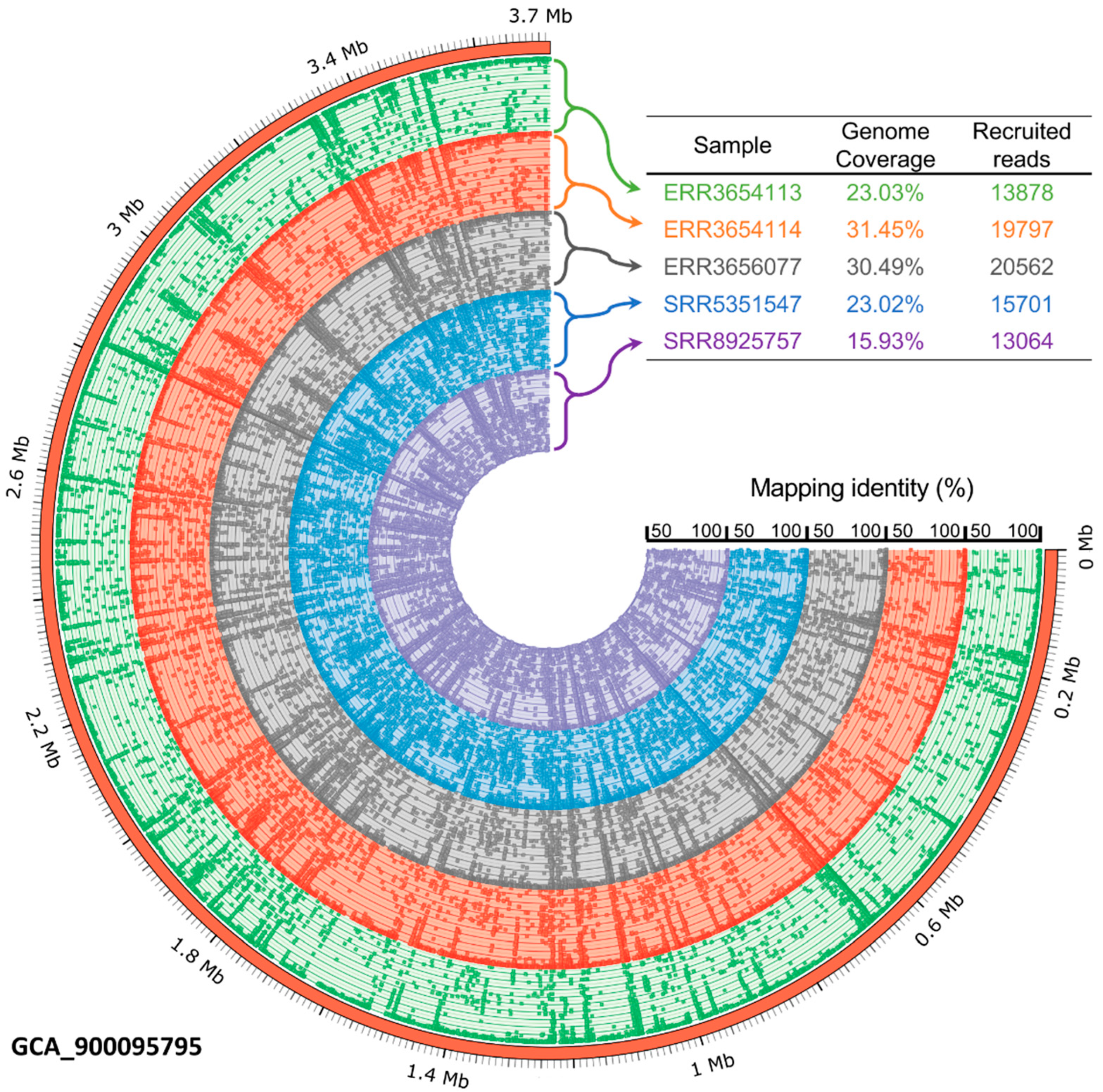
2.4. Fragment Recruitment

To determine the distribution and abundance of Petrimonas mucosa in different biogas communities, fragment recruitments using the genome sequence of Petrimonas mucosa as template were performed by application of the bioinformatics tool SparkHit [44]. Corresponding computations were scaled-up and parallelized by using the de.NBI Cloud environment (https://www.denbi.de/cloud). As a fast and sensitive fragment recruitment tool, the so-called Sparkhit-recruiter was applied. This tool extends the FR-hit pipeline [45] and is implemented natively on top of the Apache Spark. In addition, it integrates a series of analytical tools and methods for various genomic applications. The fragment recruitment option implements the q-Gram algorithm to allow more mismatches than a regular read mapping during the alignment, so that extra information is provided for the metagenomic analysis. SparkHit was applied on metagenome FASTQ files from 257 biogas metagenome datasets that were downloaded from ENA (www.ebi.ac.uk/). Randomly chosen two million reads of each FASTQ file were compared to the P. mucosa ING2-E5AT genome. The alignment identity threshold was set to > 50% similarity. The result of the fragment recruitments were visualized using Circos [46].

2.5. Petrimonas Transcriptional Profile as Deduced from Public Transcriptome Datasets

Metatranscriptome datasets pertaining to samples from different industrial- and laboratory-scale anaerobic digesters, detailed in Supporting Information Table S3, were reanalyzed to quantify transcripts of Petrimonas mucosa. Raw RNA-sequencing reads were trimmed with Trim Galore [47]. Subsequently, reads were filtered with SortMeRNA [48] to exclude ribosomal RNA from the downstream analysis. RnaSPAdes [49] was used to assemble transcripts from read datasets originating from the same biogas reactor or wastewater treatment plant. Reads from samples representing biological or technical replicates were pooled into sets. For each of these sets, the tool Kallisto [50] was used to assign read counts to the assembled metatranscriptome-contigs in case of paired-end reads. For single-end reads, Bowtie2 [51] was used to map reads to the metatranscriptome-contigs and uniquely mapping reads were counted for each contig. All contigs resulting from the transcriptome assembly were aligned to the genome of P. mucosa ING2-E5AT using nucleotide BLAST [52]. Metatranscriptome-contigs for which BLAST produced alignments covering at least 99% of the contig length with an identity of 97% or more were classified as transcripts originating from P. mucosa. The previously produced read counts for each of these contigs were assigned to features in the P. mucosa genome based on the annotated strain ING2-E5AT genome. Individual contigs may overlap multiple coding sequences of the genome. In such cases, the read counts that were computed for a contig were split up between all coding sequences which the contig is overlapping. A fraction X i of total contig-read-counts X is assigned to each coding sequence i . X i is determined by dividing the length of the sequence l i through the sum of the length of all sequences j which are overlapping the contig:
X i   =   X   *   l i j l j
In order to rank P. mucosa features based on their expression levels, transcripts per million (TPM) were calculated for each feature. Only transcripts assigned to P. mucosa were included in the TPM calculations.

2.6. Gene Expression Analysis of Petrimonas Species Based on Database Metaproteome Datasets

High-resolution metaproteomics datasets from ten industrial biogas plants and one laboratory-scale biogas fermenter (PRIDE ACCESSION: PXD009349) [53] were reanalyzed to quantify the abundance of Petrimonas mucosa proteins. For each of the biogas plants, the first replicate of the first sampling time point was searched with the database search engines X!Tandem [54] and OMMSA [55] implemented in the MetaProteomeAnalyser [56] (Version 3.0) against the metagenome database from the original study [57,58,59,60] extended by the protein sequences of P. mucosa. The remaining settings were used as described previously by Heyer and colleagues [56] with the minor change that redundant homologous proteins were grouped when they shared the same peptide.

2.7. Nucleotide Sequence Accession Number

The genome of P. mucosa ING2-E5AT was deposited in the EMBL-EBI database (European Bioinformatics Institute database; https://www.ebi.ac.uk/ena/browser/view/LT608328) under the accession number LT608328.

3. Results and Discussion

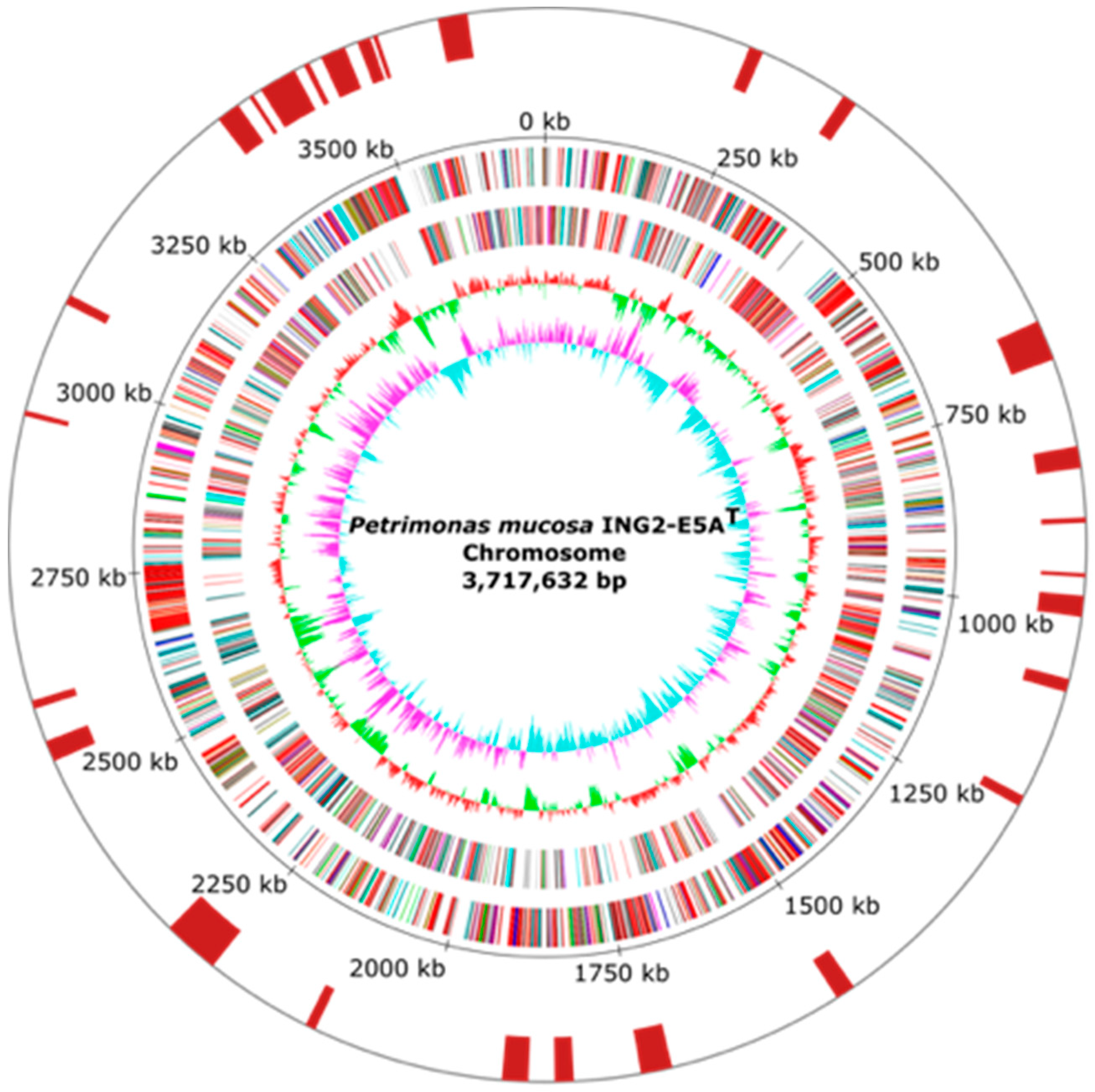
3.1. General Features of Petrimonas mucosa ING2-E5AT Genome

To deduce the importance of Petrimonas members in biogas-producing microbial communities, the genome of the P. mucosa strain ING2-E5AT (= DSM 28695T = CECT 8611T) was completely sequenced and analyzed in detail. General features of the P. mucosa ING2-E5AT genome are summarized in Table 1 and Figure 1. The sequencing approach yielded 115,545 reads with a mean read length of 8488 bp, accounting for 980,798,552 bp sequence information. Long read genome assembly followed by short read error correction resulted in one circular contig with 3,717,632 bp in size, featuring a GC-content of 47.97%. Gene prediction and annotation of the P. mucosa ING2-E5AT genome sequence resulted in the identification of 3000 coding sequences, 49 tRNA genes, and two rrn operons. Analysis of the P. mucosa ING2-E5AT genome for the presence of horizontally acquired DNA elements resulted in the identification of 27 genomic islands (Figure 1) and putative phage genes. However, no complete prophage cluster was predicted in the genome. ING2-E5AT possesses one CRISPR-cas system which may play a role in preventing invasion of phages and mobile genetic elements.

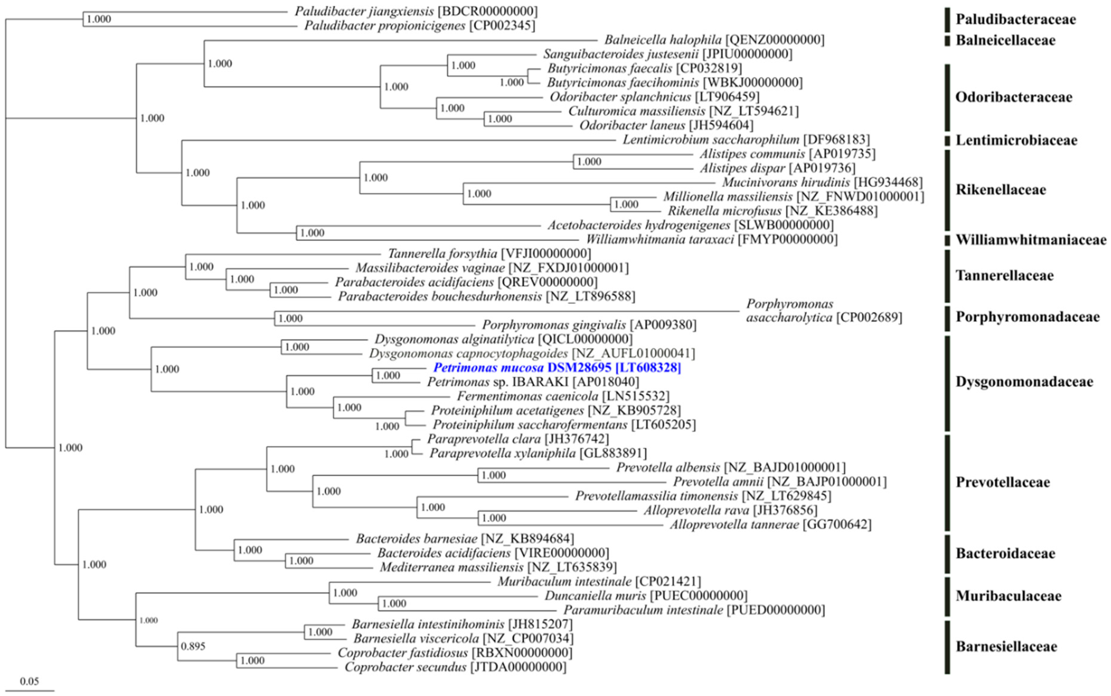
3.2. Phylogenetic Classification of Petrimonas mucosa ING2-E5AT

Phylogenetic tree reconstruction was performed based on available genomes for 47 strains of the order Bacteroidales comprising 94 core genes in total (Figure 2). The analysis resulted in a distinct cluster of the genus Petrimonas together with the genera Fermentimonas and Proteiniphilum. Originally, these genera were assigned to the order Bacteroidales, and within this order, they were affiliated with to the family Porphyromonadaceae as deduced from 16S rRNA gene analyses [25,61]. However, in the obtained core gene-based phylogenetic tree, which is in accordance with the recent taxonomic revision within the phylum Bacteroidetes [27], Porphyromonas species are located in a separated cluster with notable distance to the Petrimonas/Proteiniphilum cluster which is much closer related to the genus Dysgonomonas. Therefore, members of the genera Petrimonas, Proteiniphilum, Fermentimonas, as well as Dysgonomonas constitute the current family Dysgonomonadaceae, which was earlier suggested by Ormerod et al. [62] and has only recently been validated [27].
The genus Petrimonas was firstly described by Grabowski et al. [26] based on cultivation, classification, and characterization of the type species P. sulfuriphila. So far, P. mucosa and P. sulfuriphila were the only validly described species for this genus. Moreover, within this genus, only for the type strain P. mucosa ING2-E5AT [25] the genome sequence is currently available. By 16S rRNA gene sequence analysis and DNA–DNA hybridization, P. mucosa ING2-E5AT appeared to be closely related to P. sulfuriphila BN3T with 97% 16S rRNA gene sequence identity and a DNA–DNA relatedness of 23.8–25.7% [25].

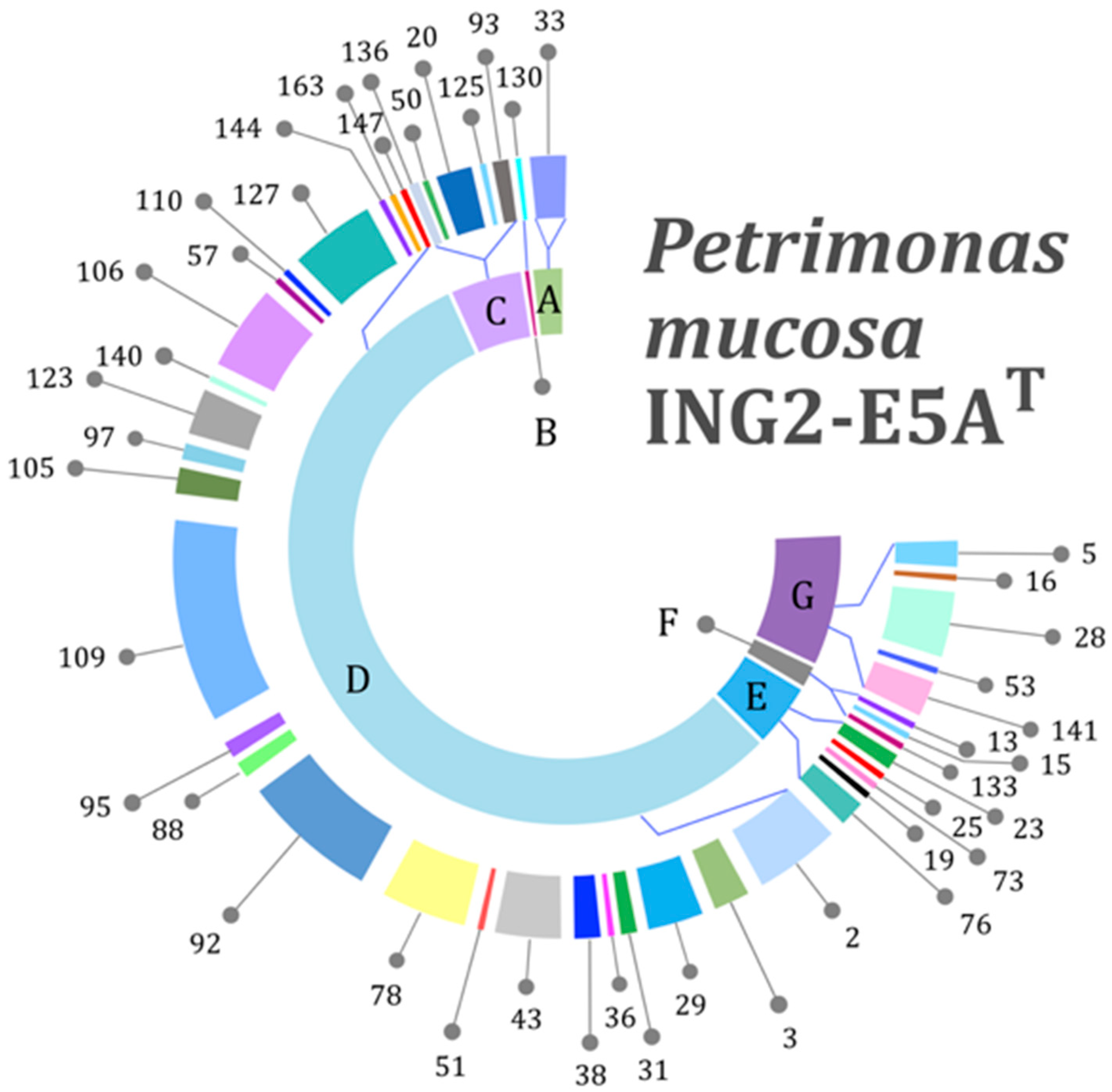
3.3. Genes Encoding Carbohydrate-Active Enzymes

To elucidate the P. mucosa ING2-E5AT genes involved in utilization of different carbohydrates and sugar components and to classify the strain’s lifestyle, the carbohydrate-active enzyme database (CAZy) annotation web-server dbCAN [42] was applied. CAZymes are a class of enzymes which synthesize, modify, or break down different carbohydrates. Their classification comprises the following families: glycoside hydrolase families (GH), polysaccharide lyase families (PL), carbohydrate esterase families (CE), glycosyltransferase families (GT), auxiliary activity families (AA), and carbohydrate-binding module (CBM) families [63]. The proportion of CAZymes predicted in the ING2-E5AT genome was calculated to represent 7.6% of all gene products encoded in the genome, indicating the strain’s ability to process various polysaccharides. Of a total of 233 CAZymes encoded in the ING2-E5AT genome, 154 GHs account for 44 different families (Figure 3). Previous studies on Gram-negative bacteria of the phylum Bacteroidetes also reported on a large variety of different GH families in these species (up to 62) [64]. Based on these observations, a broad biodegradation potential regarding carbohydrates is proposed for this phylum. The most dominant GH families in P. mucosa ING2-E5AT were the GH109, GH2, GH92, GH127, and GH106 families, listed by decreasing abundance. These families mostly encode glycosidases which hydrolyze single sugar residues from the non-reducing ends of di- and polysaccharides such as β-d-galactose (GH2), arabinosides (GH127), glycan (GH92), α-l-rhamnose (GH106), and α-(1→3)-N-acetylgalactosamine (GH109). Several other GH families identified in the ING2-E5AT genome are also associated with hydrolysis of single sugar residues indicating the importance of these substrates for this strain. Further results obtained for P. mucosa ING2-E5AT also showed that it encodes enzymes involved in cell wall degradation such as lysozyme (GH23, GH25, GH73) and chitinases (GH19, GH76). Hence, P. mucosa ING2-E5AT most probably can utilize peptidoglycan and chitin for growth. These polysaccharides are usually part of the bacterial cell wall or are found in fungi and insects, respectively, indicating the potential of P. mucosa ING2-E5AT for further biotechnological applications. As expected for a member of the phylum Bacteroidetes, no genes encoding cohesin-containing putative scaffoldins and corresponding dockerin-containing glycoside hydrolases featuring a potential for cellulosome formation were identified. However, the genome of the strain ING2-E5AT encodes enzymes predicted to degrade cellulose (GH5) and hemicellulose (GH16, GH28, GH53, and GH141).

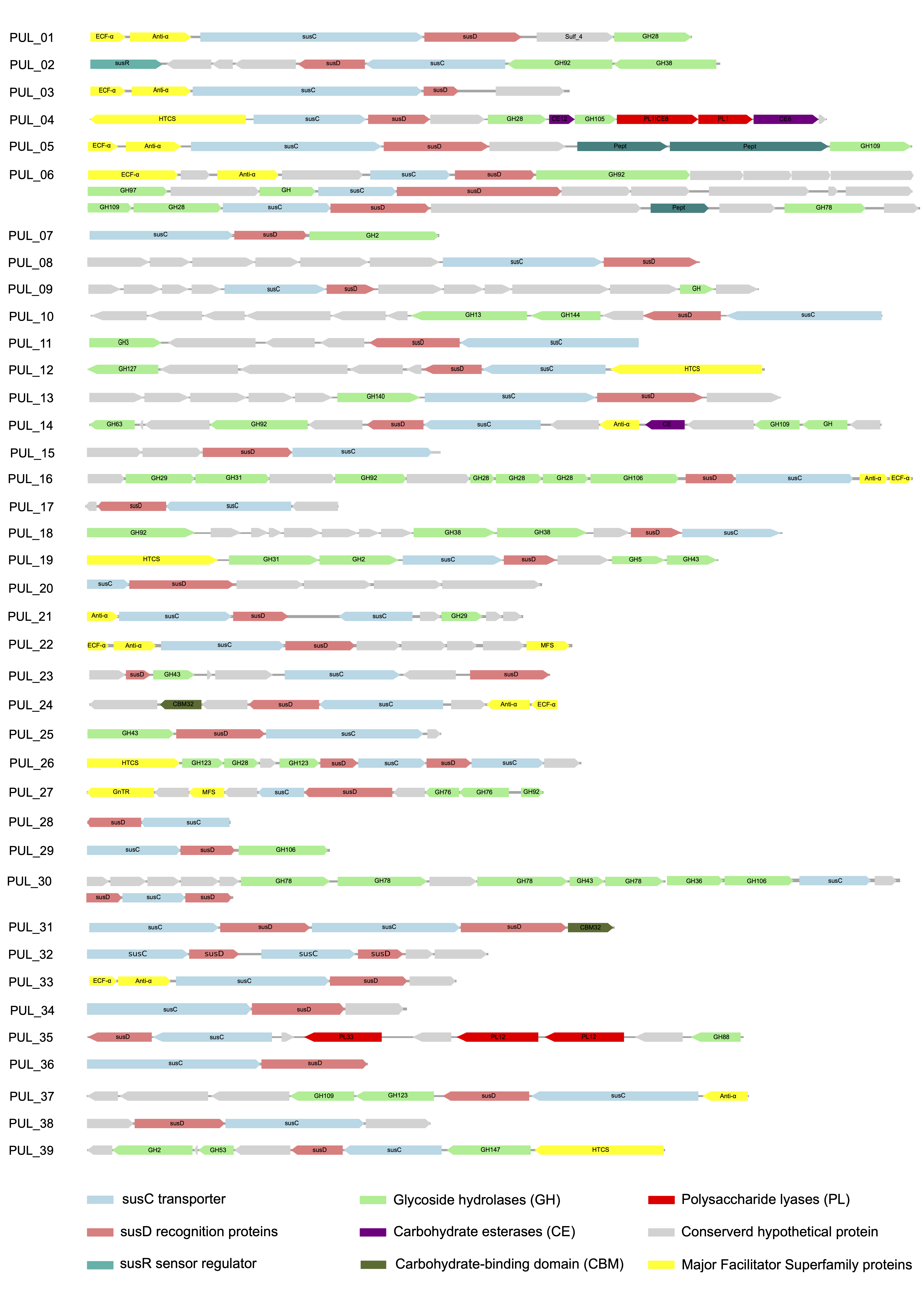
3.4. Genes Encoding Polysaccharide Utilization Loci (PUL)

Degradation of the most abundant plant polysaccharide (cellulose) has usually been affiliated to the metabolic capabilities of members of the phylum Firmicutes [65,66]. According to the current literature, the Bacteroidetes are not associated with cellulose degradation. However, various members of this phylum encode a large diversity of polysaccharide utilization loci (PUL) which were suggested to be in combination with enzymes of the GH5 family acting on cellulose, as an alternative system for cellulose degradation in Bacteroidetes members, in addition to cellulosomes and free-enzyme systems [67,68]. PUL comprise clusters of genes encoding surface glycan binding proteins, transporters, and a variety of glycan-degrading enzymes. The genetic architecture is characterized by tandem susCD-like genes encoding a TonB-dependent outer-membrane transporter (TBDT) and an N-terminally lipidated cell surface glycan-binding protein (SGBP), respectively [68].
The P. mucosa ING2-E5AT genome contains 39 PUL (Figure 4), 25 of which comprise 62 GHs belonging to 24 distinct CAZyme families, representing 25.1% of all families predicted in the genome. The predominant families are GH28 and GH92 representing enzymes involved in pectin and glycan degradation (Figure 4). Besides GH28, other families related to pectin degradation are also associated with PUL, such as GH2, GH43, GH78, GH105, GH106, CE8, CE12, and PL1. Accordingly, 29.9% of all CAZymes located in PUL are involved in pectin degradation, reflecting a pronounced specialization of the enzymatic system for the decomposition of this polysaccharide. Other PUL were predicted to be involved in degradation of arabinan (e.g., PUL_12), galactan (PUL_39), and hemi-/cellulose (PUL_11, PUL_19).
In addition to the genes for CAZymes, the P. mucosa ING2-E5AT PUL also are associated with peptidase genes. PUL_05 and PUL_06 (Figure 4) encode peptidases belonging to family M60 and S15, respectively. These enzymes were predicted to target glycoproteins [69] and serine peptidases [70]. Presence of peptidase genes in PUL suggests that strain ING2-E5AT may also be involved in protein degradation and subsequent amino acid fermentation [29,71]. However, it was recently reported that the strain ING2-E5AT was not able to grow on peptone or tryptone as sole carbon sources [25]. It may be speculated that growth on these substrates requires additional micronutrients that were not provided in the growth experiments conducted previously [25].
Approximately 46% of the P. mucosa PUL are associated with regulator system genes, for example genes encoding an extracytoplasmic function sigma factor (ECF-σ), an anti-sigma (anti-σ) factor, and a hybrid two-component system (HTCS). Involvement of corresponding regulators in controlling expression or PUL-associated genes remains to be determined [68]. Association of PUL with genes encoding ECF-σ, anti-σ pairs, HTCS, transcriptional regulators (e.g., LacI, AraC, GnTR), or sensor-regulator systems (SusR) is not unusual and has been reported for PUL of other Bacteroidetes species [68].
Similar characteristics were also observed in the CAZymes profile of Petrimonas sp. IBARAKI whose genome sequence is closely related to the one of strain ING2-E5AT (PULD database; [72]. Strain IBARAKI harbors 240 CAZymes, 33% of which are located in PUL. Approximately, 40% of its PUL encode enzymes assigned to CAZyme families involved in pectin degradation. Although there are only two genome sequences of Petrimonas species available, it can be assumed that this genus preferably degrades pectin.
In summary, genome sequencing of new Bacteroidetes species may elucidate how members of this phylum adapt PUL for decomposition of particular polysaccharides. Moreover, PUL-encoded enzymes provide exploitation possibilities for biotechnological purposes.

3.5. Genes Encoding Enzymes of the Central Fermentation Metabolism

Regarding its central metabolism, P. mucosa ING2-E5A encodes complete KEGG modules for glycolysis (Embden–Meyerhof pathway, M00001), pyruvate oxidation (M00307), the citrate cycle (TCA cycle, M00009), and the pentose phosphate pathway (M00006) allowing metabolism of glucose, pentoses, and other monosaccharides. As described above, several di-, oligo-, and polysaccharides potentially can be metabolized to the aforementioned common monosaccharides. P. mucosa was also predicted to perform degradation of proteins, oligo- and dipeptides since corresponding enzymatic activities are encoded in its genome. Amino acids resulting from peptide hydrolysis may feed fermentation pathways. For example, enzymes for conversion of l-asparagine via aspartate to fumarate are encoded in the P. mucosa genome (see Table 2). Citrate cycle enzymes catalyze the reactions from fumarate via malate to oxaloacetate which can then be decarboxylated to pyruvate by oxaloacetate decarboxylase (EC 4.1.1.112). Subsequently, ATP is generated via the phosphate-acetyltransferase/acetate kinase module (KEGG M00579) that is encoded in the P. mucosa genome.
Likewise, glutamine and glutamate may be metabolized to yield succinyl-CoA via 2-oxoglutarate (refer to Table 2). P. mucosa encodes the key enzymes methylmalonyl-CoA mutase, methylmalonyl-CoA epimerase and propionyl-CoA carboxylase catalyzing the reactions of succinyl-CoA to (R)-methylmalonyl-CoA and subsequently to propionyl-CoA, respectively. The phosphate-acetyltransferase/acetate kinase module (KEGG M00579) is also able to convert propionyl-CoA to propionate via propionyl-phosphate yielding ATP through substrate-level phosphorylation. According to ExPASy (https://enzyme.expasy.org), acetate kinase (EC 2.7.2.1) can also use propanoate as a substrate. Acetate can be converted into the intermediate acetyl-CoA by coupling the reaction of acetate kinase with phosphate acetyltransferase (EC 2.3.1.8) activity. Both, this enzyme and acetate kinase also are important in the production of propanoate. Acetate, propionate, and carbon dioxide were previously identified as fermentation end-products in P. mucosa ING2-E5AT [25]. As well as the amino acids mentioned above, other amino acids such as proline, glycine, serine, and threonine potentially can be catabolized via glutamate or pyruvate since the corresponding enzymes are encoded in the P. mucosa genome.
Genes for the butanoate pathway enzymes butyrate kinase (EC 2.7.2.7) and phosphate butyryltransferase (EC 2.3.1.19) are present in the P. mucosa genome but the pathway is incomplete at least as predicted from the genome sequence information. Genes for the enzymes involved in conversion of 3-hydroxybutanoyl-CoA to crotonoyl-CoA (or vice versa) and crotonoyl-CoA to butanoyl-CoA could not be identified in the P. mucosa ING2-E5AT genome. However, it was shown that P. mucosa ING2-E5AT produced butyric acid in trace amounts when grown on glucose [25]. So far, not all enzymes necessary for butyric acid formation were identified in P. mucosa.
Very recently, importance of the phosphotransacetylase/acetate kinase pathway for ATP production was also described in Porphyromonas gingivalis which, similar to P. mucosa, belongs to the order Bacteroidales [73,74,75,76]. In the context of the central fermentation metabolism, it is interesting to note that P. mucosa encodes a malic enzyme (EC 1.1.1.40) and pyruvate, phosphate dikinase (EC 2.7.9.1) catalyzing the NAD(P)+ dependent oxidative decarboxylation of malate to pyruvate [77] and the ATP-consuming conversion of pyruvate to phosphoenolpyruvate, respectively. Mostly, the reaction of malic enzyme contributes to NADPH-generation but under specific conditions, it may also function as anaplerotic node by maintaining suitable levels of TCA cycle intermediates [77]. For P. mucosa, the biological importance of the reaction catalyzed by malic enzyme remains to be determined.
Furthermore, two hydrogenase gene regions were identified in the P. mucosa ING2-E5AT genome by means of BLASTp analyses and consulting the Hydrogenase Database HydDB [43]. The first hydrogenase, being classified as a HydA (ING2E5A_0849), belongs to the [FeFe] Group C1; it is histidine kinase-linked and oxygen labile and its role remains unclear so far. The enzyme may serve as a hydrogen sensor in regulatory cascades. The second hydrogenase Hnd consists of four subunits (HndA, HndB, HndC, and HndD; ING2E5A_0853-0856). The hnd genes are located in close proximity to hydA. A previous study on the Hnd hydrogenase isolated from Desulfovibrio fructosovorans described the electron-bifurcating function of this enzyme coupling an exergonic redox reaction to an endergonic one allowing energy conservation in anaerobic microorganisms [78].

3.6. The Occurrence of Petrimonas Relatives in Biogas-Producing Microbial Communities as Deduced from Publicly Available Metagenome Data

To investigate the prevalence and occurrence of P. mucosa in biogas-producing microbial communities, metagenome sequences obtained from 257 publicly available datasets originating from the RefSeq collection were mapped onto the ING2-E5AT genome applying the program Sparkhit [44]. The mapping approach was aimed at the reconstruction of Petrimonas genomes from the previously published metagenome data. A further objective of this approach was to determine the degree of relatedness of species within the analyzed biogas plant microbiomes to the genome of strain ING2-E5AT. Fragment recruitments were done by mapping of at maximum ten million sequences from the corresponding metagenome representing the biogas microbial community. Only five samples (accession numbers at the NCBI: ERR3656077, ERR3654114, SRR8925757, ERR3654113, SRR5351547) showing metagenome fragment mapping results of greater than 1% were visualized in the fragment recruitment plot (Figure 5). The complete report is provided as Supporting Information Table S1.
The genus Petrimonas was detected in 146 out of 257 metagenomes analyzed (≥0.1% of all metagenome sequences) with different abundance values. Particularly high abundance values ranging between 1.03% and 3.16% of all metagenome sequences were detected in 14 out of 257 different biogas reactor metagenomes (Figure 5). In these metagenomes, the genome of P. mucosa ING2-E5AT was not only abundant, but also fully covered. The highest number of mapped sequences onto the genome of P. mucosa (3.16% of all sequences) was found for the microbiome originating from a production-scale biogas plant located in Germany (Ger-06), operated under mesophilic (40 °C) temperature regime and fed with manure (48%), maize (30%), dung (17%), and grain corn (7%) [79]. Brandt and colleagues [79] also described the widespread occurrence of the genus Petrimonas in microbiomes of biogas reactors analyzed within their study. They detected Petrimonas spp. in 13 out of 20 German and Swedish biogas plants (BGPs) and presented abundance values of up to 10% (Ger-06) of the entire community.
Petrimonas members were also detected in microbiomes originating from a pilot-scale dry anaerobic digester fed with residual biomass from the bovine husbandry (64.2% manure, 18.2% oxidation lagoon water, 3% fresh rumen fluid, 9.6% wood chips, 4.0% corn stover, and 0.9% dust mill) and operated under mesophilic conditions [80]. Interestingly, the highest number of sequences assigned to the P. mucosa genome in this reactor (2.87% of all metagenome sequences) was detected a short time after a process disturbance that occurred due to a pump failure during leachate recirculation. Most probably, P. mucosa was able to cope with corresponding stress conditions and displaced other competing bacteria that are less stress resistant. This is in line with previous studies that link the genus Petrimonas with process disorders [7,9]. Our mapping results also showed an abundance decrease of the genus Petrimonas in reactor samples taken after the process disorder (from 1.41% to 1.03%), indicating suppression by other competitive bacteria in the stabilization phase of the biogas process.
Information obtained from the fragment recruitment approach leads to the assumption that Petrimonas and its relatives are widespread in biogas plants. Petrimonas members probably proliferate when reactor conditions become unstable and the community undergoes a structural change. Under these conditions, they may play an important role in conversion of lignocellulosic biomass and also proteins for subsequent methane production.

3.7. The Transcriptional Profile of Petrimonas Species

Transcriptome sequence data reflect the transcriptional activity of the species of interest in their respective habitats. To study the Petrimonas transcriptional activity in microbiomes in which this species was found with a relative abundance of above 1% (for more details see above), corresponding metatranscriptome data were searched in the publicly available sequence databases. Unfortunately, no metatranscriptome data exist for the metagenomes presented in Figure 5 referring to the fragment recruitment analyses (see above). To gain insights into the transcriptional profile of Petrimonas members and their relatives and to understand their role in AD in general, public databases were searched for metatranscriptome datasets representing AD environments. Not all of these datasets also comprise metagenome data. For metagenome data for which also metatranscriptome data was available, only fractions of the P. mucosa genome could be assembled from the DNA reads. For this reason, a solely RNA-based analysis was carried out. Transcriptome assemblies were conducted for communities from 13 different biogas and wastewater treatment plants. Sequence reads of the corresponding studies were grouped into 29 datasets, each representing a distinct condition or sampling time point in one of the analyzed reactors. For each of these 29 datasets, reads were mapped back to the assembled transcripts. The ING2-E5AT genome was used to assign transcripts that originate from P. mucosa. Based on the number of reads that could be mapped to each P. mucosa transcript, counts and TPM values were determined for each dataset. Detailed information on process parameters and used substrates recorded for the analyzed biogas reactors were summarized in Supporting Information Table S2 as far as corresponding metadata were documented in the literature.
Transcripts assigned to Petrimonas and its relatives were detected in 20 out of 29 metatranscriptome datasets analyzed. The highest number of transcripts (0.23–0.53% of all sequences) that could be mapped onto the ING2-E5AT genome was observed for a mesophilic laboratory-scale biogas plant fed with maize silage and separated digestate from other mesophilic full-scale research biogas plants [81] (Supporting Information Table S3). Grohmann and coworkers [81] reported that the analyzed biogas plant suffered from a process disturbance leading to a strong accumulation of VFA. Acetic and propionic acid concentrations exceeded critical values and were on an alarming level. During the experiment, the reactor was continuously sampled. Petrimonas transcripts were consistently detected in all metatranscriptome datasets belonging to the latter study (on average 0.31% of all metatranscriptome sequences taking into account all metagenomes from this study). However, the highest number of transcripts (0.53%) were observed for the sample that was taken when the process had nearly stabilized.
Within the list of highly transcribed genes (Table 3 and Supporting Information Table S4), those encoding proteins associated with hydrolysis of 6-phosphogluconolactone to 6-phosphogluconic acid via the pentose phosphate pathway, such as 6-phosphogluconolactonase Pgl (EC: 3.1.1.31), were observed. Genes involved in oligopeptide utilization such as the ATP-dependent protease clp (EC: 3.4.21.-) and the peptidase IV sppA gene (EC: 3.4.21.-) were also transcribed. Furthermore, P. mucosa transcribed the mscL gene encoding the large-conductance mechanosensitive channel. This pore-forming membrane protein is an important component of the cells’ management during osmotic stress [82]. Moreover, these nanopores can be utilized for the passage of membrane-impermeable compounds or peptides [83]. The latter molecules were predicted to represent fermentation substrates of Petrimonas species. Furthermore, P. mucosa transcribed the susC gene encoding the TonB-dependent outer-membrane transporter, which is part of PUL clusters presented in Figure 4. Further copies of this gene were also highly transcribed. Transcripts encoding proteins involved in mandatory functions (basic house-keeping functions) such as transcription and translation were not further considered.
Based on genome interpretation and transcriptional profiling, a lifestyle based on carbohydrate decomposition and oligopeptide fermentation is proposed for P. mucosa ING2-E5AT. Furthermore, results obtained from metatranscriptome analyses are in line with previous findings showing increased abundance of Petrimonas members in reactors operating under stressed conditions followed by accumulation of VFA [10,19]. Results obtained within this study also indicated that Petrimonas members are competent in fermenting sugars and amino acids, with acetate and propionic acid as the major fermentation products.

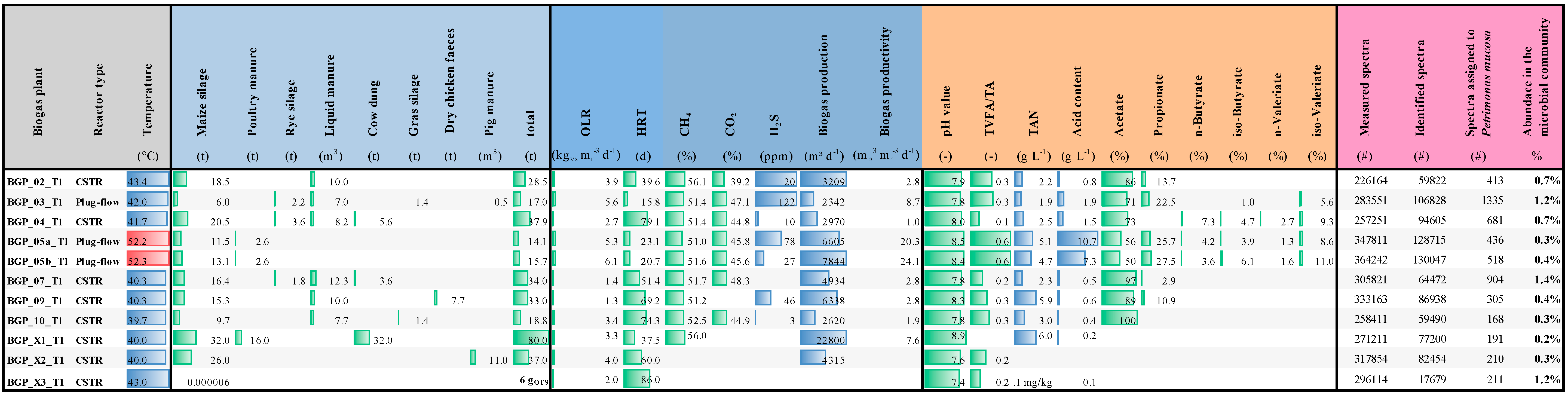
3.8. Metaproteome Analysis Revealed the Presence of Petrimonas spp. Proteins in Various Biogas-Producing Communities

Metaproteomics became a powerful technique to study the biological functions of microbial communities, to analyze and correlate the taxonomic and functional profiles of microbiomes [84] and also to evaluate the responses of microbial taxa to environmental changes [85,86]. In order to evaluate which proteins/enzymes were expressed by P. mucosa, metaproteome data originating from mesophilic and thermophilic biogas reactors (previously published by [53]) were screened for proteins/enzymes that were assigned to this taxon. Further details on operational and process parameters of the investigated BGPs as well as fed substrates are summarized in Figure 6. In total, 11 biogas microbiomes were investigated using SDS-PAGE for prefractionation of proteins and subsequent liquid chromatography (LC) coupled to a high-resolution Orbitrap Elite tandem mass spectrometer (MS/MS). Spectra assigned to P. mucosa were identified using the MetaProteomeAnalyzer software [87]. The available datasets were searched with X!tandem and OMSSA against the same protein database complemented by the protein sequences deduced from the Petrimonas mucosa genome sequence.
Based on abundances of spectral counts, peptides originating from P. mucosa proteins after tryptic digestion were detected in all biogas samples analyzed, showing abundance values in the range of 0.2–1.4% of all spectra identified. The highest number of spectra assigned to P. mucosa was detected in BGP_03_T1 (1335) representing a mesophilic plug-flow biogas reactor operating at stable process conditions as deduced from the process parameters recorded for this reactor. Among the highest expressed proteins are many of those annotated as house-keeping proteins e.g., uncharacterized outer membrane proteins, and ribosomal proteins of the 30S and 50S ribosomal subunits. Furthermore, several copies of the TonB-dependent outer-membrane transporters (SusC) were found among the most abundant spectra (all are part of different PUL clusters presented in Figure 4: PUL_02, PUL_10, PUL_11, PUL_24, PUL_29, PUL_31) and represent an essential component of P. mucosa’s PUL required for the saccharification of complex carbohydrates. This finding indicates the importance of PUL-encoded enzymes for P. mucosa’s lifestyle and provides evidence that corresponding enzymes play a central role for this species.

4. Conclusions

Integrated omics analyses are useful for a detailed characterization of both, entire microbiomes and individual microbial species enabling new ways to access the microbiological basis for AD. In this study, the genome sequence of P. mucosa ING2-E5AT isolated from a mesophilic laboratory-scale reactor was analyzed in detail including genome-based metabolic reconstruction. In addition, a polyphasic approach exploiting publicly available metagenome, metatranscriptome, and metaproteome datasets complemented the genome interpretation of the strain. Genome-based phylogenetic classification of P. mucosa ING2-E5AT positioned this strain in close proximity to Fermentimonas and Proteiniphilum species belonging to the family Dysgonomonadaceae which is in accordance with the recent taxonomic reclassification within the phylum Bacteroidetes. Dysgonomonas species are located in a sister group of the Petrimonas/Fermentimonas/Proteiniphilum cluster whereas Porphyromonas species are more distantly related to the Dysgonomonadaceae members.
Insights into the strain’s genetic repertoire and metabolic potential revealed that P. mucosa ING2-E5AT encodes a diverse set of glycosyl-hydrolyses (GH) involved in carbohydrate metabolism as well as pectin degradation. Most of the corresponding genes are organized in Polysaccharide Utilization Loci (PUL) also comprising tandem susCD-like genes for a TonB-dependent outer-membrane transporter and a cell surface glycan-binding protein. Furthermore, ING2-E5AT possesses genetic determinants needed to utilize amino acids as carbon sources. Its function in the AD process most probably is associated with acidogenesis, which is supported by reconstruction of corresponding metabolic pathways leading to production of acetate, propionate, and carbon dioxide as fermentation end-products.
Finally, publicly available metagenome, metatranscriptome, and metaproteome data were used to follow up the question on spreading of Petrimonas and its relatives in different biogas plants and subsequently to study its biological functions in the corresponding environment. The genus Petrimonas was prevalent in approximately half of the datasets investigated, supporting the importance of this genus for the AD process. Moreover, fragment recruitment results linked the genus Petrimonas with process disorders. Most probably, P. mucosa was able to cope with corresponding stress conditions and displaced other competing bacteria that are less stress resistant. Functional profiles of Petrimonas members shed new light on the importance of PUL-encoded enzymes for P. mucosa’s lifestyle, leading to the evidence that corresponding proteins are indispensable for this species.
In the context of biogas production, an integrated -omics approach is believed to provide deeper insights into functional roles of AD community members, thus facilitating the implementation of new optimization strategies for the biogas process. The knowledge gained from multi-omics analyses will provide the basis for application of the strain in demanding biotechnological processes. P. mucosa may also be used as indicator species for unbalanced AD processes in the future. However, further information on the performance of the strain in different AD microbiomes exposed to unfavorable process conditions is needed to precisely determine its functional behavior.

Supplementary Materials

The following are available online at https://www.mdpi.com/2076-2607/8/12/2024/s1, Supporting Information Table S1: List of publicly available metagenome datasets originating from the RefSeq used for fragment recruitment analysis; Supporting Information Table S2: Characteristics and fed substrates of the 13 different biogas plants analyzed in the scope of the metatranscriptome analysis; Supporting Information Table S3: Statistics of the obtained and processed metatranscriptome sequences analyzed in order to investigate the transcriptional profile of Petrimonas species; Supporting Information Table S4: P. mucosa ING2-E5AT most actively transcribed genes for enzymes involved in carbohydrate or protein utilization in 29 analyzed datasets originating from different biogas plants.

Author Contributions

Conceptualization, A.S. (Andreas Schlüter), A.S. (Alexander Sczyrba), D.W., J.H., I.M., M.K.; Methodology, G.T., I.M., L.H., R.H., T.T., V.Z.; Software, D.W., L.H., R.H., T.T.; Validation, A.S. (Andreas Schlüter), A.S. (Alexander Sczyrba), I.M., Formal Analysis, D.W., G.T., I.M., J.H., R.H., T.T., V.Z., B.B.; Investigation, A.S. (Andreas Schlüter), I.M.; Resources, A.P., A.S. (Alexander Sczyrba), A.S. (Andreas Schlüter), C.S., D.B.; Data Curation, D.W., I.M., L.H., T.T., R.H.; Writing—Original Draft Preparation, A.S. (Andreas Schlüter), G.T., I.M., M.K.; Writing—Review and Editing, all participants; Visualization, D.W., G.T., I.M., L.H., R.H., M.K.; Supervision, A.S. (Andreas Schlüter), A.S. (Alexander Sczyrba), M.K., A.P.; Project Administration, A.P., A.S. (Andreas Schlüter), M.K.; Funding Acquisition, A.P., A.S. (Andreas Schlüter), M.K. All authors have read and agreed to the published version of the manuscript.

Funding

This research was funded in the context of the joint project BIOGAS-CORE granted by the German Federal Ministry of Food and Agriculture (grant no. 22017111) and supported by the German Fachagentur Nachwachsende Rohstoffe. A.S. and J.H. gratefully acknowledge funding by the Federal Ministry of Food and Agriculture (BMP-3, GeneMining). A.S. (Alexander Sczyrba) and A.S. (Andreas Schlüter) acknowledge funding by the European Union’s Horizon 2020 Research and Innovation Programme under the Grant agreement No. 818431 (SIMBA, Sustainable Innovation of Microbiome Applications in Food Systems). The bioinformatics support of the BMBF-funded project ‘Bielefeld-Gießen Center for Microbial Bioinformatics-BiGi (grant nos. 031A533 and 031L0103)’ within the German Network for Bioinformatics Infrastructure (de.NBI) is also gratefully acknowledged. This work was also supported by the BMBF-funded de.NBI Cloud within the German Network for Bioinformatics Infrastructure (de.NBI) (031A532B, 031A533A, 031A533B, 031A534A, 031A535A, 031A537A, 031A537B, 031A537C, 031A537D, 031A538A).

Acknowledgments

The authors thank Simone Severitt and Nicole Heyer for excellent technical assistance regarding PacBio genome sequencing.

Conflicts of Interest

The authors declare that there is no conflict of interest.

References

  1. Venkiteshwaran, K.; Bocher, B.; Maki, J.; Zitomer, D. Relating Anaerobic Digestion Microbial Community and Process Function. Microbiol. Insights 2015, 8, 37–44. [Google Scholar] [PubMed] [Green Version]
  2. Schnürer, A. Biogas Production: Microbiology and Technology. Adv. Biochem. Eng. Biotechnol. 2016, 156, 195–234. [Google Scholar] [PubMed]
  3. Westerholm, M.; Dolfing, J.; Schnürer, A. Growth Characteristics and Thermodynamics of Syntrophic Acetate Oxidizers. Environ. Sci. Technol. 2019, 53, 5512–5520. [Google Scholar] [CrossRef] [PubMed]
  4. Rajagopal, R.; Saady, N.M.C.; Torrijos, M.; Thanikal, J.V.; Hung, Y.-T. Sustainable Agro-Food Industrial Wastewater Treatment Using High Rate Anaerobic Process. Water 2013, 5, 292. [Google Scholar] [CrossRef] [Green Version]
  5. Niu, Q.; Hojo, T.; Qiao, W.; Qiang, H.; Li, Y.-Y. Characterization of Methanogenesis, Acidogenesis and Hydrolysis in Thermophilic Methane Fermentation of Chicken Manure. Chem. Eng. J. 2014, 244, 587–596. [Google Scholar] [CrossRef]
  6. Lv, Z.; Wu, X.; Zhou, B.; Wang, Y.; Sun, Y.; Wang, Y.; Chen, Z.; Zhang, J. Effect of One Step Temperature Increment from Mesophilic to Thermophilic Anaerobic Digestion on the Linked Pattern between Bacterial and Methanogenic Communities. Bioresour. Technol. 2019, 292, 121968. [Google Scholar] [CrossRef]
  7. He, Q.; Li, L.; Zhao, X.; Qu, L.; Wu, D.; Peng, X. Investigation of Foaming Causes in Three Mesophilic Food Waste Digesters: Reactor Performance and Microbial Analysis. Sci. Rep. 2017, 7, 13701. [Google Scholar] [CrossRef] [Green Version]
  8. Yenigün, O.; Demirel, B. Ammonia Inhibition in Anaerobic Digestion: A Review. Process. Biochem. 2013, 48, 901–911. [Google Scholar] [CrossRef]
  9. Maus, I.; Klocke, M.; Derenkó, J.; Stolze, Y.; Beckstette, M.; Jost, C.; Wibberg, D.; Blom, J.; Henke, C.; Willenbücher, K.; et al. Impact of Process Temperature and Organic Loading Rate on Cellulolytic/Hydrolytic Biofilm Microbiomes during Biomethanation of Ryegrass Silage Revealed by Genome-Centered Metagenomics and Metatranscriptomics. Environ. Microbiome 2020, 15, 7. [Google Scholar] [CrossRef] [Green Version]
  10. He, Q.; Li, L.; Peng, X. Early Warning Indicators and Microbial Mechanisms for Process Failure Due to Organic Overloading in Food Waste Digesters. J. Environ. Eng. 2017, 143, 04017077. [Google Scholar] [CrossRef]
  11. Rajagopal, R.; Massé, D.I.; Singh, G. A Critical Review on Inhibition of Anaerobic Digestion Process by Excess Ammonia. Bioresour. Technol. 2013, 143, 632–641. [Google Scholar] [CrossRef] [PubMed]
  12. Campanaro, S.; Treu, L.; Kougias, P.G.; De Francisci, D.; Valle, G.; Angelidaki, I. Metagenomic Analysis and Functional Characterization of the Biogas Microbiome Using High Throughput Shotgun Sequencing and a Novel Binning Strategy. Biotechnol. Biofuels 2016, 9, 26. [Google Scholar] [CrossRef] [PubMed] [Green Version]
  13. Müller, B.; Sun, L.; Westerholm, M.; Schnürer, A. Bacterial Community Composition and Fhs Profiles of Low- and High-Ammonia Biogas Digesters Reveal Novel Syntrophic Acetate-Oxidising Bacteria. Biotechnol. Biofuels 2016, 9, 48. [Google Scholar] [CrossRef] [PubMed] [Green Version]
  14. Treu, L.; Kougias, P.G.; Campanaro, S.; Bassani, I.; Angelidaki, I. Deeper Insight into the Structure of the Anaerobic Digestion Microbial Community; the Biogas Microbiome Database Is Expanded with 157 New Genomes. Bioresour. Technol. 2016, 216, 260–266. [Google Scholar] [CrossRef] [Green Version]
  15. Stolze, Y.; Bremges, A.; Maus, I.; Pühler, A.; Sczyrba, A.; Schlüter, A. Targeted in Situ Metatranscriptomics for Selected Taxa from Mesophilic and Thermophilic Biogas Plants. Microb. Biotechnol. 2018, 11, 667–679. [Google Scholar] [CrossRef] [Green Version]
  16. Maus, I.; Koeck, D.E.; Cibis, K.G.; Hahnke, S.; Kim, Y.S.; Langer, T.; Kreubel, J.; Erhard, M.; Bremges, A.; Off, S.; et al. Unraveling the Microbiome of a Thermophilic Biogas Plant by Metagenome and Metatranscriptome Analysis Complemented by Characterization of Bacterial and Archaeal Isolates. Biotechnol. Biofuels 2016, 9, 171. [Google Scholar] [CrossRef] [Green Version]
  17. Fischer, M.A.; Güllert, S.; Refai, S.; Künzel, S.; Deppenmeier, U.; Streit, W.R.; Schmitz, R.A. Long-term Investigation of Microbial Community Composition and Transcription Patterns in a Biogas Plant Undergoing Ammonia Crisis. Microb. Biotechnol. 2018, 12, 305–323. [Google Scholar] [CrossRef]
  18. Campanaro, S.; Treu, L.; Rodriguez-R, L.M.; Kovalovszki, A.; Ziels, R.M.; Maus, I.; Zhu, X.; Kougias, P.G.; Basile, A.; Luo, G.; et al. The Anaerobic Digestion Microbiome: New Insights from the Biogas Microbiome by Comprehensive Genome-Resolved Metagenomics of Nearly 1600 Species Originating from Multiple Anaerobic Digesters. Biotechnol. Biofuels 2020, 13, 25. [Google Scholar] [CrossRef] [Green Version]
  19. Zhang, J.; Loh, K.-C.; Lee, J.; Wang, C.-H.; Dai, Y.; Wah Tong, Y. Three-Stage Anaerobic Co-Digestion of Food Waste and Horse Manure. Sci. Rep. 2017, 7, 1269. [Google Scholar] [CrossRef]
  20. Sun, L.; Pope, P.B.; Eijsink, V.G.H.; Schnürer, A. Characterization of Microbial Community Structure during Continuous Anaerobic Digestion of Straw and Cow Manure. Microb. Biotechnol. 2015, 8, 815–827. [Google Scholar] [CrossRef] [Green Version]
  21. Moset, V.; Poulsen, M.; Wahid, R.; Højberg, O.; Møller, H.B. Mesophilic versus Thermophilic Anaerobic Digestion of Cattle Manure: Methane Productivity and Microbial Ecology. Microb. Biotechnol. 2015, 8, 787–800. [Google Scholar] [CrossRef] [PubMed]
  22. Goux, X.; Calusinska, M.; Lemaigre, S.; Marynowska, M.; Klocke, M.; Udelhoven, T.; Benizri, E.; Delfosse, P. Microbial Community Dynamics in Replicate Anaerobic Digesters Exposed Sequentially to Increasing Organic Loading Rate, Acidosis, and Process Recovery. Biotechnol. Biofuels 2015, 8, 122. [Google Scholar] [CrossRef] [PubMed] [Green Version]
  23. Hofstad, T.; Olsen, I.; Eribe, E.R.; Falsen, E.; Collins, M.D.; Lawson, P.A. Dysgonomonas Gen. Nov. to Accommodate Dysgonomonas Gadei Sp. Nov., an Organism Isolated from a Human Gall Bladder, and Dysgonomonas Capnocytophagoides (Formerly CDC Group DF-3). Int. J. Syst. Evol. Microbiol. 2000, 50, 2189–2195. [Google Scholar] [CrossRef] [PubMed]
  24. Chen, S.; Dong, X. Proteiniphilum Acetatigenes Gen. Nov., Sp. Nov., from a UASB Reactor Treating Brewery Wastewater. Int. J. Syst. Evol. Microbiol. 2005, 55, 2257–2261. [Google Scholar] [CrossRef] [PubMed] [Green Version]
  25. Hahnke, S.; Langer, T.; Koeck, D.E.; Klocke, M. Description of Proteiniphilum Saccharofermentans Sp. Nov., Petrimonas Mucosa Sp. Nov. and Fermentimonas Caenicola Gen. Nov., Sp. Nov., Isolated from Mesophilic Laboratory-Scale Biogas Reactors, and Emended Description of the Genus Proteiniphilum. Int. J. Syst. Evol. Microbiol. 2016, 66, 1466–1475. [Google Scholar] [CrossRef] [PubMed]
  26. Grabowski, A.; Tindall, B.J.; Bardin, V.; Blanchet, D.; Jeanthon, C. Petrimonas Sulfuriphila Gen. Nov., Sp. Nov., a Mesophilic Fermentative Bacterium Isolated from a Biodegraded Oil Reservoir. Int. J. Syst. Evol. Microbiol. 2005, 55, 1113–1121. [Google Scholar] [CrossRef]
  27. García-López, M.; Meier-Kolthoff, J.P.; Tindall, B.J.; Gronow, S.; Woyke, T.; Kyrpides, N.C.; Hahnke, R.L.; Göker, M. Analysis of 1000 Type-Strain Genomes Improves Taxonomic Classification of Bacteroidetes. Front. Microbiol. 2019, 10, 2083. [Google Scholar] [CrossRef] [Green Version]
  28. Sakamoto, M. The Family Porphyromonadaceae. In The Prokaryotes: Other Major Lineages of Bacteria and the Archaea; Rosenberg, E., DeLong, E.F., Lory, S., Stackebrandt, E., Thompson, F., Eds.; Springer: Berlin/Heidelberg, Germany, 2014; pp. 811–824. [Google Scholar]
  29. Tomazetto, G.; Hahnke, S.; Wibberg, D.; Pühler, A.; Klocke, M.; Schlüter, A. Proteiniphilum Saccharofermentans Str. M3/6T Isolated from a Laboratory Biogas Reactor Is Versatile in Polysaccharide and Oligopeptide Utilization as Deduced from Genome-Based Metabolic Reconstructions. Biotechnol. Rep. 2018, 18, e00254. [Google Scholar] [CrossRef]
  30. Koeck, D.E.; Zverlov, V.V.; Liebl, W.; Schwarz, W.H. Comparative Genotyping of Clostridium Thermocellum Strains Isolated from Biogas Plants: Genetic Markers and Characterization of Cellulolytic Potential. Syst. Appl. Microbiol. 2014, 37, 311–319. [Google Scholar] [CrossRef]
  31. Cibis, K.G.; Gneipel, A.; König, H. Isolation of Acetic, Propionic and Butyric Acid-Forming Bacteria from Biogas Plants. J. Biotechnol. 2016, 220, 51–63. [Google Scholar] [CrossRef]
  32. Tejerizo, G.T.; Kim, Y.S.; Maus, I.; Wibberg, D.; Winkler, A.; Off, S.; Pühler, A.; Scherer, P.; Schlüter, A. Genome Sequence of Methanobacterium Congolense Strain Buetzberg, a Hydrogenotrophic, Methanogenic Archaeon, Isolated from a Mesophilic Industrial-Scale Biogas Plant Utilizing Bio-Waste. J. Biotechnol. 2017, 247, 1–5. [Google Scholar] [CrossRef] [PubMed]
  33. Pechtl, A.; Rückert, C.; Maus, I.; Koeck, D.E.; Trushina, N.; Kornberger, P.; Schwarz, W.H.; Schlüter, A.; Liebl, W.; Zverlov, V.V. Complete Genome Sequence of the Novel Cellulolytic, Anaerobic, Thermophilic Bacterium Herbivorax Saccincola Type Strain GGR1, Isolated from a Lab Scale Biogas Reactor as Established by Illumina and Nanopore MinION Sequencing. Genome Announc. 2018, 6, e01493-17. [Google Scholar] [CrossRef] [PubMed] [Green Version]
  34. Maus, I.; Bremges, A.; Stolze, Y.; Hahnke, S.; Cibis, K.G.; Koeck, D.E.; Kim, Y.S.; Kreubel, J.; Hassa, J.; Wibberg, D.; et al. Genomics and Prevalence of Bacterial and Archaeal Isolates from Biogas-Producing Microbiomes. Biotechnol. Biofuels 2017, 10, 264. [Google Scholar] [CrossRef] [PubMed] [Green Version]
  35. Li, H.; Durbin, R. Fast and Accurate Long-Read Alignment with Burrows-Wheeler Transform. Bioinformatics 2010, 26, 589–595. [Google Scholar] [CrossRef] [PubMed] [Green Version]
  36. Koboldt, D.C.; Zhang, Q.; Larson, D.E.; Shen, D.; McLellan, M.D.; Lin, L.; Miller, C.A.; Mardis, E.R.; Ding, L.; Wilson, R.K. VarScan 2: Somatic Mutation and Copy Number Alteration Discovery in Cancer by Exome Sequencing. Genome Res. 2012, 22, 568–576. [Google Scholar] [CrossRef] [PubMed] [Green Version]
  37. Nelkner, J.; Henke, C.; Lin, T.W.; Pätzold, W.; Hassa, J.; Jaenicke, S.; Grosch, R.; Pühler, A.; Sczyrba, A.; Schlüter, A. Effect of Long-Term Farming Practices on Agricultural Soil Microbiome Members Represented by Metagenomically Assembled Genomes (MAGs) and Their Predicted Plant-Beneficial Genes. Genes 2019, 10, 424. [Google Scholar] [CrossRef] [Green Version]
  38. Meyer, F.; Goesmann, A.; McHardy, A.C.; Bartels, D.; Bekel, T.; Clausen, J.; Kalinowski, J.; Linke, B.; Rupp, O.; Giegerich, R.; et al. GenDB—An Open Source Genome Annotation System for Prokaryote Genomes. Nucleic Acids Res. 2003, 31, 2187–2195. [Google Scholar] [CrossRef] [Green Version]
  39. Arndt, D.; Grant, J.R.; Marcu, A.; Sajed, T.; Pon, A.; Liang, Y.; Wishart, D.S. PHASTER: A Better, Faster Version of the PHAST Phage Search Tool. Nucleic Acids Res. 2016, 44, W16–W21. [Google Scholar] [CrossRef] [Green Version]
  40. Dhillon, B.K.; Laird, M.R.; Shay, J.A.; Winsor, G.L.; Lo, R.; Nizam, F.; Pereira, S.K.; Waglechner, N.; McArthur, A.G.; Langille, M.G.I.; et al. IslandViewer 3: More Flexible, Interactive Genomic Island Discovery, Visualization and Analysis. Nucleic Acids Res. 2015, 43, W104–W108. [Google Scholar] [CrossRef]
  41. Blom, J.; Kreis, J.; Spänig, S.; Juhre, T.; Bertelli, C.; Ernst, C.; Goesmann, A. EDGAR 2.0: An Enhanced Software Platform for Comparative Gene Content Analyses. Nucleic Acids Res. 2016, 44, W22–W28. [Google Scholar] [CrossRef] [Green Version]
  42. Yin, Y.; Mao, X.; Yang, J.; Chen, X.; Mao, F.; Xu, Y. DbCAN: A Web Resource for Automated Carbohydrate-Active Enzyme Annotation. Nucleic Acids Res. 2012, 40, W445–W451. [Google Scholar] [CrossRef] [PubMed]
  43. Søndergaard, D.; Pedersen, C.N.S.; Greening, C. HydDB: A Web Tool for Hydrogenase Classification and Analysis. Sci. Rep. 2016, 6, 34212. [Google Scholar] [CrossRef] [PubMed]
  44. Huang, L.; Krüger, J.; Sczyrba, A. Analyzing Large Scale Genomic Data on the Cloud with Sparkhit. Bioinformatics 2018, 34, 1457–1465. [Google Scholar] [CrossRef] [PubMed] [Green Version]
  45. Niu, B.; Zhu, Z.; Fu, L.; Wu, S.; Li, W. FR-HIT, a Very Fast Program to Recruit Metagenomic Reads to Homologous Reference Genomes. Bioinformatics 2011, 27, 1704–1705. [Google Scholar] [CrossRef] [PubMed] [Green Version]
  46. Krzywinski, M.; Schein, J.; Birol, I.; Connors, J.; Gascoyne, R.; Horsman, D.; Jones, S.J.; Marra, M.A. Circos: An Information Aesthetic for Comparative Genomics. Genome Res. 2009, 19, 1639–1645. [Google Scholar] [CrossRef] [PubMed] [Green Version]
  47. Trim Galore. Available online: https://www.psc.edu/user-resources/software/trim-galore (accessed on 20 October 2020).
  48. Kopylova, E.; Noé, L.; Touzet, H. SortMeRNA: Fast and accurate filtering of ribosomal RNAs in metatranscriptomic data. Bioinformatics 2012, 28, 3211–3217. [Google Scholar] [CrossRef]
  49. Bushmanova, E.; Antipov, D.; Lapidus, A.; Prjibelski, A.D. RnaSPAdes: A de Novo Transcriptome Assembler and Its Application to RNA-Seq Data. Gigascience 2019, 8, giz100. [Google Scholar] [CrossRef] [Green Version]
  50. Bray, N.L.; Pimentel, H.; Melsted, P.; Pachter, L. Near-Optimal Probabilistic RNA-Seq Quantification. Nat. Biotechnol. 2016, 34, 525–527. [Google Scholar] [CrossRef]
  51. Langmead, B.; Salzberg, S.L. Fast Gapped-Read Alignment with Bowtie 2. Nat. Methods 2012, 9, 357–359. [Google Scholar] [CrossRef] [Green Version]
  52. Camacho, C.; Coulouris, G.; Avagyan, V.; Ma, N.; Papadopoulos, J.; Bealer, K.; Madden, T.L. BLAST+: Architecture and Applications. BMC Bioinform. 2009, 10, 421. [Google Scholar] [CrossRef] [Green Version]
  53. Heyer, R.; Schallert, K.; Siewert, C.; Kohrs, F.; Greve, J.; Maus, I.; Klang, J.; Klocke, M.; Heiermann, M.; Hoffmann, M.; et al. Metaproteome Analysis Reveals That Syntrophy, Competition, and Phage-Host Interaction Shape Microbial Communities in Biogas Plants. Microbiome 2019, 7, 69. [Google Scholar] [CrossRef] [PubMed]
  54. Craig, R.; Beavis, R.C. TANDEM: Matching Proteins with Tandem Mass Spectra. Bioinformatics 2004, 20, 1466–1467. [Google Scholar] [CrossRef] [PubMed]
  55. Geer, L.Y.; Markey, S.P.; Kowalak, J.A.; Wagner, L.; Xu, M.; Maynard, D.M.; Yang, X.; Shi, W.; Bryant, S.H. Open Mass Spectrometry Search Algorithm. J. Proteome Res. 2004, 3, 958–964. [Google Scholar] [CrossRef] [PubMed] [Green Version]
  56. Heyer, R.; Schallert, K.; Büdel, A.; Zoun, R.; Dorl, S.; Behne, A.; Kohrs, F.; Püttker, S.; Siewert, C.; Muth, T.; et al. A Robust and Universal Metaproteomics Workflow for Research Studies and Routine Diagnostics Within 24 h Using Phenol Extraction, FASP Digest, and the MetaProteomeAnalyzer. Front. Microbiol. 2019, 10, 1883. [Google Scholar] [CrossRef] [PubMed] [Green Version]
  57. Rademacher, A.; Zakrzewski, M.; Schlüter, A.; Schönberg, M.; Szczepanowski, R.; Goesmann, A.; Pühler, A.; Klocke, M. Characterization of Microbial Biofilms in a Thermophilic Biogas System by High-Throughput Metagenome Sequencing. FEMS Microbiol. Ecol. 2012, 79, 785–799. [Google Scholar] [CrossRef] [PubMed] [Green Version]
  58. Hanreich, A.; Schimpf, U.; Zakrzewski, M.; Schlüter, A.; Benndorf, D.; Heyer, R.; Rapp, E.; Pühler, A.; Reichl, U.; Klocke, M. Metagenome and Metaproteome Analyses of Microbial Communities in Mesophilic Biogas-Producing Anaerobic Batch Fermentations Indicate Concerted Plant Carbohydrate Degradation. Syst. Appl. Microbiol. 2013, 36, 330–338. [Google Scholar] [CrossRef]
  59. Stolze, Y.; Bremges, A.; Rumming, M.; Henke, C.; Maus, I.; Pühler, A.; Sczyrba, A.; Schlüter, A. Identification and Genome Reconstruction of Abundant Distinct Taxa in Microbiomes from One Thermophilic and Three Mesophilic Production-Scale Biogas Plants. Biotechnol. Biofuels 2016, 9, 156. [Google Scholar] [CrossRef] [Green Version]
  60. Schlüter, A.; Bekel, T.; Diaz, N.N.; Dondrup, M.; Eichenlaub, R.; Gartemann, K.-H.; Krahn, I.; Krause, L.; Krömeke, H.; Kruse, O.; et al. The Metagenome of a Biogas-Producing Microbial Community of a Production-Scale Biogas Plant Fermenter Analysed by the 454-Pyrosequencing Technology. J. Biotechnol. 2008, 136, 77–90. [Google Scholar] [CrossRef]
  61. Pikuta, E.V.; Lyu, Z.; Hoover, R.B.; Liu, Y.; Patel, N.B.; Busse, H.J.; Lawson, P.A. Williamwhitmania Taraxaci Gen. Nov., Sp. Nov., a Proteolytic Anaerobe with a Novel Type of Cytology from Lake Untersee in Antarctica, Description of Williamwhitmaniaceae Fam. Nov., and Emendation of the Order Bacteroidales Krieg 2012. Int. J. Syst. Evol. Microbiol. 2017, 67, 4132–4145. [Google Scholar] [CrossRef] [Green Version]
  62. Ormerod, K.L.; Wood, D.L.A.; Lachner, N.; Gellatly, S.L.; Daly, J.N.; Parsons, J.D.; Dal’Molin, C.G.O.; Palfreyman, R.W.; Nielsen, L.K.; Cooper, M.A.; et al. Genomic Characterization of the Uncultured Bacteroidales Family S24-7 Inhabiting the Guts of Homeothermic Animals. Microbiome 2016, 4, 36. [Google Scholar] [CrossRef] [Green Version]
  63. CAZypedia Consortium. Ten Years of CAZypedia: A Living Encyclopedia of Carbohydrate-Active Enzymes. Glycobiology 2018, 28, 3–8. [Google Scholar] [CrossRef] [PubMed]
  64. Chernysheva, N.; Bystritskaya, E.; Stenkova, A.; Golovkin, I.; Nedashkovskaya, O.; Isaeva, M. Comparative Genomics and CAZyme Genome Repertoires of Marine Zobellia Amurskyensis KMM 3526T and Zobellia Laminariae KMM 3676T. Mar. Drugs 2019, 17, 661. [Google Scholar] [CrossRef] [PubMed] [Green Version]
  65. Leis, B.; Held, C.; Bergkemper, F.; Dennemarck, K.; Steinbauer, R.; Reiter, A.; Mechelke, M.; Moerch, M.; Graubner, S.; Liebl, W.; et al. Comparative Characterization of All Cellulosomal Cellulases from Clostridium Thermocellum Reveals High Diversity in Endoglucanase Product Formation Essential for Complex Activity. Biotechnol. Biofuels 2017, 10, 240. [Google Scholar] [CrossRef] [PubMed] [Green Version]
  66. Artzi, L.; Bayer, E.A.; Moraïs, S. Cellulosomes: Bacterial Nanomachines for Dismantling Plant Polysaccharides. Nat. Rev. Microbiol. 2017, 15, 83–95. [Google Scholar] [CrossRef] [PubMed]
  67. Naas, A.E.; Mackenzie, A.K.; Mravec, J.; Schückel, J.; Willats, W.G.T.; Eijsink, V.G.H.; Pope, P.B. Do Rumen Bacteroidetes Utilize an Alternative Mechanism for Cellulose Degradation? mBio 2014, 5, e01401-14. [Google Scholar] [CrossRef] [PubMed] [Green Version]
  68. Grondin, J.M.; Tamura, K.; Déjean, G.; Abbott, D.W.; Brumer, H. Polysaccharide Utilization Loci: Fueling Microbial Communities. J. Bacteriol. 2017, 199, e00860-16. [Google Scholar] [CrossRef] [PubMed] [Green Version]
  69. Nakjang, S.; Ndeh, D.A.; Wipat, A.; Bolam, D.N.; Hirt, R.P. A Novel Extracellular Metallopeptidase Domain Shared by Animal Host-Associated Mutualistic and Pathogenic Microbes. PLoS ONE 2012, 7, e30287. [Google Scholar] [CrossRef] [Green Version]
  70. Rawlings, N.D.; Barrett, A.J. Families of Serine Peptidases. Methods Enzym. 1994, 244, 19–61. [Google Scholar]
  71. Accetto, T.; Avguštin, G. Polysaccharide Utilization Locus and CAZYme Genome Repertoires Reveal Diverse Ecological Adaptation of Prevotella Species. Syst. Appl. Microbiol. 2015, 38, 453–461. [Google Scholar] [CrossRef]
  72. Terrapon, N.; Lombard, V.; Drula, É.; Lapébie, P.; Al-Masaudi, S.; Gilbert, H.J.; Henrissat, B. PULDB: The Expanded Database of Polysaccharide Utilization Loci. Nucleic Acids Res. 2018, 46, D677–D683. [Google Scholar] [CrossRef]
  73. Yoshida, Y.; Sato, M.; Nonaka, T.; Hasegawa, Y.; Kezuka, Y. Characterization of the Phosphotransacetylase-Acetate Kinase Pathway for ATP Production in Porphyromonas Gingivalis. J. Oral Microbiol. 2019, 11, 1588086. [Google Scholar] [CrossRef] [PubMed] [Green Version]
  74. Hendrickson, E.L.; Xia, Q.; Wang, T.; Lamont, R.J.; Hackett, M. Pathway Analysis for Intracellular Porphyromonas Gingivalis Using a Strain ATCC 33277 Specific Database. BMC Microbiol. 2009, 9, 185. [Google Scholar] [CrossRef] [PubMed] [Green Version]
  75. Takahashi, N.; Sato, T.; Yamada, T. Metabolic Pathways for Cytotoxic End Product Formation from Glutamate- and Aspartate-Containing Peptides by Porphyromonas Gingivalis. J. Bacteriol. 2000, 182, 4704–4710. [Google Scholar] [CrossRef] [PubMed] [Green Version]
  76. Takahashi, N.; Sato, T. Preferential Utilization of Dipeptides by Porphyromonas Gingivalis. J. Dent. Res. 2001, 80, 1425–1429. [Google Scholar] [CrossRef]
  77. Spaans, S.K.; Weusthuis, R.A.; van der Oost, J.; Kengen, S.W.M. NADPH-Generating Systems in Bacteria and Archaea. Front. Microbiol 2015, 6, 742. [Google Scholar] [CrossRef]
  78. Kpebe, A.; Benvenuti, M.; Guendon, C.; Rebai, A.; Fernandez, V.; Le Laz, S.; Etienne, E.; Guigliarelli, B.; García-Molina, G.; de Lacey, A.L.; et al. A New Mechanistic Model for an O2-Protected Electron-Bifurcating Hydrogenase, Hnd from Desulfovibrio Fructosovorans. Biochim. Biophys. Acta Bioenerg. 2018, 1859, 1302–1312. [Google Scholar] [CrossRef]
  79. Brandt, C.; Bongcam-Rudloff, E.; Müller, B. Abundance Tracking by Long-Read Nanopore Sequencing of Complex Microbial Communities in Samples from 20 Different Biogas/Wastewater Plants. Appl. Sci. 2020, 10, 7518. [Google Scholar] [CrossRef]
  80. Senés-Guerrero, C.; Colón-Contreras, F.A.; Reynoso-Lobo, J.F.; Tinoco-Pérez, B.; Siller-Cepeda, J.H.; Pacheco, A. Biogas-Producing Microbial Composition of an Anaerobic Digester and Associated Bovine Residues. Microbiol. Open 2019, 8, e00854. [Google Scholar] [CrossRef]
  81. Grohmann, A.; Fehrmann, S.; Vainshtein, Y.; Haag, N.L.; Wiese, F.; Stevens, P.; Naegele, H.-J.; Oechsner, H.; Hartsch, T.; Sohn, K.; et al. Microbiome Dynamics and Adaptation of Expression Signatures during Methane Production Failure and Process Recovery. Bioresour. Technol. 2018, 247, 347–356. [Google Scholar] [CrossRef]
  82. Bremer, E.; Krämer, R. Responses of Microorganisms to Osmotic Stress. Annu. Rev. Microbiol. 2019, 73, 313–334. [Google Scholar] [CrossRef]
  83. Booth, I.R.; Blount, P. The MscS and MscL Families of Mechanosensitive Channels Act as Microbial Emergency Release Valves. J. Bacteriol. 2012, 194, 4802–4809. [Google Scholar] [CrossRef] [PubMed] [Green Version]
  84. Heyer, R.; Kohrs, F.; Reichl, U.; Benndorf, D. Metaproteomics of Complex Microbial Communities in Biogas Plants. Microb. Biotechnol. 2015, 8, 749–763. [Google Scholar] [CrossRef] [PubMed]
  85. Kleiner, M. Metaproteomics: Much More than Measuring Gene Expression in Microbial Communities. mSystems 2019, 4, e00115-19. [Google Scholar] [CrossRef] [PubMed] [Green Version]
  86. Chiapello, M.; Zampieri, E.; Mello, A. A Small Effort for Researchers, a Big Gain for Soil Metaproteomics. Front. Microbiol. 2020, 11, 88. [Google Scholar] [CrossRef] [Green Version]
  87. Muth, T.; Behne, A.; Heyer, R.; Kohrs, F.; Benndorf, D.; Hoffmann, M.; Lehtevä, M.; Reichl, U.; Martens, L.; Rapp, E. The MetaProteomeAnalyzer: A Powerful Open-Source Software Suite for Metaproteomics Data Analysis and Interpretation. J. Proteome Res. 2015, 14, 1557–1565. [Google Scholar] [CrossRef]
Figure 1. Circular representations of the P. mucosa ING2-E5AT chromosome. From the inner to the outer circle: Circle 1: GC skew; Circle 2: GC content; Circles 3 and 4: predicted protein-coding sequences (CDS) transcribed clockwise (outer part) or anticlockwise (inner part). The CDSs are colored according to the assigned Clusters of Orthologous Groups (COG) classes. Circle 5: genomic position in kb. Circle 6: predicted genomic islands (GI) in the ING2-E5AT genome. The replication initiation gene dnaA was chosen as the first gene of the chromosome.
Microorganisms 08 02024 g001
Figure 2. Phylogenetic tree of the order Bacteroidales. The phylogenetic tree is based on 94 core genes of the selected strains as determined by means of the comparative genomics tool EDGAR 2.0 [41]. Except for the metagenome assembled genome Petrimonas sp. IBARAKI [AP018040], genome datasets for type strains were used.
Microorganisms 08 02024 g002
Figure 3. P. mucosa ING2-E5AT genes encoding for different glycoside hydrolases (GH) predicted to be involved in carbohydrate utilization as checked manually with the Carbohydrate Active Enzymes (CAZy) database (http://www.cazy.org/). The outer ring represents the diversity of the GH families identified in the genome of the strain ING2-E5AT. The inner ring indicates the predicted function of the respective GH family as follows: A: Exo-α-sialidases, B: oligosaccharide phosphorylase, C: glycosidases (hydrolysis of single sugar residues), D: glycosidases (hydrolysis of single sugar residues from nonreducing ends), E: cell wall degradation, F: starch and glycogen hydrolases, G: heteropolymers and polysaccharides including hemicellulose.
Microorganisms 08 02024 g003
Figure 4. Schematic overview of PUL (Polysaccharide Utilization Loci) predicted in the P. mucosa ING2-E5AT genome.
Microorganisms 08 02024 g004
Figure 5. Fragment recruitment of publicly available metagenome sequences derived from five different mesophilic biogas-producing microbial communities and mapped onto the P. mucosa ING2-E5AT genome sequence. Selected samples showing metagenome fragment mapping results of greater than 1% were visualized. The outer ring represents the P. mucosa ING2-E5AT genome in Mega base pairs (Mb), while remaining colored rings show the coverage of the mapped metagenome reads on the ING2-E5AT genome sequence. Abbreviations: GCA, genome collections accession; ERR and SRR stand for run accessions.
Microorganisms 08 02024 g005
Figure 6. Abundance of P. mucosa ING2-E5AT in biogas-producing communities as deduced from metaproteome analysis. Metaproteome datasets originating from eight different biogas plants represented by 11 reactors published previously [53] were investigated. The measurements were searched with X!tandem and OMSSA against the protein database derived from original data extended with the sequences of Petrimonas mucosa proteins. Subsequently, an FDR of 1% was applied and redundant peptide identifications were grouped when they shared the same peptide set. The main process parameters for the investigated biogas plants were adopted from the original publication [53]. Blue and red bars mark the mesophilic or thermophilic process temperature, respectively. Green bars visualize the values of the different parameters. CSTR: continuous stirred tank reactor; OLR: organic loading rate; HRT: hydraulic retention time; VS: volatile solids; TS: total solids; TVFA/TA: total volatile fatty acids to total alkalinity; TAN: total ammonia nitrogen; mb3: cubic meter biogas; mr3: cubic meter reactor volume; kgvs: kilogram VS.
Microorganisms 08 02024 g006
Table 1. General features of the Petrimonas mucosa ING2-E5AT genome.
FeatureChromosome
Genome size (bp)3,717,632
GC content (%)47.97
Total genes3055
Protein coding genes3000
rrn operons2
tRNA genes49
Table 2. Catabolism of amino acids and formation of acetate and propionate in P. mucosa ING2-E5AT.
Locus TagEC No.EnzymeReactionGene(s)
Catabolism of Glutamine and Glutamate
ING2E5A_12983.5.1.2Glutaminasel-glutamine + H2O <=> l-glutamate + NH3glsA
ING2E5A_23841.4.1.13Glutamate synthase (NADPH)2 l-glutamate + NADP( + ) <=> l-glutamine + 2-oxoglutarate + NADPHgltD
ING2E5A_0283,
ING2E5A_2397
1.4.1.3,
1.4.1.2
Glutamate dehydrogenase (NAD(P) +),
Glutamate dehydrogenase
l-glutamate + H2O + NAD(P)+ <=> 2-oxoglutarate + NH3 + NAD(P)H,
l-glutamate + H2O + NAD+ <=> 2-oxoglutarate + NH3 + NADH
gdhA
gdh
ING2E5A_23851.4.7.1Ferredoxin-dependent glutamate synthase 12 l-glutamate + 2 oxidized ferredoxin <=> l-glutamine + 2-oxoglutarate + 2 reduced ferredoxin + 2 H+gltB
ING2E5A_1056,
ING2E5A_1057
1.2.7.32-oxoglutarate synthase2-oxoglutarate + CoA + 2 oxidized ferredoxin <=> succinyl-CoA + CO2 + 2 reduced ferredoxin + 2 H+korB,
korA
ING2E5A_1055,
ING2E5A_1054
1.2.4.2,
2.3.1.61
Oxoglutarate dehydrogenase (succinyl-transferring),
Dihydrolipoyllysine-residue succinyltransferase
2-oxoglutarate → succinyl-CoAsucA,
sucB
ING2E5A_1515,
ING2E5A_1517
5.4.99.2Methylmalonyl-CoA mutase(R)-methylmalonyl-CoA <=> succinyl-CoAmutA,
mutB
ING2E5A_10465.1.99.1Methylmalonyl-CoA epimerase(R)-methylmalonyl-CoA <=> (S)-2-Methylmalonyl-CoA
ING2E5A_1045,
ING2E5A_1864
6.4.1.3Propionyl-CoA carboxylaseATP + propanoyl-CoA + HCO3- <=> ADP + phosphate + (S)-methylmalonyl-CoApccB1
pccB3
ING2E5A_07302.3.1.8Phosphate acetyltransferaseAcetyl-CoA + phosphate <=> CoA + acetyl phosphatepta
ING2E5A_07282.7.2.1Acetate kinaseATP + acetate <=> ADP + acetyl phosphateackA
Catabolism of Arginine and Aspartate
ING2E5A_26713.5.1.1l-asparaginase 1l-asparagine + H2O <=> l-aspartate + NH3ansA
ING2E5A_22874.3.1.1Aspartate ammonia-lyasel-aspartate <=> fumarate + NH3aspA
ING2E5A_30671.4.3.16l-aspartate oxidasel-aspartate + O2 <=> iminosuccinate + H2O2nadB
ING2E5A_13082.6.1.1Aspartate transaminasel-aspartate + 2-oxoglutarate <=> oxaloacetate + l-glutamateaspC
ING2E5A_25893.5.3.6Arginine deiminasel-arginine + H2O <=> l-citrulline + NH3arcA
ING2E5A_07376.3.4.5Argininosuccinate synthaseATP + l-citrulline + l-aspartate <=> AMP + diphosphate + N(omega)-(l-arginino)succinateargG
ING2E5A_07314.3.2.1Argininosuccinate lyase2-(N(omega)-l-arginino)succinate <=> fumarate + l-arginineargH
ING2E5A_3031,
ING2E5A_3032,
ING2E5A_3033
1.3.5.1Succinate dehydrogenase,
Succinate dehydrogenase flavoprotein subunit
Succinate + a quinone <=> fumarate + a quinolsdhA
ING2E5A_0952,
ING2E5A_0953
6.2.1.5Succinate-CoA ligase (ADP-forming)ATP + succinate + CoA <=> ADP + phosphate + succinyl-CoAsucD,
sucC
ING2E5A_06624.2.1.2Fumarate hydratase(S)-malate <=> fumarate + H2OfumA
ING2E5A_27251.1.1.37Malate dehydrogenase(S)-malate + NAD+ <=> oxaloacetate + NADHmdh
ING2E5A_1041,
ING2E5A_1042
4.1.1.112Oxaloacetate decarboxylaseOxaloacetate <=> pyruvate + CO2oadB2-1,
oadB2-2
ING2E5A_18254.1.1.112Oxaloacetate decarboxylaseOxaloacetate <=> pyruvate + CO2oadB
ING2E5A_0270,
ING2E5A_1826
6.4.1.1Pyruvate carboxylaseATP + pyruvate + HCO3- <=> ADP + phosphate + oxaloacetatepycB1,
pycB3
ING2E5A_1490,
ING2E5A_1805,
ING2E5A_1806,
1.2.4.1Pyruvate dehydrogenasePyruvate + [dihydrolipoyllysine-residue acetyltransferase] lipoyllysine <=> [dihydrolipoyllysine-residue acetyltransferase] S-acetyldihydrolipoyllysine + CO(2)pdhB1,
pdhB3,
pdhA
ING2E5A_1158,
ING2E5A_1264,
ING2E5A_1491,
ING2E5A_1804
2.3.1.12Dihydrolipoyllysine-residue acetyltransferaseAcetyl-CoA + enzyme N(6)-(dihydrolipoyl)lysine <=> CoA + enzyme N(6)-(S-acetyldihydrolipoyl)lysinepdhC1,
dlaT,
pdhC3,
pdhC5
ING2E5A_07302.3.1.8Phosphate acetyltransferaseAcetyl-CoA + phosphate <=> CoA + acetyl phosphatepta
ING2E5A_07282.7.2.1Acetate kinaseATP + acetate <=> ADP + acetyl phosphateackA
Butanoate pathway enzymes
ING2E5A_01772.7.2.7Butyrate kinaseATP + butanoate <=> ADP + butanoyl phosphatebuk
ING2E5A_01762.3.1.19Phosphate butyryltransferaseButanoyl-CoA + phosphate <=> CoA + butanoylphosphateptb
ING2E5A_07291.1.1.1573-hydroxybutyryl-CoA dehydrogenase(S)-3-hydroxybutanoyl-CoA + NADP+ <=> 3-acetoacetyl-CoA + NADPHhbd
Malic Enzyme Module
ING2E5A_25981.1.1.40Malate dehydrogenase (oxaloacetate-decarboxylating) (NADP+).(S)-malate + NADP+ <=> pyruvate + CO2 + NADPH,
Oxaloacetate <=> pyruvate + CO2
maeB
ING2E5A_30222.7.9.1Pyruvate, phosphate dikinaseATP + pyruvate + phosphate <=> AMP + phosphoenolpyruvate + diphosphateppdK
Table 3. Most actively transcribed genes for enzymes involved in carbohydrate or amino acid utilization in P. mucosa which were ranked by TPM values from highest to lowest as deduced from publicly available metatranscriptome dataset (accession numbers: ERR3010911, ERR3010910, ERR3010909, ERR3010916).
RankingGenIDGene LengthPutative Gene ProductGeneEC NumberTranscriptome Reads MappedNormalized Number of Transcripts (TPM)
7ING2E5A_13081191Aspartate aminotransferaseast2.6.1.173921,591
8ING2E5A_148411796-phosphogluconolactonasepgls3.1.1.311785255
21ING2E5A_14071500Propionyl-CoA:succinate CoA transferasescpC2.8.3.-1864328
26ING2E5A_0536720Peptidase EpepE3.4.13.21763688
32ING2E5A_30291191Aspartate aminotransferaseast2.6.1.11133315
37ING2E5A_0388657Uracil phosphoribosyltransferaseuprt2.4.2.9573049
43ING2E5A_26381521Periplasmic serine endoproteasedegP3.4.21.1071212774
50ING2E5A_040514526-phosphogluconate dehydrogenase, decarboxylatinggndA1.1.1.44972344
85ING2E5A_09143792TonB-dependent outer-membrane transportersusCn.d.2051887
94ING2E5A_06921014ATP-dependent 6-phosphofructokinasepfk2.7.1.11501725
n.d. = not determined.
Publisher’s Note: MDPI stays neutral with regard to jurisdictional claims in published maps and institutional affiliations.

