Article

Beyond Low Prevalence: Exploring Antibiotic Resistance and Virulence Profiles in Sri Lankan Helicobacter pylori with Comparative Genomics

by
Kartika Afrida Fauzia
1,2,3,
Jeewantha Rathnayake
4,
Dalla Doohan
1,3,5,
Meegahalande Durage Lamawansa
4,
Ricky Indra Alfaray
1,3,
Saruuljavkhlan Batsaikhan
1,
Bui Hoang Phuc
6,
Langgeng Agung Waskito
1,
Vo Phuoc Tuan
1,7,
Evariste Tshibangu Kabamba
1,8,
Shamshul Ansari
1,9,
Takashi Matsumoto
1,
Junko Akada
1,
Takeshi Matsuhisa
10 and
Yoshio Yamaoka
1,11,12,13,*
1
Department of Environmental and Preventive Medicine, Oita University Faculty of Medicine, Yufu 879-5593, Japan
2
Research Center for Preclinical and Clinical Medicine, National Research and Innovation Agency, Bogor 16915, Indonesia
3
Helicobacter pylori and Microbiota Study Group, Institute of Tropical Disease, Universitas Airlangga, Surabaya 60115, Indonesia
4
Department of Surgery, University of Peradeniya & Teaching Hospital Peradeniya, Kandy 2017, Sri Lanka
5
Department of Anatomy, Histology and Pharmacology, Faculty of Medicine, Universitas Airlangga, Surabaya 60115, Indonesia
6
Faculty of Applied Technology, School of Technology, Van Lang University, Ho Chi Minh 700000, Vietnam
7
Department of Endoscopy, Cho Ray Hospital, Ho Chi Minh 749000, Vietnam
8
Research Center for Infectious Sciences, Department of Parasitology, Graduate School of Medicine, Osaka City University, Osaka 585-8585, Japan
9
Health Science Division, Higher Colleges of Technology, Abu Dhabi Campus, Abu Dhabi 25026, United Arab Emirates
10
Department of Gastroenterology, Nippon Medical School Tama Nagayama Hospital, Tama 206-8512, Japan
11
Department of Internal Medicine, Faculty of Medicine, Universitas Airlangga, Surabaya 60115, Indonesia
12
Department of Medicine, Gastroenterology and Hepatology Section, Baylor College of Medicine, Houston, TX 77030, USA
13
The Research Center for GLOBAL and LOCAL Infectious Diseases (RCGLID), Oita University, Yufu 879-5593, Japan
*
Author to whom correspondence should be addressed.
Microorganisms 2025, 13(2), 420; https://doi.org/10.3390/microorganisms13020420
Submission received: 15 January 2025 / Revised: 5 February 2025 / Accepted: 8 February 2025 / Published: 14 February 2025
(This article belongs to the Section Antimicrobial Agents and Resistance)

Abstract

:
Helicobacter pylori infects at least half the population worldwide, and its highly diverse genomic content correlates with its geographic distribution because of its prolonged relationship with humans. The extremely low infection prevalence alongside low inflammation severity observed in some countries might be caused by strains with low virulence potential. Therefore, this study aimed to investigate whole-genome analysis datasets of Sri Lankan H. pylori strains. H. pylori strains were isolated from biopsy specimens and underwent whole-genome sequencing to investigate their antibiotic resistance and virulence potential. The prevalence of H. pylori infection in Sri Lanka is extremely low (1.7% in a previous study), and only six H. pylori strains were successfully isolated from bacterial culture. Antibiotic resistance analysis showed a high prevalence of metronidazole resistance (83.3%, five out of six strains), and investigation of the related genes showed truncation of the rdxA and frxA genes and single-nucleotide polymorphisms in the rdxA, frxA, ribF, omp11, and fur genes. Most virulence genes of the 144 assessed were present, except for the cag pathogenicity island (cagPAI) (absent in four out of six strains), babA/B/C, and tlpB genes. An incomplete type 4 secretion system (tfs) was found in three strains. A pan-genome analysis with non-Sri Lankan H. pylori strains showed that the htpX gene was found only in Sri Lankan strains (p-corrected = 0.0008). A phylogenetic analysis showed that the Sri Lankan strains clustered with strains from hpAsia2 and hpEurope. This comparative genomic study shows that H. pylori strains with low virulence potential are present in countries with a low prevalence of infection and disease severity, indicating a strain-type geographical pattern. The tailored guidelines for screening and treatment strategy for each region are necessary to obtain effective and efficient eradication.

1. Introduction

Around 50% of the world’s human population is infected with Helicobacter pylori, a carcinogenic pathogen [1]. H. pylori infection represents a serious public health problem because it has a causal relationship with gastric cancer and gastric mucosal-associated lymphoid tissue (MALT) lymphoma [2]. Over years of persistent H. pylori infection in humans, co-evolution has led to adaptation to the unique environment of the human gastric lumen, resulting in extreme genomic diversity [3]. The diversity of H. pylori is also reflected in its causal effect on humans. Infection prevalence varies between countries, and even between regions within a country. The probability of developing disease among H. pylori-infected patients tends to be widely variable, consistent with the geographic pattern of H. pylori distribution [4,5,6]. H. pylori infection in the South Asia region is known to be prevalent but the gastric cancer rate was low compared to the rate reported in the East Asia region [6].
The genetic variation observed in H. pylori strains is attributed to their long-term interaction with the host. Factors such as the ethnicity, residence, and antibiotic consumption patterns of the host are thought to influence the adaptation of H. pylori, as reflected in its genetic diversity [7]. This genetic variation can now be assessed more comprehensively by evaluating whole-genome sequences, as technologies become more advanced and more feasible [8,9]. Current advances in next-generation sequencing technologies have enabled researchers to assemble and screen multiple genes, evaluate the synteny, and perform comparative genomic analyses [10]. This approach is ideal for a comprehensive description of the virulence properties of H. pylori, especially in countries where H. pylori has been newly reported. Thus, this technology is beneficial for the development of precision H. pylori therapy. The most valuable genetic variation data are related to antibiotic resistance and virulence factors [9]. This information can be used to mitigate the risk of infection and assist in the development of more precise treatment guidelines tailored to specific regions or countries.
Virulence genes are required to colonize, adapt, persist, and propagate inside the host, and genetic variations contribute to the induction of inflammation [11]. One of the crucial virulence factors is cytotoxic-associated gene A (CagA), an oncogenic protein that is injected through the type 4 secretion system (Tfs) and enables H. pylori to induce a dysmorphic phenotype internally in gastric epithelial cells [12]. CagA has variable EPIYA motifs with varying affinity to SHP2; these follow a geographic pattern that partially explains the different rates of carcinogenesis between different geographical regions [12]. Vacuolating cytotoxin A (VacA) is a pore-forming toxin that causes vacuolation in gastric epithelial cells [8]. VacA enhances the capability of H. pylori to colonize the stomach and contributes to the pathogenesis of H. pylori-induced diseases [9]. The urease-family genes, flagella apparatus-encoding genes, and the chemotaxis gene are involved in early colonization and persistence in the acidic environment of the human stomach [13]. Genomic analysis has already screened H. pylori for well-known virulence genes, such as cagA, vacA, and several outer membrane proteins, including genes encoding the blood-group antigen-binding adhesin BabA [14]. There is also evidence supporting the association of insertion sequences, phage elements, and plasticity regions (such as the cag pathogenicity island, cagPAI) with disease severity [15,16]. Several genes related to antibiotic resistance have been extensively studied and can be used as biomarkers to detect antibiotic resistance. For example, 23S rRNA and gyrA are markers for clarithromycin (CLA) and levofloxacin (LEV) resistance, respectively [17].
A recent investigation on the seroprevalence of H. pylori infection in Sri Lanka, a country in South Asia that is separated from the Asian continent, revealed an extremely low H. pylori infection rate [18]. Furthermore, an endoscopy study found no cases of duodenal ulcers or gastric cancer [18]. The resistance profiles possessed by the strains found in this low-gastric-cancer environment are also crucial. A genomic dataset of Sri Lankan strains, together with histological evaluation of biopsy specimens for disease severity, could be an interesting model to understand the genomic profile of H. pylori from regions with a low prevalence of severe disease. Moreover, understanding the virulence genes possessed by these strains is equally important. Therefore, this study was conducted to elucidate the antibiotic resistance and virulence potential of Sri Lankan H. pylori strains, based on a whole-genome approach in strains associated with low infection prevalence and disease severity.

2. Materials and Methods

2.1. Sample Collection and Bacterial Strains

The isolates evaluated in this study were based on a clinical survey in Sri Lanka from 2017 [18]. In the survey, 353 patients with dyspeptic symptoms who visited Paradeniya Hospital, Sri Lanka, were recruited for endoscopy consecutively. Biopsy specimens were collected from the antrum, body, and angulus following the modified version of the Updated Sydney System to evaluate the H. pylori [19,20,21]. Patients with no previous history of H. pylori eradication and gastric resection were included and provided written informed consent for the study [18]. However, the prevalence of H. pylori infection was very low; only six strains could be isolated from all subjects. The strains were isolated from patients from different ethnicities as follows: Muslim, SLK36; Tamil, SLK231; and Sinhalese, strains SLK40, SLK91, SLK231, and SLK237.
All the study protocols of this study were approved by the ethical committee of Paradeniya Hospital, Sri Lanka, and the Faculty of Medicine, Oita University, Yufu, Japan, in accordance with the Declaration of Helsinki. The biopsy specimens for histology analysis were fixed in 10% formalin and embedded in liquid paraffin to form a block. Hematoxylin–eosin and Giemsa staining were performed to determine the gastric inflammation level according to the Updated Sydney System [20,21]. Gastritis was evaluated in the biopsy specimens collected from the antrum, body, and angulus. Neutrophil infiltration, monocyte infiltration, atrophy, and intestinal metaplasia were evaluated in each location. Each of these parameters was graded as follows: 0, none; 1, mild; 2, moderate; 3, severe. The Operative Link on Gastritis Assessment (OLGA) score was calculated by the sum of the atrophy grade/score in the antrum, body, and angulus.
For H. pylori isolation, a gastric biopsy from the antrum was taken and stored in Brucella broth medium (Becton, Dickinson and Company, Sparks, NV, USA) supplemented with 10% glycerol. After transportation to the laboratory, the biopsy specimens were homogenized and inoculated on H. pylori-selective culture plates (Eiken Chemical Co., Ltd., Tokyo, Japan). Strains growing with purple-colored colonies were collected and used for whole-genome sequencing and other in vitro analyses.

2.2. Next-Generation Sequencing and H. pylori Virulence Factors

DNA was extracted from a bacterial suspension of strains isolated from different patients using a Qiagen DNeasy Blood and Tissue Kit following the manufacturer’s instructions. Whoe-genome sequencing was performed using a high-throughput next-generation sequencer (Illumina MiSeq, Illumina, San Diego, CA, USA). The Illumina MiSeq provided paired-end short-read results that were used for further analyses. The short reads were trimmed using Trimmomatic version 0.39 and assembled using Unicycler v3.13.1 [22]. The sequences were then annotated by DFAST [23] and the open reading frames were used to determine the virulence genes. The quality of the assembly was assessed by Quast [24] and DFAST, with the minimum total length being 1.5 million bp, completeness 95%, and 0% contamination [23]. The presence of virulence genes was assessed by abricate (https://github.com/tseemann/abricate, accessed on 18 November 2023) using databases from the Virulence Factors Database (VFDB) [25] and Victors [26], with a minimum identity of 70% and minimum coverage of 50%. The cagA, vacA, and oipA genes were extracted by the BLASTN algorithm. The sequences were aligned and visualized by MEGA version 7.0 [27] to evaluate the genotype and “on” or “off” status. Plasmids, phages, and insertion sequences (ISs) were predicted by plasmidfinder 2.1.1, phaster v2.2, and ISFinder, the 2022 version, respectively [28,29,30]. The type 4 secretion system was detected by BLAST version 2.15 and visualized by CGview version 2.03 [31].

2.3. Comparative Genomic Analysis

The pan-genome analysis used GFF files from DFAST as input files for Roary [32], with a split-paralog mode and a minimum identity of 70%, to evaluate the core genomes and strain-specific genes. The results were visualized on phandango [33]. To assess the phylogenetic relatedness of cagA and vacA, a neighbor-joining tree using the Kimura-2 parameter was constructed using the cagA and vacA sequences obtained from the reference sequences, for which the genome populations had previously been determined [34]. We used complete genome sequences of H. pylori strains collected from various parts of the world as the references. The names and accession numbers of these reference genome sequences are as follows: 26695, NC_000915.1 (https://www.ncbi.nlm.nih.gov/nuccore/NC_000915.1); J99, NC_000921.1 (https://www.ncbi.nlm.nih.gov/nuccore/NC_000921.1); HPAG1, NC_008086.1 (https://www.ncbi.nlm.nih.gov/nuccore/NC_008086.1); Shi470, NC_010698.2 (https://www.ncbi.nlm.nih.gov/nuccore/NC_010698.2); G27, NC_011333.1 (https://www.ncbi.nlm.nih.gov/nuccore/NC_011333.1); P12, NC_011498.1 (https://www.ncbi.nlm.nih.gov/nuccore/NC_011498.1); F57, DDBJ: AP011945 (https://www.ncbi.nlm.nih.gov/nuccore/AP011945); F32, DDBJ: AP011943 (https://www.ncbi.nlm.nih.gov/nuccore/AP011943); F30, DDBJ: AP011941 (https://www.ncbi.nlm.nih.gov/nuccore/AP011941); F16, DDBJ: AP011940 (https://www.ncbi.nlm.nih.gov/nuccore/AP011940); B38, NC_012973.1 (https://www.ncbi.nlm.nih.gov/nuccore/NC_012973.1); 51, CP000012.1 (https://www.ncbi.nlm.nih.gov/nuccore/CP000012.1); v225d, CP001582.1 (https://www.ncbi.nlm.nih.gov/nuccore/CP001582.1); B8, NC_014256.1 (https://www.ncbi.nlm.nih.gov/nuccore/NC_014256.1); SJM180, NC_014560.1 (https://www.ncbi.nlm.nih.gov/nuccore/NC_014560.1); PeCan4, NC_014555.1 (https://www.ncbi.nlm.nih.gov/nuccore/NC_014555.1); Cuz20, CP002076.1 (https://www.ncbi.nlm.nih.gov/nuccore/CP002076.1); Sat464, CP002071.1 (https://www.ncbi.nlm.nih.gov/nuccore/CP002071.1); OK113, DDBJ: AP012600 (https://www.ncbi.nlm.nih.gov/nuccore/AP012600); OK310, DDBJ: AP012601 (https://www.ncbi.nlm.nih.gov/nuccore/AP012601); 35A, CP002096.1 (https://www.ncbi.nlm.nih.gov/nuccore/CP002096.1); 83, CP002605.1 (https://www.ncbi.nlm.nih.gov/nuccore/CP002605.1); Gambia94/24, CP002332.1 (https://www.ncbi.nlm.nih.gov/nuccore/CP002332.1); India7, CP002331.1 (https://www.ncbi.nlm.nih.gov/nuccore/CP002331.1); Lithuania75, CP002334.1 (https://www.ncbi.nlm.nih.gov/nuccore/CP002334.1); Puno120, CP002980.1 (https://www.ncbi.nlm.nih.gov/nuccore/CP002980.1); Puno135, CP002982.1 (https://www.ncbi.nlm.nih.gov/nuccore/CP002982.1); Santal49, CP002983.1 (https://www.ncbi.nlm.nih.gov/nuccore/CP002983.1); and NAB47, AJFA00000, (all URL were accessed on 5 January 2023).

2.4. Adherence Test and Hummingbird Phenotypes

An adherence and invasion test was performed based on a previous study with slight modifications [35,36]. Each strain was cultured on Brucella agar supplemented with 7% horse blood. A standardized bacterial concentration with an optical density (OD) of 0.07 of Sri Lankan strains together with reference strain 26,695 were inoculated into a gastric epithelial (AGS) cell line with a confluence of more than 80% in 12-well plates to produce a multiplicity of infection (MOI) of 50. This co-culture was incubated for 4 h before the medium was replaced with RPMI 1640 Medium (Gibco, Thermo Fisher Scientific, Waltham, MA, USA) supplemented with 10% fetal bovine serum (FBS) with 100 mg/μL gentamycin for 1 h. The cells were washed with phosphate-buffered saline (PBS) and added to RPMI supplemented with 10% FBS and 10 mg/μL gentamycin for incubation for 24 h. The cells were then incubated with 0.1% saponin (Sigma Aldrich, Darmstadt, Germany) to break the attachment between the cells and the plate. The supernatant was diluted 10, 100, and 1000 times with PBS and inoculated onto Brucella agar (Becton Dickinson, Carlsbad, CA, USA) supplemented with 7.0% FBS. The number of colonies grown was evaluated after 5 days. For hummingbird phenotype analysis, strains were co-cultured in the AGS cell line for 6 h. The hummingbird phenotype was defined as AGS cell elongation with a height–width proportion of around 1:5, evaluated at 100-times magnification [37].

2.5. Antibiotic Susceptibility Test

An antibiotic susceptibility test against five major H. pylori antibiotics—amoxicillin (AMX), clarithromycin (CLA), metronidazole (MNZ), levofloxacin (LEV), and tetracycline (TCN)—was performed using an E-test (Biomerieux, Marcy I’Etoille, France) [18]. Briefly, an H. pylori suspension from fresh culture was adjusted to be equivalent to 3.0 McFarland and then inoculated onto Mueller–Hinton II agar (Becton Dickinson, USA). An E-test strip was placed onto the middle of each agar plate and then incubated at 37 °C under microaerophilic conditions (10% O2, 5% CO2, and 85% N2). The minimum inhibitory concentration (MIC) of H. pylori strains towards each antibiotic was determined after 72 h. The determination of a strain as resistant or susceptible was made following the European Committee on Antimicrobial Susceptibility Testing (EUCAST) criteria of MIC > 0.125 mg/L for AMX, MIC > 0.5 mg/L for CAM, MIC > 8 mg/L for MNZ, MIC > 1 mg/L for LEV, and MIC > 1 mg/L for TCN [38].

3. Results

3.1. H. pylori Infection and Gastric Inflammation Severity

Of the six strains cultured, four strains were isolated from male patients and two from female patients, with a median age of 43.5 years (range: 33–62 years). All subjects infected with H. pylori were diagnosed with superficial gastritis. The scores for gastric inflammation according to the Updated Sydney System are shown in Supplementary Figure S1. Intestinal metaplasia was zero in all subjects. The patient infected with strain SLK231 had a prominent inflammation score and showed very high (severe) monocyte infiltration and high atrophy, especially in the gastric body. The highest OLGA score was observed in the patient infected with strain SLK231 (score 4), followed by those infected with strains SLK40 (score 3) and SLK237 (score 2). None of the strains showed any signs of intestinal metaplasia.

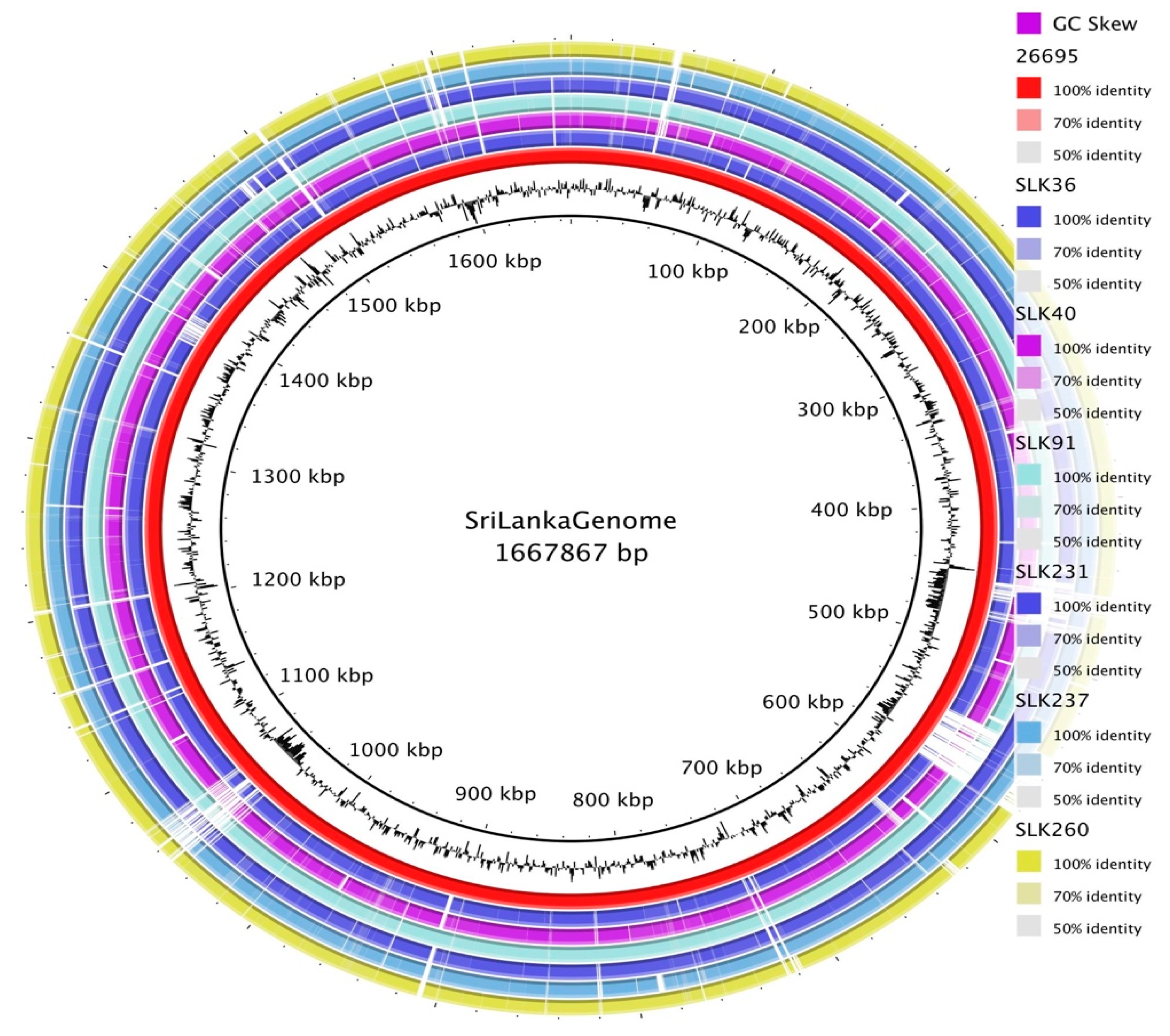
3.2. General Genomic Features of Sri Lankan Strains

All H. pylori strains isolated from Sri Lanka were sequenced by Illumina MiSeq. The sizes of the draft genomes ranged from 1,534,541 bp to 1,625,743 bp, with a GC percentage of 38.8–39.2%. Although the number of contigs ranged from 29 to 48, the longest N50 of the draft genomes was 137,496 bp (Supplementary Table S1). The average coding ratio was 89.7%, similar to the reference strain 26,695, which had a value of 90.5%. Each genome had 36 tRNA and two rRNA genes. Clustered regularly interspaced short palindromic repeats (CRISPRs) were also detected in all strains except SLK40 and SLK260. To examine the conserved genomic regions, the genomes were aligned to the genome of strain 26,695 (Figure 1). A total of 104 paralogous genes belonging to strain 26,695 were absent in all Sri Lankan strains. The alignments also showed that this missing region possessed a high GC skewness, indicating that it may be a region of plasticity.
Annotation showed that the genomes of the Sri Lankan strains had an average number of coding sequences of around 1470 genes, and that 1213 of those genes were possessed by all strains and thus defined as core genes. The total number of unique genes specific to each strain was 374 (Supplementary Figure S2).

3.3. Pan-Genome Analysis of Sri Lankan Strains

To further investigate the characteristics of the genes that were specific to only the Sri Lankan strains, a pan-genome analysis was performed in comparison with representative genomes belonging to the main H. pylori population (hpAfrica1, hpAsia2, hpEurope, and hpEastAsia) [34]. The pan-genomes were obtained by Roary with a minimum identity of 70% (Figure 2). The tree constructed from the binary accessory genes showed that the genomes of all the Sri Lankan strains were concentrated in a cluster that also included the Indian strains NAB47 and SNT49, from hpEurope and hpAsia2, respectively (Figure 2) [34,39,40]. The SLK231, SLK237, and SLK40 strains clustered with hpAsia2, while SLK36 and SLK91 clustered with hpEurope.
Data on the presence and absence of genes were used to determine the genes that are specifically present only in Sri Lankan strains. In the split-paralog mode, screening using Scoary found 122 genes with a naive p-value < 0.05. Benjamini–Hochberg correction (p-corrected) showed that six genes had a statistically significant association with the Sri Lankan strains. Genes annotated as protease HtpX (group_982, p-corrected = 0.008) and restriction endonuclease R (group_437, p-corrected = 0.046) were present in all Sri Lankan strains but absent in 93.1% (27/29) and 86.2% (25/29), respectively, of the other strains (Figure 2). Interestingly, two variant types of htpX were found, group_982 and original htpX. The original htpX gene was present in 96.6% (28/29) of the non-Sri Lankan strains and absent in all Sri Lankan strains (p-corrected = 0.008). These results suggest an adaptation at the gene level to hosts in a particular geographic location.
In the non-split-paralog mode, an outer membrane protein, Omp22 (HP0923), was absent in all Sri Lankan strains but present in the others (false discovery rate (FDR) = 0.0006). These results reflect a wide range of genomic diversity that characterizes this specific geographic location.

3.4. Presence of H. pylori Virulence Factors and Genotypes

Comparative analysis of virulence factors using databases revealed the genes with various virulence functions, summarized in Supplementary Figure S3. We examined 144 virulence genes listed in the VFDB and Victor databases. We included as virulence factors not only genes related to toxicity to the host, but also genes supporting the processes of colonization and survival in chronic infection. Most adherence genes were present, except for babA, which was absent in four of the six strains. In our previous study, the presence of babA in locus A was significantly associated with an increased severity of inflammation [41]. Hence, further analysis of the locus-wise presence of the bab genes was performed (Supplementary Figure S4). The babA genes in SLK40 and SLK231 were in locus A. The only babC gene in strain SLK260 was in locus A after the hypD gene (a marker gene for locus A), whereas four strains (SLK40, SLK231, SLK237, and SLK260) harbored the babB gene only in locus B.
Genome annotation and confirmation by BLASTN revealed that the cagA gene was only present in two strains: SLK231 and SLK237 (two out of six; 33.3%). The cagPAI-negative Sri Lankan strains clustered with hpEurope (three strains) and hpAsia2 (one strain). Meanwhile, vacA was present in all strains. vacA consists of a signal region (s), a p33 domain (i, c, and d), and a p55 (m) domain. Both the cagA-positive Sri Lankan strains possessed the s1a i1 d1 m1 c1 vacA genotype, while all cagA-negative strains possessed the s2 i2 d2 m2 c2 genotype. We constructed a phylogenetic tree of the complete vacA gene to understand its variation compared with reference genomes worldwide (Supplementary Figure S5). The vacA s2 m2 genotype formed a cluster with vacA genes from India (SNT49, India7), France (B8, B38), and Peru (Cuz20), while the vacA s1 m1 genotype was found in a large cluster with strains from Japan, separated as a distinct branch.
H. pylori virulence factors, such as oipA, babA, babB, and iceA, were also genotyped. oipA, the gene encoding the outer membrane inflammatory protein, was present in all the isolated strains; however, a variable number of CT repeats was observed (Table 1). Among the six strains, four showed oipA with an ‘on’ status, while the other two had an oipA ‘off’ status.

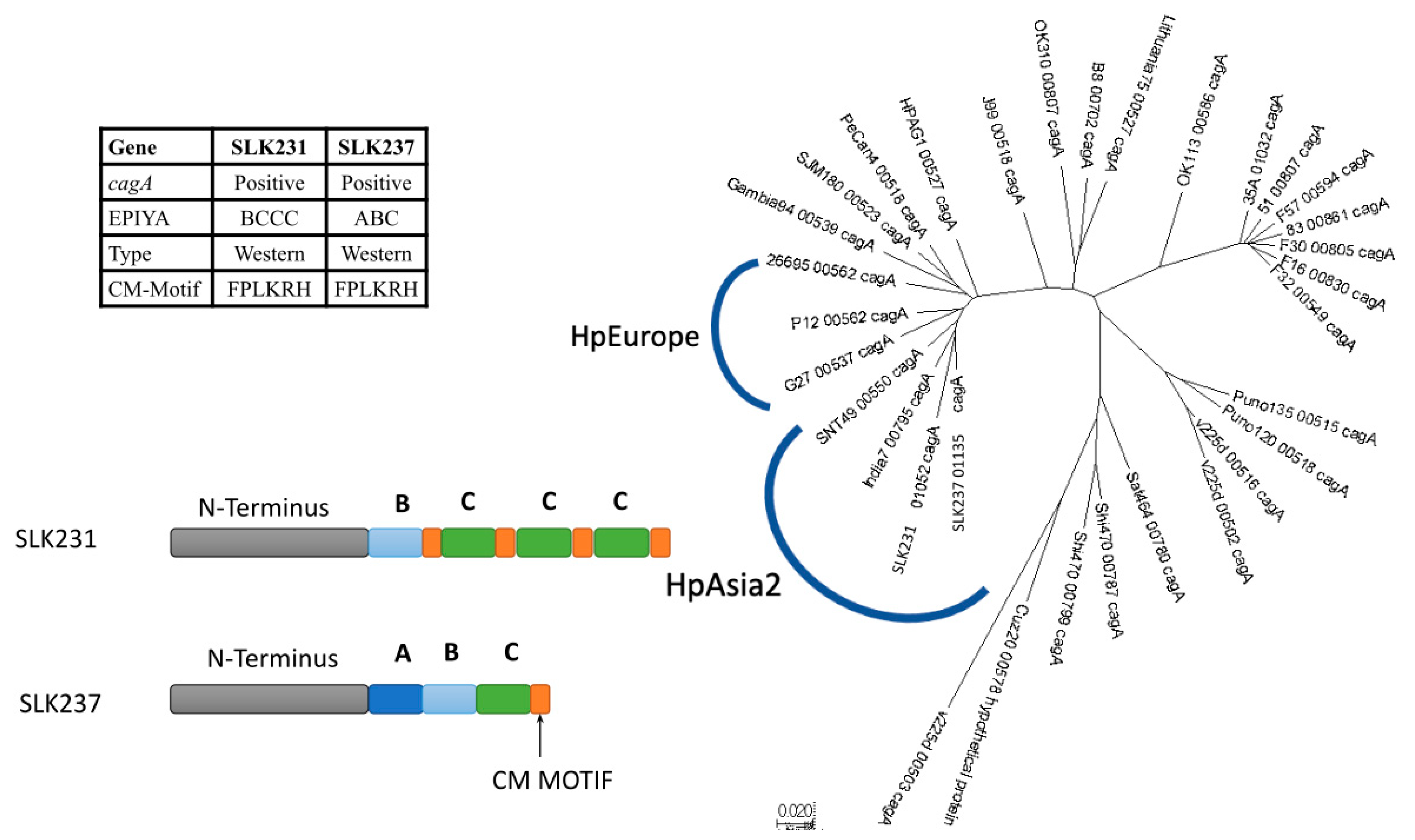
3.5. CagA Genotype Specific to Strains from South Asia

Sequence analysis of the two cagA-positive strains, focusing on the EPIYA region, showed the typical western-type CagA with an ABC-type EPIYA motif in one strain, and a BCCC-type EPIYA motif in the other (Figure 3). We identified that the sequence following the first EPIYA motif matched the B-specific sequence, and the other EPIYA motifs matched the C-specific sequence (Supplementary Figure S6). Both strains possessed the typical CagA-multimerization (CM) sequence (FPLKRHDKVVNDL) of western-type CagA.
To further understand the phylogeny of cagA, a neighbor-joining tree was constructed from the full cagA sequences of strains SLK231 and SLK237 together with the full cagA sequences extracted from the reference genomes used in the pan-genome construction (Figure 3) and vacA phylogeny analysis in the previous section. The cagA sequences of strains SLK231 and SLK237 clustered together with the Indian strains India7 and SNT49, belonging to hpAsia2 (Figure 3). Even though this phylogenetic tree was constructed using only the cagA sequences, it was concordant with the genomic population and geographical region.
To assess the ability of the strains to damage host cells, we investigated the hummingbird phenotype of AGS cells after six hours of co-culture with different CagA motifs. The results showed that strain SLK231 resulted in a greater number of AGS cells with the hummingbird phenotype and dead cells compared with strains SLK237 and SLK36. Moreover, strain SLK231 caused a greater number of cells to show the hummingbird phenotype than strain SLK237 (Supplementary Figure S7).
As most of the strains from Sri Lanka were cagPAI-negative, we further evaluated the synteny of cagPAI. cagA and cagPAI are proposed to be insertion sequences to the H. pylori genome between the dapB gene (4-hydroxy-tetrahydropicolinate reductase) and the glr gene (glutamate racemase). In cagA- and cagPAI-negative strains, this region is called the dg region, and is classified as pre-type A or pre-type B, based on the order of the genes inside the dg region. Using the SouthAfrica7 strain, which has a pre-type B dg region, our analysis showed that SLK40 and SLK91 possess pre-type A dg regions, while SLK36 and SLK260 possess pre-type B (Supplementary Figure S8). This classification is related to the evolution of cagPAI and possibly contributes to the level of virulence.

3.6. Other Virulence Determinants: Screening for Plasmids, Phages, and Secretion Systems

The plasticity region was analyzed by BLAST against the reference sequences that represent the main type of tfs, and then visualized by CGview. These regions were also indicated by the lower GC content (Supplementary Figure S9). We used the plasticity region of the reference strains for genes as follows: P12 for tfs4a, Shi470 for tfs4b, Gambia94/24 and India7 for tfs3, and 26695 for cagPAI and comB. The complete cagPAI was only present in two strains, SLK231 and SLK237, and the tfs3 and tfs4 regions were not found in either of these cagPAI-positive strains. Incomplete tfs4a, tfs4b, and tfs3 regions were observed in SLK36, SLK91, and SLK260. Strain SLK40 did not contain tfs3, tfs4, or cagPAI. The comB apparatus for DNA uptake and natural transformation was detected in all strains (Table 2).
No well-known plasmids were identified in any of the Sri Lankan genomes using the PlasmidFinder database. For the phage analysis, a well-known phage was found in only one strain, SLK237, with a GC percentage of 36.1%. However, four strains—SLK231, SLK260, SLK36, and SLK91—showed incomplete types, suggesting the possibility of phages similar to those found in other bacteria (Supplementary Table S2). No phages were detected in the SLK40 strain, in concordance with the absence of tfs3, tfs4, and cagPAI. These three regions have been proposed to play important roles in the transfer of genetic material.

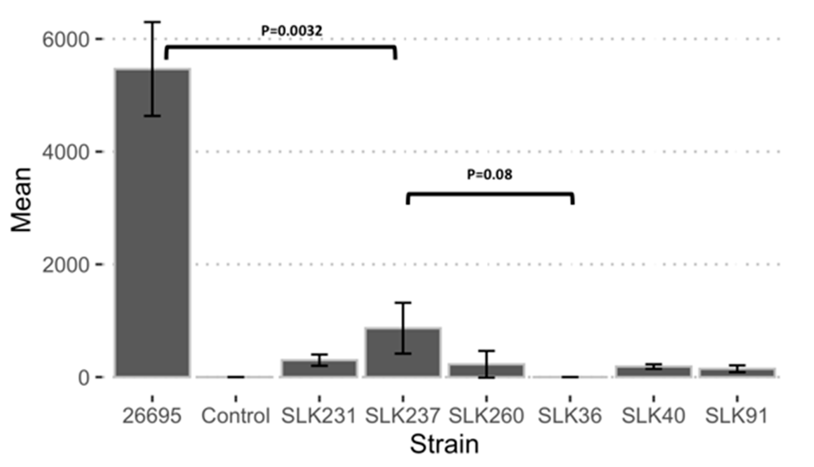
3.7. Adherence Ability of Sri Lankan Strains

Adherence and invasion are the mechanisms used by H. pylori for colonization and are associated with the development of gastric cancer and duodenal ulcers [39]. To determine the adherence and invasion abilities of the strains in gastric mucosal tissue, we carried out an adherence assay on AGS cells. The supernatant culture revealed no bacterial growth in the control group, whereas a high bacterial colony-forming unit (CFU) value was observed for strain 26695. All strains isolated from Sri Lanka showed lower adherence. A pairwise comparison between all the Sri Lankan strains showed no significant differences, except between SLK40 and SLK36 (p = 0.04). The highest adherence and invasion abilities were found in strain SLK237; however, these were still significantly lower than those of strain 26695 (p = 0.0032) (Figure 4).

3.8. Antibiotic Susceptibility Test Results

We used an E-test to test for antibiotic susceptibility to five major H. pylori antibiotics (AMX, TCN, CAM, MNZ, and LEV) on six H. pylori isolates from Sri Lanka. The antibiotic susceptibility test results of the Sri Lankan strains are shown in Table 3. All strains from Sri Lanka were sensitive to AMX, CAM, and TCN. Only one strain, SLK91, showed sensitivity to all five antibiotics tested. The prevalence of LEV resistance was 16.7% (one in six strains), while the prevalence of MNZ resistance was 83.3% (five out of six strains). We defined double-drug resistance as resistance to two of the antibiotics tested. SLK260 was the only multiple-drug-resistant strain, resistant to both LEV and MNZ.
To further investigate the cause of this antibiotic resistance of Sri Lankan strains, we checked for mutations in well-known genes related to these antibiotics. As expected, none of the strains showed any well-known mutations in the 23S rRNA, 16S rRNA, and pbp1a genes. The target protein for the quinolone drug levofloxacin is the DNA Gyrase (gyrA and gyrB), which is important for the DNA replication. For LEV resistance, we investigated the gyrA and gyrB genes to search for mutation. An Asn87Lys mutation was observed in the gyrA gene of strain SLK260, which could explain the resistance of this strain to LEV.
Metronidazole targets the nitroreductase enzyme and the loss of function of this gene is associated with resistance. However, mutations in other genes, such as frxA, fur, omp11, and ribF, have also been reported to be associated with resistance. To understand the high prevalence of resistance to metronidazole, we evaluated five genes—frxA, fur, omp11, rdxA, and ribF—and aligned these with the ATCC 26,695 control strain. We observed SNPs that were present in the resistant strains and were not observed in the MNZ-sensitive strains. The rdxA gene was investigated for any mutational changes or premature termination (Table 4). RdxA is the receptor for MNZ, and any changes to the protein by mutations or variation could affect the drug affinity. An early-stop codon was found at position 52 in SLK231, causing premature termination. The remaining strains had an intact rdxA but synonymous point mutations were observed. Alteration of histidine (H) to tyrosine (Y) or threonine (T) at locus 97 was found in 80% (four out of five) of the resistant strains, and this may play a role in metronidazole resistance. Meanwhile, the genes encoding Fur and Omp11 are the most conserved of the five genes evaluated, and the mutations to these genes that were detected are listed in Table 4. Mutations to RibF were also observed, such as Q78R, Q111K, and Q242K.

4. Discussion

H. pylori is one of the most diverse bacteria, as shown by its high recombination and mutation rates [42,43]. Persistent H. pylori infection, leading to co-evolution with humans, has contributed to the wide variety of strains, which show regional characteristics [3]. In this study, a comparative genomics analysis was performed on the genomes of H. pylori strains isolated from indigenous people of Sri Lanka. This population is unique in showing a much lower prevalence of H. pylori than the worldwide rate of 50%. In Sri Lanka, the prevalence of infection is only 1.7%, and the severity of gastric inflammation is low [18]. However, there had been no reports on the genomic features and virulence factors of H. pylori isolates from Sri Lanka. Thus, this study is the first to provide data on the antibiotic resistance, virulence factors, and disease severity of Sri Lankan H. pylori strains.
Infection by the strains isolated from Sri Lanka resulted in non-severe inflammation in the hosts, as indicated by histological analysis of the gastric mucosa. Genomic analysis also showed the absence of cagPAI as the predominant virulence factor in H. pylori, and a less virulent vacA genotype (the s2 m2 type). Moreover, the genes encoding BabA and its paralogues were also incomplete. This finding is concordant with a report showing that strains isolated from the South Asian region (Bangladesh and India) had lower pathogenicity than worldwide H. pylori, indicating that the genetic composition of the H. pylori population affects the virulence properties [44]. Furthermore, the genomes of the Sri Lankan strains were divided into two groups: one close to NAB47 (hpEurope) and one close to SNT49 (hpAsia2) [34,39,40,45]. These results indicate that Sri Lankan strains have similar characteristics to strains isolated from the South Asian region [43,46], suggesting that the H. pylori strains found in Sri Lanka may be less of a health concern than those infecting East Asian countries.
CagA and VacA are the major factors of H. pylori virulence. The delivery of CagA is dependent on the secretion system encoded by cagPAI [47]. A previous study revealed that cagA evolution is the result of horizontal gene transfer [48], which was then incorporated into the H. pylori genome in the dg region as a cluster of genes, termed cagPAI, encoding the secretion apparatus. In the genomes of Sri Lankan strains, there were no traces of the cagPAI components in the cagA-negative strains. The gene arrangement of the dg region of SLK, classed as pre-type B, is similar to that of strains from hpAfrica1. This pre-type B region can then become occupied by cagPAI type B genes, as in hpEurope strains, likely including multiple cagA genes [48]. As shown by evaluation of the phylogenetic tree, cagA from Sri Lankan strains clustered with cagA of hpAsia2 strains, close to hpEurope. This phylogenetic tree supports the suggestion that cagA evolved in concordance with the whole genome [49].
In terms of the adherence genes, some Sri Lankan strains did not possess bab-paralogous genes, such as babA, babB, and babC. Contrary to this study, when we performed targeted PCR for bab-paralogous genes in our previous study, we detected the presence of babA in 87% of strains isolated from different regions of South Asia [41]. BabA is a Lewis b binding adhesin required for the colonization of epithelial cells [50,51]. The location of babA in locus A is also related to a higher severity of inflammation [41]. The absence of babA may contribute to low adherence to AGS cells. In agreement with this, the strains in this study that possessed babA showed higher adherence, and thus higher gastric inflammation and atrophy scores.
Alongside a lack of cagA, 67% (four out of six) of Sri Lankan strains also showed a less virulent type of vacA (s2 i2 m2). There are several genotype markers for the s, i, and m regions, encoding the signal sequence, p33 domain, and p55 domain, respectively [52]. The sequence of the s2 genotype contains an insertion that inhibits vacuolization, and the m2 genotype reduces the specificity of VacA to the cellular receptor. VacA creates vacuoles, detaching the tight junction, and is required for colonization [53]. This may explain the lower severity of the gastritis, as revealed by histology score evaluation, for most of the strains in this study. Unlike cagA, the diversity of vacA matches the geographic region (Asian versus non-Asian vacA), in concordance with our previous study [52,54]. These results support the findings of our previous study, that virulence genes are fixed in indigenous people, including cagA and vacA [44]. Hence, it is not surprising that the Sri Lankan population maintains the vacA s2 m2 genotype in the cagA-negative strains.
A variety of disease outcomes are also observed among cagA-positive strains, based on the genotype for the EPIYA motifs, which affect the affinity to SHP phosphatase [12]. In this study, both cagA-positive Sri Lankan strains possessed a western-type CagA, with one strain having a BCCC-type EPIYA motif. Both EPIYA-A and EPIYA-B have recognition sites for SHP2, and so CagA function may be retained despite an absence of EPIYA-A [12,48]. The EPIYA motifs and the CM motif are binding sites for both SHP2, which induces abnormal cell proliferation, and PAR1, which destroys the gastric mucosa [55]. AGS cells showed a higher proportion of the hummingbird phenotype, with strains with BCCC-type EPIYA-motifs compared with the ABC type. Thus, even though most strains share similar virulence genes, CagA and its EPIYA motifs play integral roles in the virulence of H. pylori [56,57].
This study also upholds the consensus that the region of plasticity is a factor in virulence and is associated with more severe gastric inflammation, duodenal ulcers, and gastric cancer [15,16]. Strains with cagPAI did not possess other tfs genes, while the cagPAI-negative strains possessed tfs genes that were incomplete or absent. These genes were acquired by horizontal gene transfer, indicated by the negative skew of the GC percentage [47]. The tfs3 gene produces a protein called CtkA that can be injected into epithelial cells [58]. Moreover, tfs3 also assists in the uptake of external genetic material [59]. The lack of these tfs components is concordant with the absence of plasmids, phages, and IS elements in all strains except SLK237. SLK237 contained IS605 alongside the tnpA and tnpB genes, which play roles in genomic rearrangement and recombination [60,61].
The antibiotic susceptibility test is key, as it is the most effective way to determine the best regimen of therapy to combat H. pylori infection. To our knowledge, this is the first study evaluating the susceptibility of H. pylori strains to five major antibiotics in Sri Lanka. We found that all six strains were sensitive to AMX, TCN, and CAM. Of the six strains, five had developed resistance to MNZ, suggesting that a regimen containing MNZ might not be suitable as first-line therapy. The high prevalence of MNZ resistance was not surprising, as resistance to this antibiotic has been reported in many countries worldwide [29,30]. The results of this study suggest that triple therapy containing AMX and CAM is still an effective first-line therapy against H. pylori infection in Sri Lanka.
Our results reveal a concerning trend of increasing resistance, particularly towards metronidazole (MNZ) and levofloxacin (LEV). We identified potential mechanisms for this resistance. We detected mutations in the rdxA gene, which plays a critical role in the action of MNZ [17]. One strain had a premature-stop codon mutation in this gene, rendering it nonfunctional. Additionally, most resistant strains harbored putative mutations at locus 97, potentially changing histidine to tyrosine or threonine. These mutations might affect the protein structure and hinder MNZ binding [62]. In the case of LEV resistance, a mutation in the gyrA gene in locus 87 was identified only in the resistant strains [17,63,64].
The high prevalence of antibiotic resistance in Sri Lankan H. pylori strains is a worrying development. Antibiotic overuse is potentially contributing to this emergence of resistance [65,66]. This aligns with observations in other South Asian countries, where similar increases in H. pylori resistance have been reported [64]. For example, in Bangladesh, the prevalence of infection is 60.2%, and the resistance to metronidazole, clarithromycin, and levofloxacin is high: 94.6%, 39.3%, and 66.1%, respectively. Nepal also reported high resistance to metronidazole and clarithromycin: 88.1% and 21.4%, respectively. This finding suggests caution should be taken in the use of metronidazole in the H. pylori treatment regimens [64]. To combat this growing threat, stricter antibiotic control programs are essential. Promoting public awareness about responsible antibiotic use and exploring alternative treatment options are crucial steps. Further research is needed to understand the specific factors driving resistance in Sri Lanka and to develop effective regional strategies to manage H. pylori infections.
This study is limited by the reliability of high-throughput next-generation sequencing. There was also a discrepancy in the number of rRNA and tRNA genes, and a higher number of contigs. This draft genome used short reads to construct a de novo assembly, which might miss some overlapping regions, and the detection of repeated regions or genes with high copy numbers may be problematic [67]. Nevertheless, short-read sequencing provides higher precision and a lower error rate [68,69]. It is also sufficient to comprehensively evaluate the virulome, and this accuracy is necessary for the evaluation of the genotype and gene polymorphisms. Furthermore, the small number of strains evaluated in this study limits the capacity to make causality conclusions; additional research with a larger number of strains is required.

5. Conclusions

This investigation, using a comparative genomic method, provides a greater perspective on H. pylori virulence factors. Our comparative genomic analysis reveals genes specific to the geographic location, showing less virulent genes in most of the Sri Lankan strains, in concordance with in vitro experiments and histological assessment of gastric inflammation. This study provides a comprehensive insight into the virulence and antibiotic resistance of H. pylori. Future research using larger and more diverse population and in vitro experiments on host–pathogen interaction is necessary to establish causality.

Supplementary Materials

The following supporting information can be downloaded at: https://www.mdpi.com/article/10.3390/microorganisms13020420/s1, Supplementary Figure S1. Evaluation of gastric inflammation in antrum, angulus, and body; Supplementary Figure S2. Visualization of the core genome and shell genome of Sri Lanka strains; Supplementary Figure S3. Presence and absence of virulence genes; Supplementary Figure S4. Determination of babA, babB, and babC locus; Supplementary Figure S5. The phylogenetic tree of vacA compared to the vacA from the reference genomes; Supplementary Figure S6. Sequence alignment of cagA 26695 in the EPIYA region and CM motif; Supplementary Figure S7. The hummingbird phenotype of AGS cells infected with Sri Lanka strains; Supplementary Figure S8. cagPAI empty sites arrangement; Supplementary Figure S9. Tfs visualization by CGView; Supplementary Table S1. Quality of assembly by DFAST; Supplementary Table S2. The presence of phage among Sri Lanka isolates.

Author Contributions

Conceptualization: K.A.F. and Y.Y.; methodology: J.A. and T.M. (Takashi Matsumoto); investigation: D.D., K.A.F., R.I.A., S.B., E.T.K. and B.H.P.; resources: M.D.L., J.R., T.M. (Takeshi Matsuhisa) and Y.Y.; data curation: K.A.F., L.A.W. and V.P.T.; writing—original draft preparation: K.A.F. and Y.Y.; writing—review and editing: K.A.F., S.A., E.T.K. and Y.Y.; visualization: V.P.T. and S.B.; supervision: T.M. (Takeshi Matsuhisa) and Y.Y. All authors have read and agreed to the published version of the manuscript.

Funding

This report is based on work supported in part by Grants-in-Aid for Scientific Research from the Ministry of Education, Culture, Sports, Science, and Technology (MEXT) of Japan (22H02871 and 23K24133) (Y.Y.). This work was also supported by the Japan Agency for Medical Research and Development (AMED) [e-ASIA JRP, Science and Technology Research Partnership for Sustainable Development (SATREPS)] (Y.Y.), and Japan International Cooperation Agency (JICA) (Y.Y.).

Institutional Review Board Statement

Study approval was obtained from the ethics committees of the University of Peradeniya, Sri Lanka (2017/EC/91, 11 November 2017), and Oita University Faculty of Medicine, Japan (P-12-10, 18 January 2013).

Informed Consent Statement

Informed consent was obtained from all subjects involved in the study. Written informed consent has been obtained from the patients to publish this paper.

Data Availability Statement

Genomes of all strains were deposited in BioProject with the accession number PRJDB17566 and the BioSamples SAMD00753457_SLK231, SAMD00753458_SLK237, SAMD00753459_SLK260, SAMD00753460_SLK36, SAMD00753461_SLK40, and SAMD00753462_SLK91.

Conflicts of Interest

The authors declare no conflicts of interest.

References

  1. Mera, R.M.; Bravo, L.E.; Camargo, M.C.; Bravo, J.C.; Delgado, A.G.; Romero-Gallo, J.; Yepez, M.C.; Realpe, J.L.; Schneider, B.G.; Morgan, D.R.; et al. Dynamics of Helicobacter pylori infection as a determinant of progression of gastric precancerous lesions: 16-year follow-up of an eradication trial. Gut 2018, 67, 1239–1246. [Google Scholar] [CrossRef] [PubMed]
  2. Correa, P.; Houghton, J. Carcinogenesis of Helicobacter pylori. Gastroenterology 2007, 133, 659–672. [Google Scholar] [CrossRef] [PubMed]
  3. Kodaman, N.; Pazos, A.; Schneider, B.G.; Piazuelo, M.B.; Mera, R.; Sobota, R.S.; Sicinschi, L.A.; Shaffer, C.L.; Romero-Gallo, J.; de Sablet, T.; et al. Human and Helicobacter pylori coevolution shapes the risk of gastric disease. Proc. Natl. Acad. Sci. USA 2014, 111, 1455–1460. [Google Scholar] [CrossRef]
  4. Correa, P.; Piazuelo, M.B. Evolutionary History of the Helicobacter pylori Genome: Implications for Gastric Carcinogenesis. Gut Liver 2012, 6, 21–28. [Google Scholar] [CrossRef]
  5. de Sablet, T.; Piazuelo, M.B.; Shaffer, C.L.; Schneider, B.G.; Asim, M.; Chaturvedi, R.; Bravo, L.E.; Sicinschi, L.A.; Delgado, A.G.; Mera, R.M.; et al. Phylogeographic origin of Helicobacter pylori is a determinant of gastric cancer risk. Gut 2011, 60, 1189–1195. [Google Scholar] [CrossRef]
  6. Hooi, J.K.Y.; Lai, W.Y.; Ng, W.K.; Suen, M.M.Y.; Underwood, F.E.; Tanyingoh, D.; Malfertheiner, P.; Graham, D.Y.; Wong, V.W.S.; Wu, J.C.Y.; et al. Global Prevalence of Helicobacter pylori Infection: Systematic Review and Meta-Analysis. Gastroenterology 2017, 153, 420–429. [Google Scholar] [CrossRef] [PubMed]
  7. Fauzia, K.A.; Tuan, V.P. Rising resistance: Antibiotic choices for Helicobacter pylori infection. Lancet Gastroenterol. Hepatol. 2024, 9, 7–8. [Google Scholar] [CrossRef] [PubMed]
  8. Shanks, A.M.; El-Omar, E.M. Helicobacter pylori infection, host genetics and gastric cancer. J. Dig. Dis. 2009, 10, 157–164. [Google Scholar] [CrossRef]
  9. Dai, J.; Zhao, J.; Mao, L.; Hu, Y.; Lv, B. Study on the value of antibiotic-resistant gene detection in Helicobacter pylori in China. Exp. Ther. Med. 2022, 23, 228. [Google Scholar] [CrossRef]
  10. Balloux, F.; Brønstad Brynildsrud, O.; van Dorp, L.; Shaw, L.P.; Chen, H.; Harris, K.A.; Wang, H.; Eldholm, V. From Theory to Practice: Translating Whole-Genome Sequencing (WGS) into the Clinic. Trends Microbiol. 2018, 26, 1035–1048. [Google Scholar] [CrossRef] [PubMed]
  11. Cross, A.S. What is a virulence factor? Crit. Care 2008, 12, 196. [Google Scholar] [CrossRef]
  12. Hatakeyama, M.; Higashi, H. Helicobacter pylori CagA: A new paradigm for bacterial carcinogenesis. Cancer Sci. 2005, 96, 835–843. [Google Scholar] [CrossRef] [PubMed]
  13. Ansari, S.; Yamaoka, Y. Survival of Helicobacter pylori in gastric acidic territory. Helicobacter 2017, 22, e12386. [Google Scholar] [CrossRef] [PubMed]
  14. Yamaoka, Y.; Ojo, O.; Fujimoto, S.; Odenbreit, S.; Haas, R.; Gutierrez, O.; El-Zimaity, H.M.; Reddy, R.; Arnqvist, A.; Graham, D.Y. Helicobacter pylori outer membrane proteins and gastroduodenal disease. Gut 2006, 55, 775–781. [Google Scholar] [CrossRef]
  15. Backert, S.; Tegtmeyer, N.; Fischer, W. Composition, structure and function of the Helicobacter pylori cag pathogenicity island encoded type IV secretion system. Future Microbiol. 2015, 10, 955–965. [Google Scholar] [CrossRef] [PubMed]
  16. Backert, S.; Haas, R.; Gerhard, M.; Naumann, M. The Helicobacter pylori Type IV Secretion System Encoded by the cag Pathogenicity Island: Architecture, Function, and Signaling. Curr. Top. Microbiol. Immunol. 2017, 413, 187–220. [Google Scholar] [CrossRef] [PubMed]
  17. Tshibangu-Kabamba, E.; Yamaoka, Y. Helicobacter pylori infection and antibiotic resistance—From biology to clinical implications. Nat. Rev. Gastroenterol. Hepatol. 2021, 18, 613–629. [Google Scholar] [CrossRef] [PubMed]
  18. Doohan, D.; Fauzia, K.A.; Rathnayake, J.; Lamawansa, M.D.; Waskito, L.A.; Tuan, V.P.; Dashdorj, A.; Kabamba, E.T.; Phuc, B.H.; Ansari, S.; et al. Pepsinogen and Serum IgG Detection Is a Valuable Diagnostic Method for Helicobacter pylori Infection in a Low-Prevalence Country: A Report from Sri Lanka. Diagnostics 2021, 11, 1364. [Google Scholar] [CrossRef] [PubMed]
  19. Stolte, M.; Meining, A. The updated Sydney system: Classification and grading of gastritis as the basis of diagnosis and treatment. Can. J. Gastroenterol. 2001, 15, 591–598. [Google Scholar] [CrossRef]
  20. Dixon, M.F.; Genta, R.M.; Yardley, J.H.; Correa, P. Classification and grading of gastritis. The updated Sydney System. International Workshop on the Histopathology of Gastritis, Houston 1994. Am. J. Surg. Pathol. 1996, 20, 1161–1181. [Google Scholar] [CrossRef] [PubMed]
  21. Uchida, T.; Kanada, R.; Tsukamoto, Y.; Hijiya, N.; Matsuura, K.; Yano, S.; Yokoyama, S.; Kishida, T.; Kodama, M.; Murakami, K.; et al. Immunohistochemical diagnosis of the cagA-gene genotype of Helicobacter pylori with anti-East Asian CagA-specific antibody. Cancer Sci. 2007, 98, 521–528. [Google Scholar] [CrossRef]
  22. Wick, R.R.; Judd, L.M.; Gorrie, C.L.; Holt, K.E. Unicycler: Resolving bacterial genome assemblies from short and long sequencing reads. PLoS Comput. Biol. 2017, 13, e1005595. [Google Scholar] [CrossRef] [PubMed]
  23. Tanizawa, Y.; Fujisawa, T.; Nakamura, Y. DFAST: A flexible prokaryotic genome annotation pipeline for faster genome publication. Bioinformatics 2018, 34, 1037–1039. [Google Scholar] [CrossRef] [PubMed]
  24. Gurevich, A.; Saveliev, V.; Vyahhi, N.; Tesler, G. QUAST: Quality assessment tool for genome assemblies. Bioinformatics 2013, 29, 1072–1075. [Google Scholar] [CrossRef]
  25. Chen, L.; Zheng, D.; Liu, B.; Yang, J.; Jin, Q. VFDB 2016: Hierarchical and refined dataset for big data analysis—10 years on. Nucleic Acids Res. 2016, 44, D694–D697. [Google Scholar] [CrossRef] [PubMed]
  26. Sayers, S.; Li, L.; Ong, E.; Deng, S.; Fu, G.; Lin, Y.; Yang, B.; Zhang, S.; Fa, Z.; Zhao, B. Victors: A web-based knowledge base of virulence factors in human and animal pathogens. Nucleic Acids Res. 2019, 47, D693–D700. [Google Scholar] [CrossRef]
  27. Kumar, S.; Nei, M.; Dudley, J.; Tamura, K. MEGA: A biologist-centric software for evolutionary analysis of DNA and protein sequences. Brief. Bioinform. 2008, 9, 299–306. [Google Scholar] [CrossRef]
  28. Carattoli, A.; Hasman, H. PlasmidFinder and in silico pMLST: Identification and typing of plasmid replicons in whole-genome sequencing (WGS). In Horizontal Gene Transfer; Springer: Berlin/Heidelberg, Germany, 2020; pp. 285–294. [Google Scholar]
  29. Arndt, D.; Grant, J.R.; Marcu, A.; Sajed, T.; Pon, A.; Liang, Y.; Wishart, D.S. PHASTER: A better, faster version of the PHAST phage search tool. Nucleic Acids Res. 2016, 44, W16–W21. [Google Scholar] [CrossRef] [PubMed]
  30. Siguier, P.; Pérochon, J.; Lestrade, L.; Mahillon, J.; Chandler, M. ISfinder: The reference centre for bacterial insertion sequences. Nucleic Acids Res. 2006, 34, D32–D36. [Google Scholar] [CrossRef]
  31. Stothard, P.; Wishart, D.S. Circular genome visualization and exploration using CGView. Bioinformatics 2005, 21, 537–539. [Google Scholar] [CrossRef]
  32. Page, A.J.; Cummins, C.A.; Hunt, M.; Wong, V.K.; Reuter, S.; Holden, M.T.; Fookes, M.; Falush, D.; Keane, J.A.; Parkhill, J. Roary: Rapid large-scale prokaryote pan genome analysis. Bioinformatics 2015, 31, 3691–3693. [Google Scholar] [CrossRef] [PubMed]
  33. Hadfield, J.; Croucher, N.J.; Goater, R.J.; Abudahab, K.; Aanensen, D.M.; Harris, S.R. Phandango: An interactive viewer for bacterial population genomics. Bioinformatics 2018, 34, 292–293. [Google Scholar] [CrossRef]
  34. Yahara, K.; Furuta, Y.; Oshima, K.; Yoshida, M.; Azuma, T.; Hattori, M.; Uchiyama, I.; Kobayashi, I. Chromosome painting in silico in a bacterial species reveals fine population structure. Mol. Biol. Evol. 2013, 30, 1454–1464. [Google Scholar] [CrossRef] [PubMed]
  35. Petersen, A.M.; Blom, J.; Andersen, L.P.; Krogfelt, K.A. Role of strain type, AGS cells and fetal calf serum in Helicobacter pylori adhesion and invasion assays. FEMS Immunol. Med. Microbiol. 2000, 29, 59–67. [Google Scholar] [CrossRef] [PubMed]
  36. Chen, Y.-H.; Tsai, W.-H.; Wu, H.-Y.; Chen, C.-Y.; Yeh, W.-L.; Chen, Y.-H.; Hsu, H.-Y.; Chen, W.-W.; Chen, Y.-W.; Chang, W.-W. Probiotic Lactobacillus spp. act against Helicobacter pylori-induced inflammation. J. Clin. Med. 2019, 8, 90. [Google Scholar] [CrossRef] [PubMed]
  37. Chang, C.-C.; Kuo, W.-S.; Chen, Y.-C.; Perng, C.-L.; Lin, H.-J.; Ou, Y.-H. Fragmentation of CagA reduces hummingbird phenotype induction by helicobactor pylori. PLoS ONE 2016, 11, e0150061. [Google Scholar] [CrossRef] [PubMed]
  38. The European Committee on Antimicrobial Susceptibility Testing. European Committee on Antimicrobial Susceptibility Testing, Breakpoint Tables for Interpretation of MICs and Zone Diameters; EUCAST: Växjö, Sweden, 2015. [Google Scholar]
  39. Kumar, N.; Mukhopadhyay, A.K.; Patra, R.; De, R.; Baddam, R.; Shaik, S.; Alam, J.; Tiruvayipati, S.; Ahmed, N. Next-Generation Sequencing and De Novo Assembly, Genome Organization, and Comparative Genomic Analyses of the Genomes of Two Helicobacter pylori Isolates from Duodenal Ulcer Patients in India. J. Bacteriol. 2012, 194, 5963–5964. [Google Scholar] [CrossRef] [PubMed]
  40. Kauser, F.; Hussain, M.A.; Ahmed, I.; Ahmad, N.; Habeeb, A.; Khan, A.A.; Ahmed, N. Comparing genomes of Helicobacter pylori strains from the high-altitude desert of Ladakh, India. J. Clin. Microbiol. 2005, 43, 1538–1545. [Google Scholar] [CrossRef] [PubMed]
  41. Ansari, S.; Kabamba, E.T.; Shrestha, P.K.; Aftab, H.; Myint, T.; Tshering, L.; Sharma, R.P.; Ni, N.; Aye, T.T.; Subsomwong, P.; et al. Helicobacter pylori bab characterization in clinical isolates from Bhutan, Myanmar, Nepal and Bangladesh. PLoS ONE 2017, 12, e0187225. [Google Scholar] [CrossRef] [PubMed]
  42. Suerbaum, S.; Smith, J.M.; Bapumia, K.; Morelli, G.; Smith, N.H.; Kunstmann, E.; Dyrek, I.; Achtman, M. Free recombination within Helicobacter pylori. Proc. Natl. Acad. Sci. USA 1998, 95, 12619–12624. [Google Scholar] [CrossRef]
  43. Falush, D.; Kraft, C.; Taylor, N.S.; Correa, P.; Fox, J.G.; Achtman, M.; Suerbaum, S. Recombination and mutation during long-term gastric colonization by Helicobacter pylori: Estimates of clock rates, recombination size, and minimal age. Proc. Natl. Acad. Sci. USA 2001, 98, 15056–15061. [Google Scholar] [CrossRef]
  44. Muñoz-Ramirez, Z.Y.; Pascoe, B.; Mendez-Tenorio, A.; Mourkas, E.; Sandoval-Motta, S.; Perez-Perez, G.; Morgan, D.R.; Dominguez, R.L.; Ortiz-Princz, D.; Cavazza, M.E. A 500-year tale of co-evolution, adaptation, and virulence: Helicobacter pylori in the Americas. ISME J. 2021, 15, 78–92. [Google Scholar] [CrossRef] [PubMed]
  45. Kumar, N.; Mariappan, V.; Baddam, R.; Lankapalli, A.K.; Shaik, S.; Goh, K.-L.; Loke, M.F.; Perkins, T.; Benghezal, M.; Hasnain, S.E.; et al. Comparative genomic analysis of Helicobacter pylori from Malaysia identifies three distinct lineages suggestive of differential evolution. Nucleic Acids Res. 2014, 43, 324–335. [Google Scholar] [CrossRef] [PubMed]
  46. Falush, D.; Wirth, T.; Linz, B.; Pritchard, J.K.; Stephens, M.; Kidd, M.; Blaser, M.J.; Graham, D.Y.; Vacher, S.; Perez-Perez, G.I.; et al. Traces of Human Migrations in Helicobacter pylori Populations. Science 2003, 299, 1582–1585. [Google Scholar] [CrossRef] [PubMed]
  47. Tegtmeyer, N.; Wessler, S.; Backert, S. Role of the cag-pathogenicity island encoded type IV secretion system in Helicobacter pylori pathogenesis. FEBS J. 2011, 278, 1190–1202. [Google Scholar] [CrossRef]
  48. Furuta, Y.; Yahara, K.; Hatakeyama, M.; Kobayashi, I. Evolution of cagA oncogene of Helicobacter pylori through recombination. PLoS ONE 2011, 6, e23499. [Google Scholar] [CrossRef] [PubMed]
  49. Muñoz-Ramírez, Z.Y.; Mendez-Tenorio, A.; Kato, I.; Bravo, M.M.; Rizzato, C.; Thorell, K.; Torres, R.; Aviles-Jimenez, F.; Camorlinga, M.; Canzian, F.; et al. Whole Genome Sequence and Phylogenetic Analysis Show Helicobacter pylori Strains from Latin America Have Followed a Unique Evolution Pathway. Front. Cell. Infect. Microbiol. 2017, 7, 50. [Google Scholar] [CrossRef] [PubMed]
  50. Petzold, K.; Olofsson, A.; Arnqvist, A.; Boren, T.; Gröbner, G.; Schleucher, J. Helicobacter pylori: How is Adhesin BabA, a Blood Group Antigen Binding Membrane Protein, Involved in Bacterial Adherence? Biophys. J. 2009, 96, 409a. [Google Scholar] [CrossRef]
  51. Quintana-Hayashi, M.P.; Rocha, R.; Padra, M.; Thorell, A.; Jin, C.; Karlsson, N.G.; Roxo-Rosa, M.; Oleastro, M.; Lindén, S.K. BabA-mediated adherence of pediatric ulcerogenic H. pylori strains to gastric mucins at neutral and acidic pH. Virulence 2018, 9, 1699–1717. [Google Scholar] [CrossRef]
  52. Gangwer, K.A.; Shaffer, C.L.; Suerbaum, S.; Lacy, D.B.; Cover, T.L.; Bordenstein, S.R. Molecular evolution of the Helicobacter pylori vacuolating toxin gene vacA. J. Bacteriol. 2010, 192, 6126–6135. [Google Scholar] [CrossRef] [PubMed]
  53. Palframan, S.L.; Kwok, T.; Gabriel, K. Vacuolating cytotoxin A (VacA), a key toxin for Helicobacter pylori pathogenesis. Front. Cell. Infect. Microbiol. 2012, 2, 92. [Google Scholar] [CrossRef] [PubMed]
  54. Gutiérrez-Escobar, A.J.; Bravo, M.M.; Acevedo, O.; Backert, S. Molecular evolution of the VacA p55 binding domain of Helicobacter pylori in mestizos from a high gastric cancer region of Colombia. PeerJ 2019, 7, e6634. [Google Scholar] [CrossRef] [PubMed]
  55. Hatakeyama, M. Structure and function of Helicobacter pylori CagA, the first-identified bacterial protein involved in human cancer. Proc. Jpn. Academy. Ser. B Phys. Biol. Sci. 2017, 93, 196–219. [Google Scholar] [CrossRef] [PubMed]
  56. Matos, J.I.; de Sousa, H.A.; Marcos-Pinto, R.; Dinis-Ribeiro, M. Helicobacter pylori CagA and VacA genotypes and gastric phenotype: A meta-analysis. Eur. J. Gastroenterol. Hepatol. 2013, 25, 1431–1441. [Google Scholar] [CrossRef] [PubMed]
  57. Park, J.Y.; Forman, D.; Waskito, L.A.; Yamaoka, Y.; Crabtree, J.E. Epidemiology of Helicobacter pylori and CagA-positive infections and global variations in gastric cancer. Toxins 2018, 10, 163. [Google Scholar] [CrossRef]
  58. Alandiyjany, M.N.; Croxall, N.J.; Grove, J.I.; Delahay, R.M. A role for the tfs3 ICE-encoded type IV secretion system in pro-inflammatory signalling by the Helicobacter pylori Ser/Thr kinase, CtkA. PLoS ONE 2017, 12, e0182144. [Google Scholar] [CrossRef] [PubMed]
  59. Fischer, W.; Tegtmeyer, N.; Stingl, K.; Backert, S. Four chromosomal type IV secretion systems in Helicobacter pylori: Composition, structure and function. Front. Microbiol. 2020, 11, 1592. [Google Scholar] [CrossRef] [PubMed]
  60. He, S.; Corneloup, A.; Guynet, C.; Lavatine, L.; Caumont-Sarcos, A.; Siguier, P.; Marty, B.; Dyda, F.; Chandler, M.; Ton Hoang, B. The IS 200/IS 605 Family and “Peel and Paste” Single-Strand Transposition Mechanism. Microbiol. Spectr. 2015, 3, 4. [Google Scholar] [CrossRef] [PubMed]
  61. Abadi, A.T.B.; Mobarez, A.M.; Bonten, M.J.; Wagenaar, J.A.; Kusters, J.G. Clinical relevance of the cagA, tnpA and tnpB genes in Helicobacter pylori. BMC Gastroenterol. 2014, 14, 33. [Google Scholar] [CrossRef] [PubMed]
  62. Jenks, P.J.; Labigne, A.; Ferrero, R.L. Exposure to Metronidazole In Vivo Readily Induces Resistance in Helicobacter pylori and Reduces the Efficacy of Eradication Therapy in Mice. Antimicrob. Agents Chemother. 1999, 43, 777–781. [Google Scholar] [CrossRef] [PubMed]
  63. Fauzia, K.A.; Aftab, H.; Tshibangu-Kabamba, E.; Alfaray, R.I.; Saruuljavkhlan, B.; Cimuanga-Mukanya, A.; Matsumoto, T.; Subsomwong, P.; Akada, J.; Miftahussurur, M.; et al. Mutations Related to Antibiotics Resistance in Helicobacter pylori Clinical Isolates from Bangladesh. Antibiotics 2023, 12, 279. [Google Scholar] [CrossRef] [PubMed]
  64. Miftahussurur, M.; Aftab, H.; Shrestha, P.K.; Sharma, R.P.; Subsomwong, P.; Waskito, L.A.; Doohan, D.; Fauzia, K.A.; Yamaoka, Y. Effective therapeutic regimens in two South Asian countries with high resistance to major Helicobacter pylori antibiotics. Antimicrob. Resist. Infect. Control 2019, 8, 40. [Google Scholar] [CrossRef] [PubMed]
  65. Tillekeratne, L.G.; Bodinayake, C.K.; Dabrera, T.; Nagahawatte, A.; Arachchi, W.K.; Sooriyaarachchi, A.; Stewart, K.; Watt, M.; Østbye, T.; Woods, C.W. Antibiotic overuse for acute respiratory tract infections in Sri Lanka: A qualitative study of outpatients and their physicians. BMC Fam. Pract. 2017, 18, 37. [Google Scholar] [CrossRef] [PubMed]
  66. Gunasekera, Y.D.; Kinnison, T.; Kottawatta, S.A.; Silva-Fletcher, A.; Kalupahana, R.S. Misconceptions of antibiotics as a potential explanation for their misuse. A survey of the general public in a rural and urban community in Sri Lanka. Antibiotics 2022, 11, 454. [Google Scholar] [CrossRef]
  67. Alkan, C.; Sajjadian, S.; Eichler, E.E. Limitations of next-generation genome sequence assembly. Nat. Methods 2011, 8, 61–65. [Google Scholar] [CrossRef]
  68. Adewale, B.A. Will long-read sequencing technologies replace short-read sequencing technologies in the next 10 years? Afr. J. Lab. Med. 2020, 9, 1340. [Google Scholar] [CrossRef] [PubMed]
  69. Pearman, W.S.; Freed, N.E.; Silander, O.K. Testing the advantages and disadvantages of short- and long- read eukaryotic metagenomics using simulated reads. BMC Bioinform. 2020, 21, 220. [Google Scholar] [CrossRef] [PubMed]
Figure 1. Whoe-genome sequences of six Sri Lankan strains. The annotated genomes of the Sri Lankan strains (n = 6) were compiled and a pan-genome was built.
Microorganisms 13 00420 g001
Figure 2. Gene presence matrix in Sri Lankan strains compared with reference genomes by the split-paralog mode. The left side of the tree is constructed using accessory genes from Roary, and the presence/absence matrix on the right side shows the genes with a specific link to Sri Lanka.
Microorganisms 13 00420 g002
Figure 3. The cagA gene from Sri Lanka shows a BCCC genotype. Phylogenetic tree showing SLK231 and SLK237 clustered with the India7 cagA sequence.
Microorganisms 13 00420 g003
Figure 4. Overview of the presence of H. pylori virulence factors in the genome. The x-axis shows the strain names and the y-axis represents the mean colony-forming unit (CFU) value of three replicates.
Microorganisms 13 00420 g004
Table 1. Virulence factors of H. pylori isolates from Sri Lanka.
GeneSLK36SLK40SLK91SLK231SLK237SLK260
cagAAbsentAbsentAbsentPresentPresentAbsent
   EPIYA motif BCCCABC
   CagA type westernwestern
vacAPresentPresentPresentPresentPresentPresent
   Signal (s)s2s2s2s1as1as2
   Intermediate (i)i2i2i2 i1i1i2
   Deletion (d)d2d2d2d1d1d2
   Middle (m)m2m2m2m1am1am2
   C-region (c)c2c2c2c1c1c2
oipAPresentPresentPresentPresentPresentPresent
   CT repeat8710667
   On/off statusOnOffOnOnOnOff
iceA1PresentAbsentPresentPresentPresentAbsent
Table 2. Type IV secretion system, comB apparatus, and IS elements.
Strain tfs4atfs4btfs3cagPAIcomBIS
SLK36P12None None None 26695IS21
SLK40NoneNoneNoneNone26695IS21
SLK91NoneShi470NoneNone26695None
SLK231NoneNoneNone2669526695IS21, IS605
SLK237NoneNoneNone2669526695IS21
SLK260P12NoneGambia24None26695IS21, IS200/605
Table 3. Antibiotic susceptibility test results.
Strain NameAMXTCNCAMMNZLEV
MICSuscMICSuscMICSuscMICSuscMICSusc
SLK360.023S<0.016S0.023S>256R<0.02S
SLK400.032S0.032S0.064S192R0.094S
SLK910.023S0.032S0.023S0.75S0.19S
SLK231<0.016S0.094S0.125S48R0.5S
SLK2370.023S0.032S0.047S24R0.19S
SLK2600.19S0.094S0.125S>256R>32R
AMX, amoxicillin; TCN, tetracycline; CAM, clarithromycin; MNZ, metronidazole; LEV, levofloxacin; MIC, minimum inhibitory concentration in mg/L; S, sensitive; Susc, susceptibility; R, resistant.
Table 4. Mutations in genes related to metronidazole resistance (each row represents one locus that is mutated in one or more strains).
GeneSLK36SLK40SLK91SLK231SLK237SLK260
frxAPremature-stop codonPremature-stop codonTruncated locus 1–96-Truncated locus 1–96-
-N124S-N124SN124S-
fur---P114H-
-----C150Y
omp11-T13A----
rdxA---Premature-stop codon locus 52--
R16C---R16C-
-K64N-K64NK64N-
H97YH97T-H97TH97T-
-P106S-P106SP106S-
---V204IV204IV204I
---D205AD205AD205A
ribF-Q78R-Q78RQ78R-
D86E----D86E
-H94N--H94N-
E109G----E109G
-Q111K-Q111KQ111K-
Q242K--Q242K-Q242K
Disclaimer/Publisher’s Note: The statements, opinions and data contained in all publications are solely those of the individual author(s) and contributor(s) and not of MDPI and/or the editor(s). MDPI and/or the editor(s) disclaim responsibility for any injury to people or property resulting from any ideas, methods, instructions or products referred to in the content.

