Article

Whole Genome Sequencing and Pan-Genomic Analysis of Multidrug-Resistant Vibrio cholerae VC01 Isolated from a Clinical Sample

1
DNA Division, Directorate of Forensic Science, Gandhinagar 382007, India
2
Department of Biosciences, Veer Narmad South Gujarat University, Surat 395007, India
3
School of Applied Sciences and Technology, Gujarat Technological University, Ahmedabad 382424, India
4
Directorate of Medical & Health Services, UT of Dadra & Nagar Haveli and Daman & Diu, Silvassa 396230, India
5
Department of Biological Science and Technology, National Pingtung University of Science and Technology, Neipu, Pingtung 912, Taiwan
6
Department of Biochemical Science and Technology, National Chiayi University, Chiayi City 600, Taiwan
*
Authors to whom correspondence should be addressed.
Microorganisms 2023, 11(8), 2030; https://doi.org/10.3390/microorganisms11082030
Submission received: 1 July 2023 / Revised: 2 August 2023 / Accepted: 5 August 2023 / Published: 7 August 2023
(This article belongs to the Section Systems Microbiology)

Abstract

:
Cholera, a disease caused by the Vibrio cholerae bacteria, threatens public health worldwide. The organism mentioned above has a significant historical record of being identified as a prominent aquatic environmental pollutant capable of adapting its phenotypic and genotypic traits to react to host patients effectively. This study aims to elucidate the heterogeneity of the sporadic clinical strain of V. cholerae VC01 among patients residing in Silvasa. The study involved conducting whole-genome sequencing of the isolate obtained from patients exhibiting symptoms, including those not commonly observed in clinical practice. The strain was initially identified through a combination of biochemical analysis, microscopy, and 16s rRNA-based identification, followed by type strain-based identification. The investigation demonstrated the existence of various genetic alterations and resistance profiles against multiple drugs, particularly chloramphenicol (catB9), florfenicol (floR), oxytetracycline (tet(34)), sulfonamide (sul2), and Trimethoprim (dfrA1). The pan-genomic analysis indicated that 1099 distinct clusters were detected within the genome sequences of recent isolates worldwide. The present study helps to establish a correlation between the mutation and the coexistence of antimicrobial resistance toward current treatment.

1. Introduction

The pathogen Vibrio cholerae is widespread and has long coevolved with humans. It is the agent that causes cholera, a severe intestinal illness that can result in up to 140,000 fatalities and affects between 1 and 5 million people annually [1]. V. cholerae carries the cholera toxin and has resulted in several pandemics throughout human history. The seventh V. cholerae pandemic continues to this day, and it is even more concerning that it has evolved to present new challenges for treating cholera disease outbreaks [2]. Cholera and similar diarrheal outbreaks can be attributed to both toxigenic and non-toxigenic strains of V. cholerae. Over 200 distinct serogroups of V. cholerae can be classified based on the antigenic properties of its “O” antigenic lipopolysaccharide [3]. In recent cholera outbreaks, the causative agents have been identified as serogroups O1 and O139, which are known to produce cholera toxins [4]. Insufficient availability of current genome sequences and inadequate monitoring of mutations can lead to the increased probability of bacterial genome diversity and difficulties in identifying diseases and treatment.
The investigation of pathogenicity in strains of V. cholerae and their potential for pandemic evolution holds tremendous significance in discerning molecular differentiations in their pathogenicity. V. cholerae exhibits various stages of mechanisms in its repertoire for both colonization and infection. The development of resistance in V. cholerae species is mainly attributed to genetic recombination, genomic rearrangements, and horizontal gene transfer (HGT), which enable modifications to their genomes [5]. In addition, cholera was thought to be caused only by biotype O1; however, in 1992, a new serogroup of V. cholerae called O139 was discovered to be the root of the cholera outbreak in India and Bangladesh [6]. According to the WHO, 94 nations, including India, reported high cholera incidences [7].
The filamentous CTX bacteriophage (CTX), which encodes the cholera toxin, the TCP pathogenicity island that encodes the TCP pili, a colonization factor and receptor for CTX, and toxR, an essential virulence regulatory gene, have all been emphasized in numerous studies as being necessary for pathogenic V. cholerae strains [8]. CTX genes are responsible for controlling the primary line, and in the absence of these genes, the infection in the small intestine can be regulated by additional accessory toxic genes such as hemagglutinin protease (HAP), V. cholerae protease (PrtV), and a serine protease [9,10]. Concerning the previous incidences, one similarity between the sixth and seventh pandemics has been the CTX virus, which may have given V. cholerae access to the CTX toxin [11].
Azithromycin, fluoroquinolones, and tetracycline are just a few antimicrobials successfully used to treat cholera victims over time [12]. However, treatment failures have increased significantly in recent years due to the rapid expansion of the antimicrobial-resistant V. cholerae. The present investigation pertains to multidrug-resistant (MDR) V. cholerae. Pan-genomic analysis facilitates comprehension of the molecular assessment of particular genes within the genome [13,14]. The extensive investigation has examined the mechanisms underlying V. cholerae pathogenicity and its pandemic potential.

2. Materials and Methods

2.1. Isolation and Antibiotics Susceptibility

A female patient, aged 26, was admitted to Silvassa’s tertiary hospital due to severe symptoms of vomiting and diarrhea. A rectal swab was obtained and enriched with alkaline peptone water under the medical officer’s observation. The selective medium, thiosulfate-citrate-bile-sucrose (TCBS) agar, was used to grow V. cholerae from previously collected rectal swabs, followed by biochemical and serological typing. The disc diffusion method was used to evaluate susceptibility to antibiotics like ampicillin, doxycycline, erythromycin, nalidixic acid, chloramphenicol, ciprofloxacin, sulfamethoxazole-trimethoprim, and tetracycline based on the method suggested by Kirby-Bauer [15].

2.2. Extraction of Genomic DNA and Whole Genome Sequencing

High molecular weight genomic DNA was extracted from the isolate using a QIAamp DNA Mini Kit (QIAGEN, Hilden, Germany). The genomic DNA was quantified using a Qubit fluorometer (ThermoFisher Scientific, Waltham, MA, USA). The quality of the genomic DNA was assessed using 2% agarose gel [16]. The MinION sequencing library was prepared from 1.5 μg of input DNA as per the manufacturer’s instructions (Oxford Nanopore, Oxford, UK) and multiplexed using 1D Native barcoding kits (EXP-NBD196) followed by a ligation and sequencing kit (SQK-LSK109). The library was loaded on a SpotON flowcell R9.4.1 FLO-MIN106 and sequenced for 72 h on the MinION Mk1C device. The fast5 data from MinKNOW were converted to fastq format using the Guppy base caller in fast mode on a MinIT device (Oxford Nanopore, Oxford, UK). The fastq reads were demultiplexed using qcat v1.0.1.3 [17]. All short-read sequences were first considered to detect the contamination of the other organisms. The Kraken 2.0 tool was used to detect the source of sequences based on k-mer to detect the contamination at the stage of isolation as discussed by Luc Cornet and Denis Baurain [18].

2.3. Type Strain-Based Identification, MLST, and Whole Genome Annotation

The genome assembly was constructed using the Canu assembler on the Galaxy Australia webserver (https://usegalaxy.org.au/, accessed on 30 June 2022). The TrueBac ID system and Type (Strain) Genome Server (TYGS) were used to carry out a whole genome-based taxonomic analysis [19,20]. Based on selected closely related type strains were further compared with the MASH algorithm [21]. All pairwise comparisons among the collection of genomes for the phylogenomic inference were carried out using GBDP, and precise intergenomic distances were inferred using the algorithm “trimming” and the distance formula followed by phylogenetic inference using FASTME 2.1.6.1 after post-processing [22,23]. MLST v2.0 (https://cge.food.dtu.dk/services/MLST, accessed on 30 June 2022) was utilized to validate further species identification based on type strain-based identification [24]. Whole genome annotation of the entire genome was accomplished with the help of Prokka 1.11 [25]. The output was used for the Roary-based pan-core genome analysis in the later analysis stage. The RAST and PATRIC server was used for Gene-finding, annotation, and functional categorization [26,27]. Using the RAST annotation server, 16S rRNA genes of high quality were retrieved from the investigated genomes, and the sequences were aligned using MUSCLE. Finally, a UPGMA tree was created with MEGA11 with a bootstrap value of 500 [28].

2.4. Detection of Mobile Genetic Elements

V. cholerae has been observed to exhibit a significant proportion of mobile genetic elements (MGEs), including prophage sequences, insertion sequences, CRISPR-Cas systems, and plasmids [29]. The ICEberg v.2.0 tool was used with default parameters to detect the presence of MGEs in the isolated strain [30]. The online tools PHASTER and ISfinder were utilized to detect the prophage sequences and insertion sequences, respectively, in the genome of V. cholerae VC01 [31,32], while the identification of plasmids in the genome of isolated strains was compared and analyzed using PLSDB and NCBI-Plasmidfinder [33].

2.5. Identification of the Antimicrobial Resistance (AMR) Gene

Four approaches determined the presence of acquired resistance genes: Abricate (https://github.com/tseemann/abricate, accessed on 30 June 2022), One-Codex (https://www.onecodex.com, accessed on 30 June 2022), Blastn against Virulence factor database(VFDB) [34], and Resistance Gene Identifier (RGI) [34]. All methods utilized the default parameters with lengths ≥ 90% and identities of ≥60 except Abricate. In Abricate, the genome sequences were screened against all databases to get a detailed comparative analysis of resistance.

2.6. Pan-Genome Analysis

Using OrthoFinder 1.1.8, ortho-groups were compiled, and trees based on the presence–absence of ortho-group members were created to understand the highly diverse type of Genomes [35]. The Roary tool was used to perform a pan-genome analysis of 20 genome sequences reported from nations like Bangladesh, Haiti, and India [36]. Phandango [37] and recommended R scripts were used to view the resulting output graphs using RAxML [38].

3. Results

3.1. Isolation and Antimicrobial Susceptibility

A rectal swab sample was collected from patients with acute diarrhoeal disease in a tertiary hospital at Silvassa (India) showing sporadic cholera cases from hospitalized patients. The antimicrobial drug susceptibility exhibits a strain showing agglutination with polyvalent O1 antisera. The preliminary test results based on agglutination show similarity to the V. cholerae O1 biotype. Irrespective of their biotype, the isolated V. cholerae VC01 strain was resistant to amikacin, gentamicin, co-trimoxazole, and nalidixic acid, while retaining susceptibility towards tetracycline, chloramphenicol, and trimethoprim as observed in the organism. Only some genome sequencing studies of V. cholerae are available from the western region of India. To understand the changes in the genome sequence of the present isolate, we carried out whole genome sequencing of one isolated organism to find differences in the genomic level to compare genomes during pre- and post-COVID-19 situations.

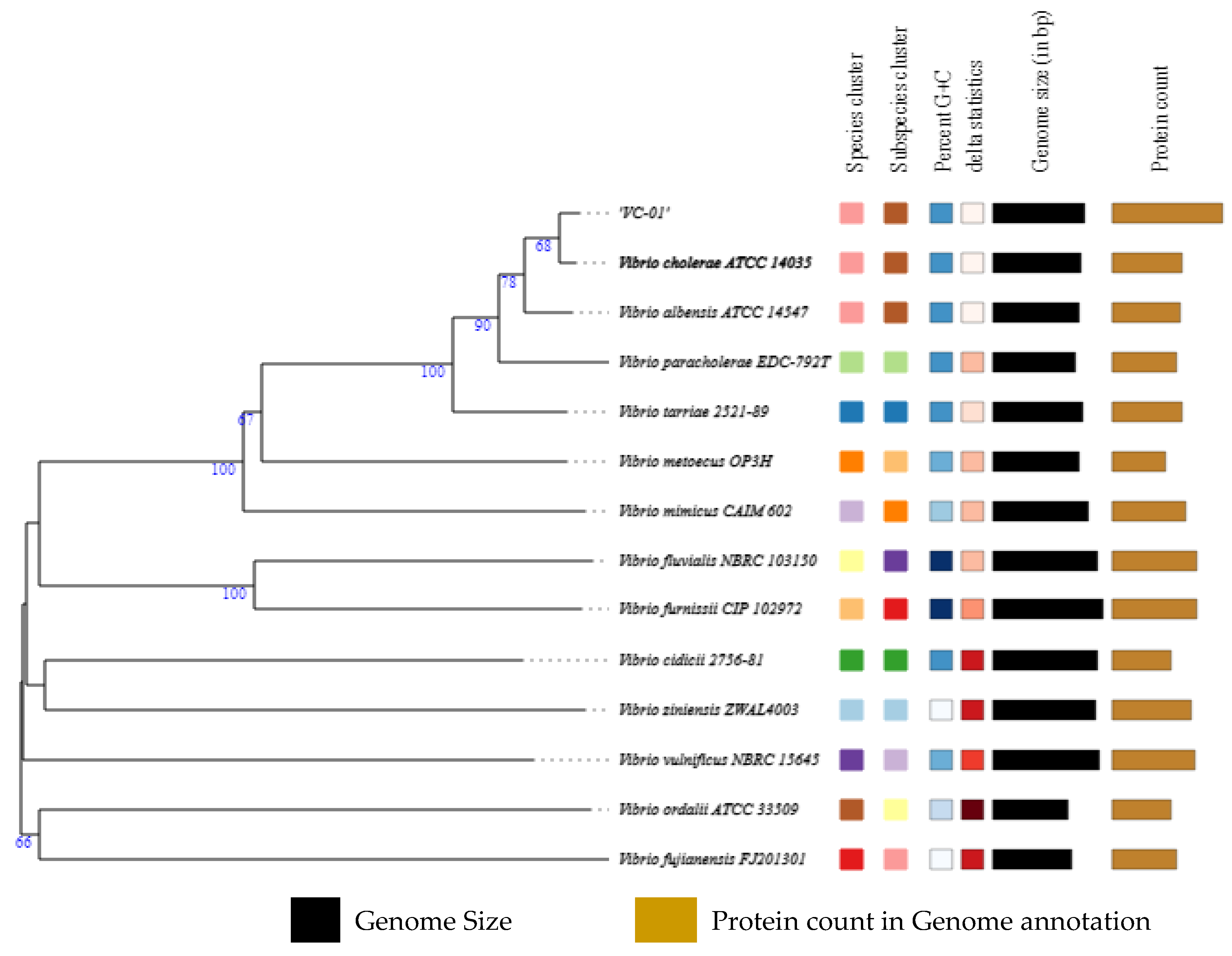
3.2. Genome Assembly and Identification

The MiniONT sequencer originally produced the raw sequences in fastq format. The results indicated no presence of contaminating sequences from other organisms. The Flye Assembler assembled two contigs with a total genome size of 4,101,607 base pairs having a 47.47% GC ratio observed. The results of QUAST indicated the 10× Genome Coverage for both assembled chromosomes. The BUSCO tool showed 82.43% genome sequence completeness based on similarity. Based on Type (Strain) Genome Server (TYGS) results, 14 strains were found with a closely related genome sequence for the isolated organism. The nearest match observed was to the V. cholerae ATCC 14035 strain, followed by Vibrio albensis ATCC 14547, Vibrio paracholerae EDC-792T, and Vibrio tarriae 2521-89 (Figure 1).
According to the findings, the isolated strain had a significant protein sequence annotation and was closely related to the V. cholerae ATCC 14035 strain. These results also show consecutive confirmation using biochemical analysis performed for the isolated strain. Based on the results, the isolate underwent MLST profiling which confirmed its close clustering with V. cholerae (Table 1).

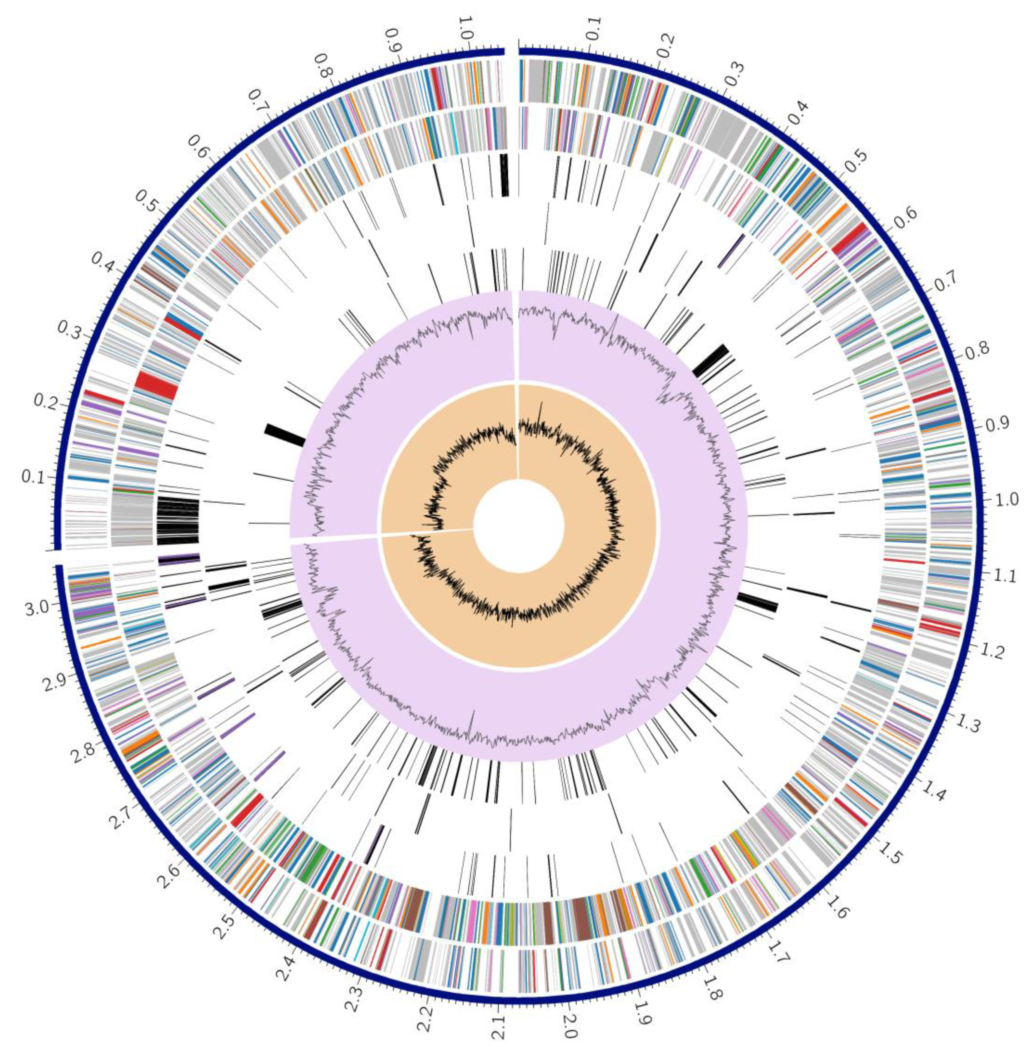
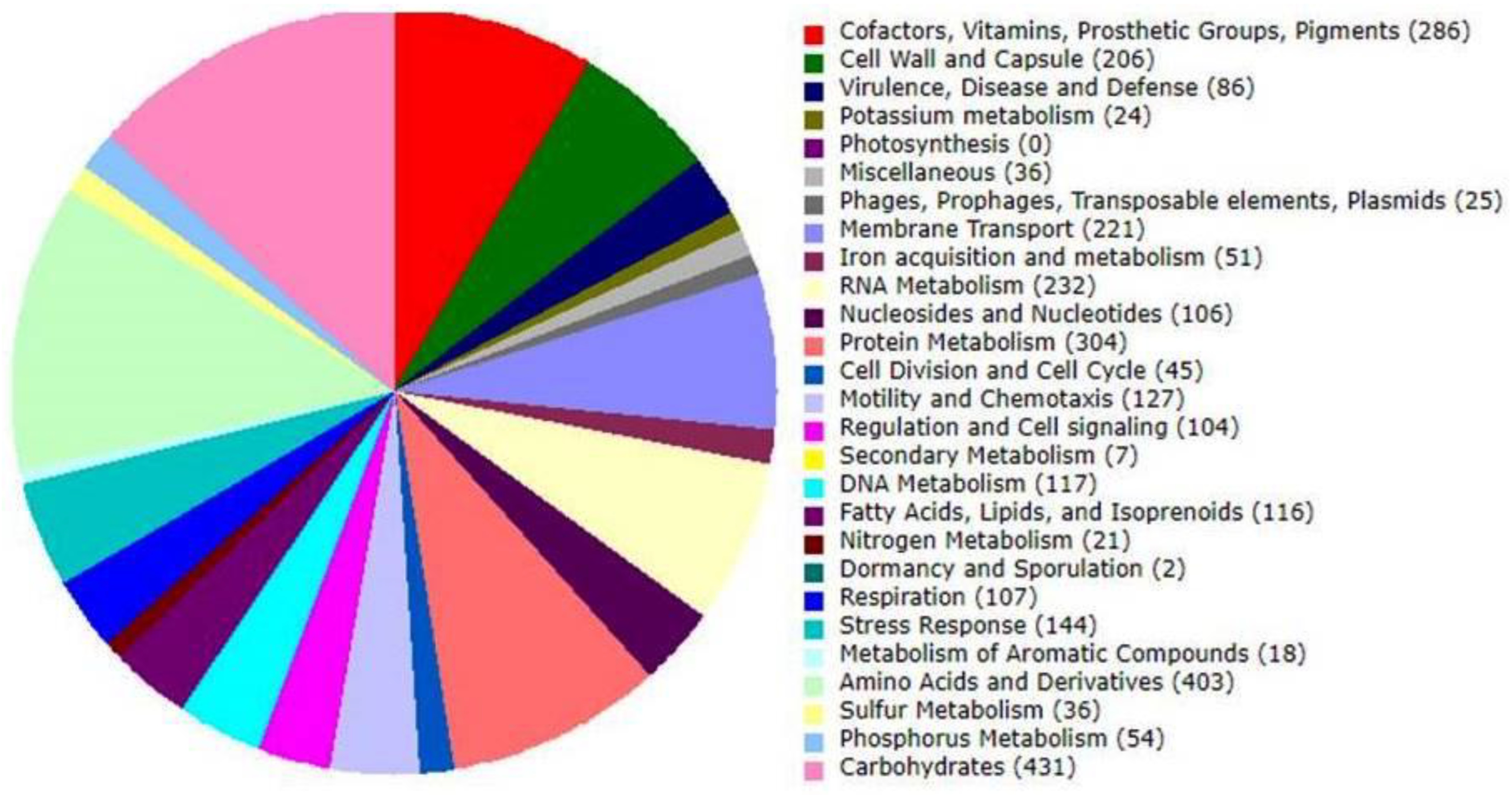
3.3. Genome Annotation

Two distinct approaches, Prokka and RAST, were utilized for the annotation of the genome. Each of these methods possesses unique advantages. Prokka-based annotation was used to analyze pan-genomes, whereas RAST-based study provided annotation based on sub-systems. A genome analysis using Prokka produced the following findings: 5310 CDS, 5520 Gene, 96 tRNA molecules, seven rRNA, 740 pseudogenes, and 914 hypothetical genes. The hypothetical proteins were mapped to their functional assignment using the PATRIC server [27]. The proteins with functional assignments included 1614 proteins with Enzyme Commission (EC) numbers [39], 1324 proteins with Gene Ontology (GO) assignments [40], and 1136 proteins that were mapped to KEGG pathways [41]. PATRIC annotation includes two types of protein families, and this genome has 5486 proteins that belong to the genus-specific protein families (PLFams) and 5579 proteins that belong to the cross-genus protein families (PGFams) (Table 2). A complete circular graphical display of the distribution of the genome annotations is provided in Figure 2.
In total, 25 features were observed for phages, prophages, transposable elements, and plasmids related to sub-systems-based annotation (Figure 3). KEGG and GO ontology-based analysis also show several precursor molecules for drug metabolism, eventually resulting in antimicrobial resistance in V. cholerae. From the current annotation, we identified two metabolic pathways that have functional importance to drug metabolism. One pathway is Azathioprine and 6-Mercaptopurine metabolism (Figure 4), and the second is the Fluorouracil metabolism pathway (Figure 5). However, the substrate of the pathway was not directly used for treating cholera; their importance was observed for cholera disease progression. A drug called azathioprine (AZA) is used to manage and treat active rheumatoid arthritis (RA), as well as to avoid kidney transplant rejection [42], Mercaptopurine and 5-Fluorourasil are extensively used for the chemotherapy treatment of cancer patients [43]. The metabolic pathway to degrade 5-Fluorourasil also damages intra-genus and inter-species quorum-sensing autoinducers, allowing the growth of V. cholerae and dispersal into the patient’s body.

3.4. Identification of Antibiotic Resistance Genes

Antibiotic resistance is a severe problem with V. cholerae. Using the Virulent Factor Database (VFDB) as a reference database, Abricate’s findings revealed 63 antibiotic resistance genes, followed by 28 from the Megares database using 75% identity/coverage. The analysis was also carried out for CARD, the NCBI antimicrobial resistance database, and the Argannot database and showed the presence of 9, 8, and 7 genes, respectively (Figure 6). The One-codex analysis revealed the presence of strB and cat9B genes associated with resistance to aminoglycosides and Chloramphenicol. At the same time, six genes (emrD, vcaM, vceA, vceB, vcmA, and vcrM) were identified as conferring resistance to the multidrug efflux drug class. Aminoglycoside, diaminopyrimidine, carbapenem, macrolide, fluoroquinolone, penam, phenicol, elfamycin, and multidrug efflux-based resistance are the main categories of antibiotic resistance that have been reported in the genome sequence of an isolated organism (Table 3).
A resistance gene was detected that belongs to Escherichia coli EF-Tu mutants conferring resistance to pulvomycin. This resistance gene could be horizontally transferred to V. cholerae. An R234F SNP was also detected in this resistance gene family; hence, the elfamycin resistance can be seen in the VC-01 strain. In addition, the homologous sequences for several organisms, including V. cholerae O1 biovar El Tor str. N16961, Yersinia enterocolitica subsp. enterocolitica 8081, Legionella pneumophila subsp. pneumophila str. Philadelphia 1, Haemophilus influenzae Rd KW20, and Neisseria meningitidis MC58 were reported upon comparison with the Virulence Factor Database (VFDB).

3.5. Identification of Integrative Conjugative Element (ICE)

ICEfinder predicted an ICE region on the analyzed genome sequence at 278,238–424,664 nucleotides covering 146,427 base pairs (Figure 7) as operons encoding a type IV secretion system (T4SS). Among the 198 ORF sequences, 28 were found to be similar to related species from the ICEberg database. The best match was observed with V. cholerae strain VC1786ICE (NCBI Accno: CP028827.1) with a maximum score of 167.5, followed by 165 for V. cholerae VC833 ICE element genomic sequence (Genbank Accno: KC886258.1) and 163.5 for V. cholerae strain VC504 ICE element genomic sequence (Genebank Accno: KC886257.1).

3.6. Identification and Comparison of Prophage Sequences

The current study aims to identify the presence of phage sequences within the genetic material of an isolated strain by utilizing the PHAge Search Tool Enhanced Release (PHASTER) [31]. The PHASTER analysis identified four prophage regions: one intact region and three incomplete regions (Table 4). All four regions were found to be distributed throughout the genome sequence. This may indicate variations of gene transfer recorded at different intervals. The prophage region predicted to be intact exhibits a significant similarity with the prophage sequence derived from Vibrio phage CTXphi (NCBI Acc No: NC_015209.1). However, three incomplete prophage regions belong to E. coli and P. aeruginosa. The co-morbid presence of V. cholerae with other filamentous organisms from clinical samples also reported such transfer [44]. Despite being a relatively underexplored phenomenon, this occurrence may offer valuable insights into the evolution and the emergence of multidrug resistance in V. cholerae-like organisms.

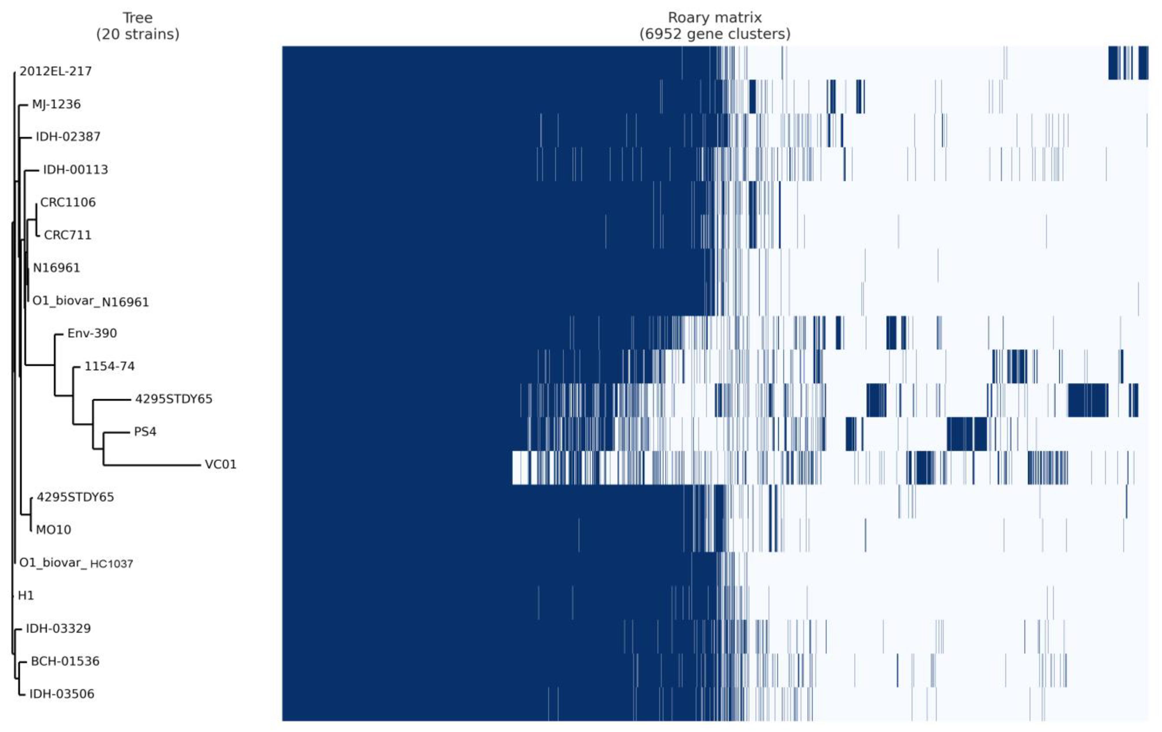
3.7. Pan-Core Genome Analysis

A total of 20 complete genomes were selected based on the similarity, occurrence, and literature survey for epidemiological importance (Table S1). These genomes were annotated using Prokka and analyzed using the Roary tool for a comprehensive analysis of pan-core genomics for pre- and post-COVID-19 implications. The Roary-based analysis findings showed 6952 clusters, including 1882 core clusters, 805 soft-core gene clusters, 3002 cloud clusters, and 1297 shell clusters of genes. Hence, the core-to-pan ratio was 54%, indicating significant gene conservation across the entire genome sequence used in the current study. The isolated V. cholerae VC01 strain is related to the V. cholerae PS4 strain as a distinct group, according to phylogenetics based on the pan-genomic study (Figure 8). Previous research indicated that the V. cholerae PS04 strain was isolated from puffer fish as a non-sucrose fermenting organism [45]. However, the strain harbored the T3SS-1 system, conferring cytotoxicity on the patient’s body.

4. Discussion

In recent years, the incidence of V. cholerae has gradually increased after the COVID-19 situation. This is attributed to the growth of global tourism, which will increase the likelihood of individuals being exposed to V. cholerae outside their home countries, leading to the development of carriers or new hosts. Despite the occurrence of seven pandemics and numerous epidemics worldwide, efforts to combat cholera remain ongoing. The disease is characterized by profuse watery diarrhea that can lead to dehydration and hypovolemic shock. The primary mode of transmission through the fecal-oral route also is a source of infection [46]. During phase 1 of COVID-19, people were more conscious regarding hygienic conditions. However, as things returned to normal and limitations eased, mass gatherings and travelling once again became common, leading to factors in the spread of diseases like cholera in subsequent stages. The emergence of antibiotic-resistant strains of V. cholerae has posed a significant challenge in the future.
This present study’s whole genome sequencing and comparative genomics methodology provide insight into the diversity of the V. cholerae strain VC01 isolated from clinical samples. A similar study was conducted to characterize the genome for pathogenic and non-pathogenic V. cholerae isolated from migratory birds at Dali Nouer Lake in China [4]. Our findings enhance and expand our understanding of the variations in the evolution of virulence determinants, antibiotic-resistant determinants, the existence of integrative and conjugative elements (ICE), mobile genetic elements (MGE), and pan-genome analysis in clinical V. cholera isolates. The integrative and conjugative element (ICE) is a critical member of the bacterial mobile genetic elements, which is integrative to the bacterial chromosome, encodes fully functioning conjugation machinery, and is thus self-transmissible between bacterial cells. The genes present in ICEs, which encode for both antibiotic resistance determinants and virulence factors, can provide the host with selective advantages. ICEfinder offers an efficient way to detect the presence of ICE sequences by comparison with its database [30]. Earlier research has documented the identification of T4SS-like elements through in silico analysis in both environmental and clinical isolates of V. cholerae O1 and non-O1/non-O139 from the Indian subcontinent and South America, as well as in Shewanella sp. from North America [47]. However, these elements appear to lack antibiotic-resistance genes. Later on, with the emergence of IDH-03944, a strain of V. cholerae O44 that exhibits resistance to clotrimoxazole has been linked to MGIVchHai6 T4SS. The comparative study regarding the ICE and its importance previously revealed that the genome of V. cholerae O1 isolates responsible for the cholera outbreak in Haiti in 2010 contained ICEVchHai1, which carried auxiliary support conferring resistance to trimethoprim (dfrA1), sulfamethoxazole (sul2), streptomycin (strAB), and chloramphenicol (floR) [48]. Similar sets of antimicrobial resistance genes have been documented in ICEs in India (1994–2005), Bangladesh (1998), Nepal (1994), and Nigeria (2010) [49].
The detected prophage sequence from an isolated organism was found in a similar sequence of Phage CTX observed in V. cholerae O1 [50]. This integration of Phage CTX into the genome might be due to gene transfer between phage CTX and the RS1 region of genome. The point mutation in the cholera toxin gene ctxB also promotes new CTX prophages into the genome sequence. Although exploring prophage sequences in various organisms has been limited, the prophage sequences in V. cholerae genomes were thoroughly investigated. The V. cholerae O1 serogroup classical biotype strains possess the cholera-toxin encoding phage CTX(cla) specific to their biotype, while the El Tor biotype strains carry CTX-1 [51]. However, the CTXΦ phage in the classical biotype is usually integrated solitarily or with a truncated copy.
Generally, Norfloxacin, trimethoprim-sulfamethoxazole (TMP-SMX), ciprofloxacin, and azithromycin have been used to treat cholera. The World Health Organization (WHO) recommends the management of cholera with oral rehydration salts in combination with antibiotics such as streptomycin, aminoglycosides, trimethoprim, fosfomycin, fluoroquinolones, sulphonamides, chloramphenicol/florfenicol, and tetracyclines [52]. Usually, bacterial species including V. cholerae can acquire resistance to antimicrobial compounds, antibiotic target replacement, and antibiotic target protection, and prompt inactivation of the antibiotic by hydrolysis, preventing access to the target site by changing membrane permeability and actively exporting antibiotics from bacterial cells [48]. In the present study, the isolate was resistant to several antibiotics, including aminoglycosides, carbapenems, and macrolides, leading to the emergence of the multidrug-resistant strain of V. cholerae. Additionally, the isolate was resistant to pulvomycin, which was horizontally transmitted to V. cholerae. A similar antibiotic resistance mechanism was previously reported in the Uganda study during 2014–2016 [53].
Although it is presently difficult to predict, the effect of treatment for infectious disease will affect antimicrobial resistance. However, most studies show extensive empirical use in contrast to comparatively infrequent bacterial co-infection and secondary infections [54]. Exposure to antimicrobial agents contributes to the escalation of microbial resistance [55]. Furthermore, prolonged hospitalization in the intensive care unit intensifies the susceptibility to infections caused by multidrug-resistant organisms [56]. The use of antibiotics may also affect the microbial flora in the digestive tract, promoting the development of resistant strains of several Infectious agents [57]. However, such a phenomenon was also reported earlier, such as gene swapping [58], quorum-sensing-based horizontal gene transfer [59], and SOS response [60], promoting multidrug resistance in V. cholerae. Antimicrobial resistance in V. cholerae is also known to be due to the acquisition of resistance genes from closely or distantly related microbial species through horizontal gene transfer [61]. Inadequate sanitation and water contamination were also discovered to have a rapid horizontal gene transfer in V. cholerae [62,63].
Based on pan-genomic analysis, out of 1099 unique clusters, 22 were identified as having a role in CRISPR (04), drug resistance (07), phage sequences (02), and toxin-related functions (09). The unique clusters (Table 5) were reported in the genome sequence of the isolated V. cholerae VC01 in the present study. The gene/clusters reported as unique were previously reported from organisms like Actinobacillus pleuropneumoniae, Aggregatibacter actinomycetemcomitans, Aliivibrio wodanis, Bacillus subtilis, Escherichia coli, Kingella kingae, Pseudoalteromonas sp., Salmonella enterica, Staphylococcus aureus, Vibrio spp. Similarly, Verma et al. (2019) also reported the cross-species transfer of resistance genes to V. cholerae genomes from 2008 to 2015 [61]. Pan-genome analysis showing a slight variation in the number of accessory and unique genes in the isolated strain revealed alteration compared to selected genomes used in present study. Additionally, the size of the pan-genome is consistently increasing in the isolated collection. This variation may be attributed to the development of mechanisms for acquired antimicrobial resistance in strains from the medical environment or the proliferation of new genes.
Over time, V. cholerae resistance to major classes of antibiotics is concerning. An example is the incidence of quinolone resistance genes in V. cholerae O139 strains from China that were conjugative and contributed due to a CIP-resistant plasmid similar to that observed in V. vulnificus [84]. Additionally, mobile genetic elements, particularly plasmids, have significantly contributed to the spread of antimicrobial resistance. Some studies also found plasmid from other strains of V. cholerae [85]. All these incidences were already reported in the past, supporting our finding regarding the antimicrobial resistance from the isolated strain. In contrast to these, researchers from Kolkata, India, also reported a Non-O1/non-O139 non-toxigenic V. cholerae strain having resistance towards more than three groups of antibiotics using genome analysis [86]. Such types of strategic analysis can simply be made more efficient through the use of complete genome sequencing and molecular characterization to track successive linked outbreaks.
In August 2020, the first round of the OCV campaign was done in the most affected districts of the Littoral, South, and Southwest regions [87]. The number of cholera cases was considerably reduced after this first round. It is unknown whether the V. cholerae vaccine can show the same scenario in the future. However, the antibiotic susceptibility data of publicly available genomes were not included in the analysis as the data were not available. Hence, there is a need for genome-based surveillance from all other currently isolated strains that might provide more insight into such antibiotic resistance. The present study provides insight into whole genome sequencing of such isolates from the patient and also can be replicated for future isolates for tracking cholera like infectious diseases.

5. Conclusions

Recently, due to COVID-19 implications, the emphasis on infectious diseases such as cholera shifted, resulting in high fatality rates worldwide due to co-morbid infection with V. cholerae. Pan-genomic profiling offers valuable insights into the presence of additional sequences in V. cholerae through gene transfer, which can confer resistance to multiple antibiotics. The findings demonstrated the need to explore alternative therapeutic approaches for managing multidrug-resistant cholera. In addition, it is beneficial to perform re-efficacy testing on presently utilized vaccines against V. cholerae. This may offer explicit knowledge of the possible existence of epitopes derived from other organisms as a potential site for the development of vaccines. The presence of antibiotic resistance genes in the whole genome and in vitro study of susceptibility towards similar antibiotics shows a correlation of gene expression in the isolated strain. This research has generated interest in further genomic profiling and understanding the dissemination and multidrug resistance using whole genome sequencing of V. cholerae. India, like other Asian countries, carries more asymptomatic V. cholerae incidences every year, leading to increased mutation of multidrug resistance. The concurrent whole genome sequencing provides a detailed understanding of the dissemination and multidrug resistance in such infectious agents.

Supplementary Materials

The following supporting information can be downloaded at: https://www.mdpi.com/article/10.3390/microorganisms11082030/s1, Table S1. List of genomes used for pan-genomic analysis.

Author Contributions

Conceptualization, V.M., R.P. and R.C.; methodology, D.Z., V.K. and M.V.; writing-original draft preparation, V.M., R.P., D.Z. and P.D.; writing-review and editing, Y.-Y.C. and D.J.H.S.; supervision, D.Z. and R.P.; project administration, V.M., D.Z. and R.P. All authors have read and agreed to the published version of the manuscript.

Funding

This research received no funding.

Informed Consent Statement

Patient consent was waived due to the use of pre-collected samples.

Data Availability Statement

Whole genome sequences have been deposited and are publicly available on NCBI with Bioproject ID: PRJNA853502 (https://www.ncbi.nlm.nih.gov/bioproject/PRJNA853502) (accessed on 30 June 2022). and SRA ID: SAMN29394033 (https://www.ncbi.nlm.nih.gov/sra/SRX15925945) (accessed on 15 July 2022).

Acknowledgments

The authors are thankful for the supercomputer facility available at the Department of Biosciences, Veer Narmad South Gujarat University, Surat for the post-genome sequence data analysis work.

Conflicts of Interest

The authors declare no conflict of interest.

References

  1. Ali, M.; Nelson, A.R.; Lopez, A.L.; Sack, D.A. Updated Global Burden of Cholera in Endemic Countries. PLoS Negl. Trop. Dis. 2015, 9, e0003832. [Google Scholar] [CrossRef] [PubMed] [Green Version]
  2. Hu, D.; Liu, B.; Feng, L.; Ding, P.; Guo, X.; Wang, M.; Cao, B.; Reeves, P.R.; Wang, L. Origins of the Current Seventh Cholera Pandemic. Proc. Natl. Acad. Sci. USA 2016, 113, E7730–E7739. [Google Scholar] [CrossRef] [PubMed]
  3. Baranova, D.E.; Levinson, K.J.; Mantis, N.J. Vibrio Cholerae O1 Secretes an Extracellular Matrix in Response to Antibody-Mediated Agglutination. PLoS ONE 2018, 13, e0190026. [Google Scholar] [CrossRef] [PubMed] [Green Version]
  4. Zheng, L.; Zhu, L.-W.W.; Jing, J.; Guan, J.Y.; Lu, G.-J.J.; Xie, L.-H.H.; Ji, X.; Chu, D.; Sun, Y.; Chen, P.; et al. Pan-Genome Analysis of Vibrio Cholerae and Vibrio Metschnikovii Strains Isolated From Migratory Birds at Dali Nouer Lake in Chifeng, China. Front. Vet. Sci. 2021, 8, 638820. [Google Scholar] [CrossRef]
  5. Devault, A.M.; Golding, G.B.; Waglechner, N.; Enk, J.M.; Kuch, M.; Tien, J.H.; Shi, M.; Fisman, D.N.; Dhody, A.N.; Forrest, S.; et al. Second-Pandemic Strain of Vibrio Cholerae from the Philadelphia Cholera Outbreak of 1849. N. Engl. J. Med. 2014, 370, 334–340. [Google Scholar] [CrossRef] [Green Version]
  6. Bhattacharya, M.K.; Bhattacharya, S.K.; Garg, S.; Saha, P.K.; Dutta, D.; Balakrish Nair, G.; Deb, B.C.; Das, K.P.; Shimada, T.; Balakrish Nair, G.; et al. Outbreak of Vibrio Cholerae Non-01 in India and Bangladesh. Lancet 1993, 341, 1346–1347. [Google Scholar] [CrossRef]
  7. Faizi, N.; Kazmi, S. Universal Health Coverage—There Is More to It than Meets the Eye. J. Fam. Med. Prim. Care 2017, 6, 169–170. [Google Scholar] [CrossRef]
  8. Ferdous, J.; Sultana, R.; Rashid, R.B.; Tasnimuzzaman, M.; Nordland, A.; Begum, A.; Jensen, P.K.M. A Comparative Analysis of Vibrio Cholerae Contamination in Point-of-Drinking and Source Water in a Low-Income Urban Community, Bangladesh. Front. Microbiol. 2018, 9, 489. [Google Scholar] [CrossRef] [Green Version]
  9. Syngkon, A.; Elluri, S.; Koley, H.; Rompikuntal, P.K.; Saha, D.R.; Chakrabarti, M.K.; Bhadra, R.K.; Wai, S.N.; Pal, A. Studies on a Novel Serine Protease of a ΔhapaΔprtV Vibrio Cholerae O1 Strain and Its Role in Hemorrhagic Response in the Rabbit Ileal Loop Model. PLoS ONE 2010, 5, e13122. [Google Scholar] [CrossRef] [Green Version]
  10. Rajpara, N.; Vinothkumar, K.; Mohanty, P.; Singh, A.K.; Singh, R.; Sinha, R.; Nag, D.; Koley, H.; Kushwaha Bhardwaj, A. Synergistic Effect of Various Virulence Factors Leading to High Toxicity of Environmental V. Cholerae Non-O1/ Non-O139 Isolates Lacking Ctx Gene: Comparative Study with Clinical Strains. PLoS ONE 2013, 8, e76200. [Google Scholar] [CrossRef] [Green Version]
  11. Takahashi, E.; Ochi, S.; Mizuno, T.; Morita, D.; Morita, M.; Ohnishi, M.; Koley, H.; Dutta, M.; Chowdhury, G.; Mukhopadhyay, A.K.; et al. Virulence of Cholera Toxin Gene-Positive Vibrio Cholerae Non-O1/Non-O139 Strains Isolated From Environmental Water in Kolkata, India. Front. Microbiol. 2021, 12, 726273. [Google Scholar] [CrossRef]
  12. Centers for Disease Control and Prevention. Recommendations for the Use of Antibiotics for the Treatment of Cholera. 2018. Available online: https://www.cdc.gov/cholera/treatment/antibiotic-treatment.html (accessed on 15 October 2022).
  13. Medini, D.; Donati, C.; Tettelin, H.; Masignani, V.; Rappuoli, R. The Microbial Pan-Genome. Curr. Opin. Genet. Dev. 2005, 15, 589–594. [Google Scholar] [CrossRef]
  14. Chaudhari, N.M.; Gupta, V.K.; Dutta, C. BPGA-an Ultra-Fast Pan-Genome Analysis Pipeline. Sci. Rep. 2016, 6, 24373. [Google Scholar] [CrossRef] [Green Version]
  15. Hudzicki, J. Kirby-Bauer Disk Diffusion Susceptibility Test Protocol. Am. Soc. Microbiol. 2009, 15, 55–63. [Google Scholar]
  16. Prussing, C.; Snavely, E.A.; Singh, N.; Lapierre, P.; Lasek-Nesselquist, E.; Mitchell, K.; Haas, W.; Owsiak, R.; Nazarian, E.; Musser, K.A. Nanopore MinION Sequencing Reveals Possible Transfer of BlaKPC–2 Plasmid Across Bacterial Species in Two Healthcare Facilities. Front. Microbiol. 2020, 11, 2007. [Google Scholar] [CrossRef]
  17. Macqueen, D.J.; Eve, O.; Gundappa, M.K.; Daniels, R.R.; Gallagher, M.D.; Alexandersen, S.; Karlsen, M. Genomic Epidemiology of Salmonid Alphavirus in Norwegian Aquaculture Reveals Recent Subtype-2 Transmission Dynamics and Novel Subtype-3 Lineages. Viruses 2021, 13, 2549. [Google Scholar] [CrossRef]
  18. Cornet, L.; Baurain, D. Contamination Detection in Genomic Data: More Is Not Enough. Genome Biol. 2022, 23, 60. [Google Scholar] [CrossRef]
  19. Salvà-Serra, F.; Jaén-Luchoro, D.; Jakobsson, H.E.; Gonzales-Siles, L.; Karlsson, R.; Busquets, A.; Gomila, M.; Bennasar-Figueras, A.; Russell, J.E.; Fazal, M.A.; et al. Complete Genome Sequences of Streptococcus Pyogenes Type Strain Reveal 100%-Match between PacBio-Solo and Illumina-Oxford Nanopore Hybrid Assemblies. Sci. Rep. 2020, 10, 11656. [Google Scholar] [CrossRef]
  20. Meier-Kolthoff, J.P.; Göker, M. TYGS Is an Automated High-Throughput Platform for State-of-the-Art Genome-Based Taxonomy. Nat. Commun. 2019, 10, 2182. [Google Scholar] [CrossRef] [Green Version]
  21. Ondov, B.D.; Treangen, T.J.; Melsted, P.; Mallonee, A.B.; Bergman, N.H.; Koren, S.; Phillippy, A.M. Mash: Fast Genome and Metagenome Distance Estimation Using MinHash. Genome Biol. 2016, 17, 132. [Google Scholar] [CrossRef] [Green Version]
  22. Meier-Kolthoff, J.P.; Auch, A.F.; Klenk, H.-P.; Göker, M. Genome Sequence-Based Species Delimitation with Confidence Intervals and Improved Distance Functions. BMC Bioinform. 2013, 14, 60. [Google Scholar] [CrossRef] [PubMed] [Green Version]
  23. Lefort, V.; Desper, R.; Gascuel, O. FastME 2.0: A Comprehensive, Accurate, and Fast Distance-Based Phylogeny Inference Program. Mol. Biol. Evol. 2015, 32, 2798–2800. [Google Scholar] [CrossRef] [PubMed] [Green Version]
  24. Larsen, M.V.; Joensen, K.G.; Zankari, E.; Ahrenfeldt, J.; Lukjancenko, O.; Kaas, R.S.; Roer, L.; Leekitcharoenphon, P.; Saputra, D.; Cosentino, S.; et al. Applied Genomics of Foodborne Pathogens. In Applied Genomics of Foodborne Pathogens; Springer: Cham, Switzerland, 2017. [Google Scholar] [CrossRef]
  25. Seemann, T. Prokka: Rapid Prokaryotic Genome Annotation. Bioinformatics 2014, 30, 2068–2069. [Google Scholar] [CrossRef] [PubMed] [Green Version]
  26. Overbeek, R.; Olson, R.; Pusch, G.D.; Olsen, G.J.; Davis, J.J.; Disz, T.; Edwards, R.A.; Gerdes, S.; Parrello, B.; Shukla, M.; et al. The SEED and the Rapid Annotation of Microbial Genomes Using Subsystems Technology (RAST). Nucleic Acids Res. 2014, 42, 206–214. [Google Scholar] [CrossRef]
  27. Wattam, A.R.; Davis, J.J.; Assaf, R.; Boisvert, S.; Brettin, T.; Bun, C.; Conrad, N.; Dietrich, E.M.; Disz, T.; Gabbard, J.L.; et al. Improvements to PATRIC, the All-Bacterial Bioinformatics Database and Analysis Resource Center. Nucleic Acids Res. 2017, 45, D535–D542. [Google Scholar] [CrossRef]
  28. Tamura, K.; Stecher, G.; Kumar, S. MEGA11: Molecular Evolutionary Genetics Analysis Version 11. Mol. Biol. Evol. 2021, 38, 3022–3027. [Google Scholar] [CrossRef]
  29. McDonald, N.D.; Regmi, A.; Morreale, D.P.; Borowski, J.D.; Boyd, E.F.; Fidelma Boyd, E. CRISPR-Cas Systems Are Present Predominantly on Mobile Genetic Elements in Vibrio Species. BMC Genom. 2019, 20, 105. [Google Scholar] [CrossRef] [Green Version]
  30. Liu, M.; Li, X.; Xie, Y.; Bi, D.; Sun, J.; Li, J.; Tai, C.; Deng, Z.; Ou, H.Y. ICEberg 2.0: An Updated Database of Bacterial Integrative and Conjugative Elements. Nucleic Acids Res. 2019, 47, D660–D665. [Google Scholar] [CrossRef]
  31. Arndt, D.; Grant, J.R.; Marcu, A.; Sajed, T.; Pon, A.; Liang, Y.; Wishart, D.S. PHASTER: A Better, Faster Version of the PHAST Phage Search Tool. Nucleic Acids Res. 2016, 44, W16–W21. [Google Scholar] [CrossRef] [Green Version]
  32. Siguier, P.; Perochon, J.; Lestrade, L.; Mahillon, J.; Chandler, M. ISfinder: The Reference Centre for Bacterial Insertion Sequences. Nucleic Acids Res. 2006, 34, 32–36. [Google Scholar] [CrossRef] [Green Version]
  33. Galata, V.; Fehlmann, T.; Backes, C.; Keller, A. PLSDB: A Resource of Complete Bacterial Plasmids. Nucleic Acids Res. 2019, 47, D195–D202. [Google Scholar] [CrossRef]
  34. Liu, B.; Zheng, D.; Jin, Q.; Chen, L.; Yang, J. VFDB 2019: A Comparative Pathogenomic Platform with an Interactive Web Interface. Nucleic Acids Res. 2019, 47, D687–D692. [Google Scholar] [CrossRef]
  35. Emms, D.M.; Kelly, S. OrthoFinder: Phylogenetic Orthology Inference for Comparative Genomics. Genome Biol. 2019, 20, 238. [Google Scholar] [CrossRef] [Green Version]
  36. Page, A.J.; Cummins, C.A.; Hunt, M.; Wong, V.K.; Reuter, S.; Holden, M.T.G.; Fookes, M.; Falush, D.; Keane, J.A.; Parkhill, J. Roary: Rapid Large-Scale Prokaryote Pan Genome Analysis. Bioinformatics 2015, 31, 3691–3693. [Google Scholar] [CrossRef] [Green Version]
  37. Hadfield, J.; Croucher, N.J.; Goater, R.J.; Abudahab, K.; Aanensen, D.M.; Harris, S.R. Phandango: An Interactive Viewer for Bacterial Population Genomics. Bioinformatics 2018, 34, 292–293. [Google Scholar] [CrossRef] [Green Version]
  38. Stamatakis, A. RAxML Version 8: A Tool for Phylogenetic Analysis and Post-Analysis of Large Phylogenies. Bioinformatics 2014, 30, 1312–1313. [Google Scholar] [CrossRef] [Green Version]
  39. Schomburg, I.; Chang, A.; Placzek, S.; Söhngen, C.; Rother, M.; Lang, M.; Munaretto, C.; Ulas, S.; Stelzer, M.; Grote, A.; et al. BRENDA in 2013: Integrated Reactions, Kinetic Data, Enzyme Function Data, Improved Disease Classification: New Options and Contents in BRENDA. Nucleic Acids Res. 2013, 41, D764–D772. [Google Scholar] [CrossRef] [Green Version]
  40. Ashburner, M.; Ball, C.A.; Blake, J.A.; Botstein, D.; Butler, H.; Cherry, J.M.; Davis, A.P.; Dolinski, K.; Dwight, S.S.; Eppig, J.T.; et al. Gene Ontology: Tool for the Unification of Biology. Nat. Genet. 2000, 25, 25–29. [Google Scholar] [CrossRef] [Green Version]
  41. Kanehisa, M.; Goto, S.; Kawashima, S.; Nakaya, A. The KEGG Databases at GenomeNet. Nucleic Acids Res. 2002, 30, 42–46. [Google Scholar] [CrossRef] [Green Version]
  42. Ladrière, M. Current indications of azathioprine in nephrology. Nephrol. Ther. 2013, 9, 8–12. [Google Scholar] [CrossRef]
  43. Longley, D.B.; Harkin, D.P.; Johnston, P.G. 5-Fluorouracil: Mechanisms of Action and Clinical Strategies. Nat. Rev. Cancer 2003, 3, 330–338. [Google Scholar] [CrossRef] [PubMed]
  44. Davis, B.M.; Waldor, M.K. Filamentous Phages Linked to Virulence of Vibrio Cholerae. Curr. Opin. Microbiol. 2003, 6, 35–42. [Google Scholar] [CrossRef] [PubMed]
  45. Das, L.; Deb, S.; Arakawa, E.; Yamasaki, S.; Das, S.K. Pufferfish (Tetraodon Cutcutia) Sampled from a Freshwater River Serves as an Intermediate Reservoir of a Sucrose Nonfermenting Variant of Vibrio Cholerae PS-4. Microbiol. Spectr. 2022, 10, e0122121. [Google Scholar] [CrossRef] [PubMed]
  46. Weil, A.A.; Becker, R.L.; Harris, J.B. Vibrio Cholerae at the Intersection of Immunity and the Microbiome. MSphere 2019, 4, E00597-19. [Google Scholar] [CrossRef] [PubMed] [Green Version]
  47. Carraro, N.; Rivard, N.; Ceccarelli, D.; Colwell, R.R.; Burrus, V. IncA/C Conjugative Plasmids Mobilize a New Family of Multidrug Resistance Islands in Clinical Vibrio Cholerae Non-O1/Non-O139 Isolates from Haiti. mBio 2016, 7, e00509-16. [Google Scholar] [CrossRef] [Green Version]
  48. Das, B.; Verma, J.; Kumar, P.; Ghosh, A.; Ramamurthy, T. Antibiotic Resistance in Vibrio Cholerae: Understanding the Ecology of Resistance Genes and Mechanisms. Vaccine 2020, 38, A83–A92. [Google Scholar] [CrossRef]
  49. Marin, M.A.; Fonseca, E.L.; Andrade, B.N.; Cabral, A.C.; Vicente, A.C.P. Worldwide Occurrence of Integrative Conjugative Element Encoding Multidrug Resistance Determinants in Epidemic Vibrio Cholerae O1. PLoS ONE 2014, 9, e108728. [Google Scholar] [CrossRef] [Green Version]
  50. Kim, E.J.; Lee, C.H.; Nair, G.B.; Kim, D.W. Whole-Genome Sequence Comparisons Reveal the Evolution of Vibrio Cholerae O1. Trends Microbiol. 2015, 23, 479–489. [Google Scholar] [CrossRef]
  51. Kim, E.J.; Lee, D.; Moon, S.H.; Lee, C.H.; Kim, D.W. CTX Prophages in Vibrio Cholerae O1 Strains. J. Microbiol. Biotechnol. 2014, 24, 725–731. [Google Scholar] [CrossRef]
  52. Hendriksen, R.S.; Price, L.B.; Schupp, J.M.; Gillece, J.D.; Kaas, R.S.; Engelthaler, D.M.; Bortolaia, V.; Pearson, T.; Waters, A.E.; Upadhyay, B.P.; et al. Population Genetics of Vibrio Cholerae from Nepal in 2010: Evidence on the Origin of the Haitian Outbreak. mBio 2011, 2, e00157-11. [Google Scholar] [CrossRef] [Green Version]
  53. Aruhomukama, D.; Sserwadda, I.; Mboowa, G. Whole-Genome Sequence Analysis of Vibrio Cholerae from Three Outbreaks in Uganda, 2014–2016. F1000Research 2019, 8, e0007330. [Google Scholar] [CrossRef] [Green Version]
  54. Vaughn, V.M.; Gandhi, T.N.; Petty, L.A.; Patel, P.K.; Prescott, H.C.; Malani, A.N.; Ratz, D.; McLaughlin, E.; Chopra, V.; Flanders, S.A. Empiric Antibacterial Therapy and Community-Onset Bacterial Coinfection in Patients Hospitalized With Coronavirus Disease 2019 (COVID-19): A Multi-Hospital Cohort Study. Clin. Infect. Dis. Off. Publ. Infect. Dis. Soc. Am. 2021, 72, e533–e541. [Google Scholar] [CrossRef] [PubMed]
  55. Marchaim, D.; Chopra, T.; Bhargava, A.; Bogan, C.; Dhar, S.; Hayakawa, K.; Pogue, J.M.; Bheemreddy, S.; Blunden, C.; Shango, M.; et al. Recent Exposure to Antimicrobials and Carbapenem-Resistant Enterobacteriaceae: The Role of Antimicrobial Stewardship. Infect. Control Hosp. Epidemiol. 2012, 33, 817–830. [Google Scholar] [CrossRef] [Green Version]
  56. Ang, H.; Sun, X. Risk Factors for Multidrug-resistant Gram-negative Bacteria Infection in Intensive Care Units: A Meta-analysis. Int. J. Nurs. Pract. 2018, 24, e12644. [Google Scholar] [CrossRef] [PubMed]
  57. Chedid, M.; Waked, R.; Haddad, E.; Chetata, N.; Saliba, G.; Choucair, J. Antibiotics in Treatment of COVID-19 Complications: A Review of Frequency, Indications, and Efficacy. J. Infect. Public Health 2021, 14, 570–576. [Google Scholar] [CrossRef] [PubMed]
  58. Borgeaud, S.; Metzger, L.C.; Scrignari, T.; Blokesch, M. The Type VI Secretion System of Vibrio Cholerae Fosters Horizontal Gene Transfer. Science 2015, 347, 63–67. [Google Scholar] [CrossRef]
  59. Antonova, E.S.; Hammer, B.K. Quorum-Sensing Autoinducer Molecules Produced by Members of a Multispecies Biofilm Promote Horizontal Gene Transfer to Vibrio Cholerae. FEMS Microbiol. Lett. 2011, 322, 68–76. [Google Scholar] [CrossRef] [Green Version]
  60. Mohanraj, R.S.; Mandal, J. Azithromycin Can Induce SOS Response and Horizontal Gene Transfer of SXT Element in Vibrio Cholerae. Mol. Biol. Rep. 2022, 49, 4737–4748. [Google Scholar] [CrossRef]
  61. Verma, J.; Bag, S.; Saha, B.; Kumar, P.; Ghosh, T.S.; Dayal, M.; Senapati, T.; Mehra, S.; Dey, P.; Desigamani, A.; et al. Genomic Plasticity Associated with Antimicrobial Resistance in Vibrio Cholerae. Proc. Natl. Acad. Sci. USA 2019, 116, 6226–6231. [Google Scholar] [CrossRef] [Green Version]
  62. Michaelis, C.; Grohmann, E. Horizontal Gene Transfer of Antibiotic Resistance Genes in Biofilms. Antibiotics 2023, 12, 328. [Google Scholar] [CrossRef]
  63. Igbinosa, E.O.; Obi, L.C.; Tom, M.; Okoh, A.I. Detection of Potential Risk of Wastewater Effluents for Transmission of Antibiotic Resistance from Vibrio Species as a Reservoir in a Peri-Urban Community in South Africa. Int. J. Environ. Health Res. 2011, 21, 402–414. [Google Scholar] [CrossRef]
  64. Maharajan, A.D.; Hjerde, E.; Hansen, H.; Willassen, N.P. Quorum Sensing Controls the CRISPR and Type VI Secretion Systems in Aliivibrio Wodanis 06/09/139. Front. Vet. Sci. 2022, 9, 799414. [Google Scholar] [CrossRef]
  65. Maruyama, A.; Kumagai, Y.; Morikawa, K.; Taguchi, K.; Hayashi, H.; Ohta, T. Oxidative-Stress-Inducible QorA Encodes an NADPH-Dependent Quinone Oxidoreductase Catalysing a One-Electron Reduction in Staphylococcus Aureus. Microbiology 2003, 149, 389–398. [Google Scholar] [CrossRef] [Green Version]
  66. Pérez-Reytor, D.; Pavón, A.; Lopez-Joven, C.; Ramírez-Araya, S.; Peña-Varas, C.; Plaza, N.; Alegría-Arcos, M.; Corsini, G.; Jaña, V.; Pavez, L.; et al. Analysis of the Zonula Occludens Toxin Found in the Genome of the Chilean Non-Toxigenic Vibrio Parahaemolyticus Strain PMC53.7. Front. Cell. Infect. Microbiol. 2020, 10, 482. [Google Scholar] [CrossRef]
  67. DeAngelis, C.M.; Nag, D.; Withey, J.H.; Matson, J.S. Characterization of the Vibrio Cholerae Phage Shock Protein Response. J. Bacteriol. 2019, 201, e00761-18. [Google Scholar] [CrossRef] [Green Version]
  68. Nichols, B.P.; Guay, G.G. Gene Amplification Contributes to Sulfonamide Resistance in Escherichia Coli. Antimicrob. Agents Chemother. 1989, 33, 2042–2048. [Google Scholar] [CrossRef] [Green Version]
  69. Thompson, S.E.; Smith, M.; Wilkinson, M.C.; Peek, K. Identification and Characterization of a Chitinase Antigen from Pseudomonas Aeruginosa Strain 385. Appl. Environ. Microbiol. 2001, 67, 4001–4008. [Google Scholar] [CrossRef] [Green Version]
  70. De, R. Mobile Genetic Elements of Vibrio Cholerae and the Evolution of Its Antimicrobial Resistance. Front. Trop. Dis. 2021, 2, 691604. [Google Scholar] [CrossRef]
  71. Jeong, H.; Kim, H.J.; Lee, S.J. Complete Genome Sequence of Escherichia Coli Strain BL21. Genome Announc. 2015, 3, e00134-15. [Google Scholar] [CrossRef] [Green Version]
  72. Colavecchio, A.; D’Souza, Y.; Tompkins, E.; Jeukens, J.; Freschi, L.; Emond-Rheault, J.-G.; Kukavica-Ibrulj, I.; Boyle, B.; Bekal, S.; Tamber, S.; et al. Prophage Integrase Typing Is a Useful Indicator of Genomic Diversity in Salmonella Enterica. Front. Microbiol. 2017, 8, 1283. [Google Scholar] [CrossRef] [Green Version]
  73. De Jonge, N.; Hohlweg, W.; Garcia-Pino, A.; Respondek, M.; Buts, L.; Haesaerts, S.; Lah, J.; Zangger, K.; Loris, R. Structural and Thermodynamic Characterization of Vibrio Fischeri CcdB. J. Biol. Chem. 2010, 285, 5606–5613. [Google Scholar] [CrossRef] [PubMed] [Green Version]
  74. Lehours, P.; Freydière, A.-M.; Richer, O.; Burucoa, C.; Boisset, S.; Lanotte, P.; Prère, M.F.; Ferroni, A.; Lafuente, C.; Vandenesch, F.; et al. The RtxA Toxin Gene of Kingella Kingae: A Pertinent Target for Molecular Diagnosis of Osteoarticular Infections. J. Clin. Microbiol. 2011, 49, 1245–1250. [Google Scholar] [CrossRef] [PubMed] [Green Version]
  75. Schindler, B.D.; Patel, D.; Seo, S.M.; Kaatz, G.W. Mutagenesis and Modeling to Predict Structural and Functional Characteristics of the Staphylococcus Aureus MepA Multidrug Efflux Pump. J. Bacteriol. 2013, 195, 523–533. [Google Scholar] [CrossRef] [PubMed] [Green Version]
  76. Prakash, B.P.; Davis, B.M.; Jie, Y.; Waldor, M.K. Characterization of a HigBA Toxin-Antitoxin Locus in Vibrio Cholerae. J. Bacteriol. 2007, 189, 491–500. [Google Scholar] [CrossRef] [Green Version]
  77. Ohki, R.; Murata, M. Bmr3, a Third Multidrug Transporter Gene of Bacillus Subtilis. J. Bacteriol. 1997, 179, 1423–1427. [Google Scholar] [CrossRef] [Green Version]
  78. Crosby, J.A.; Kachlany, S.C. TdeA, a TolC-like Protein Required for Toxin and Drug Export in Aggregatibacter (Actinobacillus) Actinomycetemcomitans. Gene 2007, 388, 83–92. [Google Scholar] [CrossRef] [Green Version]
  79. Mudrak, B.; Tamayo, R. The Vibrio Cholerae Pst2 Phosphate Transport System Is Upregulated in Biofilms and Contributes to Biofilm-Induced Hyperinfectivity. Infect. Immun. 2012, 80, 1794–1802. [Google Scholar] [CrossRef] [Green Version]
  80. Foote, S.J.; Bossé, J.T.; Bouevitch, A.B.; Langford, P.R.; Young, N.M.; Nash, J.H.E. The Complete Genome Sequence of Actinobacillus Pleuropneumoniae L20 (Serotype 5b). J. Bacteriol. 2008, 190, 1495–1496. [Google Scholar] [CrossRef] [Green Version]
  81. Amitai, S.; Yassin, Y.; Engelberg-Kulka, H. MazF-Mediated Cell Death in Escherichia Coli: A Point of No Return. J. Bacteriol. 2004, 186, 8295–8300. [Google Scholar] [CrossRef] [Green Version]
  82. Moch, C.; Fromant, M.; Blanquet, S.; Plateau, P. DNA Binding Specificities of Escherichia Coli Cas1-Cas2 Integrase Drive Its Recruitment at the CRISPR Locus. Nucleic Acids Res. 2017, 45, 2714–2723. [Google Scholar] [CrossRef] [Green Version]
  83. Blattner, F.R.; Plunkett, G., 3rd; Bloch, C.A.; Perna, N.T.; Burland, V.; Riley, M.; Collado-Vides, J.; Glasner, J.D.; Rode, C.K.; Mayhew, G.F.; et al. The Complete Genome Sequence of Escherichia Coli K-12. Science 1997, 277, 1453–1462. [Google Scholar] [CrossRef] [Green Version]
  84. Zhou, Y.-Y.; Ma, L.-Y.; Yu, L.; Lu, X.; Liang, W.-L.; Kan, B.; Su, J.-R. Quinolone Resistance Genes and Their Contribution to Resistance in Vibrio Cholerae Serogroup O139. Antibiotics 2023, 12, 416. [Google Scholar] [CrossRef]
  85. Nohejl, T.; Valcek, A.; Papousek, I.; Palkovicova, J.; Wailan, A.M.; Pratova, H.; Minoia, M.; Dolejska, M. Genomic Analysis of Qnr-Harbouring IncX Plasmids and Their Transferability within Different Hosts under Induced Stress. BMC Microbiol. 2022, 22, 136. [Google Scholar] [CrossRef]
  86. Morita, D.; Takahashi, E.; Morita, M.; Ohnishi, M.; Mizuno, T.; Miyoshi, S.; Dutta, D.; Ramamurthy, T.; Chowdhury, G.; Mukhopadhyay, A.K.; et al. Genomic Characterization of Antibiotic Resistance-Encoding Genes in Clinical Isolates of Vibrio Cholerae Non-O1/Non-O139 Strains from Kolkata, India: Generation of Novel Types of Genomic Islands Containing Plural Antibiotic Resistance Genes. Microbiol. Immunol. 2020, 64, 435–444. [Google Scholar] [CrossRef]
  87. Mogasale, V.; Levin, A.; Maskery, B.; DeRoeck, D.; Kim, Y.E.; Clemens, J.; Lopez, A.L.; Burgess, C.; Wierzba, T. Oral Cholera Vaccines to Control Endemic Disease: An Economic and Epidemiological Modelling Analysis. Lancet 2013, 382, 6. [Google Scholar] [CrossRef]
Figure 1. Type Strain-based maximum likelihood organisms reported compared to isolated Vibrio cholerae VC-01 Strain.
Microorganisms 11 02030 g001
Figure 2. Complete genome annotation of Vibrio cholerae VC01. (The components mentioned in Figure 2, arranged in a hierarchical order from the outermost to the innermost rings, comprise the contigs, CDS sequences located on the forward strand, CDS on the reverse strand, RNA genes, CDS with homology to known antimicrobial resistance genes, CDS with homology to known virulence factors, as well as the GC content and GC skew).
Microorganisms 11 02030 g002
Figure 3. Sub-system-based annotation.
Microorganisms 11 02030 g003
Figure 4. Azathioprine and 6-Mercaptopurine metabolism pathway.
Microorganisms 11 02030 g004
Figure 5. Fluorouracil metabolism pathway.
Microorganisms 11 02030 g005
Figure 6. Antibiotic resistance/virulence factors evaluated against various databases.
Microorganisms 11 02030 g006
Figure 7. Integrative Conjugative Element (ICE) in the genome sequence of Vibrio cholerae VC01 (Region 1 specifies the location of ICE identified in the circular genome). (a) Circular view of the genome representing ICE region. (b) Alignment view of whole genome sequence using ICEberg database.
Microorganisms 11 02030 g007aMicroorganisms 11 02030 g007b
Figure 8. Pan-core genome analysis of selected genomes.
Microorganisms 11 02030 g008
Table 1. Multilocus sequence typing (MLST) profile of Vibrio cholerae VC01.
LocusIdentityCoverageAlignment LengthAllele LengthGapsOrganism
adk1001004164160V. cholerae
gyrB99.76899.7684314311
mdh99.049999.04994214214
metE1001005915910
pntA1001004314310
purM99.579899.57984764762
pyrC99.55461004494490
Table 2. Protein feature analysis of Vibrio cholerae VC01 using PATRIC tool.
NoProtein FeatureCount
1Hypothetical proteins812
2Proteins with functional assignments4870
3Proteins with EC number assignments1614
4Proteins with GO assignments1324
5Proteins with Pathway assignments1136
6Proteins with PATRIC genus-specific family (PLfam) assignments5486
7Proteins with PATRIC cross-genus family (PGfam) assignments5579
Table 3. Resistance against different drug classes, AMR gene families, and resistance mechanisms.
Resistance MechanismGeneAMR Gene FamilyDrug ClassIdentity (%)Coverage (%)
Antibiotic effluxCRPresistance-nodulation-cell division (RND) antibiotic efflux pumpmacrolide antibiotic, fluoroquinolone antibiotic, penam95.24100
Antibiotic inactivationAPH (3″)-IbAPH (3″)aminoglycoside98.8194.38
catB9chloramphenicol acetyltransferase (CAT)phenicol antibiotic99.5299.52
Vibrio cholerae varGSubclass B1 Vibrio cholerae varG beta-lactamasecarbapenem99.784.87
Antibiotic target alterationE. coli EF-Tuelfamycin resistant EF-Tuelfamycin antibiotic87.2896.33
dfrA1trimethoprim-resistant dihydrofolate reductase dfrdiaminopyrimidine99.36100
Antibiotics resistanceSUL2, DFRA1dihydropteroate synthase (DHPS)Sulfonamide and trimethoprim-resistant dihydropteroate synthase Sul299.1498.9
Efflux RegulatorEMRDDrug and biocide resistanceMultidrug efflux100100
VCEADrug and biocide resistanceMultidrug efflux98.2499.75
VCEBDrug and biocide resistanceMultidrug efflux98.3199.93
VCERDrug and biocide resistanceMultidrug efflux99.8399.83
VCMADrug and biocide resistanceMultidrug efflux99.7199.86
Table 4. Prophage regions in Vibrio cholerae VC01.
RegionLengthCompletenessScoreTotal ProteinsRegion PositionMost Common PhageGC (%)
110.7 Kbincomplete6014315,989–326,748PHAGE_Escher_500465_1_
NC_049342(7)
47.13
212.1 Kbintact15030966,779–978,912PHAGE_Vibrio_CTX_
NC_015209(13)
45.67
312.7 Kbincomplete50261,339,522–1,352,283PHAGE_Pseudo_JBD18_
NC_027986(3)
42.96
420.6 Kbincomplete60133,749,246–3,769,881PHAGE_Escher_RCS47_
NC_042128(2)
45.76
Table 5. Unique clusters identified from Vibrio cholerae VC01.
NoCluster GeneAnnotationFunction OrganismReference
1casCcasCCRISPR system Cascade subunit CasCCRISPRVibrio spp.[29]
2csy3csy3CRISPR-associated protein Csy3CRISPRAliivibrio wodanis[64]
3group_1026qorAQuinone oxidoreductase 1ResistanceStaphylococcus aureus[65]
4group_1096zotZona occludens toxinToxinVibrio parahaemolyticus[66]
5group_1408pspAPhage shock protein APhageVibrio cholerae[67]
6group_1432bcrBicyclomycin resistance proteinResistanceEscherichia coli K12[68]
7group_146endo IChitodextrinaseResistancePseudoalteromonas sp. P1-9[69]
8group_1966tcpEToxin coregulated pilus biosynthesis protein EToxinVibrio cholerae[70]
9group_2021group_2021IS3 family transposase ISVpa4ResistanceEscherichia coli[71]
10group_307intAProphage integrase IntAPhageSalmonella enterica[72]
11group_3154ccdBToxin CcdBToxinVibrio fischeri[73]
12group_33rtxAMultifunctional-autoprocessing repeats-in-toxinToxinKingella kingae[74]
13group_373mepAMultidrug export protein MepAResistanceStaphylococcus aureus[75]
14group_3960higB-1Toxin HigB-1ToxinVibrio cholerae[76]
15group_3962higA-1Antitoxin HigA-1Toxin [76]
16group_442bmr3Multidrug resistance protein 3ResistanceBacillus subtilis[77]
17group_461ltxBLeukotoxin export ATP-binding protein LtxBToxinAggregatibacter actinomycetemcomitans[78]
18group_655fruBMultiphosphoryl transfer proteinResistanceVibrio cholerae Pst2[79]
19group_76apxIBToxin RTX-I translocation ATP-binding proteinToxinActinobacillus pleuropneumoniae[80]
20mazFmazFEndoribonuclease toxin MazFToxinEscherichia coli[81]
21ygbFygbFCRISPR-associated endoribonuclease Cas2CRISPREscherichia coli[82]
22ygbTygbTCRISPR-associated endonuclease Cas1CRISPREscherichia coli K12[83]
Disclaimer/Publisher’s Note: The statements, opinions and data contained in all publications are solely those of the individual author(s) and contributor(s) and not of MDPI and/or the editor(s). MDPI and/or the editor(s) disclaim responsibility for any injury to people or property resulting from any ideas, methods, instructions or products referred to in the content.

