Abstract
A non-motile, Gram-staining-negative, orange-pigmented bacterium called herbae pc1-10T was discovered in Tibet in the soil around Pyrola calliantha H. Andres’ roots. The isolate thrived in the temperature range of 10–30 °C (optimal, 25 °C), pH range of 5.0–9.0 (optimum, pH = 6.0), and the NaCl concentration range of 0–1.8% (optimal, 0%). The DNA G+C content of the novel strain was 37.94 mol%. It showed the function of dissolving organophosphorus, acquiring iron from the environment by siderophore and producing indole acetic acid. Moreover, the genome of strain herbae pc1-10T harbors two antibiotic resistance genes (IND-4 and AdeF) encoding a β-lactamase, and the membrane fusion protein of the multidrug efflux complex AdeFGH; antibiotic-resistance-related proteins were detected using the Shotgun proteomics technology. The OrthoANIu values between strains Chryseobacterium herbae pc1-10T; Chryseobacterium oleae CT348T; Chryseobacterium kwangjuense KJ1R5T; and Chryseobacterium vrystaatense R-23566T were 90.94%, 82.96%, and 85.19%, respectively. The in silico DDH values between strains herbae pc1-10T; C. oleae CT348T; C. kwangjuense KJ1R5T; and C. vrystaatense R-23566T were 41.7%, 26.6%, and 29.7%, respectively. Chryseobacterium oleae, Chryseobacterium vrystaatense, and Chryseobacterium kwangjuense, which had 16S rRNA gene sequence similarity scores of 97.80%, 97.52%, and 96.75%, respectively, were its closest phylogenetic relatives. Chryseobacterium herbae sp. nov. is proposed as the designation for the strain herbae pc1-10T (=GDMCC 1.3255 = JCM 35711), which represented a type species based on genotypic and morphological characteristics. This study provides deep knowledge of a Chryseobacterium herbae characteristic description and urges the need for further genomic studies on microorganisms living in alpine ecosystems, especially around medicinal plants.
1. Introduction
The genus Chryseobacterium is included within the family Weeksellaceae [1], the main bacterial lineage in the phylum Bacteroidetes. It is well known that strains of the plant-related Chryseobacterium species have the ability to promote plant growth [2,3,4,5,6] and degrade pesticide residues [7,8], and many of them have genes associated with antibiotic resistance [6,9,10,11,12]. According to the “List of Prokaryotic Names with Standing in Nomenclature” (http://www.bacterio.net/ accessed on 20 May 2023) which interprets the list of all validly published names within this genus, 169 species with validly published names were at the time of writing considered to be members of the genus Chryseobacterium. Animals [13,14,15,16], plants [17,18,19,20,21,22], water [23,24,25], sediment [23,26,27], and air [28] are just a few of the diverse settings in which members of the genus Chryseobacterium can be isolated.
In the southeast of the Tibetan Plateau, where alpine forest ecosystems play a significant role in maintaining the water balance and carbon sequestration, there are abundant forestry resources to be discovered [29]. Soil microbes play a vital role in biogeochemical cycling processes in alpine forest ecosystems [30,31,32]. In China, Pyrola calliantha is found in many provinces, including Shaanxi, Qinghai, Gansu, and Henan. P. calliantha often inhabits broad-leaved woods, coniferous and broad-leaved mixed forests, or mountain coniferous forests between 700 and 4100 m above sea level. Pyrola calliantha has been used medicinally for a very long time in China. It is a traditional Chinese medication that, after drying, has a wide range of therapeutic characteristics. It is frequently used to cure disorders including bleeding, coughing up blood, inflamed sores, and discomfort in tendons and bones [33].
This study aims to characterize a new species, Chryseobacterium herbae pc1-10T, which inhabits the rhizosphere of Pyrola calliantha, with genomics and proteomics-assisted characterization.
2. Materials and Methods
2.1. Bacterial Isolation and Cultivation
The strain of bacterium Chryseobacterium sp. pc1-10T. was isolated from the root system soil of Pyrola rotundifolia (Pyrola calliantha H. Andres.) in Segrila Mountain, geographically located in Nyingchi District, Tibet Autonomous Region, China (94°34′34.67″ E, 29°33′41.85″ N). The samples were kept in the lab at 4 °C until use and transported at low temperatures. In a sterile mineral buffer, the root system soil samples were suspended and homogenized [34]. On Nutrient Agar (NA; Difco, Beirut, Lebanon) adjusted to pH 7.0, serial dilutions were inoculated and incubated for 5 days at 30 °C. A total of 29 morphologically distinct colonies appeared on nutrient agar plates. We obtained 15 strains, strain herbae pc1-10T being one of them. The pure culture was then kept in glycerol suspensions (25%, v/v) at −80 °C and on NA slants at 4 °C.
2.2. Phylogenetic and Genotypic Analysis
Using bacterial universal primers 27F (5′-AGAGTTTGATCCTGGCTCAG-3′) and 1492R (5′-GGTTACCTTGTTACGACTT- 3′), the 16S rRNA gene of the herbae pc1-10T was amplified by PCR [35]. The Life Technologies Company (Shenzhen, China) sequenced purified PCR products (approximately 1.4 kb). The virtually complete sequences of 16S rRNA genes were put together using the DNAMan version 6.0.3.99 software (DNASTAR Inc., Madison, WI, USA). All of the 16S rRNA gene sequences of the closest phylogenetic members were used to construct phylogenetic trees using neighbor-joining (NJ), maximum-likelihood (ML), and silva alignment methods by the Mega 7 software [36].
2.3. Physiological, Biochemical, and Chemotaxonomic Analysis
An exponentially developing culture’s cell morphology was investigated using a Nikon 80i light microscope and a transmission electron microscope (Hitachi 7500, Tokyo, Japan). The isolates were grown on Nutrient Agar (NA; Difco) in a variety of environments to assess physiological traits. Five temperatures of 10, 20, 25, 30, and 40 °C were set to determine the growth temperature. The NaCl-free NA broth (Difco, made according to the NA recipe but without agar and NaCl) with various NaCl concentrations (0, 0.1–2.0% at 0.1% increments, and 2–12% at 1% increments, w/v) was used to investigate the optimum concentration of NaCl for growth. The hydrolysis of Tweens 20, 40, 60, and 80 was discovered using conventional techniques [37]. Herbae pc1-10T was inoculated into an NA medium with a 0.05% L-tryptophan with the NA medium serving as a control in order to determine the production of indole acetic acid (IAA) [38]. The culture was incubated for 48 h at 25 °C. One milliliter of bacterial culture was centrifuged at 10,000 rpm for 6 min at 4 °C, and 0.6 mL of supernatant was mixed with 0.6 milliliters of Salkowski’s reagent (17.5 milliliters of HClO4, 32.5 milliliters of distilled water, 1 milliliter of 0.5 M FeCl3·6H2O) and the mixture was then incubated for thirty minutes at room temperature and in the dark [39]. At 530 nm, color absorbance was measured, and a quantitative analysis was performed by comparing the results to the IAA standard (5–40 mg/L). Chrome azurol S (CAS) agar medium was used to test herbae pc1-10T’s capacity to produce siderophores [40]. Briefly, in order to examine color change, a 5 μL inoculum of the herbae pc1-10T was spot plated and stored at 25 °C. To test its nitrogen fixation capacity, we inoculated it on the ASHBY medium by observing if it grows. Additional physiological and biochemical features were determined as follows.
Transmission electron microscopy (TEM) was used to analyze the strain herbae pc1-10T’s morphological characteristics, including the size and shape of the cells and their surface ornamentation. Gram staining was performed according to Halebian et al. [41]. Filter-paper disks (Hopebio, Qingdao, China) impregnated with a 1% solution of N,N-Dimethyl-p-phenylenediamine dihydrochloride were used to measure the activity of oxidase, a positive test resulted in the production of a blue-purple coating on the filter paper biomass within two minutes. In order to evaluate the effects of starch degradation, plates containing nutrient agar (NA Difco) (0.8%), starch (1%), and agarose (1.5%) were used. These plates were developed by flooding them with iodine solution (1%) after 5 days of incubation, and hydrolysis rings around the strain were then observed to form. According to the manufacturer’s (bioMérieux, Marcy-l’Étoile, France) instructions, API ZYM galleries were used to assess the strain herbae pc1-10T’s enzyme activity. Other biochemical assays were carried out in accordance with Tindall et al.’s instructions, including the methyl red (method 15.2.52) and the Voges–Proskauer (method 15.2.82) tests [42]. The utilization of carbon compounds was measured using API 20NE test strips (bioMérieux) according to the manufacturer’s instructions, incubated at 25 °C. The growth of bacteria on various carbohydrates and their derivatives (heterosides, polyalcohols, and uronic acids) was examined using the API 50CH test.
Well-grown cells of strain herbae pc1-10T grown on tryptic soy agar (TSA) medium for 24 h at 25 °C were used for fatty acid analysis. Separation and identification of whole-cell esters were carried out with the Sherlock Microbial Identification System (MIDI version 3.0) as represented by Vandamme et al. [43]. Polar lipids were extracted using the method of Xu et al. [44] and separated by two-dimensional TLC using silica gel 60 F254 aluminum-backed thin-layer plates (Merck, Darmstadt, Germany) [45]. Isoprenoid quinones of strain herbae pc1-10T were extracted from freeze-dried cells and analyzed as previously described using LC-MS [46]. Using a molybdophosphoric acid hydrate ethanol solution, the total lipids were identified. Utilizing the ninhydrin reagent, aminolipids were identified. With the help of the Zinzadze reagent, phospholipids were identified, and the naphthol reagent was used to find glycolipids. The data were interpreted as described by Tindall et al. [42].
2.4. Genome Analyses
Genomic DNA was collected according to the method of Marmur [47]. The genomic G+C content and the organism overview of strain herbae pc1-10T were determined using the RAST Server [48] by genome sequence, which was performed on the Illumina MiSeq platform by the Guangzhou Magigene Company (Guangzhou, China). The reads were put together using the SOAPdenovo version 1 [49]. According to the minimal requirements suggested by Chun et al., the average nucleotide identity (ANI) and in silico DNA–DNA hybridization (DDH) values were determined [50]. The OrthoANIu algorithm (https://www.ezbiocloud.net/tools/ani accessed on 17 July 2022) was used to determine the ANI value between the two genomes [49]. The Genome-to-Genome Distance Calculator 3.0 (https://ggdc.dsmz.de/ggdc.php# accessed on 17 July 2022) was used to determine the in silico DDH values [51]. Under the accession number JAOAMU000000000, strain herbae pc1-10T’s Whole-Genome Shotgum project was deposited at GenBank.
2.5. Shutgun Proteomics Analyses
We used shotgun proteomics [52,53,54] to detect the global profile of the protein/polypeptide complement within the mixture expression in the cell and culture supernatant of strain herbae pc1-10 T. The cells and the supernatant of herbae pc1-10T were separated by centrifugation (8000 rpm, 10 min). The cells were washed three times with a 0.85% NaCl, immediately frozen in liquid nitrogen, and transported to Personal Biotechnology Co., Ltd., Shanghai, China at −80 °C until use. The protein sequence database is QLDBPSN025.
2.6. Pigment Analyses
We experimentally validate the production of flexirubin by herbae pc1-10T as described by Siddaramappa et al. [55] by exposing a 2 mL pure culture of strain herbae pc1-10T, which was cultured in an NA liquid medium for 24 h, to a 500 μL 3% KOH, which resulted in a change from yellow to orange/red if flexirubin pigments were present, followed by a neutralization step with a 500 μL 1.5 N HCl which resulted in a return to yellow pigmentation.
3. Results and Discussion
3.1. Phylogenetic and Genotypic Analysis
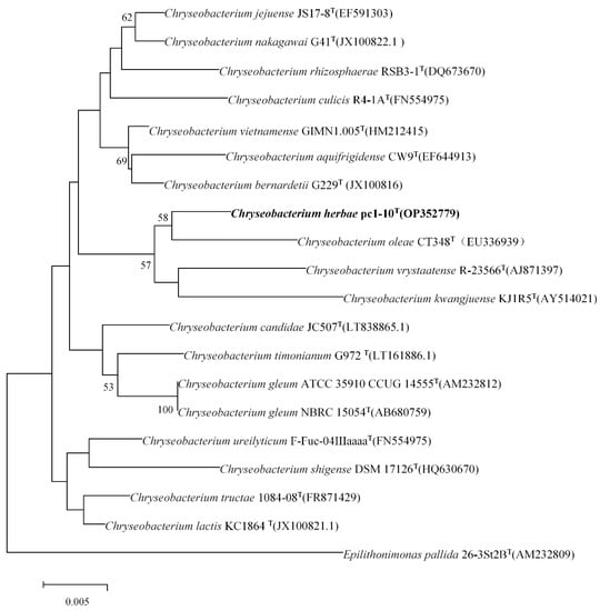
The 16S rRNA gene sequence (1517 bp) of strain herbae pc1-10T (OP352779) was acquired. Comparative analysis from this 16S rRNA gene sequence showed that the novel strain stands for a member of the genus Chryseobacterium. Strains Chryseobacterium oleae CT348T, Chryseobacterium vrystaatense RJ-7-14T, and Chryseobacterium kwangjuense KJ1R5T, were prominently picked out as reference strains for comparative studies. Phylogenetic analysis grounded on the NJ, ML methods showed that strain herbae pc1-10T formed a stable subclade with C. oleae CT348T in the phylogenetic tree (Figure 1 and Figure S3).

Figure 1.
Neighbor-joining phylogenetic tree based on a comparison of the 16S rRNA gene sequences of strain herbae pc1-10T and its closest relatives. GenBank accession numbers are given in parentheses. Bar, 0.005 nucleotide changes per 1000 nucleotides. Bootstrap values (>50%) based on 1000 replications are shown at branch nodes. Bar, 0.005 substitutions per nucleotide position.
3.2. Physiological, Biochemical, and Chemotaxonomic Analysis
Cells (0.9–2.4 µm long and 0.5–0.7 µm wide) were observed to be aerobic (Figure S1), Gram-staining-negative, rod-shaped, and non-motile. Colonies on nutrient agar were detected as orange-pigmented, entire, convex, and circular (Figure S4). On the NA agar, a colony with a size of 0.5–1 mm was seen for 5 days at 25 °C. At pH 5.0–9.0 and 10–30 °C (optimal: 25 °C), cells grew; pH 6.0 was optimum. Cells were found to grow most effectively in the absence of NaCl yet tolerated 1.8% NaCl. Catalase and oxidase, on the other hand, were determined to be negative. The strain herbae pc1-10T significantly grew well and was observed to generate a halo of organophosphorus-dissolving circles but no degradant halo of phosphate on their respective media. The strain was cultured in an NA-tryptophan liquid medium for 2 days and an IAA yield was detected to be 3.53 mg/L (Figure S4, Table S1). Unfortunately, the strain herbae pc1-10T did not grow on the CAS medium (with nutrient agar providing the essential elements for growth) and the ASHBY medium. In the pigment detection experiment, after adding the KOH solution to the bacterial suspension, the color changes from yellow to orange–red, and the addition of HCl does not change color (Figure S7), which can prove its production of the flexirubin-type pigment.
Iso-C15:0 (42.82%), iso-C17:0 3-OH (21.03%), and Summed features 9 (C16:1 w6c and/or C16:1 w7c; 12.43%) were the strain herbae pc1-10T’s main cellular fatty acids (>10%). The amount of summed feature 9 (C16:1 w6c and/or C16:1 w7c; 12.43%) in strain herbae pc1-10T was lower than that tested in C. oleae CT348T; C. kwangjuense KJ1R5T; C. vrystaatense R-23566T. However, strain herbae pc1-10T stood apart from other phylogenetically related members of the genus Chryseobacterium due to quantitative variations in major and minor fatty acids and the presence of specific minor fatty acids like C16:0 3-OH, iso-C15:0 3-OH, iso-C16:0, etc. (Table 1). Menaquinone-6 (MK-6) was found to be the only respiratory quinone that fit the description of the genus Chryseobacterium. Phosphatidylethanolamine (PE) contains the main polar lipid. Additionally, two lipids (L1-L2), three glycolipids, and five aminolipids (AL1-AL5) were found (Figure S2).

Table 1.
Cellular fatty acid contents of strain herbae pc1-10T and its relative reference strains in the genus Chryseobacterium. Strains: 1, C. herbae pc1-10T; 2, C. oleae CT348T; 3, C. kwangjuense KJ1R5T; 4, C. vrystaatense R-23566T. The data of other strains are from previous studies. Values were percentages of total fatty acids. Tr, Trace amount (<0.5%); ND, not detected; NA, not available.
Summed Features 3: including C16:1 ω6c and/or C16:1 ω7c, Summed Features 9: 10-methyl C16:0 and/or iso-C17:1 ω9c.
In the API 50CH strip, weak acid production from Erythritol, Glucose, Fructose, Mannose, Trehalose, and Gentiobiose, and acid production from Glycerol, d-Arabinose, L-Arabinose, D-Ribose, D-Xylose, L-Xylose, D-Adonitol, Methyl-β-D-xylopyranoside, D-Galactose, L-Sorbose, L-Rhamnose, Dulcitol, Inositol, Mannitol, Sorbitol, Methyl-α-D-mannopyranoside, Methyl-α-D-glucopyranoside, N-Acetyl-glucosamine, Amygdalin, Arbutin, Esculin, Salicin, Cellobiose, Maltose, Lactose, Melibiose, Sucrose, Inulin, Melezitose, Raffinose, Amyloid, Glycogen, Xylitol, D-Turanose, D-Lyxose, D-Tagatose, D-Fucose, L-Fucose, D-Arabitol, L-Arabitol, Glucoheptonate, 2-Ketogluconate, 5-Ketogluconate were negative. In the API ZYM strip, there were observations of positive results for Alkaline phosphatase, Esterase (C4), Esterase Lipase (C8), Lipase (C14), Leucine arylamidase, Acid phosphatase, Naphthol-AS-BI-phosphohydrolase, α-Galactosidase, β-Glucosidase, N-Acetyl-β-glucosaminidase, α-Fucosidase. However, other negative results for Valine arylamidase, Cystine arylamidase, Trypsin, Chymotrypsin, β-Galactosidase, β-Glucuronidase, α-Glucosidase, and α-Mannosidase were obtained. Strain herbae pc1-10T showed negative results for Nitrate reduction, D-Glucose fermentation, Arginine dihydrolase, Urease, β-Galactosidase, Arabinose, N-Acetyl-D-glucosamine, D-Maltose, Gluconate, Capric acid, Adipic acid, Malic acid, Citrate, p-hydroxy-Phenylacetic acid and positive results for β-Glucosidase, Indole production, Gelatinase and weakly positive for Glucose, Mannitol, mannose in the API 20NE strip test. Table 2 compares strains from the genus Chryseobacterium that are phylogenetically linked to strain herbae pc1-10T in terms of how well they thrive on various carbohydrates and their derivatives.

Table 2.
Differential characteristics of strain herbae pc1-10T and its relative reference strain in the genus Chryseobacterium. Strains: 1, C. herbae pc1-10T; 2, C. oleae CT348T; 3, C. kwangjuense KJ1R5T; 4, C. vrystaatense R-23566T. The data of other strains are from previous studies. Symbols: +, positive; −, negative; w, weak reaction. nm, not mentioned.
3.3. Genome Analyses
The genome of strain herbae pc1-10T was 5,142,603 bp long including 36 contigs with an N50 value of 306,357 and a genome coverage of 81.0×. The DNA G+C content was 37.94 mol%, the mean length was 142,836.5 bp, and 71 tRNA genes, 3 rRNA genes, and 1 tmRNA were predicted. The strain herbae pc1-10T full-length 16S rRNA scaffold was retrieved from the genome assembly to confirm that the genomic data is legitimate and uncontaminated, and one 16S rRNA sequence was received. The result was consistent with that of the PCR sequence. The OrthoANIu values between strains C. herbae pc1-10T; C. oleae CT348T; C. kwangjuense KJ1R5T; and C. vrystaatense R-23566T were 90.94%, 82.96%, and 85.19%, respectively. The in silico DDH values between strains herbae pc1-10T; C. oleae CT348T; C. kwangjuense KJ1R5T; and C. vrystaatense R-23566T were 41.7%, 26.6%, and 29.7%, respectively.
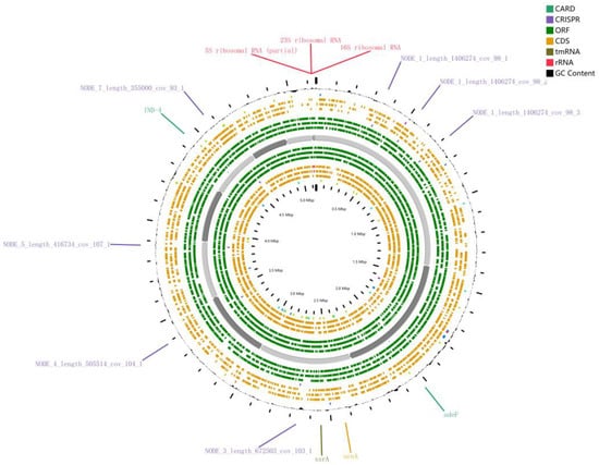
The herbae pc1-10T genome’s RAST annotation showed 264 subsystems with 4733 genes and an 18% subsystem coverage, totaling 264 subsystems (see Figure 2 below for details). It provides initial annotations of gene functions for herbae pc1-10T’s genomes, such as producing siderophores, plant hormones, beta-lactamase, etc.

Figure 2.
Annotation of the genome of herbae pc1-10T through Proksee. CARD: The comprehensive antibiotic resistance database, CRISPER: CRISPR arrays, ORF: Open reading frames, CDS: Coding sequence, tmRNA: Transfer-messenger RNA, rRNA: Ribosomal RNA.
Furthermore, we annotated the genome of herbae pc1-10T through Proksee (https://proksee.ca/projects/new accessed on 21 May 2023) (Figure 2). Antibiotic resistance gene predictions showed that herbae pc1-10T contained two antibiotic resistance genes: IND-4, which is a beta-lactamase found in Chryseobacterium indologenes, and AdeF, which is the membrane fusion protein of the multidrug efflux complex AdeFGH; the protein-coding information is provided by CARD [59] (https://card.mcmaster.ca/ accessed on 21 May 2023). According to a study, bacteria that live in soil or water naturally have ARGs that they can use to compete with other organisms that share the same habitat [60]. We used CRISPRCasFinder [61] to identify CRISPR (clustered regularly interspaced short palindromic repeats) arrays; 5 of the 23 analysis sequences were found to contain CRISPR but no Cas genes were found in the vicinity of CRISPRs. Components of these biological systems can be used in multiple applications in genetic engineering [62,63].
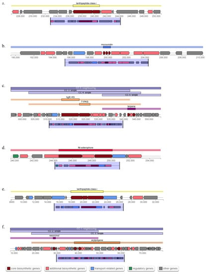
Secondary metabolites (Table 3) were identified using antiSMASH version 7.0.0 (https://antismash.secondarymetabolites.org/, accessed on 14 May 2023). One gene cluster with arylpolyene and resorcinol biosynthesis domains was found in strain herbae pc1-10T, and it showed a 75% similarity to a known biosynthetic gene cluster generating flexirubin, the major pigment of Chryseobacterium [64]. Strain herbae pc1-10T additionally had a gene cluster with NI-siderophore biosynthetic domains (Figure 3d) showing a 33% similarity to fulvivirgamide A2 biosynthetic gene cluster from Fulvivirga marina [65] and lanthipeptide class I (Figure 3e) biosynthetic domains showing a 17% similarity to pinensins biosynthetic gene cluster from Chitinophaga pinensis DSM 2588 [66], which is the first antifungal lantibiotics [67]. Other gene clusters containing microviridin (Figure 3b), hglE-KS, T1PKS, terpene (Figure 3c), and lanthipeptide class I (Figure 3a) biosynthetic were observed with no similarity to known clusters. The serine proteases chymotrypsin, trypsin, and elastase are just a few of the ones that microviridins are effective and selective inhibitors of [68]. Furthermore, it was discovered that microviridin J was poisonous to Crustacean Daphnia [69].

Table 3.
Analysis of secondary metabolite biosynthesis gene clusters of herbae pc1-10T through antiSMASH.

Figure 3.
Biosynthetic gene clusters in the genome of Chryseobacterium herbae pc1-10T retrieved using antiSMASH version 7.0.0. (a) Lanthipeptide class i. (b) Microviridin. (c) hglE-KS, T1PKS, terpene. (d) NI-siderophore. (e) Lanthipeptide class i. (f) Arylpolyene and resorcinol.
3.4. Shut-Gun Proteomics Results
The qualitative statistical results of proteins are shown in the attached Table S2. Finally, we identified 2455 (cell) and 2650 (supernatant) proteins and 19,568 (cell) and 23,825 (supernatant) peptides from the two samples, respectively (see Supplementary Materials Table S3).
The TonB-dependent siderophore receptor (MCT2563047.1), the biopolymer transporter ExbD (MCT2562352.1, MCT2562353.1, MCT2563473.1), and the MotA/TolQ/ExbB proton channel family protein (MCT2562351.1) were detected in both cell and supernatant, which means that herbae pc1-10T could uptake siderophore from the outside environment [70]. The pinensin family lanthipeptide (MCT2564031.1) was detected in the supernatant, and it was regarded as the first lantibiotics isolated from a Gram-negative native producer [67]. It is famous for its significant resistance to some fungi; however, we found that herbae pc1-10T only had a weak inhibitory effect on the growth of pathogenic fungi (Magnaporthe oryzae) of rice blast (Figure S6). Terpene synthase family protein (MCT2561927.1) was detected in the supernatant and cell. Penicillin-binding transpeptidase domain-containing protein (MCT2563304.1), the TolC family protein (MCT2563484.1, MCT2561713.1, MCT2564157.1), nitroreductase (MCT2564731.1) [71], and the efflux transporter outer membrane subunit (MCT2563723.1) were detected in cell samples. These proteins are closely related to antibiotic resistance. However, microviridin was not detected in the culture environment and cells, inferring that the predicted result is unreliable, or that the gene is not expressed in our culture environment, or the expression level does not reach the proteome detection threshold, etc.
4. Description of Chryseobacterium herbae sp. nov.
Chryseobacterium herbae (her’bae. L. gen. n. herbae, of a herb)
Cells of a new strain herbae pc1-10T are rod-shaped, aerobic, Gram-stain-negative, and non-motile, measuring 0.9 to 2.4 μm in length and 0.5 to 0.7 μm in width. Nutrient agar colonies are rounded, whole, convex, and orange-colored (Flexirubin-type pigment). The colony size on the NA agar is 0.5–1 mm for 5 days at 25 °C. The optimum temperature for cell growth is 25 °C, while the optimum pH range is 5.0 to 9.0. NaCl is not necessary for good cell growth, but a 1.8% NaCl is acceptable. Catalase and oxidase are positive and negative, respectively. Starch, Tweens 20, 40, 60, and 80 are hydrolyzed but arabinose or urea are not decomposed. It produces gelatinase and β-glucosidase but not β-galactosidase and arginine dihydrolase. Iso-C15:0, iso-C17:0 3-OH, and summed features 9 (C16:1 ω6c and/or C16:1 ω7c) were the main cellular fatty acids (>10%) of strain herbae pc1-10T, while menaquinone- 6 (MK-6) was the predominate respiratory quinone.
Weak acid production from Erythritol, Glucose, Fructose, Mannose, Trehalose, and Gentiobiose, and acid production from Glycerol, d-Arabinose, L-Arabinose, D-Ribose, D-Xylose, L-Xylose, D-Adonitol, Methyl-β-D-xylopyranoside, D-Galactose, L-Sorbose, L-Rhamnose, Dulcitol, Inositol, Mannitol, Sorbitol, Methyl-α-D-mannopyranoside, Methyl-α-D-glucopyranoside, N-Acetyl-glucosamine, Amygdalin, Arbutin, Esculin, Salicin, Cellobiose, Maltose, Lactose, Melibiose, Sucrose, Inulin, Melezitose, Raffinose, Amyloid, Glycogen, Xylitol, D-Turanose, D-Lyxose, D-Tagatose, D-Fucose, L-Fucose, D-Arabitol, L-Arabitol, Glucoheptonate, 2-Ketogluconate, 5-Ketogluconate was negative. In the API ZYM strip, there were observations of positive results for Alkaline phosphatase, Esterase (C4), Esterase Lipase (C8), Lipase (C14), Leucine arylamidase, Acid phosphatase, Naphthol-AS-BI-phosphohydrolase, α-Galactosidase, β-Glucosidase, N-Acetyl-β-glucosaminidase, α-Fucosidase. However, other negative results for Valine arylamidase, Cystine arylamidase, Trypsin, Chymotrypsin, β-Galactosidase, β-Glucuronidase, α-Glucosidase, and α-Mannosidase were obtained. Strain herbae pc1-10T showed negative results for Nitrate reduction, D-Glucose fermentation, Arginine dihydrolase, Urease, β-Galactosidase, Arabinose, N-Acetyl-D-glucosamine, D-Maltose, Gluconate, Capric acid, Adipic acid, Malic acid, Citrate, p-hydroxy-Phenylacetic acid and positive results for β-Glucosidase, Indole production, Gelatinase and weakly positive for Glucose, Mannitol, mannose in the API 20NE strip test.
The type strain’s genomic DNA has a G+C concentration of 37.9 mol%. Genome sequence accession number: JAOAMU000000000. 16S rRNA gene accession number: OP352779. The type strain, herbae pc1-10T (=GDMCC 1.3255T = JCM 35711T), was isolated from the root system soil of Pyrola calliantha H. in Tibet.
5. Discussion
Rhizosphere microbial colonization often has plant genotype specific selectivity [72]. With technological progress, genomics and proteomics provide more sufficient technical support for microbial molecular level research [54]. The strain herbae pc1-10T initially showed the function of dissolving organophosphorus, siderophore-mediated iron acquisition systems, and producing indole acetic acid (3.53 mg/L in 2 days), which may have a positive effect on plant growth. Moreover, the genome of strain herbae pc1-10T harbors two antibiotic resistance genes (IND-4 and AdeF) encoding a β-lactamase, which is responsible for β-lactam antibiotic resistance, and encoding the membrane fusion protein of the multidrug efflux complex AdeFGH, respectively. They were confirmed in Shut-gun proteomic testing. Due to the fact that bacteria that live naturally in soil or water have inbuilt ARGs to combat the chemical substances created by rivals living in the same environment, we speculate that the antibiotic resistance gene possessed by strain herbae pc1-10T is caused by related chemicals present in the rhizosphere of Pyrola calliantha H. Andres. In a word, analyzing the internal characteristics of microorganisms in special habitats may provide new ideas for future production practices and scientific research.
Supplementary Materials
The following supporting information can be downloaded at: https://www.mdpi.com/article/10.3390/microorganisms11082017/s1, Figure S1: Transmission electron micrograph of the cell of strain herbae pc1-10T. Bar, 500 nm. Figure S2: The polar lipids of strain herbae pc1-10T. Figure S3: Maximum-likelihood phylogenetic tree of strain herbae pc1-10T and its relatives based on the comparison of the 16S rRNA gene sequences. GenBank accession numbers given in parentheses; Figure S4: Growth morphology of strains on NA plates. Figure S5: The indole acetic acid (IAA) standard (5–40 mg/L) curve. Figure S6: Antagonistic experiment of against pathogenic fungi (Magnaporthe oryzae) of rice blast. Figure S7: Reversible colour change in pigment of strain herbae pc1-10T. Figure S8: Pie-diagram showing the genes of Chryseobacterium herbae pc1-10T genome distributed in various subsystem categories. Data for the subsystem categories were obtained by SEED viewer in the RAST server. Table S1: The standard curve raw data and OD530 values of the strain herbae pc1-10T to be tested. 16S rRNA gene sequence of Chryseobacterium herbae sp.nov. (from the genome). Table S2: Statistics of protein identification results. Table S3: The worksheet Cells_Proteome.id.anno.all.inte: Proteins and peptides identified in the cell of strain herbae pc1-10T, The worksheet Supernatant_Proteome.id.anno.al: Proteins and peptides identified in the supernatant of strain herbae pc1-10T.
Author Contributions
Conceptualization: L.Z., D.K. and Q.M.; Formal analysis and investigation: L.Z.; Performing research: L.Z. and Y.W.; Writing—original draft: L.Z. and D.K.; Writing—review and editing: Y.L., Z.X. and Z.R.; Supervision: Y.L., Z.X., Z.R. and Q.M. All authors have read and agreed to the published version of the manuscript.
Funding
This research was supported by the National Natural Science Foundation of China (NSFC 3211101206). This research was also supported by Agricultural Resources and Environment Discipline Construction Project (533320003), the Fundamental Research Funds for Central Non-profit Scientific Institution (1610132020009) and Central Public-interest Scientific Institution Basal Research Fund (No. Y2022GH18).
Data Availability Statement
The datasets generated during and/or analyzed during the current study are available from the corresponding author on reasonable request.
Conflicts of Interest
The authors declare no conflict of interest.
References
- García-López, M.; Meier-Kolthoff, J.P.; Tindall, B.J.; Gronow, S.; Woyke, T.; Kyrpides, N.C.; Hahnke, R.L.; Göker, M. Analysis of 1000 Type-Strain Genomes Improves Taxonomic Classification of Bacteroidetes. Front. Microbiol. 2019, 10, 2083. [Google Scholar] [CrossRef] [PubMed]
- Kim, H.; Yu, S.M. Chryseobacterium salivictor sp. nov., a Plant-Growth-Promoting Bacterium Isolated from Freshwater. Antonie van Leeuwenhoek 2020, 113, 989–995. [Google Scholar] [CrossRef]
- Kumar, V.; Patial, V.; Thakur, V.; Singh, R.; Singh, D. Genomics Assisted Characterization of Plant Growth-Promoting and Metabolite Producing Psychrotolerant Himalayan Chryseobacterium Cucumeris PCH239. Arch. Microbiol. 2023, 205, 108. [Google Scholar] [CrossRef]
- Montero-Calasanz, M.D.C.; Göker, M.; Rohde, M.; Spröer, C.; Schumann, P.; Busse, H.J.; Schmid, M.; Tindall, B.J.; Klenk, H.P.; Camacho, M. Chryseobacterium hispalense sp. nov., a Plantgrowth- Promoting Bacterium Isolated from a Rainwater Pond in an Olive Plant Nursery, and Emended Descriptions of Chryseobacterium defluvii, Chryseobacterium indologenes, Chryseobacterium wanjuense and Chryseobacterium gregarium. Int. J. Syst. Evol. Microbiol. 2013, 63, 4386–4395. [Google Scholar] [CrossRef]
- Dardanelli, M.S.; Manyani, H.; González-Barroso, S.; Rodríguez-Carvajal, M.A.; Gil-Serrano, A.M.; Espuny, M.R.; López-Baena, F.J.; Bellogín, R.A.; Megías, M.; Ollero, F.J. Effect of the Presence of the Plant Growth Promoting Rhizobacterium (PGPR) Chryseobacterium balustinum Aur9 and Salt Stress in the Pattern of Flavonoids Exuded by Soybean Roots. Plant Soil 2010, 328, 483–493. [Google Scholar] [CrossRef]
- Anderson, M.; Habiger, J. Characterization and Identification of Productivity-Associated Rhizobacteria in Wheat. Appl. Environ. Microbiol. 2012, 78, 4434–4446. [Google Scholar] [CrossRef]
- Xiang, X.; Yi, X.; Zheng, W.; Li, Y.; Zhang, C.; Wang, X.; Chen, Z.; Huang, M.; Ying, G.G. Enhanced Biodegradation of Thiamethoxam with a Novel Polyvinyl Alcohol (PVA)/Sodium Alginate (SA)/Biochar Immobilized Chryseobacterium sp. H5. J. Hazard. Mater. 2023, 443, 130247. [Google Scholar] [CrossRef] [PubMed]
- Zhang, W.; Chen, W.-J.; Chen, S.-F.; Lei, Q.; Li, J.; Bhatt, P.; Mishra, S.; Chen, S. Cellular Response and Molecular Mechanism of Glyphosate Degradation by Chryseobacterium sp. Y16C. J. Agric. Food Chem. 2023, 71, 6650–6661. [Google Scholar] [CrossRef]
- Dahal, R.H.; Chaudhary, D.K.; Kim, D.U.; Pandey, R.P.; Kim, J. Chryseobacterium antibioticum sp. nov. with Antimicrobial Activity against Gram-Negative Bacteria, Isolated from Arctic Soil. J. Antibiot. 2021, 74, 115–123. [Google Scholar] [CrossRef]
- Kaur, H.; Mohan, B.; Hallur, V.; Raj, A.; Basude, M.; Mavuduru, R.S.; Taneja, N. Increased Recognition of Chryseobacterium Species as an Emerging Cause of Nosocomial Urinary Tract Infection Following Introduction of Matrix-Assisted Laser Desorption/Ionisation-Time of Flight for Bacterial Identification. Indian J. Med. Microbiol. 2017, 35, 610–616. [Google Scholar] [CrossRef] [PubMed]
- Abdalhamid, B.; Elhadi, N.; Alsamman, K.; Aljindan, R. Chryseobacterium Gleum Pneumonia in an Infant with Nephrotic Syndrome. IDCases 2016, 5, 34–36. [Google Scholar] [CrossRef] [PubMed]
- Mirza, I.A.; Khalid, A.; Hameed, F.; Imtiaz, A.; Ashfaq, A.; Tariq, A. Chryseobacterium Indologenes as a Novel Cause of Bacteremia in a Neonate. J. Coll. Phys. Surg. Pak. 2019, 29, 375–378. [Google Scholar] [CrossRef]
- Loch, T.P.; Faisal, M. Chryseobacterium Aahli sp. nov., Isolated from Lake Trout (Salvelinus namaycush) and Brown Trout (Salmo trutta), and Emended Descriptions of Chryseobacterium ginsenosidimutans and Chryseobacterium gregarium. Int. J. Syst. Evol. Microbiol. 2014, 64, 1573–1579. [Google Scholar] [CrossRef]
- Kämpfer, P.; Fallschissel, K.; Avendaño-Herrera, R. Chryseobacterium chaponense sp. nov., Isolated from Farmed Atlantic Salmon (Salmo salar). Int. J. Syst. Evol. Microbiol. 2011, 61, 497–501. [Google Scholar] [CrossRef]
- Zamora, L.; Fernández-Garayzábal, J.F.; Palacios, M.A.; Sánchez-Porro, C.; Svensson-Stadler, L.A.; Domínguez, L.; Moore, E.R.; Ventosa, A.; Vela, A.I. Chryseobacterium oncorhynchi sp. nov., Isolated from Rainbow Trout (Oncorhynchus mykiss). Syst. Appl. Microbiol. 2012, 35, 24–29. [Google Scholar] [CrossRef]
- Kirk, K.E.; Hoffman, J.A.; Smith, K.A.; Strahan, B.L.; Failor, K.C.; Krebs, J.E.; Gale, A.N.; Do, T.D.; Sontag, T.C.; Batties, A.M.; et al. Chryseobacterium angstadtii sp. nov., Isolated from a Newt Tank. Int. J. Syst. Evol. Microbiol. 2013, 63, 4777–4783. [Google Scholar] [CrossRef] [PubMed]
- Kook, M.; Son, H.M.; Ngo, H.T.T.; Yi, T.H. Chryseobacterium camelliae sp. nov., Isolated from Green Tea. Int. J. Syst. Evol. Microbiol. 2014, 64, 851–857. [Google Scholar] [CrossRef]
- Lin, S.Y.; Hameed, A.; Liu, Y.C.; Hsu, Y.H.; Hsieh, Y.T.; Lai, W.A.; Young, C.C. Chryseobacterium endophyticum sp. nov., Isolated from a Maize Leaf. Int. J. Syst. Evol. Microbiol. 2017, 67, 570–575. [Google Scholar] [CrossRef]
- Kämpfer, P.; Poppel, M.T.; Wilharm, G.; Busse, H.J.; McInroy, J.A.; Glaeser, S.P. Chryseobacterium gallinarum sp. nov., Isolated from a Chicken, and Chryseobacterium contaminans sp. nov., Isolated as a Contaminant from a Rhizosphere Sample. Int. J. Syst. Evol. Microbiol. 2014, 64, 1419–1427. [Google Scholar] [CrossRef] [PubMed]
- Cho, S.H.; Lee, K.S.; Shin, D.S.; Han, J.H.; Park, K.S.; Lee, C.H.; Park, K.H.; Kim, S.B. Four New Species of Chryseobacterium from the Rhizosphere of Coastal Sand Dune Plants, Chryseobacterium elymi sp. nov., Chryseobacterium hagamense sp. nov., Chryseobacterium lathyri sp. nov. and Chryseobacterium rhizosphaerae sp. nov. Syst. Appl. Microbiol. 2010, 33, 122–127. [Google Scholar] [CrossRef]
- Nguyen, N.L.; Kim, Y.J.; Hoang, V.A.; Yang, D.C. Chryseobacterium ginsengisoli sp. nov., Isolated from the Rhizosphere of Ginseng and Emended Description of Chryseobacterium gleum. Int. J. Syst. Evol. Microbiol. 2013, 63, 2975–2980. [Google Scholar] [CrossRef] [PubMed]
- Zhang, X.; Guo, X.; Kahaer, M.; Tian, T.; Sun, Y. Chryseobacterium endalhagicum sp. nov., Isolated from Seed of Leguminous Plant. Int. J. Syst. Evol. Microbiol. 2021, 71, 005077. [Google Scholar] [CrossRef]
- Kim, K.K.; Bae, H.S.; Schumann, P.; Lee, S.T. Chryseobacterium daecheongense sp. nov., Isolated from Freshwater Lake Sediment. Int. J. Syst. Evol. Microbiol. 2005, 55, 133–138. [Google Scholar] [CrossRef]
- Yoon, J.H.; Kang, S.J.; Oh, T.K. Chryseobacterium daeguense sp. nov., Isolated from Wastewater of a Textile Dye Works. Int. J. Syst. Evol. Microbiol. 2007, 57, 1355–1359. [Google Scholar] [CrossRef] [PubMed][Green Version]
- Kämpfer, P.; Dreyer, U.; Neef, A.; Dott, W.; Busse, H.J. Chryseobacterium defluvii sp. nov., Isolated from Wastewater. Int. J. Syst. Evol. Microbiol. 2003, 53, 93–97. [Google Scholar] [CrossRef]
- Pires, C.; Carvalho, M.F.; De Marco, P.; Magan, N.; Castro, P.M.L. Chryseobacterium palustre sp. nov. and Chryseobacterium humi sp. nov., Isolated from Industrially Contaminated Sediments. Int. J. Syst. Evol. Microbiol. 2010, 60, 402–407. [Google Scholar] [CrossRef] [PubMed]
- Xu, L.; Huo, Y.Y.; Li, Z.Y.; Wang, C.S.; Oren, A.; Xu, X.W. Chryseobacterium profundimaris sp. nov., a New Member of the Family Flavobacteriaceae Isolated from Deep-Sea Sediment. Antonie Van Leeuwenhoek 2015, 107, 979–989. [Google Scholar] [CrossRef]
- Heidler von Heilborn, D.; Nover, L.L.; Weber, M.; Hölzl, G.; Gisch, N.; Waldhans, C.; Mittler, M.; Kreyenschmidt, J.; Woehle, C.; Hüttel, B.; et al. Polar Lipid Characterization and Description of Chryseobacterium capnotolerans sp. nov., Isolated from High CO2-Containing Atmosphere and Emended Descriptions of the Genus Chryseobacterium, and the Species C. Balustinum, C. Daecheongense, C. Formosense, C. Gleum, C. Indologenes, C. Joostei, C. Scophthalmum and C. Ureilyticum. Int. J. Syst. Evol. Microbiol. 2022, 72, 005372. [Google Scholar] [CrossRef]
- Yue, K.; Yang, W.Q.; Peng, Y.; Huang, C.P.; Zhang, C.; Wu, F.Z. Effects of Streams on Lignin Degradation during Foliar Litter Decomposition in an Alpine Forest. Chin. J. Plant Ecol. 2016, 40, 893. [Google Scholar] [CrossRef]
- Ma, T.; Zhang, X.; Wang, R.; Liu, R.; Shao, X.; Li, J.; Wei, Y. Linkages and Key Factors between Soil Bacterial and Fungal Communities along an Altitudinal Gradient of Different Slopes on Mount Segrila, Tibet, China. Front. Microbiol. 2022, 13, 1024198. [Google Scholar] [CrossRef]
- Chen, Q.; Lei, T.; Wu, Y.; Si, G.; Xi, C.; Zhang, G. Comparison of Soil Organic Matter Transformation Processes in Different Alpine Ecosystems in the Qinghai-Tibet Plateau. J. Geophys. Res. Biogeosci. 2019, 124, 33–45. [Google Scholar] [CrossRef]
- Wang, X.; Zhang, Z.; Yu, Z.; Shen, G.; Cheng, H.; Tao, S. Composition and Diversity of Soil Microbial Communities in the Alpine Wetland and Alpine Forest Ecosystems on the Tibetan Plateau. Sci. Total Environ. 2020, 747, 141358. [Google Scholar] [CrossRef]
- He, C.; Liu, J.; Ke, T.; Luo, Y.; Zhang, S.; Mao, T.; Li, Z.; Qin, X.; Jin, S. Pyrolae Herba: A Review on Its Botany, Traditional Uses, Phytochemistry, Pharmacology and Quality Control. J. Ethnopharmacol. 2022, 298, 115584. [Google Scholar] [CrossRef] [PubMed]
- Vincent, J.M. A Manual for the Practical Study of the Root-Nodule Bacteria; CAB International: Wallingford, UK, 1970. [Google Scholar]
- Weisburg, W.G.; Barns, S.M.; Pelletier, D.A.; Lane, D.J. 16S Ribosomal DNA Amplification for Phylogenetic Study. J. Bacteriol. 1991, 173, 697–703. [Google Scholar] [CrossRef] [PubMed]
- Pruesse, E.; Peplies, J.; Glöckner, F.O. SINA: Accurate High-Throughput Multiple Sequence Alignment of Ribosomal RNA Genes. Bioinformatics 2012, 28, 1823–1829. [Google Scholar] [CrossRef] [PubMed]
- Wood, W.A.; Krieg, N.R. Methods for General and Molecular Bacteriology. Am. Soc. Microbiol. 1989. [Google Scholar]
- Rahman, A.; Sitepu, I.R.; Tang, S.-Y.; Hashidoko, Y. Salkowski’s Reagent Test as a Primary Screening Index for Functionalities of Rhizobacteria Isolated from Wild Dipterocarp Saplings Growing Naturally on Medium-Strongly Acidic Tropical Peat Soil. Biosci. Biotechnol. Biochem. 2010, 74, 2202–2208. [Google Scholar] [CrossRef]
- Libbert, E.; Silhengst, P. Interactions between Plants and Epiphytic Bacteria Regarding Their Auxin Metabolism: VIII. Transfer of 14C-Indoleacetic Acid from Epiphytic Bacteria to Corn Coleotiles. Physiol. Plant 1970, 23, 480–487. [Google Scholar] [CrossRef]
- Schwyn, B.; Neilands, J.B. Universal Chemical Assay for the Detection and Determination of Siderophores. Anal. Biochem. 1987, 160, 47–56. [Google Scholar] [CrossRef]
- Halebian, S.; Harris, B.; Finegold, S.M.; Rolfe, R.D. Rapid Method That Aids in Distinguishing Gram-Positive from Gram-Negative Anaerobic Bacteria. J. Clin. Microbiol. 1981, 13, 444–448. [Google Scholar] [CrossRef]
- Tindall, B.J.; Sikorski, J.; Smibert, R.A.; Krieg, N.R. Phenotypic Characterization and the Principles of Comparative Systematics. In Methods for General and Molecular Microbiology, 3rd ed.; John Wiley & Sons, Inc.: Hoboken, NJ, USA, 2007; pp. 330–393. [Google Scholar]
- Vandamme, P.; Vancanneyt, M.; Pot, B.; Mels, L.; Hoste, B.; Dewettinck, D.; Vlaes, L.; Van Den Borre, C.; Higgins, R.; Hommez, J. Polyphasic Taxonomic Study of the Emended Genus Arcobacter with Arcobacter butzleri comb. nov. and Arcobacter skirrowii sp. nov., an Aerotolerant Bacterium Isolated from Veterinary Specimens. Int. J. Syst. Evol. Microbiol. 1992, 42, 344–356. [Google Scholar] [CrossRef][Green Version]
- Xu, X.-W.; Huo, Y.-Y.; Wang, C.-S.; Oren, A.; Cui, H.-L.; Vedler, E.; Wu, M. Pelagibacterium halotolerans gen. nov., sp. nov. and Pelagibacterium luteolum sp. nov., Novel Members of the Family Hyphomicrobiaceae. Int. J. Syst. Evol. Microbiol. 2011, 61, 1817–1822. [Google Scholar] [CrossRef]
- Kates, M. Techniques of Lipidology. 2. Rev. In Laboratory Techniques in Biochemistry and Molecular Biology; Elsevier: Amsterdam, The Netherlands, 1986. [Google Scholar]
- Minnikin, D.E.; O’donnell, A.G.; Goodfellow, M.; Alderson, G.; Athalye, M.; Schaal, A.; Parlett, J.H. An Integrated Procedure for the Extraction of Bacterial Isoprenoid Quinones and Polar Lipids. J. Microbiol. Methods 1984, 2, 233–241. [Google Scholar] [CrossRef]
- Marmur, J. A Procedure for the Isolation of Deoxyribonucleic Acid from Micro-Organisms. J. Mol. Biol. 1961, 3, 208–2018. [Google Scholar] [CrossRef]
- Aziz, R.K.; Bartels, D.; Best, A.A.; DeJongh, M.; Disz, T.; Edwards, R.A.; Formsma, K.; Gerdes, S.; Glass, E.M.; Kubal, M.; et al. The RAST Server: Rapid Annotations Using Subsystems Technology. BMC Genom. 2008, 9, 75. [Google Scholar] [CrossRef] [PubMed]
- Yoon, S.-H.; Ha, S.-M.; Lim, J.; Kwon, S.; Chun, J. A Large-Scale Evaluation of Algorithms to Calculate Average Nucleotide Identity. Antonie Van Leeuwenhoek 2017, 110, 1281–1286. [Google Scholar] [CrossRef] [PubMed]
- Chun, J.; Oren, A.; Ventosa, A.; Christensen, H.; Arahal, D.R.; da Costa, M.S.; Rooney, A.P.; Yi, H.; Xu, X.-W.; De Meyer, S. Proposed Minimal Standards for the Use of Genome Data for the Taxonomy of Prokaryotes. Int. J. Syst. Evol. Microbiol. 2018, 68, 461–466. [Google Scholar] [CrossRef] [PubMed]
- Meier-Kolthoff, J.P.; Carbasse, J.S.; Peinado-Olarte, R.L.; Göker, M. TYGS and LPSN: A Database Tandem for Fast and Reliable Genome-Based Classification and Nomenclature of Prokaryotes. Nucleic Acids Res. 2021, 50, D801–D807. [Google Scholar] [CrossRef]
- Wilhelm, M.; Schlegl, J.; Hahne, H.; Gholami, A.M.; Lieberenz, M.; Savitski, M.M.; Ziegler, E.; Butzmann, L.; Gessulat, S.; Marx, H.; et al. Mass-Spectrometry-Based Draft of the Human Proteome. Nature 2014, 509, 582–587. [Google Scholar] [CrossRef]
- Omenn, G.S.; Lane, L.; Overall, C.M.; Pineau, C.; Packer, N.H.; Cristea, I.M.; Lindskog, C.; Weintraub, S.T.; Orchard, S.; Roehrl, M.H.A.; et al. The 2022 Report on the Human Proteome from the HUPO Human Proteome Project. J. Proteome Res. 2023, 22, 1024–1042. [Google Scholar] [CrossRef]
- Wu, C.C.; MacCoss, M.J. Shotgun Proteomics: Tools for the Analysis of Complex Biological Systems. Curr. Opin. Mol. Ther. 2002, 4, 242–250. [Google Scholar] [PubMed]
- Siddaramappa, S.; Narjala, A.; Viswanathan, V.; Maliye, C.; Lakshminarayanan, R. Phylogenetic Insights into the Diversity of Chryseobacterium Species. Access Microbiol. 2019, 1, e000019. [Google Scholar] [CrossRef] [PubMed]
- Del Carmen Montero-Calasanz, M.; Göker, M.; Rohde, M.; Spröer, C.; Schumann, P.; Busse, H.-J.; Schmid, M.; Klenk, H.-P.; Tindall, B.J.; Camacho, M. Chryseobacterium oleae sp. nov., an Efficient Plant Growth Promoting Bacterium in the Rooting Induction of Olive Tree (Olea europaea L.) Cuttings and Emended Descriptions of the Genus Chryseobacterium, C. Daecheongense, C. Gambrini, C. Gleum, C. Joostei, C. Jejuense, C. Luteum, C. Shigense, C. Taiwanense, C. Ureilyticum and C. Vrystaatense. Syst. Appl. Microbiol. 2014, 37, 342–350. [Google Scholar]
- Sang, M.K.; Kim, H.-S.; Myung, I.-S.; Ryu, C.-M.; Kim, B.S.; Kim, K.D. Chryseobacterium kwangjuense sp. nov., Isolated from Pepper (Capsicum annuum L.) Root. Int. J. Syst. Evol. Microbiol. 2013, 63, 2835–2840. [Google Scholar] [CrossRef] [PubMed]
- De Beer, H.; Hugo, C.J.; Jooste, P.J.; Willems, A.; Vancanneyt, M.; Coenye, T.; Vandamme, P.A.R. Chryseobacterium vrystaatense sp. nov., Isolated from Raw Chicken in a Chicken-Processing Plant. Int. J. Syst. Evol. Microbiol. 2005, 55, 2149–2153. [Google Scholar] [CrossRef] [PubMed]
- Alcock, B.P.; Raphenya, A.R.; Lau, T.T.Y.; Tsang, K.K.; Bouchard, M.; Edalatmand, A.; Huynh, W.; Nguyen, A.V.; Cheng, A.A.; Liu, S.; et al. CARD 2020: Antibiotic Resistome Surveillance with the Comprehensive Antibiotic Resistance Database. Nucleic Acids Res. 2020, 48, D517–D525. [Google Scholar] [CrossRef]
- Klimkaitė, L.; Ragaišis, I.; Krasauskas, R.; Ružauskas, M.; Sužiedėlienė, E.; Armalytė, J. Novel Antibiotic Resistance Genes Identified by Functional Gene Library Screening in Stenotrophomonas maltophilia and Chryseobacterium spp. Bacteria of Soil Origin. Int. J. Mol. Sci. 2023, 24, 6037. [Google Scholar] [CrossRef]
- Couvin, D.; Bernheim, A.; Toffano-Nioche, C.; Touchon, M.; Michalik, J.; Néron, B.; Rocha, E.P.C.; Vergnaud, G.; Gautheret, D.; Pourcel, C. CRISPRCasFinder, an Update of CRISRFinder, Includes a Portable Version, Enhanced Performance and Integrates Search for Cas Proteins. Nucleic Acids Res. 2018, 46, W246–W251. [Google Scholar] [CrossRef]
- Barrangou, R.; Doudna, J.A. Applications of CRISPR Technologies in Research and Beyond. Nat. Biotechnol. 2016, 34, 933–941. [Google Scholar] [CrossRef]
- Hille, F.; Charpentier, E. CRISPR-Cas: Biology, Mechanisms and Relevance. Philos. Trans. R Soc. Lond. B Biol. Sci. 2016, 371, 5569–5576. [Google Scholar] [CrossRef]
- Reichenbach, H.; Kohl, W.; Böttger-Vetter, A.; Achenbach, H. Flexirubin-Type Pigments in Flavobacterium. Arch. Microbiol. 1980, 126, 291–293. [Google Scholar] [CrossRef]
- Wang, Z.J.; Zhou, H.; Zhong, G.; Huo, L.; Tang, Y.J.; Zhang, Y.; Bian, X. Genome Mining and Biosynthesis of Primary Amine-Acylated Desferrioxamines in a Marine Gliding Bacterium. Org. Lett. 2020, 22, 939–943. [Google Scholar] [CrossRef] [PubMed]
- Glavina Del Rio, T.; Abt, B.; Spring, S.; Lapidus, A.; Nolan, M.; Tice, H.; Copeland, A.; Cheng, J.F.; Chen, F.; Bruce, D.; et al. Complete Genome Sequence of Chitinophaga Pinensis Type Strain (UQM 2034). Stand. Genom. Sci. 2010, 2, 87–95. [Google Scholar] [CrossRef]
- Mohr, K.I.; Volz, C.; Jansen, R.; Wray, V.; Hoffmann, J.; Bernecker, S.; Wink, J.; Gerth, K.; Stadler, M.; Müller, R. Pinensins: The First Antifungal Lantibiotics. Angew. Chem. Int. Ed. 2015, 54, 11254–11258. [Google Scholar] [CrossRef]
- Murakami, M.; Sun, Q.; Ishida, K.; Matsuda, H.; Okino, T.; Yamaguchi, K. Microviridins, Elastase Inhibitors from the Cyanobacterium Nostoc Minutum (NIES-26). Phytochemistry 1997, 45, 1197–1202. [Google Scholar] [CrossRef]
- Rohrlack, T.; Christoffersen, K.; Kaebernick, M.; Neilan, B.A. Cyanobacterial Protease Inhibitor Microviridin J Causes a Lethal Molting Disruption in Daphnia pulicaria. Appl. Environ. Microbiol. 2004, 70, 5047–5050. [Google Scholar] [CrossRef] [PubMed]
- Ferguson, A.D.; Deisenhofer, J. TonB-Dependent Receptors—Structural Perspectives. Biochim. Biophys. Acta Biomembr. 2002, 1565, 318–332. [Google Scholar] [CrossRef] [PubMed]
- Boddu, R.S.; Perumal, O.; Divakar, K. Microbial Nitroreductases: A Versatile Tool for Biomedical and Environmental Applications. Biotechnol. Appl. Biochem. 2021, 68, 1518–1530. [Google Scholar] [CrossRef]
- Zhang, R.; Vivanco, J.M.; Shen, Q. The Unseen Rhizosphere Root–Soil–Microbe Interactions for Crop Production. Curr. Opin. Microbiol. 2017, 37, 8–14. [Google Scholar] [CrossRef]
Disclaimer/Publisher’s Note: The statements, opinions and data contained in all publications are solely those of the individual author(s) and contributor(s) and not of MDPI and/or the editor(s). MDPI and/or the editor(s) disclaim responsibility for any injury to people or property resulting from any ideas, methods, instructions or products referred to in the content. |
© 2023 by the authors. Licensee MDPI, Basel, Switzerland. This article is an open access article distributed under the terms and conditions of the Creative Commons Attribution (CC BY) license (https://creativecommons.org/licenses/by/4.0/).