Article

Comparative Microbiome Analysis of Three Epidemiologically Important Tick Species in Latvia

Latvian Biomedical Research and Study Centre, Ratsupites Street 1, k-1, LV-1067 Riga, Latvia
*
Author to whom correspondence should be addressed.
Microorganisms 2023, 11(8), 1970; https://doi.org/10.3390/microorganisms11081970
Submission received: 10 July 2023 / Revised: 25 July 2023 / Accepted: 26 July 2023 / Published: 31 July 2023
(This article belongs to the Section Public Health Microbiology)

Abstract

:
(1) Background: Amplicon-based 16S rRNA profiling is widely used to study whole communities of prokaryotes in many niches. Here, we comparatively examined the microbial composition of three tick species, Ixodes ricinus, Ixodes persulcatus and Dermacentor reticulatus, which were field-collected in Latvia. (2) Methods: Tick DNA samples were used for microbiome analysis targeting bacterial 16S rDNA using next-generation sequencing (NGS). (3) Results: The results showed significant differences in microbial species diversity and composition by tick species and life stage. A close similarity between microbiomes of I. ricinus and I. persulcatus ticks was observed, while the D. reticulatus microbiome composition appeared to be more distinct. Significant differences in alpha and beta microbial diversity were observed between Ixodes tick life stages and sexes, with lower taxa richness indexes obtained for female ticks. The Francisella genus was closely associated with D. reticulatus ticks, while endosymbionts Candidatus Midichlorii and Candidatus Lariskella were associated with I. ricinus and I. persulcatus females, respectively. In I. ricinus females, the endosymbiont load negatively correlated with the presence of the Rickettsia genus. (4) Conclusions: The results of this study revealed important associations between ticks and their microbial community and highlighted the microbiome features of three tick species in Latvia.

1. Introduction

Ticks are important vectors of pathogens that affect both humans and animals worldwide. In addition to the pathogens they transmit, ticks harbor a diverse group of commensals and symbiotic microorganisms that collectively comprise the tick microbiome [1]. The development of high-throughput sequencing technologies has enabled the in-depth microbiome profiling of metagenomic samples, and is widely used nowadays to study whole communities of prokaryotes in many niches [2]. During the last decade, the microbiome of several tick species belonging to the genera Ixodes, Dermacentor, Amblyomma, Hyalomma, Haemaphysalis and Rhipicephalus has been analyzed, and the obtained information has altered our understanding of tick–microbe interactions [3]. In particular, microbiome studies have highlighted the complexity and dynamic variability of tick microbial communities, as well as the significance of the tick microbiota in tick biology [1,3]. It was shown that tick symbionts and commensals can play various roles in nutritional adaptation, development, reproduction, defense against environmental stress and immunity, as well as in vector competence and pathogen transmission dynamics [4,5,6].
Recent studies have increasingly shown that the tick microbiome varies by geographical origin, species, sex, life stages, environmental stress, tick immunity, host and blood meal [7,8,9,10,11,12,13,14]. In particular, the results showed that tick-associated bacterial communities are largely species-specific, and microbiota of nymphs and males appeared to be more diverse than those of adult females [7,9,10,12]. The findings of several studies revealed that tick samples originating from geographically close locations had shown higher microbiome similarity [11,12,13,14]. In addition, the characterization of the bacterial microbiota in Ixodes ricinus ticks along a replicated elevational gradient revealed a lower variation in microbial community composition at higher elevations; also, a higher microbial diversity later in the season was reported [9].
Studying the tick microbiome is also a critical step in understanding how tick-borne diseases are transmitted, and how to prevent them. By studying and understanding the tick microbiome, we can identify environmental factors that promote the growth of disease-causing bacteria, which, in turn, may potentially lead to the development of new treatments and preventatives. To address this question, the concepts of scale and temporality were highlighted as crucial when studying tick microbial communities, as it allows for achieving better understanding of the tick microbial community ecology and pathogen/microbiota interactions [3]. In Latvia, a Baltic state in northern Europe, three epidemiologically important tick species are present: I. ricinus is widespread throughout the territory, while Ixodes persulcatus is restricted to the Latgale and eastern and northeastern Vidzeme regions, and Dermacentor reticulatus is located in the western, southern and central regions of Latvia, including the Riga region [15]. Despite Ixodes and Dermacentor ticks being of great public health relevance, their microbial communities, apart from intensively studied tick-borne infections, are still largely unexplored. In this study, we aimed to compare the bacterial communities present within Latvian I. ricinus, I. persulcatus and D. reticulatus ticks under natural field conditions by using 16S rRNA high-throughput sequencing technology. Both immature and adult ticks of all three species were studied, and microbiome data were explored in the context of ecological studies.

2. Materials and Methods

2.1. Sampling Design and Tick Sampling

Ticks were collected using the flagging method in different regions according to the tick species distribution and sympatric area patterns in Latvia [15] in order to avoid location-dependent biases in microbiome analysis. The collection sites were geolocated and maps were created using the Google Earth platform (http://www.google.co.uk/intl/en_uk/earth (accessed on 27 July 2022) (Supplementary Figure S1)). Tick samples were preserved in 70% ethanol. Ticks were identified to the species level, and their stage and sex were identified based on morphological characteristics [16,17] After morphological identification, the ticks were individually stored at −20 °C.

2.2. DNA Extraction and Tick Species Identification

DNA was extracted via the phenol/chloroform method as described previously [15]. In total, 126 field-collected tick samples were studied: 53 I. ricinus, 40 I. persulcatus and 33 D. reticulatus ticks. Tick species of the samples were confirmed using the quantitative real-time PCR (qPCR) method based on the detection of the ITS2 gene. Primers and probes were described previously [18]. For I. ricinus and I. persulcatus species identification, a multiplex reaction was performed in a final volume of 12 μL containing TaqMan™ Fast Advanced Master Mix (Applied Biosystems, Waltham, MA, USA), 150 nM of each primer, 50 nM of I. ricinus probe, 100 nM of I. persulcatus probe and 2 μL of tick DNA. Thermal cycling conditions were as follows: 50 °C for 2 min, 95 °C for 20 s, followed by 40 cycles of a 2-step amplification profile at 95 °C for 3 s and 65 °C for 30 s. For the D. reticulatus species identification, qPCR mix contained TaqMan™ Fast Advanced Master Mix (Applied Biosystems, USA), 280 nM of each primer and probe and 2 μL of control DNA. Thermal cycling conditions were as follows: 50 °C for 2 min, 95 °C fo 3 min, followed by 40 cycles of a 2-step amplification profile of 10 s at 95 °C and 20 s at 60 °C. Negative controls (nuclease-free water instead of DNA) were included in every reaction. The reactions were performed using a ViiA 7 real-time PCR system (Thermo Fisher Scientific, Waltham, MA, USA). The results were analyzed using QuantStudio software (Thermo Fisher Scientific, USA).

2.3. Library Preparation and Sequencing

Extracted DNA underwent the process of amplicon library preparation using Ion 16STM Metagenomics Kit (Life Technologies, Carlsbad, CA, USA) following manufacturer instructions. Two sets of primers for the amplification of bacterial 16S rRNA were used: the first primer set was used to amplify 16S rRNA variable regions 2–4–8 (V 2–4–8), whereas the second primer set targeted 16S rRNA variable regions 3–6 and 7–9 (V 3–6; V 7–9). Prior to sequencing, the obtained amplicons of both primer sets were combined together for each sample. Immediately after preparation, libraries were examined for size, quality and concentration using Agilent High-Sensitivity DNA Kit and Bioanalyser 2100 instrument (Agilent Technologies, Santa Clara, CA, USA).
16S rRNA amplicon sequencing was performed using the Ion PGM System and 318 v2 chip (Thermo Fisher Scientific, Waltham, MA, USA) according to the manufacturer instructions.
To control laboratory contamination, tick 16S rRNA amplicon sequencing procedures were accompanied by corresponding blank samples, which were also treated equally and underwent the same library preparation steps. However, blank control samples showed no detectable amplification (Supplementary Figure S2).
Raw sequencing reads have been submitted to the European Nucleotide Archive, project accession PRJEB63277.

2.4. 16S rRNA Amplicon Sequence Analysis

Sequencing data were further analyzed with a variety of computational methods including freely available programs and in-house scripts. Sequencing data preprocessing on the local Ion Torrent Proton server included initial quality control steps as well as data assignment to each individual sample. Barcodes and sequencing adapters, together with polyclonal and low-quality sequences, were filtered by Proton software during the first post-sequencing data handling step. The resultant data were exported for further manipulations in the form of BAM files. First-stage quality control was performed using Galaxy Public Server, overrepresented adapter sequences were removed, and sequencing reads were filtered based on quality score, i.e., sequences with PHRED quality score <20 were excluded from further manipulations [19,20]. Taxonomic profiling was performed using Parallel-META 3 using Greengenes 13_8 (16S rRNA, 97% level) database [21].

2.5. Statistical Analysis

Visual representation, abundance, diversity and statistical analysis of tick microbiome samples were performed using Microbiome Analyst online server (https://www.microbiomeanalyst.ca/) [22,23]. The analysis was run for all samples together, as well as for each tick species separately. For alpha diversity analysis, Chao 1 index was used to determine differences between the sample groups. Beta diversity of the samples was assessed by principal coordinate analysis (PCoA) and permutational multivariate analysis of variance (PERMANOVA) to show microbial composition of the sample groups. Dendrogram analysis using the Bray–Curtis Index and the Ward clustering method was used to determine the separation of the samples. Additionally, the Linear Discriminant Analysis Effect Size (LefSe) analysis was performed.

3. Results

3.1. Tick Sample Characteristics

In total, 126 tick samples were included in this study: 53 samples were I. ricinus, 40 were I. persulcatus and 33 were D. reticulatus (Table 1). For each tick species, samples of unfed male, female and nymph ticks were analyzed. Tick samples were collected in two biotopes: in a woodland/grassy area ecotone (N = 70) and in a mixed forest (N = 56). The vast majority of D. reticulatus ticks were found in woodland/grassy area ecotones (87.9%) (Table 1).

3.2. Microbial Diversity and Microbiome Composition of Three Tick Species

For tick samples, on average, 85769 taxonomically assigned reads were obtained per sample at the genus level; the maximum and minimum read count were 954453 and 2083 reads, respectively (Supplementary Figure S3). In total, at the genus level, 96 OTUs were discovered within the samples. For further analysis, all samples were rarefied to even sequencing depth. Among all tick samples, ten of the most abundant microbial genera were Ca. Lariskella, Ca. Midichloria, Corynebacterium, Francisella, Halomonas, Methylobacterium, Mycobacterium, Propionibacterium, Pseudomonas, Rickettsia and Sphingomonas; however, significant differences in microbial species diversity and composition by tick species, sex and life stage were observed (Figure 1A).
Alpha diversity analysis by Chao1 index showed a statistically significant difference between three sample groups based on tick species (p = 0.0191), and I. ricinus tick samples appeared to have the greatest taxonomical diversity (Figure 1B). The results of beta diversity analysis by principal coordinate analysis (PCoA) and permutational multivariate analysis of variance (PERMANOVA) showed that the microbial composition of three tick species was significantly different (p < 0.001), with I. ricinus and I. persulcatus sample clusters being more related to each other, while D. reticulatus samples formed a more distant group (Figure 1C). Also, a dendrogram analysis using the Bray–Curtis Index and the Ward clustering method showed, with a few exceptions, a separation of the samples based of the tick species, sex and developmental stage (Supplementary Figure S4).
Based on the linear discriminant analysis effect size (LefSe) analysis, three microbial taxa with significantly different (p < 0.05) abundance were detected among the three tick species (Figure 1D). Among these genera, Ca. Lariskella was more abundant in I. persulcatus, Ca. Midichloria—in I. ricinus, and Francisella—in D. reticulatus ticks.

3.3. Microbiome Composition of I. ricinus Ticks

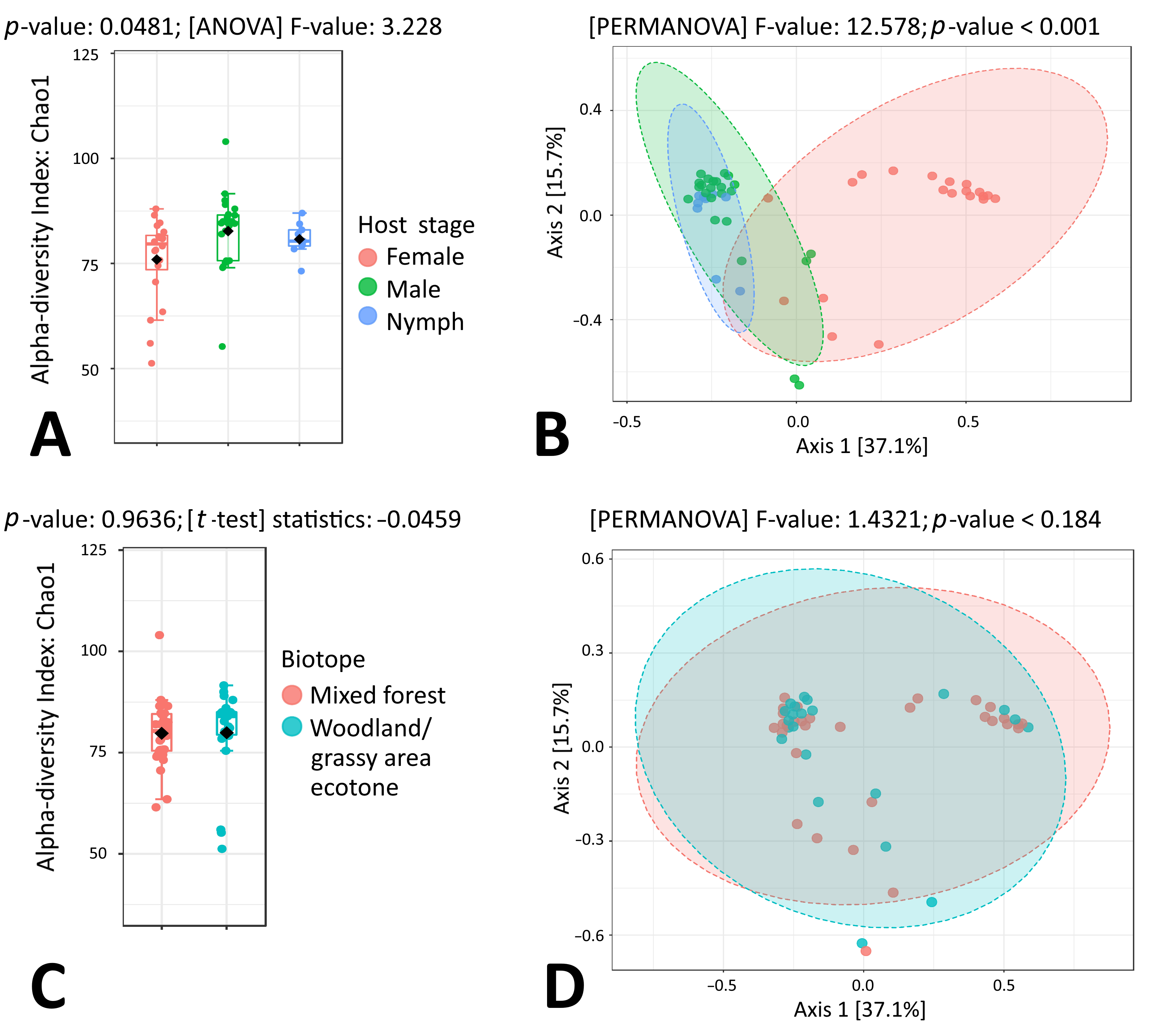
When taking a closer look at microbial taxonomic data in the context of individual tick species, a statistically significant difference between I. ricinus sex groups was observed for both alpha and beta microbial diversity (p = 0.0481 and p < 0.001, respectively) (Figure 2A,B). On the other hand, no differences were observed for microbiome composition when tick samples representing mixed forest and woodland/grassy area ecotones were compared (Figure 2C,D). In I. ricinus nymphs, ten of the most abundant microbial genera were Sphingomonas (11.67%), Halomonas (7.80%), Rickettsia (6.52%), Propionibacterium (5.30%), Novosphingobium (4.92%), Methylobacterium (4.21%), Bradyrhizobium (3.56%), Staphylococcus (3.36%), Corynebacterium (3.16%) and Streptococcus (3.16%) (Supplementary Table S1). Quite similarly, in I. ricinus males, ten of the most abundant microbial genera were Sphingomonas (12.44%), Mycobacterium (9.85%), Rickettsia (7.82%), Francisella (5.14%), Rickettsiella (3.45%), Propionibacterium (3.44%), Halomonas (3.37%), Methylobacterium (3.26%), Pseudomonas (3.14%) and Williamsia (2.86%) (Supplementary Table S1). The results of beta diversity analysis by PCoA and PERMANOVA showed that I. ricinus nymph and male samples formed tightly overlapping clusters, while I. ricinus female samples formed a distant group (Figure 2B). Indeed, in I. ricinus females, the most abundant microbial genus Ca. Midichloria comprised more than half of all sequencing reads (58.38%), followed by Rickettsia (9.12%), Sphingomonas (4.88%), Mycobacterium (4.59%), Williamsia (2.31%), Propionibacterium (1.25%), Methylobacterium (1.20%), Pseudomonas (1.00%), Acinetobacter (0.96%) and Corynebacterium (0.92%) (Figure 1A, Supplementary Table S1).
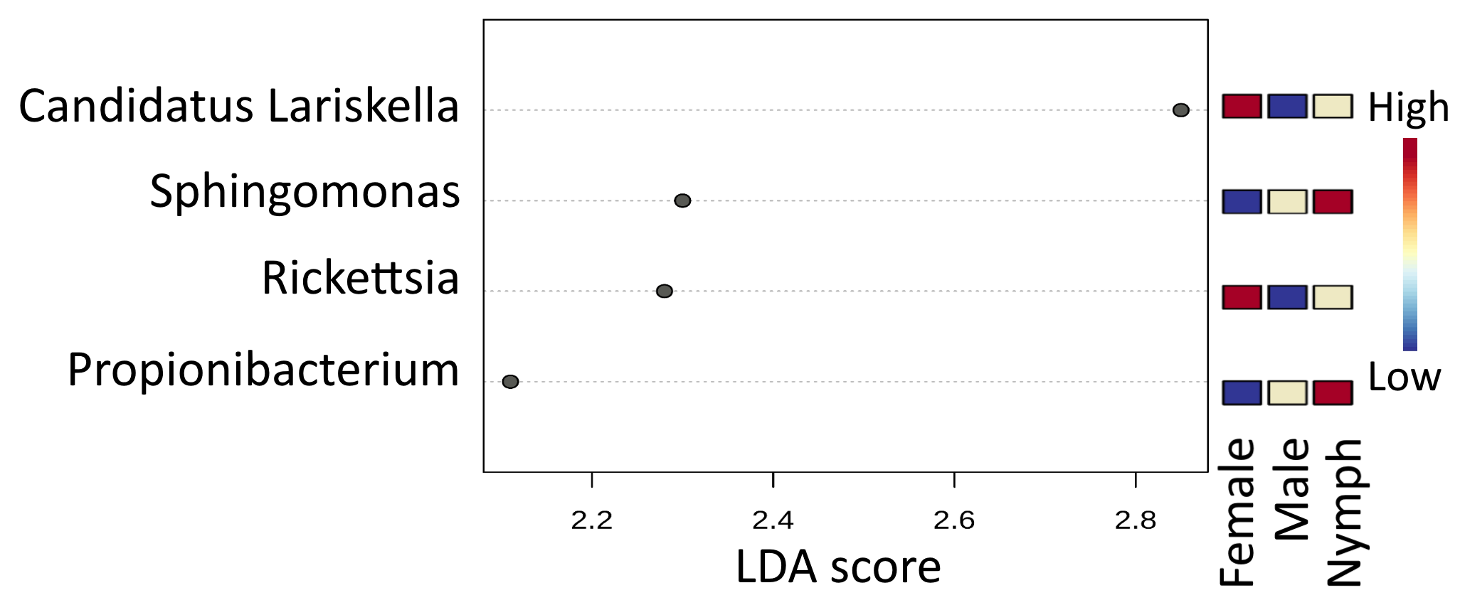
LefSe analysis revealed seven microbial taxa with significantly different (p < 0.05) abundance: Ca. Midichloria was more abundant in females and Sphingomonas was more abundant in males, while Halomonas, Propionibacterium, Bradyrhizobium, Staphylococcus and Streptococcus were highly associated with I. ricinus nymphs (Figure 3).

3.4. Microbiome Composition of I. persulcatus Ticks

Similarly to I. ricinus, a statistically significant difference between I. persulcatus sex and development stage groups was observed for both alpha and beta microbial diversity (p = 0.0425 and p < 0.001, respectively), while no differences were detected for microbiome composition when tick samples representing mixed forest and woodland/grassy area ecotones were compared (Figure 4A–D). The results of beta diversity analysis by PCoA and PERMANOVA showed that I. persulcatus nymph, male and female samples formed three clusters (Figure 4B).
In I. persulcatus nymphs, ten of the most abundant microbial genera were Sphingomonas (15.81%), Propionibacterium (10.62%), Rickettsia (8.90%), Halomonas (7.00%), Ca. Lariskella (6.43%), Corynebacterium (4.90%), Pelomonas (4.31%), Staphylococcus (4.06%), Methylobacterium (2.93%) and Streptococcus (2.48%). In I. persulcatus males, ten of the most abundant microbial genera were Spiroplasma (8.32%), Borrelia (7.88%), Sphingomonas (6.65%), Propionibacterium (5.71%), Acinetobacter (5.15%), Corynebacterium (5.15%), Halomonas (5.00%), Rickettsia (4.84%), Pseudomonas (4.04%) and Mycobacterium (4.01%). In I. persulcatus females, ten of the most abundant microbial genera were Ca. Lariskella (50.07%), Rickettsia (18.19%), Spiroplasma (7.41%), Pseudomonas (5.18%), Mycobacterium (2.40%), Propionibacterium (1.58%), Borrelia (1.40%), Sphingomonas (1.40%), Halomonas (1.21%) and Luteibacter (0.81%). LefSe analysis revealed four microbial taxa with significantly different (p < 0.05) abundance: Ca. Lariskella and Rickettsia were more abundant in females, and Sphingomonas and Propionibacterium were more abundant in I. persulcatus nymphs (Figure 5).

3.5. Microbiome Composition of D. reticulatus Ticks

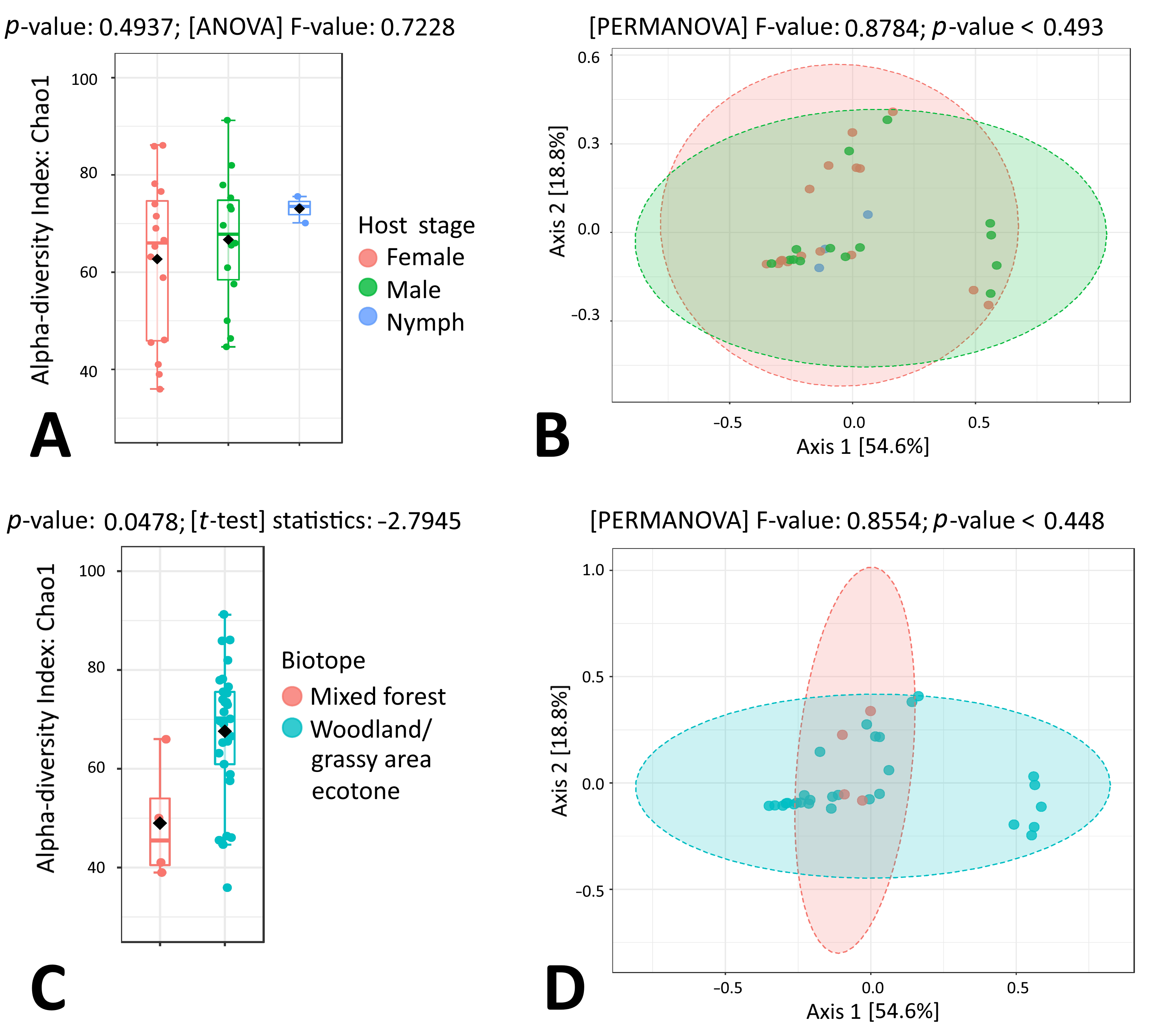
In contrast to the Ixodes species, in this study, a statistically significant difference between D. reticulatus sex and development stage groups was not observed for alpha and beta microbial diversity (p > 0.05) (Figure 6A,B). However, the microbial alpha diversity of the D. reticulatus ticks collected in a woodland/grassy area ecotone was slightly higher than for the samples representing mixed forest areas, and this difference reached statistical significance (p = 0.0478) (Figure 6C). The results of beta diversity analysis by PCoA and PERMANOVA showed that D. reticulatus samples from both biotopes formed overlapped clusters (p = 0.448; Figure 6D).
In D. reticulatus nymphs, ten of the most abundant microbial genera were Francisella (47.66%), Sphingomonas (11.67%), Methylobacterium (5.12%), Rickettsia (3.94%), Mycobacterium (2.16%), Pelomonas (1.95%), Nocardioides (1.81%), Propionibacterium (1.01%), Jatrophihabitans (0.96%) and Sphingomonadaceae Group (0.85%). In D. reticulatus males, ten of the most abundant microbial genera were Francisella (39.57%), Rickettsia (10.29%), Sphingomonas (5.42%), Methylobacterium (3.18%), Halomonas (2.63%), Propionibacterium (1.98%), Pseudomonas (1.90%), Nesterenkonia (1.84%), Corynebacterium (1.71%) and Mycobacterium (1.58%). Quite similarly, in D. reticulatus females, ten of the most abundant microbial genera were Francisella (52.30%), Rickettsia (14.30%), Sphingomonas (3.64%), Methylobacterium (2.51%), Mycobacterium (1.30%), Ca. Endoecteinascidia (1.23%), Haemophilus (1.22%), Propionibacterium (1.20%), Nocardioides (1.20%) and Pseudomonas (0.99%) (Figure 1A, Supplementary Table S1). Accordingly, no differentially abundant microbial taxa were identified between D. reticulatus tick sample groups.

3.6. Endosymbiont Species Abundance in Female Ixodes Ticks

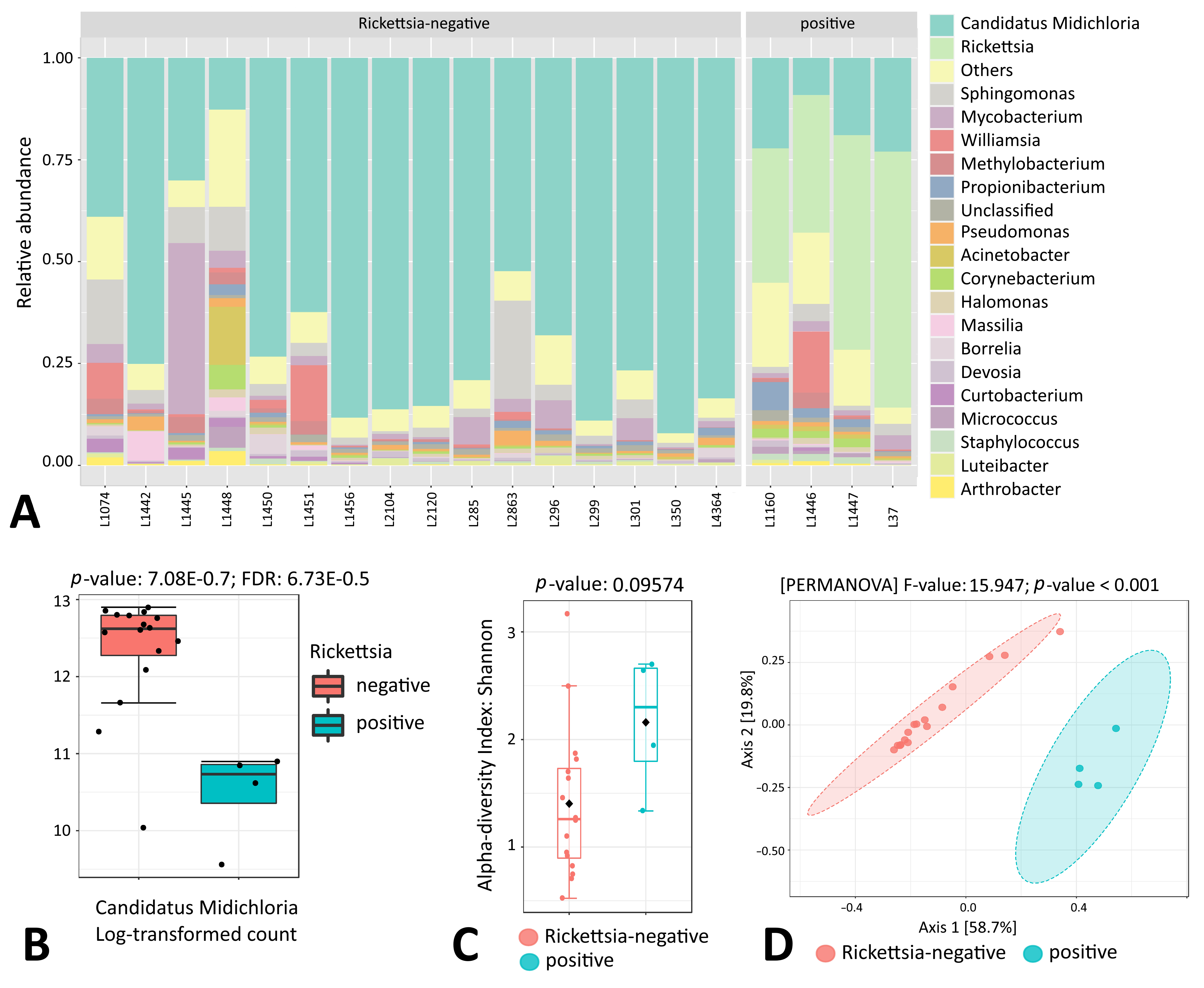
In I. ricinus, endosymbiont Ca. Midichloria was detected almost exclusively in female tick samples; only a small portion of sequencing reads of male ticks and nymphs were attributed to this genus (0.58% and 2.04%, respectively) (Supplementary Table S1). The prevalence of Ca. Midichloria in I. ricinus female ticks was 100%; however, the number of endosymbiont reads was not uniform and, as appeared, was largely dependent on the presence of the Rickettsia genus (Figure 7A,B). Further analysis indicated statistically significant differences in microbial beta diversity between Rickettsia-positive and -negative I. ricinus females (p < 0.001), while the alpha diversity was not affected (Figure 7C,D).
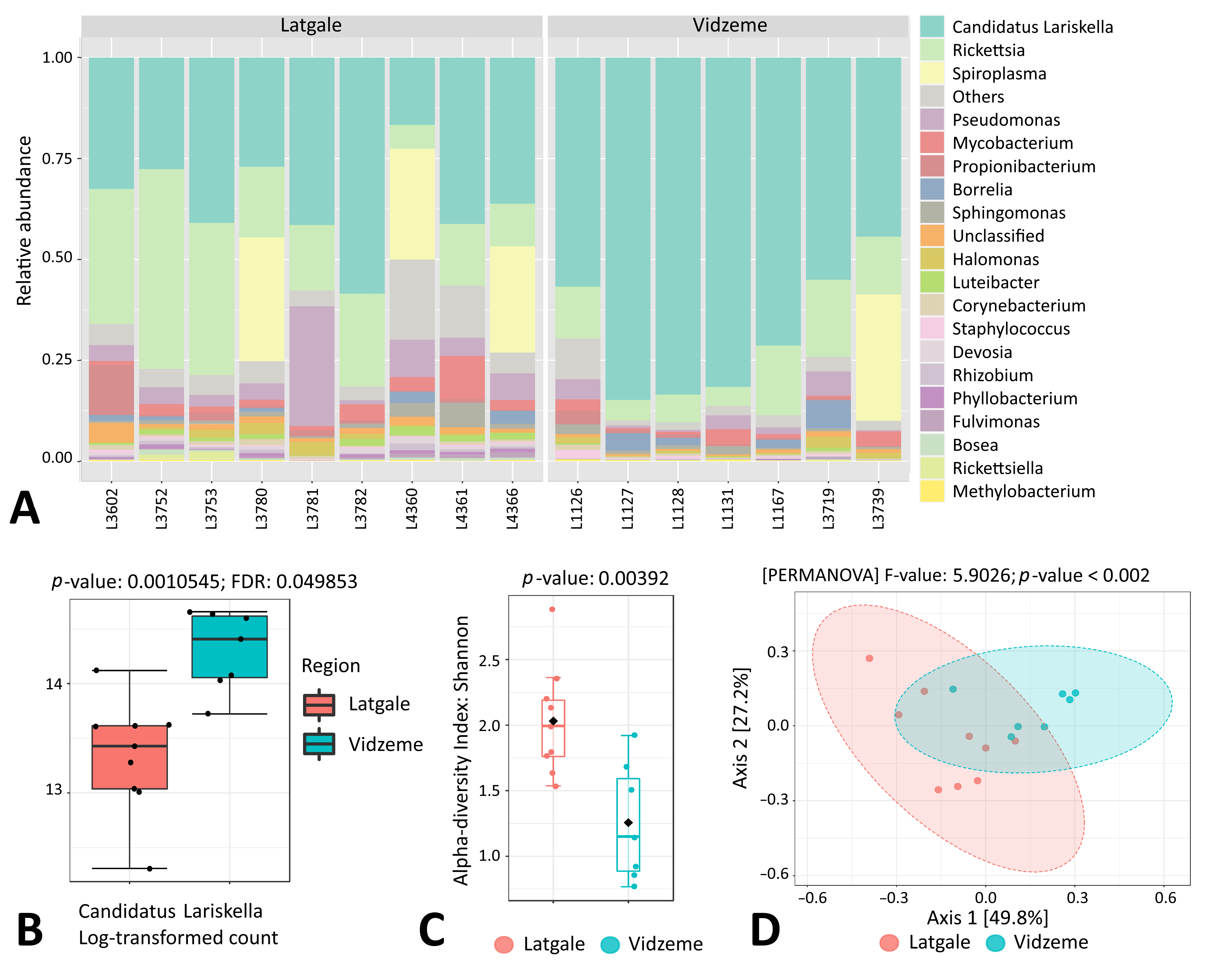
Similarly, in I. persulcatus, endosymbiont Ca. Lariskella was mainly associated with female ticks, as only a small fraction of sequencing reads in male and nymph ticks belonged to this genus (50.07% versus 0.13% and 6.43%, respectively) (Supplementary Table S1). However, in contrast to I. ricinus, all I. persulcatus females appeared to be Rickettsia-positive in this study; thus, the presence of Rickettsia alone did not explain the variability in the endosymbiont load observed in the I. persulcatus female samples (Figure 8A). On the other hand, the Ca. Lariskella genus was significantly more abundant in I. persulcatus females collected in the Vidzeme region in comparison to the Latgale region (Figure 8A,B). In addition, statistically significant differences in both alpha and beta microbial diversity were observed between Vidzeme and Latgale I. persulcatus female ticks (Figure 8C,D). On the contrary, for I. ricinus females, we did not observe any relations between tick collection site (i.e., geographical region or biotope) and the number of endosymbionts in this study.

4. Discussion

In Latvia, I. ricinus is widespread throughout the territory, while I. persulcatus is present in the eastern region and D. reticulatus is located in the western and southern regions; thus, sympatric populations of D. reticulatus and I. ricinus ticks, as well as D. reticulatus, I. ricinus and I. persulcatus ticks, exist in the country [15]. Despite the sympatric occurrence of the tick species, the obtained results clearly indicated the diversity in tick microbiome composition: while a close similarity between the microbiome of I. ricinus and I. persulcatus ticks was observed, D. reticulatus microbiome composition appeared to be significantly different. Similarly to our observations, significant differences in microbial diversity and composition were reported in the study in the far-western United States between several Ixodes, Dermacentor and Haemaphysalis leporispalustris tick species [24].
There is strong evidence that ticks acquire a significant portion of their microbiome through exposure to their environment [25]; thus, the observed differences could be, at least partly, driven by specific tick-species-related ecological relationships and habitat parameters. Indeed, I. ricinus and I. persulcatus ticks occur in several ecoregions, commonly found in deciduous and coniferous woodland and mixed forests of European type, while D. reticulatus is a typical open country tick species, preferring meadows and pastures [8].
Recently, the important role of small-scale ecological variation and microbe–microbe interactions in shaping tick microbial communities was efficiently highlighted [7]. In our study, we looked at the possible microbiome correlation with different habitats, such as forest and woodland/grassy area ecotones. For Ixodes ticks, no such correlation was detected, while in D. reticulatus, a possible impact of the habitat type on the microbiome composition was observed. However, a small sample number of D. reticulatus ticks collected in the forest areas in this study should be acknowledged; thus, additional studies with larger sample sets should be conducted to verify this result.
Statistically significant differences in alpha and beta microbial diversity were observed between Ixodes tick life stages and sexes, with lower taxa richness indexes obtained for female ticks. This result is similar to other studies conducted in different tick species in Europe and America [24,26]. More specifically, a decrease in both microbial species richness and evenness was reported as the Ixodes tick matures from larva to adult [24,25]. Interestingly, this phenomenon was not apparently present in D. reticulatus ticks in our study, as no statistically significant differences in microbial composition were detected between life stages and sexes. These findings could be partly explained by the endosymbiont loads, as a high load of endosymbionts in Ixodes female ticks was detected. In contrast, D. reticulatus tick samples did not show the presence of a single dominant microbial species in either males, females or nymphs, indicating the unique physiology of this tick species or its interaction with the surrounding environment. The most abundant microbial genus in D. reticulatus ticks was Francisella, which was previously recognized as the endosymbiont for this tick species [27]. In addition, our results showed that the Francisella genus was closely associated with D. reticulatus ticks, while endosymbionts Ca. Midichlorii and Ca. Lariskella were associated with I. ricinus and I. persulcatus females, respectively. Indeed, Ca. Midichloria mitochondrii is a well-known main endosymbiont of the tick I. ricinus, which appears to be ubiquitous in females across the tick’s distribution, while a lower prevalence is observed in males [28]. In our study, the prevalence of Ca. Midichloria in I. ricinus females was 100% with an overall abundancy of 58.38%, while only a small fraction of sequencing reads in males and nymphs was attributed to this genus. Also, in females, the endosymbiont load negatively correlated with the presence of the Rickettsia genus, as a significantly higher abundance of Ca. Midichloria was observed in the Rickettsia-negative I. ricinus samples. This observation is in contrary to a recent study, where a positive correlation between Ca. Midichloria and the presence of Rickettsia was detected in I. ricinus nymphs [29]. However, the observed difference could be explained by the different tick stage; in addition, the Rickettsia genus was previously reported to be involved in antagonist relationships with pathogenic or symbiotic microbial genera in I. ricinus and Rhipicephalus sanguineus ticks [7,30]. On the other hand, a positive correlation between Ca. Midichloria mitochondrii load and Rickettsia parkeri presence in the tick Amblyomma maculatum was described recently [31]. Also, evidence of a competition between pathogenic and endosymbiotic Rickettsia species exists [32,33]. Unfortunately, the sequencing approach of our study, similarly to other high-throughput 16S-amplicon-sequencing-based studies, did not allow us to identify Rickettsia at the species level; thus, it is unclear whether the detected sequences belonged to pathogenic or parasitoid species. Additional studies are needed to address the question of the role and/or interplay of different Rickettsia species within tick microbiomes.
Another bacteria of the family Candidatus Midichloriaceae, Ca. Lariskella arthropodarum (provisionally named ‘Montezuma’), have been detected in I. persulcatus and I. pavlovskyi ticks, as well as in human blood in the Russian Far East [34,35,36]. The hypothesis about the loss of this endosymbiont during I. persulcatus male development has been proposed [37]; however, the role of Ca. Lariskella in the host ticks has not yet been clearly determined [38]. The previously reported Ca. Lariskella prevalence in adult I. persulcatus females in different studies in Russia and Estonia was very diverse, ranging from 8.4% up to 97.1% [35,37,39,40]. In our study, all I. persulcatus females were Ca. Lariskella-positive; however, a great variability in the endosymbiont load was observed. In contrary to I. ricinus females, there was no connection between the Ca. Lariskella load and the presence of Rickettsia genus. However, interestingly, significant differences in the microbiome composition, including Ca. Lariskella amounts, were observed between I. persulcatus female samples collected in different regions of Latvia. Previously, differences in the bacterial community structure and composition of ticks across habitats [8] and geographical sites [26] have been documented; thus, this result could indicate the existence of specific interactions between ticks, hosts and the surrounding environment within different ecological niches.

5. Conclusions

The present study has assessed the microbiome composition of three endemic tick species in Latvia in the context of tick sex, geographical location, biotope and endosymbionts. Collectively, the obtained results revealed important associations between ticks and their microbial community and highlighted several tick microbiome features. We showed that, despite the sympatric occurrence of the tick species, tick microbiota and endosymbiont content were largely tick-species- and sex-specific, and were influenced by geographical location. Also, our data revealed possible interactions between Rickettsia and Ca. Midichloria in I. ricinus females. The obtained data provide the basis for additional studies to decipher the roles of, and interactions among, specific bacteria, which, in turn, could help to understand the factors that affect the tick microbial communities, and the consequences of microbiome variation. Further studies on the microbial communities of ticks within and between different ecosystems, and the influence of microbial diversity on the persistence and transmission of various medically important pathogens are of great importance.

Supplementary Materials

The following supporting information can be downloaded at https://www.mdpi.com/article/10.3390/microorganisms11081970/s1, Figure S1: Tick collection sites in Latvia. Data were mapped using Google Earth. (A) I. ricinus samples, (B) I. persulcatus samples. Samples from Vidzeme region are encircled by blue, and samples from Latgale region are encircled by red line. (C) D. reticulatus samples. Figure S2: Quality assessment and sequence length control of 16S rRNA amplicons obtained in this study. Representative plots are shown. E7, E8, E10—tick samples; “-“—blank sample, bk—barcode used. The analysis was performed using a Bioanalyzer 2100 instrument with an Agilent High-Sensitivity DNA Kit (Agilent Technologies, United States). Figure S3: Taxonomically assigned sequencing reads per sample at the genus level. Figure S4: Clustering dendrogram based on the Bray–Curtis Index and the Ward clustering method. Samples with more similar genus profiles were clustered closer together. Table S1: Microbial genera detected in tick samples.

Author Contributions

Conceptualization, A.B., V.C. and R.R.; methodology, A.N. and A.K. (Alisa Kazarina); investigation, A.N., A.K. (Alisa Kazarina), M.L., S.A., V.U., A.K. (Agnija Kivrane), L.F., D.S., J.K., A.B. and V.C.; resources, A.N.; data curation, A.N., A.K. (Alisa Kazarina), M.L. and J.K.; writing—original draft preparation, A.N.; writing—review and editing, A.K. (Alisa Kazarina), M.L. and R.R.; supervision, R.R.; funding acquisition, R.R. All authors have read and agreed to the published version of the manuscript.

Funding

This research was funded by the European Regional Development Fund (ERDF), project No. 1.1.1.1/16/A/044.

Institutional Review Board Statement

Not applicable.

Informed Consent Statement

Not applicable.

Data Availability Statement

Raw sequencing reads have been submitted to the European Nucleotide Archive, project accession PRJEB63277.

Conflicts of Interest

The authors declare no conflict of interest. The funders had no role in the design of the study; in the collection, analyses, or interpretation of data; in the writing of the manuscript; or in the decision to publish the results.

References

  1. Narasimhan:, S.; Fikrig, E. Tick Microbiome: The Force Within. Trends Parasitol. 2015, 31, 315–323. [Google Scholar] [CrossRef] [PubMed] [Green Version]
  2. Reuter, J.A.; Spacek, D.; Snyder, M.P. High-Throughput Sequencing Technologies. Mol. Cell 2015, 58, 586–597. [Google Scholar] [CrossRef] [Green Version]
  3. Pollet, T.; Sprong, H.; Lejal, E.; Krawczyk, A.I.; Moutailler, S.; Cosson, J.-F.; Vayssier-Taussat, M.; Estrada-Peña, A. The Scale Affects Our View on the Identification and Distribution of Microbial Communities in Ticks. Parasites Vectors 2020, 13, 36. [Google Scholar] [CrossRef] [Green Version]
  4. Bonnet, S.I.; Binetruy, F.; Hernández-Jarguín, A.M.; Duron, O. The Tick Microbiome: Why Non-Pathogenic Microorganisms Matter in Tick Biology and Pathogen Transmission. Front. Cell. Infect. Microbiol. 2017, 7, 236. [Google Scholar] [CrossRef]
  5. Bonnet, S.I.; Pollet, T. Update on the Intricate Tango between Tick Microbiomes and Tick-Borne Pathogens. Parasite Immunol. 2021, 43, e12813. [Google Scholar] [CrossRef] [PubMed]
  6. Narasimhan, S.; Swei, A.; Abouneameh, S.; Pal, U.; Pedra, J.H.F.; Fikrig, E. Grappling with the Tick Microbiome. Trends Parasitol. 2021, 37, 722–733. [Google Scholar] [CrossRef] [PubMed]
  7. Aivelo, T.; Norberg, A.; Tschirren, B. Bacterial Microbiota Composition of Ixodes Ricinus Ticks: The Role of Environmental Variation, Tick Characteristics and Microbial Interactions. PeerJ 2019, 7, e8217. [Google Scholar] [CrossRef] [Green Version]
  8. Estrada-Peña, A.; Cabezas-Cruz, A.; Pollet, T.; Vayssier-Taussat, M.; Cosson, J.-F. High Throughput Sequencing and Network Analysis Disentangle the Microbial Communities of Ticks and Hosts Within and Between Ecosystems. Front. Cell. Infect. Microbiol. 2018, 8, 236. [Google Scholar] [CrossRef] [Green Version]
  9. Aivelo, T.; Lemoine, M.; Tschirren, B. Elevational Changes in Bacterial Microbiota Structure and Diversity in an Arthropod-Disease Vector. Microb. Ecol. 2022, 84, 868–878. [Google Scholar] [CrossRef]
  10. Brinkerhoff, R.J.; Clark, C.; Ocasio, K.; Gauthier, D.T.; Hynes, W.L. Factors Affecting the Microbiome of Ixodes Scapularis and Amblyomma Americanum. PLoS ONE 2020, 15, e0232398. [Google Scholar] [CrossRef]
  11. Batool, M.; Blazier, J.C.; Rogovska, Y.V.; Wang, J.; Liu, S.; Nebogatkin, I.V.; Rogovskyy, A.S. Metagenomic Analysis of Individually Analyzed Ticks from Eastern Europe Demonstrates Regional and Sex-Dependent Differences in the Microbiota of Ixodes Ricinus. Ticks Tick-Borne Dis. 2021, 12, 101768. [Google Scholar] [CrossRef]
  12. Van Treuren, W.; Ponnusamy, L.; Brinkerhoff, R.J.; Gonzalez, A.; Parobek, C.M.; Juliano, J.J.; Andreadis, T.G.; Falco, R.C.; Ziegler, L.B.; Hathaway, N.; et al. Variation in the Microbiota of Ixodes Ticks with Regard to Geography, Species, and Sex. Appl. Environ. Microbiol. 2015, 81, 6200–6209. [Google Scholar] [CrossRef] [PubMed] [Green Version]
  13. Krawczyk, A.I.; Röttjers, L.; Fonville, M.; Takumi, K.; Takken, W.; Faust, K.; Sprong, H. Quantitative Microbial Population Study Reveals Geographical Differences in Bacterial Symbionts of Ixodes Ricinus. Microbiome 2022, 10, 120. [Google Scholar] [CrossRef] [PubMed]
  14. Duncan, K.T.; Elshahed, M.S.; Sundstrom, K.D.; Little, S.E.; Youssef, N.H. Influence of Tick Sex and Geographic Region on the Microbiome of Dermacentor Variabilis Collected from Dogs and Cats across the United States. Ticks Tick-Borne Dis. 2022, 13, 102002. [Google Scholar] [CrossRef] [PubMed]
  15. Capligina, V.; Seleznova, M.; Akopjana, S.; Freimane, L.; Lazovska, M.; Krumins, R.; Kivrane, A.; Namina, A.; Aleinikova, D.; Kimsis, J.; et al. Large-Scale Countrywide Screening for Tick-Borne Pathogens in Field-Collected Ticks in Latvia during 2017–2019. Parasites Vectors 2020, 13, 351. [Google Scholar] [CrossRef] [PubMed]
  16. Filippova, N.A. Fauna SSSR Paukoobraznye. Arachnida Class: Ixodid Ticks of the Subfamily Ixodinae; Nauka: Leningrad, Russia, 1977. [Google Scholar]
  17. Estrada-Peña, A.; Mihalca, A.D.; Petney, T.N. (Eds.) Ticks of Europe and North Africa; Springer International Publishing: Cham, Switzerland, 2017; ISBN 978-3-319-63759-4. [Google Scholar]
  18. Sprong, H.; Fonville, M.; Docters van Leeuwen, A.; Devillers, E.; Ibañez-Justicia, A.; Stroo, A.; Hansford, K.; Cull, B.; Medlock, J.; Heyman, P.; et al. Detection of Pathogens in Dermacentor Reticulatus in Northwestern Europe: Evaluation of a High-Throughput Array. Heliyon 2019, 5, e01270. [Google Scholar] [CrossRef] [Green Version]
  19. Goecks, J.; Nekrutenko, A.; Taylor, J. Galaxy Team Galaxy: A Comprehensive Approach for Supporting Accessible, Reproducible, and Transparent Computational Research in the Life Sciences. Genome Biol. 2010, 11, R86. [Google Scholar] [CrossRef] [Green Version]
  20. Kosakovsky Pond, S.; Wadhawan, S.; Chiaromonte, F.; Ananda, G.; Chung, W.-Y.; Taylor, J.; Nekrutenko, A. Windshield Splatter Analysis with the Galaxy Metagenomic Pipeline. Genome Res. 2009, 19, 2144–2153. [Google Scholar] [CrossRef] [Green Version]
  21. Jing, G.; Sun, Z.; Wang, H.; Gong, Y.; Huang, S.; Ning, K.; Xu, J.; Su, X. Parallel-META 3: Comprehensive Taxonomical and Functional Analysis Platform for Efficient Comparison of Microbial Communities. Sci. Rep. 2017, 7, 40371. [Google Scholar] [CrossRef] [Green Version]
  22. Chong, J.; Liu, P.; Zhou, G.; Xia, J. Using MicrobiomeAnalyst for Comprehensive Statistical, Functional, and Meta-Analysis of Microbiome Data. Nat. Protoc. 2020, 15, 799–821. [Google Scholar] [CrossRef] [PubMed]
  23. Dhariwal, A.; Chong, J.; Habib, S.; King, I.L.; Agellon, L.B.; Xia, J. MicrobiomeAnalyst: A Web-Based Tool for Comprehensive Statistical, Visual and Meta-Analysis of Microbiome Data. Nucleic Acids Res. 2017, 45, W180–W188. [Google Scholar] [CrossRef] [PubMed]
  24. Chicana, B.; Couper, L.I.; Kwan, J.Y.; Tahiraj, E.; Swei, A. Comparative Microbiome Profiles of Sympatric Tick Species from the Far-Western United States. Insects 2019, 10, 353. [Google Scholar] [CrossRef] [PubMed] [Green Version]
  25. Kwan, J.Y.; Griggs, R.; Chicana, B.; Miller, C.; Swei, A. Vertical vs. Horizontal Transmission of the Microbiome in a Key Disease Vector, Ixodes Pacificus. Mol. Ecol. 2017, 26, 6578–6589. [Google Scholar] [CrossRef]
  26. Carpi, G.; Cagnacci, F.; Wittekindt, N.E.; Zhao, F.; Qi, J.; Tomsho, L.P.; Drautz, D.I.; Rizzoli, A.; Schuster, S.C. Metagenomic Profile of the Bacterial Communities Associated with Ixodes Ricinus Ticks. PLoS ONE 2011, 6, e25604. [Google Scholar] [CrossRef]
  27. de Carvalho, I.L.; Santos, N.; Soares, T.; Zé-Zé, L.; Núncio, M.S. Francisella-like Endosymbiont in Dermacentor Reticulatus Collected in Portugal. Vector Borne Zoonotic Dis. Larchmt. N 2011, 11, 185–188. [Google Scholar] [CrossRef] [Green Version]
  28. Sassera, D.; Beninati, T.; Bandi, C.; Bouman, E.A.P.; Sacchi, L.; Fabbi, M.; Lo, N. “Candidatus Midichloria Mitochondrii”, an Endosymbiont of the Tick Ixodes Ricinus with a Unique Intramitochondrial Lifestyle. Int. J. Syst. Evol. Microbiol. 2006, 56, 2535–2540. [Google Scholar] [CrossRef] [PubMed]
  29. Lejal, E.; Chiquet, J.; Aubert, J.; Robin, S.; Estrada-Peña, A.; Rue, O.; Midoux, C.; Mariadassou, M.; Bailly, X.; Cougoul, A.; et al. Temporal Patterns in Ixodes Ricinus Microbial Communities: An Insight into Tick-Borne Microbe Interactions. Microbiome 2021, 9, 153. [Google Scholar] [CrossRef]
  30. René-Martellet, M.; Minard, G.; Massot, R.; Tran Van, V.; Valiente Moro, C.; Chabanne, L.; Mavingui, P. Bacterial Microbiota Associated with Rhipicephalus Sanguineus (s.l.) Ticks from France, Senegal and Arizona. Parasites Vectors 2017, 10, 416. [Google Scholar] [CrossRef] [Green Version]
  31. Budachetri, K.; Kumar, D.; Crispell, G.; Beck, C.; Dasch, G.; Karim, S. The Tick Endosymbiont Candidatus Midichloria Mitochondrii and Selenoproteins Are Essential for the Growth of Rickettsia Parkeri in the Gulf Coast Tick Vector. Microbiome 2018, 6, 141. [Google Scholar] [CrossRef] [Green Version]
  32. Burgdorfer, W.; Hayes, S.; Mavros, A.J. Nonpathogenic Rickettsiae in Dermacentor Andersoni: A Limiting Factor for the Distribution of Rickettsia Rickettsii. In Conference on Rickettsiae and Rickettsial Diseases (1980: Rocky Mountain Laboratories); Rickettsiae and rickettsial diseases; Academic Press: New York, NY, USA, 1981; pp. 585–594. ISBN 0-12-143150-9. [Google Scholar]
  33. Macaluso, K.R.; Sonenshine, D.E.; Ceraul, S.M.; Azad, A.F. Rickettsial Infection in Dermacentor Variabilis (Acari: Ixodidae) Inhibits Transovarial Transmission of a Second Rickettsia. J. Med. Entomol. 2002, 39, 809–813. [Google Scholar] [CrossRef] [Green Version]
  34. Matsuura, Y.; Kikuchi, Y.; Meng, X.Y.; Koga, R.; Fukatsu, T. Novel Clade of Alphaproteobacterial Endosymbionts Associated with Stinkbugs and Other Arthropods. Appl. Environ. Microbiol. 2012, 78, 4149–4156. [Google Scholar] [CrossRef] [PubMed] [Green Version]
  35. Mediannikov, O.I.; Ivanov, L.I.; Nishikawa, M.; Saito, R.; Sidel’nikov, I.N.; Zdanovskaia, N.I.; Mokretsova, E.V.; Tarasevich, I.V.; Suzuki, H. Microorganism “Montezuma” of the order Rickettsiales: The potential causative agent of tick-borne disease in the Far East of Russia. Zhurnal Mikrobiol. Epidemiol. I Immunobiol. 2004, 1, 7–13. [Google Scholar]
  36. Kurilshikov, A.; Livanova, N.N.; Fomenko, N.V.; Tupikin, A.E.; Rar, V.A.; Kabilov, M.R.; Livanov, S.G.; Tikunova, N.V. Comparative Metagenomic Profiling of Symbiotic Bacterial Communities Associated with Ixodes Persulcatus, Ixodes Pavlovskyi and Dermacentor Reticulatus Ticks. PLoS ONE 2015, 10, e0131413. [Google Scholar] [CrossRef] [Green Version]
  37. Eremeeva, M.E.; Oliveira, A.; Robinson, J.B.; Ribakova, N.; Tokarevich, N.K.; Dasch, G.A. Prevalence of Bacterial Agents in Ixodes Persulcatus Ticks from the Vologda Province of Russia. Ann. N. Y. Acad. Sci. 2006, 1078, 291–298. [Google Scholar] [CrossRef] [PubMed]
  38. Duron, O.; Binetruy, F.; Noël, V.; Cremaschi, J.; McCoy, K.D.; Arnathau, C.; Plantard, O.; Goolsby, J.; Pérez de León, A.A.; Heylen, D.J.A.; et al. Evolutionary Changes in Symbiont Community Structure in Ticks. Mol. Ecol. 2017, 26, 2905–2921. [Google Scholar] [CrossRef] [PubMed] [Green Version]
  39. Eremeeva, M.E.; Oliveira, A.; Moriarity, J.; Robinson, J.B.; Tokarevich, N.K.; Antyukova, L.P.; Pyanyh, V.A.; Emeljanova, O.N.; Ignatjeva, V.N.; Buzinov, R.; et al. Detection and Identification of Bacterial Agents in Ixodes Persulcatus Schulze Ticks from the North Western Region of Russia. Vector Borne Zoonotic Dis. Larchmt. N 2007, 7, 426–436. [Google Scholar] [CrossRef]
  40. Mukhacheva, T.A.; Kovalev, S.Y. Bacteria of the Family ‘Candidatus Midichloriaceae’ in Sympatric Zones of Ixodes Ticks: Genetic Evidence for Vertical Transmission. Microb. Ecol. 2017, 74, 185–193. [Google Scholar] [CrossRef]
Figure 1. Genus-level comparison of bacterial profiles in Ixodes ricinus, I. persulcatus and Dermacentor reticulatus tick samples: (A) Stacked plots of the taxonomic classification. The abundances of the most abundant genera are shown. N: nymph, F: female, M: male. (B) Chao1 diversity analysis. DR: D. reticulatus, IP: I. persulcatus, IR: I. ricinus. (C) Principal coordinate analysis (PCoA) derived from Bray–Curtis distances among samples of the three groups (p < 0.001 by PERMANOVA). For each axis, in square brackets, the percent of variation explained was reported. (D) Linear discriminant analysis (LDA) combined with effect size measurements (LEfSe) identified microbial genera that enabled discrimination between the microbiotas of three tick species. False Discovery Rate (FDR)-adjusted p-value cutoff: 0.05; logarithmic LDA score ≥ 2.0.
Microorganisms 11 01970 g001
Figure 2. Genus-level comparison of bacterial profiles in I. ricinus tick samples: (A) Chao1 diversity analysis of nymph, male and female samples. (B) PCoA derived from Bray–Curtis distances among tick samples of the three groups: nymphs, males and females (p < 0.001 by PERMANOVA). (C) Chao1 diversity analysis of tick samples from different biotopes. (D) PCoA derived from Bray–Curtis distances among tick samples of the two biotope groups (p-value by PERMANOVA).
Microorganisms 11 01970 g002
Figure 3. LEfSe identified microbial genera that enabled discrimination between the microbiotas of I. ricinus nymph, male and female samples. False Discovery Rate (FDR)-adjusted p-value cutoff: 0.05; logarithmic LDA score ≥ 2.0.
Microorganisms 11 01970 g003
Figure 4. Genus-level comparison of bacterial profiles in I. persucatus tick samples: (A) Chao1 diversity analysis of nymph, male and female samples. (B) PCoA derived from Bray–Curtis distances among tick samples of the three groups: nymphs, males and females (p < 0.001 by PERMANOVA). (C) Chao1 diversity analysis of tick samples from different biotopes. (D) PCoA derived from Bray–Curtis distances among tick samples of the two biotope groups (p-value by PERMANOVA).
Microorganisms 11 01970 g004
Figure 5. LEfSe identified microbial genera that enabled discrimination between the microbiotas of I. persulcatus nymph, male and female samples. False Discovery Rate (FDR)-adjusted p-value cutoff: 0.05; logarithmic LDA score ≥ 2.0.
Microorganisms 11 01970 g005
Figure 6. Genus-level comparison of bacterial profiles in D. reticulatus tick samples: (A) Chao1 diversity analysis of nymph, male and female samples. (B) PCoA derived from Bray–Curtis distances among tick samples of the three groups: nymphs, males and females (p-value by PERMANOVA). (C) Chao1 diversity analysis of tick samples from different biotopes. (D) PCoA derived from Bray–Curtis distances among tick samples of the two biotope groups (p-value by PERMANOVA).
Microorganisms 11 01970 g006
Figure 7. Genus-level comparison of bacterial profiles in I. ricinus female tick samples: (A) Stacked plots of the taxonomic classification. The abundances of the most abundant genera are shown. (B) Comparison of the relative abundance of Ca. Midichloria between Rickettsia-positive and -negative samples. p value is indicated. (C) Shannon diversity analysis of Rickettsia-positive and -negative samples. (D) PCoA derived from Bray–Curtis distances among Rickettsia-positive and -negative samples (p < 0.001 by PERMANOVA).
Microorganisms 11 01970 g007
Figure 8. Genus-level comparison of bacterial profiles in I. persulcatus female tick samples: (A) Stacked plots of the taxonomic classification. The abundances of the most abundant genera are shown. (B) Comparison of the relative abundance of Ca. Lariskella between samples from Latgale and Vidzeme regions. p value is indicated. (C) Shannon diversity analysis of samples from Latgale and Vidzeme regions. (D) PCoA derived from Bray–Curtis distances among samples from Latgale and Vidzeme regions (p < 0.001 by PERMANOVA).
Microorganisms 11 01970 g008
Table 1. Tick samples (N = 126).
Tick SpeciesTick StageNo (%) of SamplesBiotope, No. (%) of Tick Samples
Mixed ForestWoodland/Grassy Area Ecotone
I. ricinusFemale20 (37.7)14 (70.0)6 (30.0)
Male24 (45.3)10 (41.7)14 (58.3)
Nymph9 (17.0)7 (77.8)2 (22.2)
Total5331 (58.5)22 (41.5)
I. persulcatusFemale16 (40.0)10 (62.5)6 (37.5)
Male17 (42.5)7 (41.2)10 (58.8)
Nymph7 (17.5)4 (57.1)3 (42.9)
Total4021 (52.5)19 (47.5)
D. reticulatusFemale16 (48.5)2 (12.5)14 (87.5)
Male14 (42.4)2 (14.3)12 (85.7)
Nymph3 (9.1)03 (100.0)
Total334 (12.1)29 (87.9)
Total 1265670
Disclaimer/Publisher’s Note: The statements, opinions and data contained in all publications are solely those of the individual author(s) and contributor(s) and not of MDPI and/or the editor(s). MDPI and/or the editor(s) disclaim responsibility for any injury to people or property resulting from any ideas, methods, instructions or products referred to in the content.