Article

The Immune System Response to Porphyromonas gingivalis in Neurological Diseases

by
Raffaella Franciotti
1,*,
Pamela Pignatelli
2,
Domenica Lucia D’Antonio
3,4,
Rosa Mancinelli
1,
Stefania Fulle
1,
Matteo Alessandro De Rosa
1,5,
Valentina Puca
6,
Adriano Piattelli
7,8,
Astrid Maria Thomas
1,5,
Marco Onofrj
1,5,
Stefano Luca Sensi
1,5,9 and
Maria Cristina Curia
3
1
Department of Neuroscience, Imaging and Clinical Science, G. d’Annunzio University of Chieti-Pescara, 66100 Chieti, Italy
2
COMDINAV DUE, Nave Cavour, Italian Navy, Stazione Navale Mar Grande, Viale Jonio, 74122 Taranto, Italy
3
Department of Medical, Oral and Biotechnological Sciences, G. d’Annunzio University of Chieti-Pescara, 66100 Chieti, Italy
4
Fondazione Villaserena per la Ricerca, 65013 Città Sant’Angelo, Pescara, Italy
5
Center for Advanced Studies and Technology (CAST), G. d’Annunzio University of Chieti-Pescara, 66100 Chieti, Italy
6
Department of Pharmacy, G. d’Annunzio University of Chieti-Pescara, 66100 Chieti, Italy
7
School of Dentistry, Saint Camillus International University for Health Sciences, 00131 Rome, Italy
8
Facultad de Medicina, UCAM Universidad Católica San Antonio de Murcia, Guadalupe, 30107 Murcia, Spain
9
Institute for Advanced Biomedical Technologies, G. d’Annunzio University of Chieti-Pescara, 66100 Chieti, Italy
*
Author to whom correspondence should be addressed.
Microorganisms 2023, 11(10), 2555; https://doi.org/10.3390/microorganisms11102555
Submission received: 31 July 2023 / Revised: 3 October 2023 / Accepted: 10 October 2023 / Published: 13 October 2023

Abstract

:
Previous studies have reported an association between oral microbial dysbiosis and the development and progression of pathologies in the central nervous system. Porphyromonas gingivalis (Pg), the keystone pathogen of the oral cavity, can induce a systemic antibody response measured in patients’ sera using enzyme-linked immunosorbent assays. The present case–control study quantified the immune system’s response to Pg abundance in the oral cavities of patients affected by different central nervous system pathologies. The study cohort included 87 participants: 23 healthy controls (HC), 17 patients with an acute neurological condition (N-AC), 19 patients with a chronic neurological condition (N-CH), and 28 patients with neurodegenerative disease (N-DEG). The results showed that the Pg abundance in the oral cavity was higher in the N-DEG patients than in the HC (p = 0.0001) and N-AC patients (p = 0.01). In addition, the Pg abundance was higher in the N-CH patients than the HCs (p = 0.005). Only the N-CH patients had more serum anti-Pg antibodies than the HC (p = 0.012). The inadequate response of the immune system of the N-DEG group in producing anti-Pg antibodies was also clearly indicated by an analysis of the ratio between the anti-Pg antibodies quantity and the Pg abundance. Indeed, this ratio was significantly lower between the N-DEG group than all other groups (p = 0.0001, p = 0.002, and p = 0.03 for HC, N-AC, and N-CH, respectively). The immune system’s response to Pg abundance in the oral cavity showed a stepwise model: the response diminished progressively from the patients affected with an acute condition to the patients suffering from chronic nervous system disorders and finally to the patients affected by neurodegenerative diseases.

1. Introduction

The oral cavity is characterized by distinctive features, making it an ideal habitat for a collection of microorganisms [1]. The mouth harbors more than 700 bacterial species or phylotypes, which are site- and subject-specific [2].
Porphyromonas gingivalis (Pg) is a Gram-negative, nonmotile short rod, and an anaerobic bacterium, considered to be the keystone pathogen of the oral cavity because it is implicated in the biofilm formation of bacterial plaques. With other oral microbes, it forms a biofilm that alters tight epithelial junctions and promotes inflammation by mucosal immune cells. Pg can modulate the host immune response, at first increasing nutrient availability and biofilm growth, and subsequently facilitating bacterial resistance by destroying complement factors [3]. Pg produces many virulence factors, such as proteases, endotoxins, organic acids, and key enzymes, including the proteases known as gingipains [4]. Gingipains adhere to the epithelium, affecting its permeability and upregulating inflammation. The infiltration of immune cells—macrophages, neutrophils, and lymphocytes—leads to an altered antigen presentation, which overturns the equilibrium between the host and the microorganisms, creating dysbiosis [5]. Pg inhibits the conversion of M2 macrophages by redirecting them to the M1 inflammatory subtype, worsening the inflammatory environment. M1 may exhibit cytotoxic and M2 neuroprotective effects [6]. Chronic inflammation damages epithelial cells through the production of reactive oxidative species (ROS), reactive nitrogen species (RNS), and proinflammatory cytokines. These products cause DNA damage and cell death, further destroying the epithelial barrier. Pg promotes the onset of chronic periodontal disease, which is characterized by a systemic antibody response, measured by the serum levels of antibodies against periodontal pathogens [7,8].
The immune system preserves the microenvironment for the microbiota maintaining local and systemic homeostasis and preserving host biological integrity [9]. The presence of serum antibodies to major periodontal pathogens has been associated with heart disease, stroke [10,11,12], and Alzheimer’s Disease (AD) [13,14]. In particular, elevated levels of immunoglobulin G (IgG) against Pg were detected in subjects before cognitive impairment [15,16]. Anti-Pg antibodies were also detected in neurological patients’ sera using enzyme-linked immunosorbent assays (ELISA) [17].
Specific oral bacterial species have also been implicated in several systemic diseases, such as bacterial endocarditis [18], aspiration pneumonia [19], osteomyelitis in children [20], pre-term low birth weight [21], obesity, diabetes [22], cardiovascular disease [23], rheumatoid arthritis, osteoporosis [24,25,26], and neurological and psychiatric disorders such as autism spectrum disorder, bipolar disorder, post-traumatic stress disorder, schizophrenia, and major depressive disorder [27,28,29]. In recent years, many researchers have introduced the oral microbiota–brain axis, given that the oral cavity is the origin of the gastrointestinal tract and could represent an extension of the microbiota gut–brain axis. In normal conditions, the oral and gut microbiome profiles are well-segregated due to the oral–gut barrier, physical distance, and chemical impediments, such as gastric acid and bile. In pathological conditions, oral–gut barrier impairment can allow for bidirectional communication between the oral and gut microbes, reshaping the microbial ecosystems of both habitats [30]. Thus, the mechanisms through which oral bacterial composition can impact the brain can be direct or indirect. The direct mechanism could be related to the trigeminal/olfactory/facial nervous system and the bloodstream [29]. The indirect mechanism could be related to the involvement of gut microbiota dysbiosis and systemic inflammation [31]. Both mechanisms could promote the release of proinflammatory cytokines, such as interleukin-IL-1β, IL-6, tumor necrosis factor (TNF)-α, chemokines, inflammasome NLRP3, and ROS, exacerbating neuroinflammation and provoking synaptic toxicity and neuronal death [29]. The microbiota–gut–brain axis and the oral microbiota–brain axis play important roles in maintaining homeostasis and their dysfunctions have been linked to various central nervous system (CNS) diseases, such as multiple sclerosis, AD, and Parkinson’s Disease (PD) [32,33,34,35]. It is well known that Pg-induced inflammation in the oral cavity can be transferred to the CNS. Pg lipopolysaccharide (LPS) can reach the brain-resident microglia via the leptomeningeal cells, which express Pg-LPS receptors (TLR2 and TLR4). Liu et al. demonstrated that these cells can transmit inflammatory signals from peripheral macrophages to the brain-resident microglia due to Pg-LPS stimulation [36]. Furthermore, more LPS from Pg has been detected in AD patients’ brain tissues compared to healthy volunteers [37]. AD incidence and mortality risk have been linked to a composite of Pg titers and neuroinflammation processes [38].
Recently, a novel pathogenic model, brain-first versus body-first, was proposed to classify two different subtypes of PD. According to this model, in the brain-first (top-down) type, α-synuclein pathology could initially arise in the brain with secondary spreading to the peripheral autonomic nervous system. In the body-first (bottom-up) type, the pathology could originate in the enteric or peripheral autonomic nervous system and then spread to the brain. However, these findings are still under further investigation [39].
Proactive approaches to balancing oral dysbiosis include the use of probiotics, paraprobiotics, postbiotics, and ozone therapy [40].
In this study, we evaluated the immune system’s response to Pg abundance in the oral cavities of patients affected by pathologies in the CNS. In particular, anti-Pg antibodies in the serum and the Pg abundance in the oral cavity were quantified in patients with neurodegenerative diseases compared to healthy controls (HC) and patients affected by acute or chronic neurological diseases.

2. Materials and Methods

2.1. Study Cohort

The study was conducted according to the guidelines of the Declaration of Helsinki and approved by the local Ethics Committee of the “G. d’Annunzio” University of Chieti-Pescara on May 2020 (ethics code: 1909).
A previous pilot study [17] found a significant difference (p = 0.037, with a statistical power of 60.4% considering the probability of type I error α = 0.05) between 15 patients affected by neurodegenerative diseases and 21 patients affected by neurological non-neurodegenerative diseases in the ratio between anti-Pg antibodies’ quantity and Pg abundance. In the same condition, the statistical power could reach 80% with α = 0.05 if a sample size of 33 was considered for each group. In addition, also considering the HC group, we estimated that, with a low percentage increase of 2.4% in the ratio values, we could obtain a statistical power of 80% and α = 0.05 with a sample size of 30 for each group. Accordingly, the eligibility criteria were evaluated in 30 HC and 90 neurological patients, who could be subdivided into three groups, obtaining 1:1 matching among the groups. HCs free from neurological diseases were recruited from a list of HCs. In contrast, from May 2020 to July 2022, all patients were enrolled in the Neurology Clinic of “SS Annunziata” Hospital of Chieti. Individuals under antibiotic therapy (2 HCs and 8 patients) or those using daily chlorhexidine mouthwash within the last 3 months (3 HCs and 7 patients) were excluded from the study. In addition, 5 patients were excluded for concomitant tumors, and 2 HCs and 6 patients declined to participate. Thus, the final cohort included 23 HCs, 17 patients with an acute neurological condition (N-AC), 19 patients with a chronic neurological condition (N-CH), and 28 patients with a neurodegenerative disease (N-DEG). Written informed consent was obtained from all 87 participants. For each individual, tongue biofilm was used to quantify the Pg abundance and a blood sample was used to quantify the anti-Pg antibody levels.
The N-AC group included patients referred to the Neurology Clinic for the following reasons: ischemic stroke (n = 7), head trauma (n = 6), encephalitis (n = 2), loss of consciousness (n = 1), and cerebrospinal fluid hypotension (n = 1).
The N-CH group included patients with a diagnosis of a chronic neurological condition such as: epilepsy (n = 7), myasthenia (n = 6), polyneuropathy (n = 4), brain tumor (n = 1), and vascular leukoencephalopathy (n = 1).
The N-DEG group included patients with the following diagnoses: Parkinson’s disease (n = 10, of which n = 5 had dementia), multiple sclerosis (n = 6), sclerosis lateral amyotrophic (n = 4), mixed dementia (n = 3), Alzheimer’s disease (n = 2), Frontotemporal dementia (n = 2), and Huntington’s disease (n = 1).
Figure 1 shows a flow diagram of the case–control study from the eligibility screening to the collection of samples and oral indices. STROBE Statement for observational studies was provided in Table S1 of Supplementary Materials.

2.2. Collection of the Samples

Tongue biofilm was taken from each patient and each HC 8 h after they last brushed their teeth. The swab was collected by brushing from the middle third of the tongue dorsum 5 times. After shaking it vigorously for 30 s, the swab was immediately transferred into 5.0 mL of phosphate-buffered saline (PBS) and then kept at +4 °C until the nucleic acid extraction. A blood sample was also taken from all the participants on the same day of the brushing and stored at −80 °C.

2.3. Oral Examinations

Oral examinations were performed by a trained dentist (P.P.) using mirrors (MIR3HD, Hu-Friedy, Chicago, IL, USA), a dental probe (PCP-UNC 15, Hu-Friedy, Chicago, IL, USA), and an intra-oral light. The teeth number, plaque index (PI), and gingival index (GI) were recorded using the standardized Oral Health Questionnaire [41]. All other indices were evaluated as described in Franciotti et al., 2021 [17].

2.4. Bacterial DNA Quantification on Brushing

In this study, the reference bacterial strain Pg ATCC 33277 (LGC Standards S.r.l., Sesto San Giovanni, Milano, Italy) was used and cultivated as previously reported [42]. Briefly, the bacterium was cultivated in Fastidious Anaerobe Agar (Neogen, Lansing, MI, USA) supplemented with 5% of defibrinated horse sterile blood (Oxoid Limited, Hampshire, UK) for 48 h in anaerobiosis (Anaerogen Pak Jar, Oxoid, UK). After incubation, all the colonies were collected, washed in PBS, and centrifugated to obtain a pellet corresponding to 6 × 10¹² CFU. Molecular analyses were performed to quantify the Pg in the oral cavities of all the participants. The total genomic DNA was isolated from the samples and the reference bacterial strain using a Quick DNA miniPrep Plus KIT (Zymo Research, Irvine, CA, USA). StepOne™ 2.0 (Applied Biosystems, Thermo Fisher Scientific, Waltham, MA, USA) was used in a qPCR analysis to quantify the Pg abundance in each sample. A TaqMan-based assay that recognizes Pg 16S rRNA, the gene encoding the small subunit of 16S ribosomal RNA, was used, as previously reported [43]. A standard curve passing through 5 points was constructed, indicating the cycle threshold values versus the Pg 16S rRNA gene. This method allowed us to estimate the bacterial quantity based on the amount of total DNA isolated from the oral samples [43].

2.5. Antibody Assay on Serum

The ELISA technique assayed the serum IgG antibody responses to the bacterial pathogens from Pg using the ChonBlockTM buffer system (Human Anti-Bacteria & Toxins Antibody ELISA Kits, #6119, Chondrex, Woodinville, WA, USA, an ELISA protocol to improve the accuracy and reliability of serological antibody assays) as follows [44]. Specific 96-well plates were used to perform the assay at room temperature. Dilution samples less than 1:1000 required a parallel run on both antigen-coated and uncoated plates; conversely, for dilution samples more than 1:1000, only antigen-coated plates were run. In detail, sera were collected from each participant and stored at −80 °C. The sera were then thawed and centrifuged at 10,000 rpm for 5 min to perform the assay. The supernatants were recovered and suitably diluted with ChonBlockTM standard/sample dilution buffer. Moreover, Blocking Buffer was added to each well and incubated for 1 h; the plate was washed using Wash Buffer; the standards and samples were loaded and incubated for 2 h; the plate was washed using Wash Buffer; secondary antibody solution was loaded and incubated for 1 h; the plate was washed using Wash Buffer; TMB solution was added and incubated for 25 min; and finally, Stop Solution was added and the OD values at 450 nm were recorded within 5 min. The antibody levels were determined by comparing them to standard levels and were analyzed using a regression analysis. The antibody levels were expressed as EU (103 units/mL).

2.6. Statistical Analyses

The data were compared among the groups using non-parametric Kruskal–Wallis tests. Levene’s test was used to test the homogeneity of the variance among the groups. A post hoc test was applied for pairwise comparisons. Spearman’s correlation tests were performed on all the participants to evaluate the associations between the variables. Spearman’s correlation tests were also performed separately for each group to evaluate the effect of age and sex on the Pg abundance and anti-Pg antibody quantity. The level of significance for all the statistical analyses was set at 0.05.

3. Results

3.1. Participants’ Characteristics

The demographic and oral characteristics of the study cohort are shown in Table 1.
Age significantly differed among the groups (χ2(3) = 10.4, p = 0.02). The post hoc comparisons showed that only the N-AC group was significantly older than HCs (p = 0.02). Sex, smoker status (%), and former smoker status (%) were not significantly different among the groups (χ2(3) = 4.4, p = 0.22, χ2(3) = 1.3, p = 0.73, and χ2(3) = 4.6, p = 0.21, respectively). The teeth number significantly differed among the groups (χ2(3) = 13.9, p = 0.003). The post hoc comparisons showed that HC group had a higher teeth number than the N-AC (p = 0.008), N-CH (p = 0.04), and N-DEG (p = 0.02) groups. No significant differences among the groups were found in the number of fixed prostheses (p = 0.27) and removable dentures (p = 0.17). Due to the low number of participants who completed the dental visit, statistical comparisons among the groups were not performed on other oral indices.

3.2. Pg Bacteria and Antibody Quantification

The means by group of the Pg abundance, anti-Pg antibodies’ quantity, and ratio between the anti-Pg antibodies’ quantity and Pg abundance are shown in Table 2.
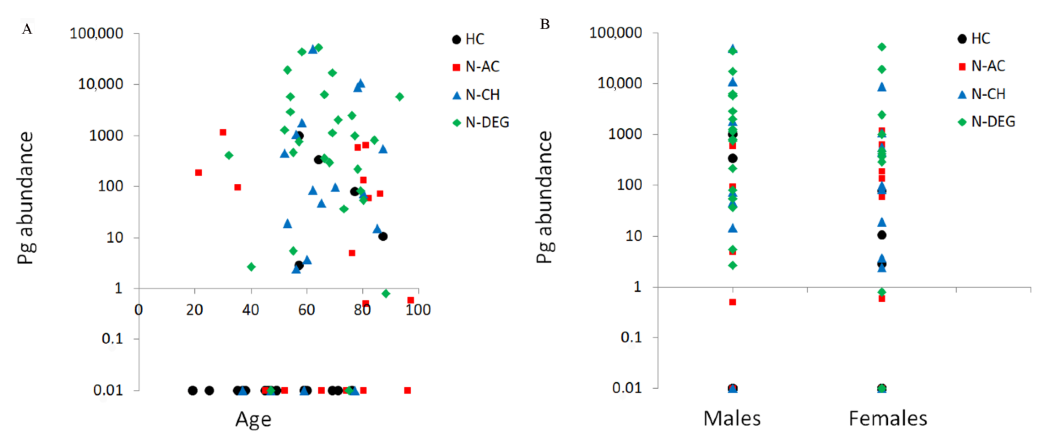
Spearman’s correlation showed a significant positive correlation between the abundance of Pg and the anti-Pg quantity (ρ = 0.61, p = 0.0001). A significant correlation was also found between the Pg abundance, age (ρ = 0.24, p = 0.029), and sex (ρ = 0.21, p = 0.047). These correlations are shown in Figure 2. The abundance of Pg in the oral cavity increased with age and in males. A significant correlation between age and Pg abundance was found for the HC group only (ρ = 0.47, p = 0.025). Indeed, the N-AC, N-CH, and N-DEG groups showed no significant correlations between Pg abundance and age (ρ = −0.13, p = 0.62; ρ = 0.34, p = 0.15; and ρ = −0.11, p = 0.59, respectively). No significant correlation was found between Pg abundance and sex at the group level (ρ = 0.11, p = 0.60 for HC; ρ = 0.24, p = 0.35 for N-AC; ρ = 0.28, p = 0.26 for N-CH; and ρ = 0.11, p = 0.59 for N-DEG). No significant correlation was found between the abundance of Pg and smoking (p = 0.35), prior smoking (p = 0.99), teeth number (p = 0.44), number of fixed prostheses (p = 0.54), and number of removable dentures (p = 0.22). No significant correlation was found between anti-Pg antibodies’ quantity and age (p = 0.90), sex (p = 0.07), smoking (p = 0.54), or former smoking (p = 0.53).
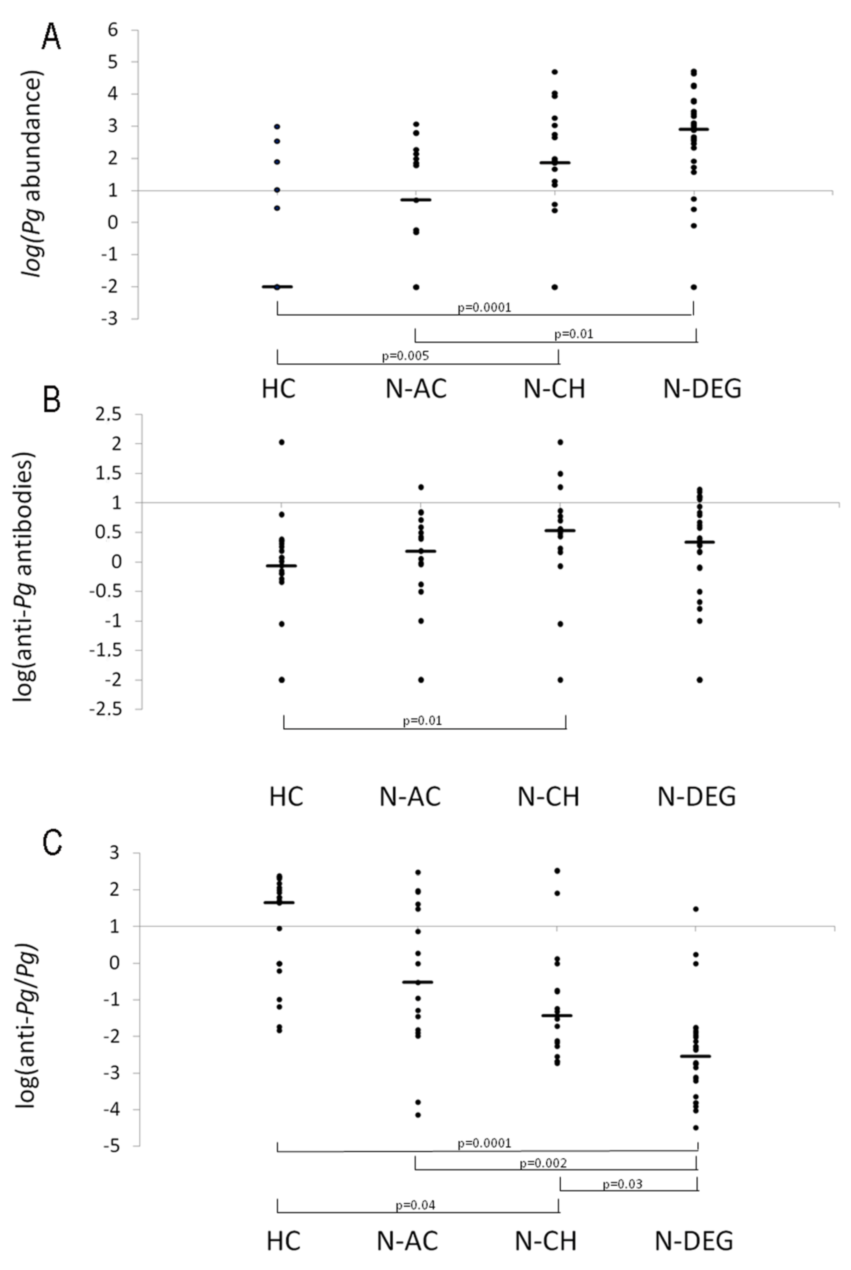
Non-parametric Kruskal–Wallis tests were performed to compare the Pg bacteria and antibody quantities among the groups. Specifically, logarithmic transformations were applied to the Pg abundance value, anti-Pg antibodies’ quantity, and the ratio between anti-Pg and Pg to respect the homogeneity of the variance among the groups, as assessed by Levene’s test. The Kruskal–Wallis H test showed that the log abundance of Pg in the oral cavity was significantly different among the groups (χ2(3) = 31.9, p = 0.0001), with a mean rank value of 23.1 for the HC, 38.2 for the N-AC, 48.8 for the N-CH, and 61.4 for the N-DEG groups. The post hoc test showed that the Pg abundance in the oral cavity was higher in the N-DEG group than the HC (p = 0.0001) and N-AC (p = 0.01) groups, and it was also higher in the N-CH group than the HCs (p = 0.005).
The log of anti-Pg antibodies was significantly different among the groups (χ2(3) = 10.0, p = 0.020), with mean rank values of 31.8, 42.8, 55.9, and 46.7 for the HC, N-AC, N-CH, and N-DEG groups, respectively. The post hoc test showed that the anti-Pg antibodies in the serum were higher in the N-CH group than the HCs (p = 0.012).
The log of the ratio between the anti-Pg antibodies’ quantity and the Pg abundance was significantly different among the groups (χ2(3) = 37.5, p = 0.0001), with mean rank values of 65.4 for the HC, 50.4 for the N-AC, 43.8 for the N-CH, and 22.7 for the N-DEG groups. The post hoc test showed that the log of the ratio between the anti-Pg antibodies’ quantity and the Pg abundance was lower in the N-DEG group than the HC (p = 0.0001), N-AC (p = 0.002), and N-CH (p = 0.03) groups, and it was also lower in the N-CH group than the HCs (p = 0.04).
Figure 3 shows the scatterplots, the medians, and the significant results of the Pg abundance logarithm, the anti-Pg antibodies’ logarithm, and the logarithm of the ratio between the anti-Pg antibodies and Pg abundance for each group.

4. Discussion

This study showed that the Pg abundance in the oral cavity was higher in people with chronic neurological conditions than in the HCs or people affected by an acute neurological condition. Previous studies have shown that the oral microbiota composition originating from buccal and sublingual mucosa and salivary samples differs between healthy subjects and PD patients [45,46,47,48]. Saliva represents a collection of the microbiota from all the ecological niches in the oral cavity and is easy to collect. However, it has the major disadvantage of lacking specificity compared to samples collected from a single oral site. The use of brushes allows for a noninvasive, simple, and repeatable way of managing abundant site-specific microbiota [49,50]. Unlike the salivary microbiota, the supragingival dental microbiota are subject to more variation, probably due to brushing habits [51].
Periodontal pathogens, such as Pg and Treponema denticola, are increased in subjects with cognitive impairment or AD [52,53]. The action of accessory pathogens could facilitate Pg colonization, and commensal-turned pathobionts could contribute to inflammation [3]. It has been suggested that oral dysbiosis is unlikely to result from dental or periodontal complications in patients affected by cognitive impairment or AD. Instead, it could be a primary event related to the pathogenesis of neurodegenerative diseases [54]. The mechanism by which bacteria cross the blood–brain barrier remains unclear. A possible explanation may lie in gingipains, the class of Pg cysteine proteases. A Pg increase in neurological patients may lead to a higher delivery of gingipains, which are involved in the degradation of the tight junction proteins of cerebral microvascular endothelial cells (i.e., Zonula occludens-1 and occluding), promoting damage to the blood–brain barrier [55]. Pg produces two forms of gingipains: the free form and the outer membrane vesicles (OMVs)-associated form. Probably, via the OMVs, gingipains may be transported and released, increasing the permeability of human cerebral microvascular endothelial cells [42].
In our study, Pg abundance was also increased with age, confirming previous findings [56]. However, the analysis of single groups indicated that the correlation between Pg abundance and age was significant for the HCs only. This result suggests that neurological conditions bother the relationship between age and the abundance of Pg bacteria in the oral cavity. The sex effect could have been related to the greater number of males in the N-DEG group, which could have biased the result toward an increased abundance of Pg in males compared to females. Indeed, when the correlation analyses were performed on single groups, the correlation between Pg abundance and sex was lost.
A significant positive correlation was found between the abundance of Pg in the oral cavity and the quantity of anti-Pg antibodies in the serum of all the participants, confirming the hypothesis that oral bacteria can cause the production of anti-Pg antibodies, as previously reported [17,57]. In addition, this relationship confirms that the number of bacteria is also influenced by the immune system’s ability to manage the host’s state [58].
Interestingly, despite the higher amount of Pg in the N-DEG group, the quantity of anti-Pg antibodies was not higher in this group than the others. The inadequate response of the immune system of the N-DEG group in producing anti-Pg antibodies was revealed by the analysis of the ratio between the anti-Pg antibodies’ quantity and the Pg abundance. Thus, a low level of antibodies could result in a chronic inflammatory state inside and outside the oral cavity, which might be involved in the processes of neurodegeneration. Elevated IgG antibodies against periodontal disease bacteria have been found in subjects years before cognitive impairment, whereas IgG against Pg was decreased in patients who developed AD [15]. Chronic inflammation is an important factor in neurodegeneration, causing the dysregulation of circulating inflammatory molecules and the innate immune response [59,60]. Thereby, in turn, the LPS and gingipain of Pg can regulate immune responses by creating an inflammatory environment through the stimulation of TLR2-PI3K-mediated signaling, which results in a reduction in bactericidal activity, an increase in proinflammatory cytokines (e.g., IL-1β, IL-6, and TNF-α), and the inhibition of phagosome formation and maturation [61].
The N-CH group showed a higher quantity of anti-Pg antibodies than the HCs. However, the amount of these antibodies was inadequate compared to the HCs, as indicated by the significant difference between the N-CH and HC groups in the ratio between the anti-Pg antibodies and Pg. However, this ratio was higher in the N-CH than in the N-DEG group, suggesting that the low production of anti-Pg antibodies with a high abundance of Pg in the oral cavity was related more to neurodegeneration than to chronic neurological disease. On the contrary, the patients affected by an acute neurological condition (i.e., ischemic stroke or head trauma) included in the N-AC group showed no significant difference compared to the HC group. Indeed, the HC and N-AC groups showed higher antibodies in the presence of a low abundance of Pg in the oral cavity.
The accumulation of bacterial plaques by periodontopathogen bacteria, in addition to cigarette smoking, is the most important risk factor in causing an inflammatory response [62,63]. To reduce the bacterial load of the orange and red complex and to avoid consequent tissue damage, ozone-based therapies have been proposed in recent years and have been tested as antimicrobial therapy. Ozone produces free radicals that penetrate inside bacterial DNA and RNA, destroying their structure, thus accelerating curative action [64,65]. Ozone therapy as an adjunct to mechanical therapy reduces the gingival index in generalized chronic periodontitis patients [66].
Other therapeutic approaches include prebiotics, probiotics, paraprobiotics, and post-biotics. An example of probiotic therapy is Lactobacillus reuteri, which can reduce the amount of periodontopathogens and proinflammatory cytokines [55,67]. All these therapies exert an immunomodulatory and anti-inflammatory action on the periodontal tissues due to their antioxidant action [68,69,70].
The main limitation of our study is its relatively small sample size and the heterogeneity of the patient groups. Unfortunately, a considerable amount of participants were excluded or dropped out and the recruitment could not continue. Thus, we could not draw definitive conclusions about this oral bacterium’s influence and the altered immune response in the pathogenesis of neurodegenerative diseases. Future follow-up studies could monitor the microbiota profile and the immune response over time and at different stages of disease to investigate the influence of periodontopathogen bacteria on the onset and progression of neurological and neurodegenerative diseases.
In conclusion, the response of the immune system to Pg abundance in the oral cavity, assessed by the quantification of anti-Pg antibodies in the serum, showed a stepwise model. The immune system response decreased progressively in patients affected by an acute condition such as ischemic stroke or head trauma, less in patients with epilepsy or peripheral nervous system disorders, and finally, even less in patients affected by neurodegenerative diseases such as Parkinson’s or multiple sclerosis.

Supplementary Materials

The following supporting information can be downloaded at: https://www.mdpi.com/article/10.3390/microorganisms11102555/s1, Table S1: STROBE Statement—checklist of items that should be included in reports of observational studies.

Author Contributions

Conceptualization, R.F. and M.C.C.; methodology, D.L.D., R.M., S.F., V.P. and M.C.C.; formal analysis, R.F.; investigation, M.A.D.R. and P.P.; writing—original draft preparation, R.F., S.L.S. and P.P.; writing—review and editing, R.M., S.F., V.P., A.P., A.M.T., S.L.S. and M.C.C.; visualization, R.F.; supervision, R.F. and M.O. All authors have read and agreed to the published version of the manuscript.

Funding

This research received no external funding.

Institutional Review Board Statement

The study was approved by the local Ethics Committee of the “G. d’Annunzio” University of Chieti-Pescara on May 2020 (ethics code: 1909).

Informed Consent Statement

Not applicable.

Data Availability Statement

The data supporting this study’s findings are available upon reasonable request.

Conflicts of Interest

The authors declare no conflict of interest.

References

  1. Marcotte, H.; Lavoie, M.C. Oral microbial ecology and the role of salivary immunoglobulin A. Microbiol. Mol. Biol. Rev. 1998, 62, 71–109. [Google Scholar] [PubMed]
  2. Aas, J.A.; Paster, B.J.; Stokes, L.N.; Olsen, I.; Dewhirst, F.E. Defining the normal bacterial flora of the oral cavity. J. Clin. Microbiol. 2005, 43, 5721–5732. [Google Scholar]
  3. Hajishengallis, G.; Lamont, R.J. Breaking bad: Manipulation of the host response by Porphyromonas gingivalis. Eur. J. Immunol. 2014, 44, 328–338. [Google Scholar] [PubMed]
  4. Xu, W.; Zhou, W.; Wang, H.; Liang, S. Roles of Porphyromonas gingivalis and its virulence factors in periodontitis. Adv. Protein Chem. Struct. Biol. 2020, 120, 45–84. [Google Scholar] [PubMed]
  5. Olsen, I.; Lambris, J.D.; Hajishengallis, G. Porphyromonas gingivalis disturbs host-commensal homeostasis by changing complement function. J. Oral Microbiol. 2017, 9, 1340085. [Google Scholar] [CrossRef]
  6. Wang, W.; Zheng, C.; Yang, J.; Li, B. Intersection between macrophages and periodontal pathogens in periodontitis. J. Leukoc. Biol. 2021, 110, 577–583. [Google Scholar]
  7. Meka, A.; Bakthavatchalu, V.; Sathishkumar, S.; Lopez, M.C.; Verma, R.K.; Wallet, S.; Bhattacharyya, I.; Boyce, B.F.; Handfield, M.; Lamont, R.J.; et al. Porphyromonas gingivalis infection-induced tissue and bone transcriptional profiles. Mol. Oral Microbiol. 2010, 25, 61–74. [Google Scholar] [PubMed]
  8. Vernal, R.J.; Díaz-Zúñiga, J.; Melgar-Rodríguez, S.; Pujol, M.; Diaz-Guerra, E.; Silva, A.; Sanz, M.; Garcia-Sanz, J.A. Activation of RANKL-induced osteoclasts and memory T lymphocytes by Porphyromonas gingivalIs is serotype-dependant. J. Clin. Periodontol. 2014, 41, 451–459. [Google Scholar] [PubMed]
  9. Jack, C.; Yu, H.K.; Baban, B. Innate immunity and oral microbiome: A personalized, predictive, and preventive approach to the management of oral diseases. EPMA J. 2019, 10, 43–50. [Google Scholar]
  10. Aoki, S.; Hosomi, N.; Nishi, H.; Nakamori, M.; Nezu, T.; Shiga, Y.; Kinoshita, N.; Ueno, H.; Ishikawa, K.; Imamura, E.; et al. Serum IgG Titers to Periodontal Pathogens Predict 3-Month Outcome in Ischemic Stroke Patients. PLoS ONE 2020, 15, e0237185. [Google Scholar]
  11. Aoyama, N.; Kure, K.; Minabe, M.; Izumi, Y. Increased Heart Failure Prevalence in Patients with a High Antibody Level Against Periodontal Pathogen. Int. Heart J. 2019, 60, 1142–1146. [Google Scholar]
  12. Pussinen, P.J.; Alfthan, G.; Rissanen, H.; Reunanen, A.; Asikainen, S.; Knekt, P. Antibodies to Periodontal Pathogens and Stroke Risk. Stroke 2004, 35, 2020–2023. [Google Scholar] [PubMed]
  13. Kamer, A.R.; Craig, R.G.; Pirraglia, E.; Dasanayake, A.P.; Norman, R.G.; Boylan, R.J.; Nehorayoff, A.; Glodzik, L.; Brys, M.; de Leon, M.J. TNF-Alpha and Antibodies to Periodontal Bacteria Discriminate between Alzheimer’s Disease Patients and Normal Subjects. J. Neuroimmunol. 2009, 216, 92–97. [Google Scholar]
  14. Noble, J.M.; Borrell, L.N.; Papapanou, P.N.; Elkind, M.S.V.; Scarmeas, N.; Wright, C.B. Periodontitis Is Associated with Cognitive Impairment among Older Adults: Analysis of NHANES-III. J. Neurol. Neurosurg. Psychiatry 2009, 80, 1206–1211. [Google Scholar]
  15. Sparks Stein, P.; Steffen, M.J.; Smith, C.; Jicha, G.; Ebersole, J.L.; Abner, E.; Dawson, D. Serum Antibodies to Periodontal Pathogens Are a Risk Factor for Alzheimer’s Disease. Alzheimers Dement. 2012, 8, 196–203. [Google Scholar] [PubMed]
  16. Zhang, J.; Yu, C.; Zhang, X.; Chen, H.; Dong, J.; Lu, W.; Song, Z.; Zhou, W. Porphyromonas gingivalis Lipopolysaccharide Induces Cognitive Dysfunction, Mediated by Neuronal Inflammation via Activation of the TLR4 Signaling Pathway in C57BL/6 Mice. J. Neuroinflamm. 2018, 15, 37. [Google Scholar]
  17. Franciotti, R.; Pignatelli, P.; Carrarini, C.; Romei, F.M.; Mastrippolito, M.; Gentile, A.; Mancinelli, R.; Fulle, S.; Piattelli, A.; Onofrj, M.; et al. Exploring the Connection between Porphyromonas gingivalis and Neurodegenerative Diseases: A Pilot Quantitative Study on the Bacterium Abundance in Oral Cavity and the Amount of Antibodies in Serum. Biomolecules 2021, 11, 845. [Google Scholar]
  18. Berbari, E.F.; Cockerill, F.R.; Steckelberg, J.M. Infective endocarditis due to unusual or fastidious microorganisms. Mayo Clin. Proc. 1997, 72, 532–542. [Google Scholar] [PubMed]
  19. Scannapieco, F.A. Role of oral bacteria in respiratory infection. J. Periodontol. 1999, 70, 793–802. [Google Scholar] [PubMed]
  20. Dodman, T.; Robson, J.; Pincus, D. Kingella kingae infections in children. J. Paediatr. Child Health 2000, 36, 87–90. [Google Scholar] [PubMed]
  21. Buduneli, N.; Baylas, H.; Buduneli, E.; Turkoglu, O.; Kose, T.; Dahlen, G. Periodontal infections and pre-term low birth weight: A case-control study. J. Clin. Periodontol. 2005, 32, 174–181. [Google Scholar] [PubMed]
  22. Idris, A.; Hasnain, S.Z.; Huat, L.Z.; Koh, D. Human diseases, immunity and the oral microbiota—Insights gained from metagenomic studies. Oral Sci. Int. 2017, 14, 27–32. [Google Scholar]
  23. Wu, T.; Trevisan, M.; Genco, R.J.; Dorn, J.P.; Falkner, K.L.; Sempos, C.T. Periodontal disease and risk of cerebrovascular disease: The first national health and nutrition examination survey and its follow-up study. Arch. Intern. Med. 2000, 160, 2749–2755. [Google Scholar] [PubMed]
  24. Kharlamova, N.; Sherina, N.; Quirke, A.M.; Eriksson, K.; Israelsson, L.; Potempa, J.; Venables, P.; Lindberg, T.L.; Lundberg, K. A6.8 Elevated antibody levels to Porphyromonas gingivalis detected in rheumatoid arthritis patients with a specific anticitrullinated protein/peptide antibody profile. Ann. Rheum. Dis. 2014, 73, A73–A74. [Google Scholar]
  25. Contaldo, M.; Itro, A.; Lajolo, C.; Gioco, G.; Inchingolo, F.; Serpico, R. Overview on osteoporosis, periodontitis and oral dysbiosis: The emerging role of oral microbiota. Appl. Sci. 2020, 10, 6000. [Google Scholar]
  26. Li, X.; Kolltveit, K.M.; Tronstad, L.; Olsen, I. Systemic diseases caused byoral infection. Clin. Microbiol. Rev. 2000, 13, 547–558. [Google Scholar]
  27. Cunha, F.A.; Cota, L.O.M.; Cortelli, S.C.; Miranda, T.B.; Neves, F.S.; Cortelli, J.R.; Costa, F.O. Periodontal condition and levels of bacteria associated with periodontitis in individuals with bipolar affective disorders: A case-control study. J. Periodontal Res. 2019, 54, 63–72. [Google Scholar]
  28. Yang, I.; Arthur, R.A.; Zhao, L.; Clark, J.; Hu, Y.; Corwin, E.J.; Lah, J. The oral microbiome and inflammation in mild cognitive impairment. Exp. Gerontol. 2021, 147, 111273. [Google Scholar]
  29. Scassellati, C.; Marizzoni, M.; Cattane, N.; Lopizzo, N.; Mombelli, E.; Riva, M.A.; Cattaneo, A. The Complex Molecular Picture of Gut and Oral Microbiota-Brain-Depression System: What We Know and What We Need to Know. Front. Psychiatry 2021, 12, 722335. [Google Scholar]
  30. Park, S.Y.; Hwang, B.O.; Lim, M.; Ok, S.H.; Lee, S.K.; Chun, K.S.; Park, K.K.; Hu, Y.; Chung, W.Y.; Song, N.Y. Oral-gut microbiome axis in gastrointestinal disease and cancer. Cancers 2021, 13, 2124. [Google Scholar]
  31. Narengaowa Kong, W.; Lan, F.; Awan, U.F.; Qing, H.; Ni, J. The oral-gut-brain AXIS: The influence of microbes in Alzheimer’s disease. Front. Cell. Neurosci. 2021, 15, 633735. [Google Scholar]
  32. Dogra, N.; Mani, R.J.; Katare, D.P. The Gut-Brain Axis: Two Ways Signaling in Parkinson’s Disease. Cell. Mol. Neurobiol. 2022, 42, 315–332. [Google Scholar]
  33. Tan, A.H.; Lim, S.Y.; Lang, A.E. The microbiome-gut-brain axis in Parkinson disease-from basic research to the clinic. Nat. Rev. Neurol. 2022, 18, 476–495. [Google Scholar]
  34. Tremlett, H.; Bauer, K.C.; Appel-Cresswell, S.; Finlay, B.B.; Waubant, E. The gut microbiome in human neurological disease: A review. Ann. Neurol. 2017, 81, 369–382. [Google Scholar] [PubMed]
  35. Bowland, G.B.; Weyrich, L.S. The Oral-Microbiome-Brain Axis and Neuropsychiatric Disorders: An Anthropological Perspective. Front. Psychiatry 2022, 13, 810008. [Google Scholar] [PubMed]
  36. Liu, Y.; Wu, Z.; Zhang, X.; Ni, J.; Yu, W.; Zhou, Y.; Nakanishi, H. Leptomeningeal cells transduce peripheral macrophages inflammatory signal to microglia in reponse to Porphyromonas gingivalis LPS. Mediat. Inflamm. 2013, 2013, 407562. [Google Scholar]
  37. Poole, S.; Singhrao, S.K.; Kesavalu, L.; Curtis, M.A.; Crean, S. Determining the presence of periodontopathic virulence factors in short-term postmortem Alzheimer’s disease brain tissue. J. Alzheimers Dis. 2013, 36, 665–677. [Google Scholar] [PubMed]
  38. Beydoun, M.A.; Beydoun, H.A.; Hossain, S.; El-Hajj, Z.W.; Weiss, J.; Zonderman, A.B. Clinical and bacterial markers of periodontitis and their association with incident all-cause and Alzheimer’s disease dementia in a large national survey. J. Alzheimer’s Dis. 2020, 75, 157–172. [Google Scholar]
  39. Horsager, J.; Andersen, K.B.; Knudsen, K.; Skjærbæk, C.; Fedorova, T.D.; Okkels, N.; Schaeffer, E.; Bonkat, S.K.; Geday, J.; Otto, M.; et al. Brain-first versus body-first Parkinson’s disease: A multimodal imaging case-control study. Brain 2020, 143, 3077–3088. [Google Scholar]
  40. Scribante, A.; Butera, A.; Alovisi, M. Customized Minimally Invasive Protocols for the Clinical and Microbiological Management of the Oral Microbiota. Microorganisms 2022, 10, 675. [Google Scholar] [CrossRef]
  41. Löe, H. The Gingival Index, the Plaque Index and the Retention Index Systems. J. Periodontol. 1967, 38, 610–616. [Google Scholar] [CrossRef]
  42. Nonaka, S.; Kadowaki, T.; Nakanishi, H. Secreted gingipains from Porphyromonas gingivalis increase permeability in human cerebral microvascular endothelial cells through intracellular degradation of tight junction proteins. Neurochem. Int. 2022, 154, 105282. [Google Scholar] [PubMed]
  43. Pignatelli, P.; Iezzi, L.; Pennese, M.; Raimondi, P.; Cichella, A.; Bondi, D.; Grande, R.; Cotellese, R.; Di Bartolomeo, N.; Innocenti, P.; et al. The Potential of Colonic Tumor Tissue Fusobacterium nucleatum to Predict Staging and Its Interplay with Oral Abundance in Colon Cancer Patients. Cancers 2021, 13, 1032. [Google Scholar]
  44. Waritani, T.; Chang, J.; McKinney, B.; Terato, K. An ELISA Protocol to Improve the Accuracy and Reliability of Serological Antibody Assays. MethodsX 2017, 4, 153–165. [Google Scholar]
  45. Li, Z.; Lu, G.; Luo, E.; Wu, B.; Li, Z.; Guo, J.; Xia, Z.; Zheng, C.; Su, Q.; Zeng, Y.; et al. Oral, Nasal, and Gut Microbiota in Parkinson’s Disease. Neuroscience 2022, 480, 65–78. [Google Scholar]
  46. Mihaila, D.; Donegan, J.; Barns, S.; LaRocca, D.; Du, Q.; Zheng, D.; Vidal, M.; Neville, C.; Uhlig, R.; Middleton, F.A. The oral microbiome of early stage Parkinson’s disease and its relationship with functional measures of motor and non-motor function. PLoS ONE 2019, 14, e0218252. [Google Scholar]
  47. Nowak, J.M.; Kopczyński, M.; Friedman, A.; Koziorowski, D.; Figura, M. Microbiota Dysbiosis in Parkinson Disease-In Search of a Biomarker. Biomedicines 2022, 10, 2057. [Google Scholar]
  48. Pereira, P.A.B.; Aho, V.T.E.; Paulin, L.; Pekkonen, E.; Auvinen, P.; Scheperjans, F. Oral and nasal microbiota in Parkinson’s disease. Park. Relat. Disord. 2017, 38, 61–67. [Google Scholar]
  49. Vogtmann, E.; Chen, J.; Kibriya, M.G.; Amir, A.; Shi, J.; Chen, Y.; Islam, T.; Eunes, M.; Ahmed, A.; Naher, J.; et al. Comparison of Oral Collection Methods for Studies of Microbiota. Cancer Epidemiol. Biomark. Prev. 2019, 28, 137–143. [Google Scholar]
  50. Fan, X.; Peters, B.A.; Min, D.; Ahn, J.; Hayes, R.B. Comparison of the oral microbiome in mouthwash and whole saliva samples. PLoS ONE 2018, 13, e0194729. [Google Scholar]
  51. Noiri, Y.; Ozaki, K.; Nakae, H.; Matsuo, T.; Ebisu, S. An immunohistochemical study on the localization of Porphyromonas gingivalis, Campylobacter rectus and Actinomyces viscosus in human periodontal pockets. J. Periodontal Res. 1997, 32, 598–607. [Google Scholar] [CrossRef] [PubMed]
  52. Bathini, P.; Foucras, S.; Dupanloup, I.; Imeri, H.; Perna, A.; Berruex, J.-L.; Doucey, M.-A.; Annoni, J.-M.; Alberi, L.A. Classifying dementia progression using microbial profiling of saliva. Alzheimers Dement. (Amst.) 2020, 12, e12000. [Google Scholar] [CrossRef] [PubMed]
  53. Holmer, J.; Aho, V.; Eriksdotter, M.; Paulin, L.; Pietiäinen, M.; Auvinen, P.; Schultzberg, M.; Pirkko, J.; Pussinen, P.J.; Buhlin, K. Subgingival microbiota in a population with and without cognitive dysfunction. J. Oral Microbiol. 2021, 13, 1854552. [Google Scholar] [CrossRef] [PubMed]
  54. Fleury, V.; Zekeridou, A.; Lazarevic, V.; Gaïa, N.; Giannopoulou, C.; Genton, L.; Cancela, J.; Girard, M.; Goldstein, R.; Bally, J.F.; et al. Oral Dysbiosis and Inflammation in Parkinson’s Disease. J. Park. Dis. 2021, 11, 619–631. [Google Scholar] [CrossRef]
  55. Joint Food and Agriculture Organization (FAO); World Health Organization (WHO) Working Group. Working Group Report on Drafting Guidelines for the Evaluation of Probiotics in Food; World Health Organization: London, ON, Canada, 2002.
  56. Zeller, I.; Hutcherson, J.A.; Lamont, R.J.; Demuth, D.R.; Gumus, P.; Nizam, N.; Buduneli, N.; Scott, D.A. Altered antigenic profiling and infectivity of Porphyromonas gingivalis in smokers and non-smokers with periodontitis. J. Periodontol. 2014, 85, 837–844. [Google Scholar] [CrossRef] [PubMed]
  57. Dominy, S.S.; Lynch, C.; Ermini, F.; Benedyk, M.; Marczyk, A.; Konradi, A.; Nguyen, M.; Haditsch, U.; Raha, D.; Griffin, C.; et al. Porphyromonas gingivalis in Alzheimer’s Disease Brains: Evidence for Disease Causation and Treatment with Small-Molecule Inhibitors. Sci. Adv. 2019, 5, eaau3333. [Google Scholar] [CrossRef]
  58. Cho, T.; Nagao, J.-I.; Imayoshi, R.; Tanaka, Y. Importance of diversity in the oral microbiota including candida species revealed by high-throughput technologies. Int. J. Dent. 2014, 2014, 454391. [Google Scholar] [CrossRef]
  59. Kannarkat, G.T.; Boss, J.M.; Tansey, M.G. The Role of Innate and Adaptive Immunity in Parkinson’s Disease. J. Park. Dis. 2013, 3, 493–514. [Google Scholar] [CrossRef]
  60. Mou, Y.; Du, Y.; Zhou, L.; Yue, J.; Hu, X.; Liu, Y.; Chen, S.; Lin, X.; Zhang, G.; Xiao, H.; et al. Gut Microbiota Interact with the Brain Through Systemic Chronic Inflammation: Implications on Neuroinflammation, Neurodegeneration, and Aging. Front. Immunol. 2022, 13, 796288. [Google Scholar] [CrossRef]
  61. Boeri, L.; Perottoni, S.; Izzo, L.; Giordano, C.; Albani, D. Microbiota-Host Immunity Communication in Neurodegenerative Disorders: Bioengineering Challenges for In Vitro Modeling. Adv. Healthc. Mater. 2021, 10, e2002043. [Google Scholar] [CrossRef] [PubMed]
  62. Kinane, D.F.; Chestnutt, I.G. Smoking and periodontal disease. Crit. Rev. Oral Biol. Med. 2000, 11, 356–365. [Google Scholar] [CrossRef] [PubMed]
  63. Marsh, P.D. Dental plaque: Biological significance of a biofilm and community life-style. J. Clin. Periodontol. 2005, 32, 7–15. [Google Scholar] [CrossRef]
  64. Arita, M.; Nagayoshi, M.; Fukuizumi, T.; Okinaga, T.; Masumi, S.; Morikawa, M.; Kakinoki, Y.; Nishihara, T. Microbicidal efficacy of ozonated water against Candida albicans adhering to acrylic denture plates. Oral Microbiol. Immunol. 2005, 20, 206–210. [Google Scholar] [CrossRef]
  65. Smith, N.L.; Wilson, A.L.; Gandhi, J.; Vatsia, S.; Khan, S.A. Ozone therapy: An overview of pharmacodynamics, current research, and clinical utility. Med. Gas Res. 2017, 7, 212–219. [Google Scholar]
  66. Ramirez-Peña, A.M.; Sánchez-Pérez, A.; Campos-Aranda, M.; Hidalgo-Tallón, F.J. Ozone in Patients with Periodontitis: A Clinical and Microbiological Study. J. Clin. Med. 2022, 11, 2946. [Google Scholar] [CrossRef] [PubMed]
  67. Szkaradkiewicz, A.K.; Stopa, J.; Karpiński, T.M. Effect of oral administration involving a probiotic strain of Lactobacillus reuteri on pro- inflammatory cytokine response in patients with chronic periodontitis. Arch. Immunol. Ther. Exp. 2014, 62, 495–500. [Google Scholar] [CrossRef]
  68. Żółkiewicz, J.; Marzec, A.; Ruszczyński, M.; Feleszko, W. Postbiotics-A Step Beyond Pre- and Probiotics. Nutrients 2020, 12, 2189. [Google Scholar] [CrossRef]
  69. Ince, G.; Gürsoy, H.; Ipçi, S.D.; Cakar, G.; Emekli-Alturfan, E.; Yılmaz, S. Clinical and Biochemical Evaluation of Lozenges Containing Lactobacillus reuteri as an Adjunct to Non-Surgical Periodontal Therapy in Chronic Periodontitis. J. Periodontol. 2015, 86, 746–754. [Google Scholar] [CrossRef] [PubMed]
  70. Butera, A.; Gallo, S.; Maiorani, C.; Preda, C.; Chiesa, A.; Esposito, F.; Pascadopoli, M.; Scribante, A. Management of Gingival Bleeding in Periodontal Patients with Domiciliary Use of Toothpastes Containing Hyaluronic Acid, Lactoferrin, or Paraprobiotics: A Randomized Controlled Clinical Trial. Appl. Sci. 2021, 11, 8586. [Google Scholar] [CrossRef]
Figure 1. Flowchart of the study cohort.
Microorganisms 11 02555 g001
Figure 2. Pg abundance in logarithmic scale as a function of age (A) and sex (B).
Microorganisms 11 02555 g002
Figure 3. Scatterplot and significant results of the Pg abundance logarithm (A), the anti-Pg antibodies’ logarithm (B), and the logarithm of the ratio between anti-Pg antibodies and Pg abundance (C) for all the groups. Horizontal bars indicate the median value for each group.
Microorganisms 11 02555 g003
Table 1. Demographic characteristics and oral indices of all participants of the groups included in the study. Otherwise, the number of patients included in the calculation is shown in parentheses.
HC
(n = 23)
N-AC
(n = 17)
N-CH
(n = 19)
N-DEG
(n = 28)
Age51.9 ± 3.868.2 ± 5.664.4 ± 3.165.5 ± 2.7
Sex (% male)34.835.342.160.7
Smoker (%)
Former smoker (%)
4.3
13.0
11.8
17.6
6.3 (n = 16)
31.3 (n = 16)
12.5 (n = 24)
17.5 (n = 24)
Teeth number 24.4 ± 1.8 (n = 20)13.8 ± 3.0 (n = 16)17.2 ± 2.2 (n = 15)16.8 ± 1.9 (n = 26)
Plaque index 1.6 ± 0.4 (n = 5)0.1 ± 0.0 (n = 1)2.2 ± 0.2 (n = 7)2.1 ± 0.3 (n = 8)
Gingival index 0.8 ± 0.6 (n = 5)0.0 ± 0.0 (n = 1)1.1 ± 0.2(n = 7)1.1 ± 0.3(n = 7)
Presence of gingivitis (%) 60.0 (n = 5)60.0 (n = 10)75.0 (n = 12)86.7 (n = 15)
Lingual patina index 1.2 ± 0.5 (n = 5)0.8 ± 0.2 (n = 10)0.8 ± 0.3 (n = 12)1.9 ± 0.3 (n = 15)
Presence of oral infection (%)60.0 (n = 5)40.0 (n = 10)66.7 (n = 11)80.0 (n = 14)
Oral hygiene index 40.0 (n = 5)77.8 (n = 9)36.4 (n = 11)14.3 (n = 14)
Presence of fissured tongue (%)0.0 (n = 5)20.0 (n = 10)18.2 (n = 11)0.0 (n = 15)
Number of fixed prostheses 0.2 ± 0.1 (n = 20)0.3 ± 0.1 (n = 16)0.6 ± 0.2 (n = 14)0.4 ± 0.1 (n = 25)
Number of removable dentures 0.2 ± 0.1 (n = 19)0.7 ± 0.3 (n = 14)0.3 ± 0.2 (n = 14)0.3 ± 0.1 (n = 24)
Values are expressed as mean ± standard error.
Table 2. Pg abundance and anti-Pg antibodies’ quantity of all participants of the groups included in the study.
HC (n = 23)N-AC (n = 17)N-CH (n = 19)N-DEG (n = 28)
Pg abundance (CFU/mL)63.4 ± 46.1178.4 ± 80.93961.4 ± 2698.86013.3 ± 2488.3
Anti-Pg antibodies (units/mL)5.8 ± 4.63.2 ± 1.110.9 ± 5.64.7 ± 1.0
Anti-Pg/Pg (units/CFU)69.5 ± 17.834.2 ± 18.940.1 ± 24.41.2 ± 1.1
Values are expressed as mean ± standard error.
Disclaimer/Publisher’s Note: The statements, opinions and data contained in all publications are solely those of the individual author(s) and contributor(s) and not of MDPI and/or the editor(s). MDPI and/or the editor(s) disclaim responsibility for any injury to people or property resulting from any ideas, methods, instructions or products referred to in the content.