Article

A Robust One-Step Recombineering System for Enterohemorrhagic Escherichia coli

1
Institute of Hygiene, University of Münster, 48149 Münster, Germany
2
Institute of Food Science and Biotechnology, University of Hohenheim, 70599 Stuttgart, Germany
3
National Consulting Laboratory for Hemolytic Uremic Syndrome (HUS), 48149 Münster, Germany
4
Department of Biological Sciences, University at Buffalo, Buffalo, NY 14203, USA
*
Author to whom correspondence should be addressed.
These authors contributed equally to this work.
Microorganisms 2022, 10(9), 1689; https://doi.org/10.3390/microorganisms10091689
Submission received: 14 July 2022 / Revised: 9 August 2022 / Accepted: 18 August 2022 / Published: 23 August 2022
(This article belongs to the Section Food Microbiology)

Abstract

:
Enterohemorrhagic Escherichia coli (EHEC) can cause severe diarrheic in humans. To improve therapy options, a better understanding of EHEC pathogenicity is essential. The genetic manipulation of EHEC with classical one-step methods, such as the transient overexpression of the phage lambda (λ) Red functions, is not very efficient. Here, we provide a robust and reliable method for increasing recombineering efficiency in EHEC based on the transient coexpression of recX together with gam, beta, and exo. We demonstrate that the genetic manipulation is 3–4 times more efficient in EHEC O157:H7 EDL933 Δstx1/2 with our method when compared to the overexpression of the λ Red functions alone. Both recombineering systems demonstrated similar efficiencies in Escherichia coli K-12 MG1655. Coexpression of recX did not enhance the Gam-mediated inhibition of sparfloxacin-mediated SOS response. Therefore, the additional inhibition of the RecFOR pathway rather than a stronger inhibition of the RecBCD pathway of SOS response induction might have resulted in the increased recombineering efficiency by indirectly blocking phage induction. Even though additional experiments are required to unravel the precise mechanistic details of the improved recombineering efficiency, we recommend the use of our method for the robust genetic manipulation of EHEC and other prophage-carrying E. coli isolates.

1. Introduction

Enterohemorrhagic Escherichia coli (EHEC) are zoonotic pathogens that can cause haemorrhagic colitis, haemolytic uremic syndrome (HUS), and eventually death [1,2]. EHEC have caused large foodborne outbreaks with thousands of infections worldwide that were associated with hundreds of hospitalizations and significant numbers of deaths [3,4,5]. The cardinal virulence factor of EHEC are Shiga toxins (Stx) that are encoded in lambdoid Stx prophages which inhibit eukaryotic protein synthesis by cleaving an adenine residue from the 28S ribosomal subunit [6].
All antibiotics were believed to increase stx expression [1], thereby resulting in an increased frequency of HUS [7]. Consequently, there is no causative cure for EHEC infections available. Nonetheless, it is clear that antibiotics that inhibit the bacterial gene expression apparatus do not enhance stx expression in vitro [8,9], and that these antibiotics were beneficial in a mouse infection model [10]. Regardless, the use of these antibiotics in therapy remains controversial.
Given these therapeutic limitations, it is necessary to develop alternative treatment options for EHEC infections. These developments will require a better understanding of the gene regulatory pathways that control pathogen behaviour. Therefore, efficient methods to genetically manipulate EHEC are essential. One of the most frequently applied site-directed mutagenesis methods in Escherichia coli (E. coli) is based on the overexpression of the phage lambda (λ) Red functions [11]. However, we and others had only sporadic success using the transient gam, beta, and exo overexpression system of Datsenko and Wanner [12]. In order to be able to more efficiently genetically modify EHEC, empirical improvements of the method, as well as more time-consuming two-step protocols have been developed [13,14].
We hypothesized that the process of transforming a linear PCR product, which is the essential step in the Datsenko and Wanner gene replacement protocol, may induce the bacterial SOS response. Induction of SOS would be especially problematic for genetically manipulating EHEC carrying Stx-encoding lambdoid Stx prophage, since the autoproteolysis of the CI-like repressors of lambdoid Stx prophages is stimulated by the coprotease activity of RecA, which is activated during the SOS response. Activated RecA [15] stimulated autocleavage of the CI-like repressor results in prophage and stx induction.
The formation of RecA nucleofilament on single-stranded DNA, and thereby activation of RecA depends on the activities of either two enzyme complexes, each of which respond to different types of DNA damage; RecBCD in the case of DNA double-strand breaks and RecFOR in the case of DNA gaps [16]. Neither RecA, nor RecBCD are necessary for recombineering using the λ Red functions [11,17]. Although the activation of RecA via RecBCD should be already sufficiently inhibited by Gam [18], the transformation of PCR products containing nicks or gaps could still activate RecA via the RecFOR pathway, resulting in the overall reduced efficiency of λ Red-based mutagenesis procedures.
The E. coli RecX protein is encoded in an operon with recA. It inhibits the coprotease activities of RecA in vitro and in vivo [19]. It also interacts with RecF [20] and its structure was used as the basis for the design of small molecule inhibitors of the SOS response [21]. We, therefore, decided to test if the transient coexpression of recX together with gam, beta, and exo would increase the frequency of recombinants in EHEC when compared to the gam, beta, and exo expression alone. We demonstrate here that the recX coexpression resulted in a significant 3–4-fold increase in the number of recombinants in the EHEC O157:H7 strain EDL933 Δstx1/2 (EHEC O157:H7 EDL933 Δstx1/2), but not in the E. coli K-12 strain MG1655 (E. coli K-12 MG1655). The inhibition of a sparfloxacin-induced SOS response was not further enhanced in cells expressing recX in addition to gam, suggesting that the observed effect resulted from an inhibition of the RecFOR pathway.

2. Materials and Methods

2.1. Bacterial Strains and Plasmids Used in This Study

The bacterial strains used in this study are listed in Supplementary Table S1 and the plasmids used in this study are listed in Supplementary Table S2.

2.2. Construction of Plasmids and Strains

The plasmid pKD46recX was constructed by amplifying the kanamycin resistance cassette from pKD4 [12] with primers MBP214 and MBP215 with Q5 polymerase (NEB, Ipswich, MA, USA) according to the manufacturer’s instructions. The resulting recombination substrate was integrated 3′ of recX in E. coli K-12 MG1655 with the help of pKD46 by Red recombineering [12]. RecX-kanR was afterwards amplified from the resulting strain by PCR primers MBP216 (which included the gam-beta spacer 5′-TAAAACGA-3′) and MBP217 with Q5 polymerase (NEB, Ipswich, MA, USA). The resulting PCR product was used to generate a recombination substrate with primers MBP218 and MBP219 with Q5 polymerase (NEB, Ipswich, MA, USA). This recombination substrate was afterwards used to transcriptionally fuse recX to exo of pKD46 by Red recombineering [12] in E. coli K-12 MG1655. The correctness of the exo-recX fusion was afterwards verified by Sanger sequencing (Eurofins Scientific SE, Luxembourg). The plasmid was thereafter named pKD46recX.
The plasmid pLP1 was constructed by removing the kanamycin resistance cassette of pKD46recX by digestion with XbaI (NEB, Ipswich, MA, USA) and ligation with T4 DNA ligase (NEB, Ipswich, MA, USA) after gel purification.
The plasmid pLP2 was constructed by amplifying cat of pMB54 [22] with primers MBPD80 (containing a PstI site) and MBPD81 (containing a KpnI site) and Q5 polymerase (NEB, Ipswich, MA, USA) according to the manufacturer’s instructions. The PCR product was afterwards digested with PstI (NEB, Ipswich, MA, USA) and KpnI (NEB, Ipswich, MA, USA) and, after purification, was cloned into a PstI (NEB, Ipswich, MA, USA) and KpnI (NEB, Ipswich, MA, USA) digested pUC18 [23] plasmid with T4 DNA ligase (NEB, Ipswich, MA, USA). The junction of the plasmid and cat was afterwards confirmed by Sanger sequencing (Eurofins Scientific SE, Luxembourg) and the plasmid was named pLP2.
The plasmid pWKSPfrr was constructed by amplifying the promoter of frr (Pfrr) of E. coli K-12 MG1655 with primers MBP272 and MBP273 with Q5 polymerase (NEB, Ipswich, MA, USA) according to the manufacturer’s instructions. The PCR product was afterwards cloned into the SmaI (NEB, Ipswich, MA, USA) digested plasmid pWKS30 [24]. The plasmid was named pWKSfrr.
The plasmid pWKSPfrr-cfp-aph(3′)-Ia was constructed by amplifying cfp (cyan fluorescent protein)-aph(3′)-Ia with primers MBP274 and MBP275 and Q5 polymerase (NEB, Ipswich, MA, USA) and plasmid pMB47 [9] as a template. The fusion to Pfrr in pWKSPfrr was then generated by Red recombineering [12] in E. coli K-12 MG1655. The junction of Pfrr and cfp was confirmed by Sanger sequencing (Eurofins Scientific SE, Luxembourg) and the plasmid was named pWKSPfrr-cfp-aph(3′)-Ia.
E. coli K-12 MG1655 att::Pfrr-cfp-aph(3′)-Ia was constructed by amplifying Pfrr-cfp-aph(3′)-Ia from the plasmid pWKSPfrr-cfp-aph(3′)-Ia with primers MBP276 and MBP277 and Q5 polymerase (NEB, Ipswich, MA, USA) according to the manufacturer’s instructions. The integration of the recombination substrate in the attB site of E. coli K-12 MG1655 was achieved with Red recombineering [12]. The junctions to the chromosome were afterwards verified by PCR with primers MBP280 and MBP273 (5′ junction) and MBP278 and MBP279 (3′ junction). The PCR products were in addition verified by Sanger sequencing (Eurofins Scientific SE, Luxembourg).
E. coli K-12 MG1655 PrecA SENSOR was constructed by amplifying PrecA-yfp (yellow fluorescent protein)-cat with primers MC204 and MC205 and Q5 polymerase (NEB, Ipswich, MA, USA) and pMBM22 [25] as a template. This recombination substrate was integrated convergent to the Pfrr-cfp cassette in E. coli K-12 MG1655 att::Pfrr-cfp-aph(3′)-Ia by Red recombineering [12]. The junction of the chromosome with the PrecA-yfp cassette was confirmed by PCR and the PCR product was verified by Sanger sequencing (Eurofins Scientific SE, Luxembourg). The strain was thereafter named E. coli K-12 MG1655 PrecA SENSOR.

2.3. Comparison of the Recombination Efficiency of pKD46recX with pKD46recX

A detailed protocol for the generation of the recombination substrates and a step-by-step protocol for the preparation of recombination proficient competent cells are provided in the Supplementary Materials. Briefly, pKD46 and pKD46recX were transformed in E. coli K-12 MG1655 and EHEC O157:H7 EDL933 Δstx1/2 and glycerol stocks were made. For each experiment, the strains were plated on LB plates containing ampicillin [100 µg/mL] and incubated at 30 °C overnight. The next day, a single colony was picked and inoculated in 2 mL dYT containing ampicillin [100 µg/mL] and incubated at 180 rpm at 30 °C overnight. The next day, the overnight cultures were diluted 1:200 in 100 mL dYT containing ampicillin [100 µg/mL] and incubated at 180 rpm at 30 °C. When the OD600nm reached 0.4, 10% L-arabinose solution was added to a final concentration (f. c.) of 0.3% and the incubation was continued at 180 rpm at 37 °C for 1 h. Afterwards, the bacteria were chilled on ice, harvested by centrifugation, and washed three times with ice-cold water. The pellet was resuspended in a small volume of ice-cold water and the OD600nm of a 1:100 dilution was determined. Next, the bacterial suspension was adjusted to an OD600nm = 45 − 54. Seventy µL of this suspension was used to transform four aliquots of the indicated amounts of the recombination substrates (stored at −20 °C) by electroporation (Bio-Rad Micropulser, settings 1.8 kV/6 ms). Afterwards, the bacteria were washed out of the cuvette with 1 mL dYT medium and incubated for 1 h at 37 °C with shaking. Afterwards, the bacteria were harvested by centrifugation, resuspended in 100 µL fresh dYT and spread on an LB plate containing chloramphenicol [12.5 µg/mL]. The plates were incubated overnight at 37 °C and the next day the colonies per plate were counted. The technical replicate that deviated the most from the average of colony numbers was excluded and the remaining three technical replicates were used for further evaluation. From these plates, ten randomly chosen colonies were used for the PCR analysis (for the details see the Supplementary Materials). The whole procedure was repeated three times for each recombination substrate.

2.4. Test of pKD46recX in E. coli O104:H4

The plasmid pKD46recX was transformed into electrocompetent E. coli O104:H4 cells and recombination proficient cells were prepared (see Supplementary Materials, step-by-step protocol). In order to replace stx2, a recombination substrate was generated with primers MBP223 and MBP364 and pLP1 as a template using Q5 polymerase (NEB, Ipswich, MA, USA) and purified as described for the other recombination substrates described above. The recombination substrate was transformed in the recombination proficient E. coli O104:H4 cells and after one-hour recovery at 37 °C, the cells were spread on LB agar plates containing chloramphenicol [12.5 µg/mL] and incubated overnight at 37 °C. The replacement of stx2 by cat was afterwards confirmed by PCR with primers MBP226 and MBP260 (5′ junction) and MBP5 and MBP227 (3′ junction) and whole-genome sequencing (PacBio, Menlo Park, CA, USA).

2.5. Measurement of SOS Response

Plasmids pKD46 and pKD46recX were transformed in the SOS response reporter strain E. coli K-12 MG1655 PrecA SENSOR. The bacteria were grown overnight in the M9 medium supplemented with 0.4% glucose containing ampicillin [100 µg/mL] at 30 °C and 180 rpm in an INFORS HT Multitron incubator. The next day, the bacteria were diluted 1 to 10 in M9 medium supplemented with 0.4% arabinose or 0.4% glucose, 0.4% casamino acids and [100 µg/mL ampicillin] (150 µL final volume) in black µ-clear plates with a transparent bottom and lid (Greiner Bio-One, Frickenhausen, Germany) and incubated in a Tecan infinite 2000pro instrument at 30 °C with shaking. Sparfloxacin [15 µg/mL f. c.] was added after 1 h of incubation. Every ten minutes the optical density was automatically recorded at 595 nm and the YFP signal was recorded with excitation at 514 nm, emission at 550 nm, and with a gain 100. The optical density was corrected for a medium blank and the fluorescence signal for background fluorescence (E. coli K-12 MG1655 transformed with pKD46). The corrected fluorescence signal was normalized to the corrected optical density and the promoter activity of PrecA was calculated as an increase of the normalized fluorescence signal (F/OD) over time: d(F/OD) = (F/OD)n − (F/OD)n−1. Negative values for d(F/OD) were set to zero. The overall SOS response was calculated by F/OD response = F/OD (250 min) − F/OD (60 min) for each biological replicate. The average values for the overall SOS response were calculated by using three biological replicates.

2.6. Statistics and Data Visualization

Statistical analysis was performed using GraphPad Prism 8.0.1 statistical analysis software (www.graphpad.com, accessed on 12 May 2022). A Student’s t-test was used with a p-value < 0.05 considered as statistically significant. Figures were created in R v.4.0.3 computing environment (R Core Team, 2021) using R package ggplot2 (Wickham, 2016) and Inkscape v1.1 (Inkscape Project, 2020). DNA sequence alignments were completed with CLC Workbench 6.

3. Results

3.1. Construction of Plasmids

The plasmid pKD46recX was constructed by transcriptionally fusing recX to the exo gene in pKD46 [12] by λ Red recombineering. In order to ensure the coexpression of recX with gam, beta, and exo, the gam-beta spacer (5′-TAAAACGA-3′) was introduced 5′ of the ATG of recX (for the details see Materials and Methods). The plasmid pLP2 was constructed by cloning cat of pMB54 [22] into the multiple cloning site of pUC18 [23]. The plasmid pLP2 was used for the generation of all chloramphenicol resistance cassette (cat) encoding recombination substrates that were used in this work.

3.2. Selection of Conserved Chromosomal Loci for a Direct Comparison of Recombination Efficiency in EHEC O157:H7 EDL933 Δstx1/2 and E. coli K-12 MG1655 and Generation of Recombination Substrates

We wanted to directly compare the effect of pKD46recX on recombination efficiencies in an EHEC background vs. E. coli K-12 MG1655. For safety reasons, we used an stx negative derivative of EHEC O157:H7 EDL933 for these experiments [26]. In order to make the relevant comparisons, we needed to use the same recombination substrates and targets in both genetic backgrounds, meaning we needed to identify genetic loci that were conserved on the nucleotide level in between both strains. We identified chromosomal regions of perfect chromosomal homology (CH) to design primers with 50 bp overhangs in order to delete lacI and lacZYA in EHEC O157:H7 EDL933 Δstx1/2 as well as in E. coli K-12 MG1655 (Supplementary Figure S1A). We also wanted to test the effect of pKD46recX on the efficiency of inserting the cat gene into the chromosome. The region upstream of attB (with respect to the direction of replication) is highly conserved on the nucleotide level between both strains (Supplementary Figure S1B). Therefore, we targeted the chromosomal region upstream of attB for the design of a recombination substrate. The positions of the CH with respect to lacI, lacZYA and attB are graphically depicted in Figure 1.
The recombination substrates for the deletion of lacI and lacZYA and the insertion upstream of attB were generated by PCR using the primers listed in Supplementary Table S3 and pLP2 as templates. This first PCR product was then purified from an agarose gel and served as a template for a second PCR to generate the recombination substrates with the same primers. The recombination substrates were analysed qualitatively by agarose gel electrophoresis, quantified, and afterwards stored in aliquots at −20 °C until use in the experiments.

3.3. Test for the Optimal Induction Time of the λ Red Functions

Expression of the λ Red genes, gam, beta, and exo, which are required for recombineering, is induced by adding arabinose during the preparation of electrocompetent target cells bearing the pKD46 plasmid [12]. The prolonged expression of the λ Red functions is mutagenic [13]. However, expressing these functions for too short a time may have reduced the recombination frequency. Therefore, to ensure we identified the best possible conditions for recombination, we determined the optimal time of induction. For these experiments, pKD46 [12] containing E. coli K-12 MG1655 cells were exposed to 0.3% arabinose for various time periods and then electrotransformed with 0.5 ng of the lacZYA locus recombination substrate (Figure 1A). We obtained the highest number of colonies after 1 h of induction of the expressions of gam, beta, and exo (Supplementary Figure S2). Longer induction times were deemed not to be needed since 1 h of exposure to the arabinose inducer provided both adequate numbers of transformants while not increasing off-target mutations [13]. All of the subsequent experiments were therefore completed with 1 h of induction time.

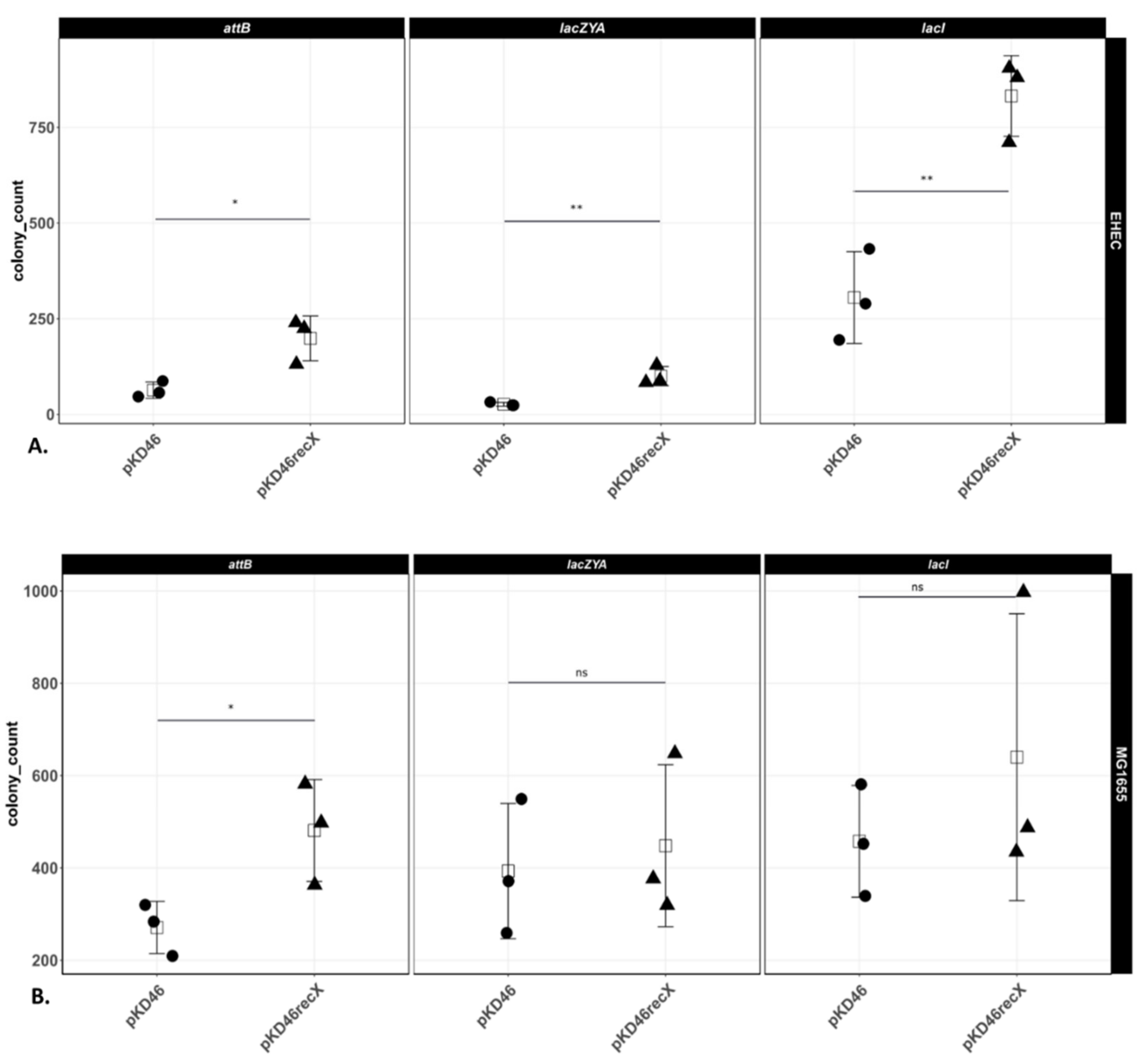
3.4. Comparison of the Numbers of Recombinants in E. coli K-12 MG1655 and EHEC O157:H7 EDL933 Δstx1/2

Preliminary observations indicated that, for any given locus, genetic modification of EHEC O157:H7 EDL933 Δstx1/2 chromosome required substantially higher amounts of the same recombination substrate than E. coli K-12 MG1655. The amounts of recombination substrate needed to successfully modify the chromosome also varied between the chromosomal loci within each individual genetic background. Therefore, in order to obtain appropriate numbers of recombinants on selective agar plates for enumeration, the amounts of recombination substrate were adjusted to genetic background and chromosomal position. For the details, see the Materials and Methods section (a detailed step-by-step protocol is additionally provided in the Supplementary Materials). Figure 2A shows that at each chromosomal locus, we obtained significantly higher numbers of colonies (3–4-fold) in EHEC O157:H7 EDL933 Δstx1/2 bearing pKD46recX compared to cells bearing pKD46 (the technical replicates for each biological replicate are shown in Supplementary Figure S3). In contrast, the effect of expressing recX from pKD46 on recombination efficiency varied between the three individual targeted E. coli K-12 MG1655 loci. In this strain, only the integration of the resistance cassette upstream of attB was significantly more efficient when pKD46recX was used, albeit less than two-fold (Figure 2B; Supplementary Figure S4 shows the technical replicates for each biological replicate).
To determine the accuracy of recombination, we performed a PCR analysis to determine the location of the insertion or deletion in both EHEC O157:H7 EDL933 Δstx1/2 and E. coli K-12 MG1655. For these experiments, we picked 10 random colonies for each biological replicate, plasmid, and chromosomal locus and performed a PCR analysis (Supplementary Figure S5). The PCR analysis indicated that the resistance cassette was integrated at the correct chromosomal locus in both genetic backgrounds in almost all cases.
In addition to that, we have also used pKD46recX to replace stx2 of E. coli O104:H4 by cat (Supplementary Figure S6; see Materials and Methods).

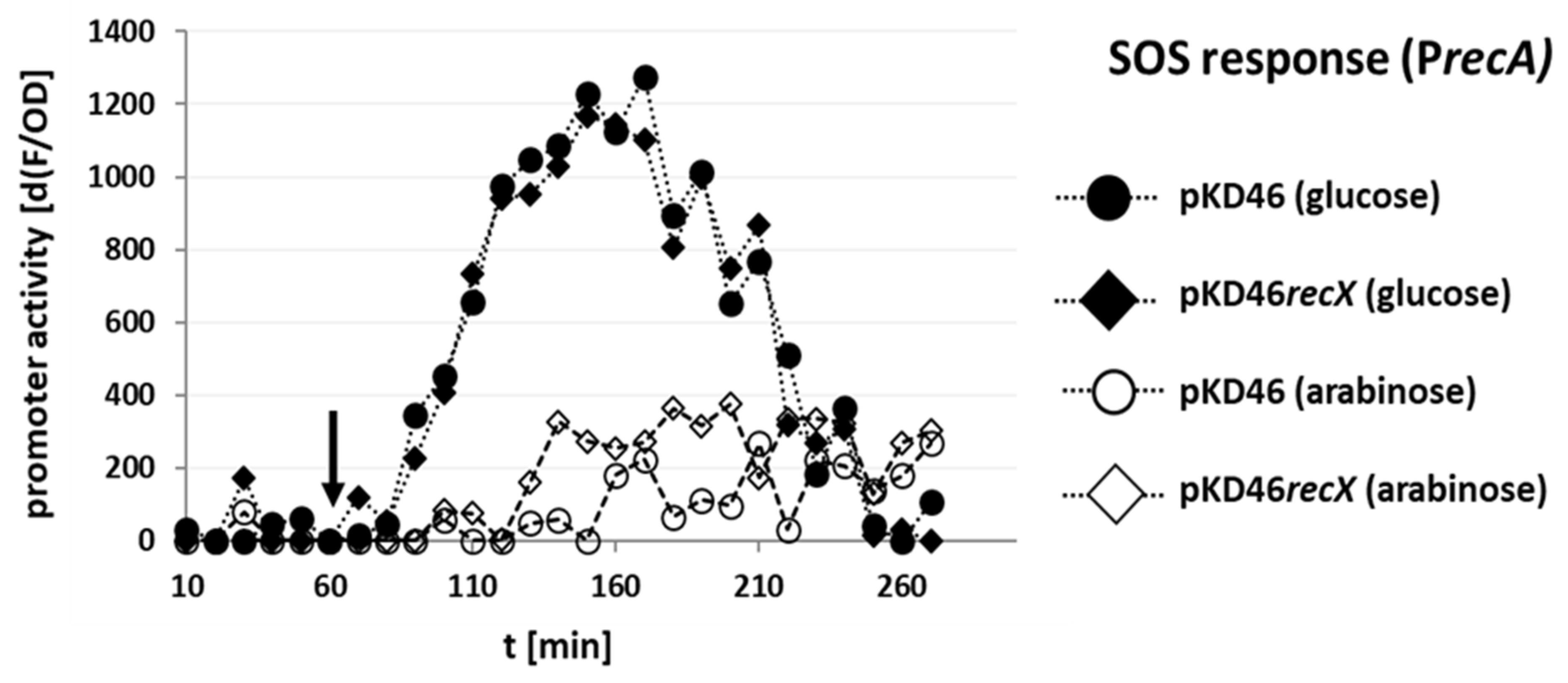
3.5. Coexpression of RecX with Gam, Beta, and Exo Does Not Further Repress the Sparfloxacin-Dependent SOS Response Induction

Sparfloxacin is a quinolone antibiotic that inhibits DNA gyrase [27,28], thereby causing double-stranded breaks and ultimately arresting DNA replication and activating RecA [29]. To investigate the mechanism by which recX impacts recombination efficiency, we decided to test if the coexpression of recX together with the λ Red functions would further repress a sparfloxacin-induced SOS response. For these experiments, we constructed an E. coli K-12 MG1655 strain with a recA promoter (PrecA)-yfp transcriptional fusion chromosomally integrated into attB (E. coli K-12 MG1655 PrecA SENSOR; for the details, see Materials and Methods). Subsequently, the reporter strain was transformed separately with plasmids pKD46 and pKD46recX. The encoded enzymes in both pKD46 and its derivative pKD46recX are under the control of the araBAD promoter. This promoter is activated by the addition of arabinose to the growth medium as described above but strongly repressed by the addition of glucose [30]. We measured the effect of sparfloxacin addition on the PrecA fluorescent reporter activity in cells grown in M9 + glucose (uninduced) or M9 + arabinose (induced).
When E. coli K-12 MG1655 PrecA SENSOR transformed with either pKD46 or pKD46recX were grown with glucose, the fluorescence signal started to increase shortly after sparfloxacin was added to the medium (black arrow) and the signal further increased until ~250 min. This demonstrated that PrecA was activated by sparfloxacin, as expected (Figure 3, filled symbols) [27]. In contrast, when these cells were grown in the presence of arabinose, the increase in the fluorescence signal in response to the sparfloxacin addition was substantially lower (Figure 3, open symbols) in both pKD46 and pKD46recX strains. The coexpression of recX together with gam, beta, and exo (Figure 3, open diamonds) did not further reduce the sparfloxacin-induced SOS response when compared to the expression of gam, beta, and exo alone (Figure 3, open circles). In contrast, the coexpression of recX slightly, but significantly attenuated the SOS response repression by Gam, as judged by the overall response of PrecA (Supplementary Figure S7).

4. Discussion

EHEC infections still constitute a significant worldwide health problem [3,4,5,31]. Even though the use of appropriate antibiotics in therapy is recently again more frequently discussed [2,9,32], it remains controversial [7,33]. In order to develop alternative therapy options, a better understanding of EHEC is essential. However, the genetic manipulation of EHEC with classical methods has only achieved sporadic success. To overcome this problem, several other groups have relied on empirically optimized or more time-consuming two-step recombineering methods [13,14]. The goal of our work was to provide a reliable method for increasing recombineering efficiency in EHEC strains. Even though also other important factors, e.g., the overexpression system itself are known to affect recombination efficiencies and may also further improve our system (for review, see [34]), we used pKD46 as the starting point for our investigations, as it is one of the most commonly used plasmids for recombineering [12,34].
Figure 2 shows that genetic modification of three conserved chromosomal loci in EHEC O157:H7 EDL933 Δstx1/2 was 3–4 times more efficient in cells coexpressing recX together with gam, beta, and exo, as compared to the same strain that only expressed the λ Red functions. Importantly, we found that recombination took place accurately, regardless of locus and whether the product of the reaction was an insertion or deletion, demonstrating that this coexpression did not increase the frequency of off-target mutagenic recombination. Significantly, in addition to EHEC O157:H7 EDL933 Δstx1/2, we have also used pKD46recX to successfully modify clinically relevant EHEC wild-type strains, strains in which successful recombineering was rather sporadic. For example, we used pKD46recX to delete the stx2 gene of the highly virulent E. coli O104:H4 [5].
The mechanism by which coexpression of recX with gam, beta, and exo increased recombination efficiency in EHEC O157:H7 EDL933 Δstx1/2 is, so far, not clear. We considered several possibilities. First, it is possible that the higher number of chloramphenicol-resistant colonies obtained when using pKD46recX in EHEC O157:H7 EDL933 Δstx1/2 may not result from an increased recombination efficiency, but instead result from an improvement in transformation efficiency. However, we routinely tested each batch of competent cells by transforming 0.5 ng of pLP2 and plating an aliquot of the transformation reaction. We did not observe an improvement in the transformation efficiency. Instead, we observed the opposite, i.e., when pKD46recX was used, the transformation efficiency dropped in both E. coli K-12 MG1655 and to a lesser degree in EHEC O157:H7 EDL933 Δstx1/2. This drop in transformation efficiency following recX overexpression was expected since activation of RecA is known to increase the transformation efficiency [35]. However, the number of recombinants in EHEC O157:H7 EDL933 Δstx1/2 increased, while the recombination efficiency in E. coli K-12 MG1655 did not drop to the same degree as the transformation efficiency in this strain. Therefore, either the transformation efficiency of linear DNA was less affected by recX expression, or the drop in transformation efficiency was outweighed by a concomitant general increase in recombination efficiency.
We speculated that electroporation of linear DNA into cells may activate RecA and the bacterial SOS response impacting, directly or indirectly, recombination frequency, thereby activating lytic growth of prophage resident within EHEC strains, eliminating them from the population of possible recombination targets. This suggestion is consistent with the observation that expression of recX together with λ Red function had no significant impact on recombination efficiency in E. coli K-12 MG1655, a strain that lacks inducible prophage (Figure 2; Supplementary Figures S3 and S4).
Depending on the type of DNA damage, the SOS response and RecA activation can be stimulated by either or both of two enzyme complexes RecBCD and RecFOR. Quinolone antibiotics stimulate the SOS response via RecBCD and Gam protein inhibits the activation of RecA by interacting with the DNA binding site of the RecBCD complex [18]. If RecX affected SOS activation and thereby recombineering efficiency via the RecBCD pathway, coexpression of this protein would be predicted to further reduce Gam inhibition of recA promoter activity. However, recX coexpression did not cause further inhibition of the quinolone-induced SOS response (Figure 3; Supplementary Figure S7), indicating that this potential pathway of SOS response induction by the transformation of a linear recombination substrate is already sufficiently blocked by gam expression alone. This finding suggests that RecX does not act to increase recombination via inhibition of the RecBCD pathway.
The RecFOR pathway of the SOS response induction, however, is not blocked by Gam. This pathway has a role in mediating the recombination of damaged plasmid DNA with the bacterial chromosome [36]. Given our findings, we suggest that the process of transformation of recombination substrates may induce the RecFOR pathway of SOS induction, especially when mechanically damaged DNA or DNA damaged by the freezing–thawing process is used [37]. In this case, the increase in recombination efficiency in EHEC O157:H7 EDL933 Δstx1/2 that was observed here might be indeed due to the RecX-dependent inhibition of the RecFOR pathway of SOS response induction and the prevention of subsequent phage-mediated lysis [15,20]. Despite the attractiveness of this idea, further experiments are necessary to clarify the exact mechanism. Nevertheless, we have already used pKD46recX successfully in clinical non-EHEC E. coli isolates in which we have failed to construct mutants with pKD46. Whether or not this is due to the presence of uncharacterised SOS-inducible prophages as well, is not clear. However, it indicates that using pKD46recX might be of advantage in case the phage content of a strain is not known [38].
We also found that the amount of recombination substrate needed to obtain enumerable numbers of recombinant colonies varied between chromosomal loci and genetic backgrounds. Interestingly, we observed that insertion at the attB locus required the addition of the highest amount of recombination substrate, regardless of genetic background and even though the product of the reaction resulted in a small, ten base pair deletion. The attB locus is more distant from the origin of replication than lacI and lacZYA. This finding is consistent with earlier work demonstrating that phage P1 transduction efficiency also decreases with increasing distance of the target from the origin of replication [39]. Since logarithmically growing cells are used for recombineering, this may therefore simply reflect the general relative increase of the copy numbers of the target loci, or a relatively higher number of replication forks at the specific locus, which are required for Red recombination [40]. However, a more systematic investigation would be necessary to clarify the question if a phage P1-like bias also exists for recombineering.

5. Conclusions

The work presented here describes a robust and easy-to-implement strategy for increasing the efficiency of genetically engineering clinically important E. coli. Even though some elements of the pKD46recX recombineering system and of its derivative pLP1 (lacking the kanamycin resistance) may be subject to further optimization [34], even the relatively modest, but robust increases in recombination efficiency in EHEC or any other hard-to-manipulate E. coli background facilitated by this strategy clearly can make a difference between failure and success.

Supplementary Materials

The following supporting information can be downloaded at: https://www.mdpi.com/article/10.3390/microorganisms10091689/s1, Figure S1: (A) Shown is an alignment of the nucleotide sequences of the regulatory gene lacI and the metabolic operon lacZYA (−/+ 499 bp) of EHEC O157:H7 EDL933 and E. coli K-12 MG1655. (B) Shown is an alignment of the nucleotide sequences of the conserved 15 bp phage lambda attachment site (attB) and the DNA sequences 1000 bp upstream and downstream of attB of EHEC O157:H7 EDL933 and E. coli K-12 MG1655; Figure S2: Induction time test. Shown is the average number of colonies obtained with 1 ng of recombination substrate for the lacZYA locus when using pKD46 in E. coli K-12 MG1655. Shown are the average colony numbers and the standard deviations from three technical replicates for each time point; Figure S3: Numbers of chloramphenicol-resistant colonies obtained for the indicated genetic locus for EHEC O157:H7 EDL933 Δstx1/2. Shown are the numbers and standard deviations of colonies obtained from each of the three technical replicates in each experiment. Statistically significant t test comparisons are indicated by * (p < 0.05), ** (p < 0.01), *** (p < 0.001) and “ns” if not statistically significant; Figure S4: Numbers of chloramphenicol-resistant colonies obtained for the indicated genetic locus for E. coli K-12 MG1655. Shown are the numbers and standard deviations of colonies obtained from each of the three technical replicates in each experiment. Statistically significant t test comparisons are indicated by * (p < 0.05), ** (p < 0.01), *** (p < 0.001) and “ns” if not statistically significant; Figure S5: PCR analysis. Shown are the results of the colony PCR for each experiment. 10 randomly picked colonies per plasmid and biological replicate were analyzed by PCR using the primers that are indicated graphically on top of the images. Indicated are the numbers of colonies analyzed (#1–#10) and the negative control (−) that was the same strain containing either pKD46 or pKD46recX. (A) attB locus insertion in E. coli K-12 MG1655. (B) lacZYA deletion in E. coli K-12 MG1655. (C) lacI deletion in E. coli K-12 MG1655. (D) attB locus insertion in EHEC O157:H7 EDL933 Δstx1/2. (E) lacZYA deletion in EHEC O157:H7 EDL933 Δstx1/2. (F) lacI deletion in EHEC O157:H7 EDL933 Δstx1/2. The vast majority of chloramphenicol-resistant colonies was PCR-positive. There was no difference in PCR-negative colonies between pKD46 and pKD46recX containing strains. For the details, see the text; Figure S6: Verification of E. coli O104:H4 stx2::cat by whole genome sequencing. (A) Coverage of the BAM files generated by mapping sequencing reads using the E. coli O104:H4 genome GCF_000299455.1 as a reference genome and visualized in Integrative Genomics Viewer. No reads could be mapped to stx2. (B) Sequence of the stx2::cat region from the de novo assembly. The sequence of the insert carrying the resistance cassette (bold) flanked by 200 nt of the Stx2 phage region is shown. The cat ORF is marked in yellow and t-RNA O3K_RS16215 in green; Figure S7: Overall E. coli K-12 MG1655 SENSOR SOS response to sparfloxacin challenge. Shown are values and standard deviations of three biological replicates. Statistically significant t test comparisons are indicated by * (p < 0.05), *** (p < 0.001) and “ns” if not statistically significant; Table S1: Bacterial strains used in this study; Table S2: Plasmids used in this study; Table S3: Primers used in this study. References in Supplementary Materials can be seen in [9,12,22,23,24,26,41,42].

Author Contributions

M.B. conceived and designed the research. L.P., N.H., M.C. and R.M.D. performed the experiments. P.B., H.S., A.M. and U.D. contributed with reagents, material, and analysis tools. M.B., L.P. and R.M.D. analysed the data. M.B., P.B., G.K., H.S., A.M. and U.D. authored the paper. All authors reviewed the manuscript and approved the final draft. All authors have read and agreed to the published version of the manuscript.

Funding

This work was funded by the Deutsche Forschungsgemeinschaft (DFG, German Research Foundation)—project number 276606594, SFB1009 B04, and SFB1009 B05 and by the Interdisciplinary Center for Clinical Research, Medical Faculty Münster (grant number Dob2/022/16). The funding bodies had no role in the design of the study, the collection, analysis, and interpretation of data, and in writing the manuscript. We acknowledge support from the Open Access Publishing Fund of University of Münster.

Institutional Review Board Statement

Not applicable.

Informed Consent Statement

Not applicable.

Data Availability Statement

The data presented in this study are available in article and Supplementary Materials.

Conflicts of Interest

The authors declare no conflict of interest. The funders had no role in the design of the study; in the collection, analyses, or interpretation of data; in the writing of the manuscript, or in the decision to publish the results.

References

  1. Nguyen, Y.; Sperandio, V. Enterohemorrhagic E. coli (EHEC) pathogenesis. Front. Cell. Infect. Microbiol. 2012, 2, 90. [Google Scholar] [CrossRef] [PubMed] [Green Version]
  2. Kampmeier, S.; Berger, M.; Mellmann, A.; Karch, H.; Berger, P. The 2011 German enterohemorrhagic Escherichia coli O104:H4 outbreak—The danger is still out there. Curr. Top. Microbiol. Immunol. 2018, 416, 117–148. [Google Scholar] [CrossRef] [PubMed]
  3. Michino, H.; Araki, K.; Minami, S.; Takaya, S.; Sakai, N.; Miyazaki, M.; Ono, A.; Yanagawa, H. Massive outbreak of Escherichia coli O157:H7 infection in schoolchildren in Sakai City, Japan, associated with consumption of white radish sprouts. Am. J. Epidemiol. 1999, 150, 787–796. [Google Scholar] [CrossRef] [Green Version]
  4. Xiong, Y.; Wang, P.; Lan, R.; Ye, C.; Wang, H.; Ren, J.; Jing, H.; Wang, Y.; Zhou, Z.; Bai, X.; et al. A novel Escherichia coli O157:H7 clone causing a major hemolytic uremic syndrome outbreak in China. PLoS ONE 2012, 7, e36144. [Google Scholar] [CrossRef]
  5. Karch, H.; Denamur, E.; Dobrindt, U.; Finlay, B.B.; Hengge, R.; Johannes, L.; Ron, E.Z.; Tønjum, T.; Sansonetti, P.J.; Vicente, M. The enemy within us: Lessons from the 2011 European Escherichia coli O104:H4 outbreak. EMBO Mol. Med. 2012, 4, 841–848. [Google Scholar] [CrossRef]
  6. Melton-Celsa, A.R. Shiga toxin (Stx) classification, structure, and function. Microbiol. Spectr. 2014, 2, EHEC-0024-2013. [Google Scholar] [CrossRef] [PubMed] [Green Version]
  7. Wong, C.S.; Mooney, J.C.; Brandt, J.R.; Staples, A.O.; Jelacic, S.; Boster, D.R.; Watkins, S.L.; Tarr, P.I. Risk factors for the hemolytic uremic syndrome in children infected with Escherichia coli O157:H7: A multivariable analysis. Clin. Infect. Dis. 2012, 55, 33–41. [Google Scholar] [CrossRef] [Green Version]
  8. Kimmitt, P.T.; Harwood, C.R.; Barer, M.R. Toxin gene expression by Shiga toxin-producing Escherichia coli: The role of antibiotics and the bacterial SOS response. Emerg. Infect. Dis. 2000, 6, 458–465. [Google Scholar] [CrossRef] [Green Version]
  9. Berger, M.; Aijaz, I.; Berger, P.; Dobrindt, U.; Koudelka, G. Transcriptional and translational inhibitors block SOS response and Shiga toxin expression in enterohemorrhagic Escherichia coli. Sci. Rep. 2019, 9, 18777. [Google Scholar] [CrossRef]
  10. Mühlen, S.; Ramming, I.; Pils, M.C.; Koeppel, M.; Glaser, J.; Leong, J.; Flieger, A.; Stecher, B.; Dersch, P. Identification of antibiotics that diminish disease in a murine model of enterohemorrhagic Escherichia coli infection. Antimicrob. Agents Chemother. 2020, 64, e02159-19. [Google Scholar] [CrossRef]
  11. Murphy, K.C. Use of bacteriophage lambda recombination functions to promote gene replacement in Escherichia coli. J. Bacteriol. 1998, 180, 2063–2071. [Google Scholar] [CrossRef] [PubMed] [Green Version]
  12. Datsenko, K.A.; Wanner, B.L. One-Step Inactivation of Chromosomal Genes in Escherichia coli K-12 Using PCR Products. Proc. Natl. Acad. Sci. USA 2000, 97, 6640–6645. [Google Scholar] [CrossRef] [PubMed] [Green Version]
  13. Murphy, K.C.; Campellone, K.G. Lambda red-mediated recombinogenic engineering of enterohemorrhagic and enteropathogenic E. coli. BMC Mol. Biol. 2003, 4, 11. [Google Scholar] [CrossRef] [Green Version]
  14. Lee, D.J.; Bingle, L.E.; Heurlier, K.; Pallen, M.J.; Penn, C.W.; Busby, S.J.; Hobman, J.L. Gene doctoring: A method for recombineering in laboratory and pathogenic Escherichia coli strains. BMC Microbiol. 2009, 9, 252. [Google Scholar] [CrossRef] [Green Version]
  15. Koudelka, A.P.; Hufnagel, L.A.; Koudelka, G.B. Purification and characterization of the repressor of the Shiga toxin-encoding bacteriophage 933W: DNA binding, gene regulation, and autocleavage. J. Bacteriol. 2004, 186, 7659–7669. [Google Scholar] [CrossRef] [Green Version]
  16. Baharoglu, Z.; Mazel, D. SOS, the formidable strategy of bacteria against aggressions. FEMS Microbiol. Rev. 2014, 38, 1126–1145. [Google Scholar] [CrossRef] [PubMed] [Green Version]
  17. Muyrers, J.P.; Zhang, Y.; Buchholz, F.; Stewart, A.F. RecE/RecT and Redalpha/Redbeta initiate double-stranded break repair by specifically interacting with their respective partners. Genes Dev. 2000, 14, 1971–1982. [Google Scholar] [CrossRef]
  18. Wilkinson, M.; Troman, L.; Wan Nur Ismah, W.A.; Chaban, Y.; Avison, M.B.; Dillingham, M.S.; Wigley, D.B. Structural basis for the inhibition of RecBCD by gam and its synergistic antibacterial effect with quinolones. eLife 2016, 5, e22963. [Google Scholar] [CrossRef] [Green Version]
  19. Stohl, E.A.; Brockman, J.P.; Burkle, K.L.; Morimatsu, K.; Kowalczykowski, S.C.; Seifert, H.S. Escherichia coli RecX inhibits RecA recombinase and coprotease activities in vitro and in vivo. J. Biol. Chem. 2003, 278, 2278–2285. [Google Scholar] [CrossRef] [Green Version]
  20. Lusetti, S.L.; Hobbs, M.D.; Stohl, E.A.; Chitteni-Pattu, S.; Inman, R.B.; Seifert, H.S.; Cox, M.M. The RecF protein antagonizes RecX function via direct interaction. Mol. Cell 2006, 21, 41–50. [Google Scholar] [CrossRef] [Green Version]
  21. Yakimov, A.; Pobegalov, G.; Bakhlanova, I.; Khodorkovskii, M.; Petukhov, M.; Baitin, D. Blocking the RecA activity and SOS-response in bacteria with a short α-helical peptide. Nucleic Acids Res. 2017, 45, 9788–9796. [Google Scholar] [CrossRef] [PubMed] [Green Version]
  22. Berger, M.; Gerganova, V.; Berger, P.; Rapiteanu, R.; Lisicovas, V.; Dobrindt, U. Genes on a wire: The nucleoid-associated protein HU insulates transcription units in Escherichia coli. Sci. Rep. 2016, 6, 31512. [Google Scholar] [CrossRef] [PubMed] [Green Version]
  23. Norrander, J.; Kempe, T.; Messing, J. Construction of improved M13 vectors using oligodeoxynucleotide-directed mutagenesis. Gene 1983, 26, 101–106. [Google Scholar] [CrossRef]
  24. Wang, R.F.; Kushner, S.R. Construction of versatile low-copy-number vectors for cloning, sequencing and gene expression in Escherichia coli. Gene 1991, 100, 195–199. [Google Scholar] [CrossRef]
  25. Chittò, M.; Berger, M.; Klotz, L.; Dobrindt, U. Sub-inhibitory concentrations of SOS-response inducing antibiotics stimulate integrase expression and excision of pathogenicity islands in uropathogenic Escherichia coli strain 536. Int. J. Med. Microbiol. 2020, 310, 151361. [Google Scholar] [CrossRef]
  26. Gobert, A.P.; Vareille, M.; Glasser, A.-L.; Hindré, T.; de Sablet, T.; Martin, C. Shiga toxin produced by enterohemorrhagic Escherichia coli inhibits PI3K/NF-KB signaling pathway in globotriaosylceramide-3-negative human intestinal epithelial cells. J. Immunol. 2007, 178, 8168–8174. [Google Scholar] [CrossRef] [Green Version]
  27. Piddock, L.J.; Zhu, M. Mechanism of action of Sparfloxacin against and mechanism of resistance in gram-negative and gram-positive bacteria. Antimicrob. Agents Chemother. 1991, 35, 2423–2427. [Google Scholar] [CrossRef] [Green Version]
  28. Pan, X.S.; Fisher, L.M. Targeting of DNA gyrase in Streptococcus pneumoniae by Sparfloxacin: Selective targeting of gyrase or topoisomerase IV by quinolones. Antimicrob. Agents Chemother. 1997, 41, 471–474. [Google Scholar] [CrossRef] [Green Version]
  29. Drlica, K.; Zhao, X. DNA gyrase, topoisomerase IV, and the 4-quinolones. Microbiol. Mol. Biol. Rev. 1997, 61, 377–392. [Google Scholar] [CrossRef]
  30. Guzman, L.M.; Belin, D.; Carson, M.J.; Beckwith, J. Tight regulation, modulation, and high-level expression by vectors containing the arabinose PBAD promoter. J. Bacteriol. 1995, 177, 4121–4130. [Google Scholar] [CrossRef] [Green Version]
  31. Tuttle, J.; Gomez, T.; Doyle, M.P.; Wells, J.G.; Zhao, T.; Tauxe, R.V.; Griffin, P.M. Lessons from a large outbreak of Escherichia coli O157:H7 infections: Insights into the infectious dose and method of widespread contamination of hamburger patties. Epidemiol. Infect. 1999, 122, 185–192. [Google Scholar] [CrossRef] [PubMed]
  32. Joseph, A.; Cointe, A.; Mariani Kurkdjian, P.; Rafat, C.; Hertig, A. Shiga toxin-associated hemolytic uremic syndrome: A narrative review. Toxins 2020, 12, 67. [Google Scholar] [CrossRef] [PubMed] [Green Version]
  33. Wong, C.S.; Jelacic, S.; Habeeb, R.L.; Watkins, S.L.; Tarr, P.I. The risk of the hemolytic-uremic syndrome after antibiotic treatment of Escherichia coli O157:H7 infections. N. Engl. J. Med. 2000, 342, 1930–1936. [Google Scholar] [CrossRef] [PubMed] [Green Version]
  34. Murphy, K.C. λ recombination and recombineering. EcoSal Plus 2016, 7, ESP-0011-2015. [Google Scholar] [CrossRef] [PubMed] [Green Version]
  35. Vericat, J.A.; Guerrero, R.; Barbé, J. Increase in plasmid transformation efficiency in SOS-induced Escherichia coli cells. Mol. Gen. Genet. 1988, 211, 526–530. [Google Scholar] [CrossRef]
  36. Abbott, P.J. Stimulation of recombination between homologous sequences on carcinogen-treated plasmid DNA and chromosomal DNA by induction of the SOS response in Escherichia coli K12. Mol. Gen. Genet. 1985, 201, 129–132. [Google Scholar] [CrossRef]
  37. Chung, W.-J.; Cui, Y.; Chen, C.-S.; Wei, W.H.; Chang, R.-S.; Shu, W.-Y.; Hsu, I.C. Freezing shortens the lifetime of DNA molecules under tension. J. Biol. Phys. 2017, 43, 511–524. [Google Scholar] [CrossRef]
  38. Casjens, S. Prophages and bacterial genomics: What have we learned so far? Mol. Microbiol. 2003, 49, 277–300. [Google Scholar] [CrossRef]
  39. Masters, M. The frequency of P1 transduction of the genes of Escherichia coli as a function of chromosomal position: Preferential transduction of the origin of replication. Mol. Gen. Genet. 1977, 155, 197–202. [Google Scholar] [CrossRef]
  40. Fels, U.; Gevaert, K.; Van Damme, P. Bacterial genetic engineering by means of recombineering for reverse genetics. Front. Microbiol. 2020, 11, 548410. [Google Scholar] [CrossRef]
  41. Blattner, F.R.; Plunkett, G.; Bloch, C.A.; Perna, N.T.; Burland, V.; Riley, M.; Collado-Vides, J.; Glasner, J.D.; Rode, C.K.; Mayhew, G.F.; et al. The Complete Genome Sequence of Escherichia Coli K-12. Science 1997, 277, 1453–1462. [Google Scholar] [CrossRef] [PubMed] [Green Version]
  42. Grant, S.G.; Jessee, J.; Bloom, F.R.; Hanahan, D. Differential Plasmid Rescue from Transgenic Mouse DNAs into Escherichia coli Methylation-Restriction Mutants. Proc. Natl. Acad. Sci. USA 1990, 87, 4645–4649. [Google Scholar] [CrossRef] [PubMed] [Green Version]
Figure 1. Schematic representation of the positions of chromosomal homology (CH) between EHEC O157:H7 EDL933 and E. coli K-12 MG1655 that were used to design the primers for the recombination substrates. (A) CH for the deletion of the lacZYA operon. (B) CH for the deletion of the regulatory gene lacI. (C) CH for the insertion upstream of the phage lambda attachment site attB.
Microorganisms 10 01689 g001
Figure 2. Numbers of chloramphenicol-resistant colonies obtained for the indicated genetic locus. (A) Number of chloramphenicol-resistant colonies in EHEC O157:H7 EDL933 Δstx1/2. (B) Number of chloramphenicol-resistant colonies in E. coli K-12 MG1655. Shown are the average values of three technical replicates of three independent experiments. Statistically significant t-test comparisons are indicated by * (p < 0.05), ** (p < 0.01) and “ns” if not statistically significant.
Microorganisms 10 01689 g002
Figure 3. Sparfloxacin-dependent induction of the SOS response in E. coli MG1655 PrecA SENSOR. The promoter activity of PrecA as the increase of fluorescence over time in the presence of the indicated plasmid and the indicated sugar is shown. The black arrow indicates the time point of the addition of sparfloxacin (f. c. 15 µg/mL).
Microorganisms 10 01689 g003
Publisher’s Note: MDPI stays neutral with regard to jurisdictional claims in published maps and institutional affiliations.

