Article

Unveiling the Occupational Exposure to Microbial Contamination in Conservation–Restoration Settings

1
H&TRC—Health & Technology Research Center, ESTeSL—Escola Superior de Tecnologia da Saúde de Lisboa, Instituto Politgy Research Center, 1990-096 Lisbon, Portugal
2
Public Health Research Centre, NOVA National School of Public Health, Universidade NOVA de Lisboa, 1099-085 Lisboa, Portugal
3
Comprehensive Health Research Center (CHRC), NOVA Medical School, Universidade NOVA de Lisboa, 1169-056 Lisboa, Portugal
4
Department of Physiology and Toxicology, Institute of Experimental Biology, Faculty of Natural Sciences, Kazimierz Wielki University, Chodkiewicza 30, 85-064 Bydgoszcz, Poland
5
Research Institute for Medicines (iMed.uLisboa), Faculty of Pharmacy, University of Lisbon, 1649-003 Lisbon, Portugal
6
HERCULES Laboratory—Cultural Heritage Protection Studies, University of Evora, Palácio do Vimioso, Largo Marquês de Marialva, 8, 7000-809 Evora, Portugal
*
Author to whom correspondence should be addressed.
Microorganisms 2022, 10(8), 1595; https://doi.org/10.3390/microorganisms10081595
Submission received: 2 July 2022 / Revised: 26 July 2022 / Accepted: 4 August 2022 / Published: 8 August 2022

Abstract

:
Assuring a proper environment for the fulfillment of professional activities is one of the Sustainable Development Goals and is contemplated in the One Health approach assumed by the World Health Organization. This particular study is applied to an often neglected sector of our society—the conservators/restorers—despite the many health issues reported by these professionals. Three different specialties (textiles, paintings and wood sculpture) and locations were selected for evaluation by placement of electrostatic dust cloths. After treatment of the samples, bacterial and fungal contamination were assessed, as well as mycotoxin determination, the presence of azole-resistant strains and cytotoxicity of the microorganisms encountered. Bacteria were only present in one of medias used and showed relatively low numbers. The highest level of contamination by fungi was identified in one of the textiles settings. The textile area also showed the highest variability for fungi. Aspergillus sp. are one indicator of possible environmental issues, and A. sections Fumigati and Circumdati were particularly relevant in two of the settings and identified in all of them. No mycotoxins were detected and the large majority of the fungi identified were non-cytotoxic. Overall, these can be considered low-contaminated environments but attention should be given to the Aspergillus sp. contamination. Additional studies are needed not only to make these results more robust, but also to test if the environmental sampling alone is the best approach in a setting where there is very little movement and dust displacement and where professionals are in very close proximity to the artefacts being treated, which may suggest the existence of a micro-atmosphere worth evaluating and comparing to the obtained results.

1. Introduction

Among all microorganisms, fungi cause the degradation of cultural heritage sites to the greatest extent [1]. In the case of archives, some of the fungi present in paper documents, surfaces and air from archives, libraries and museums are also a threat to human health [2]. Due to their enormous enzymatic activity and their ability to grow at low water activity levels values, fungi are able to inhabit and to decay paintings, textiles, paper, parchment, leather, oil, casein, glue and other materials used for historical art objects. In museums and their storage rooms, climate control, regular cleaning and microbiological monitoring are essential in order to prevent fungal contamination.
It is mandatory for Portuguese employers to assess and prevent occupational exposure to chemical, physical and biological risks, as in all European countries [3]. Several studies have reported that exposure to microorganisms, such as bacteria and fungi, can originate respiratory diseases [4,5,6]. Nevertheless, those of biological origin are less recognized and reported than chemicals [7].
Conservators–restorers—professionals that handle priceless cultural heritage artefacts maintaining them for future generations—can work in a myriad of settings. From small private ateliers to large, state-run facilities, handling organic substrates, such as paintings or books or, conversely, working with stone or metals, doing their job high on a scaffold or bent over a textile. Whatever the location, position or material being handled, the exposure to the different hazards carried by each specialty should be addressed.
The biological hazards are, presumably, more relevant in the settings where the conservator–restorer handles organic substrates. Anyone who has ever entered an archive or library can recall a particular scent, and part of this aroma comes from paper, rag, leather or parchment degradation [8,9]. In addition, some of these deterioration issues come from the activity of microorganisms, acting on the organic substrate. Handling potentially contaminated cultural heritage artefacts may provoke a diverse array of health effects on the staff, due to the increased exposure [2,10,11,12]. In fact, high microbiological contamination, previously reported in museums, libraries and archives, may be harmful to workers [13,14,15].
Conservation measures and treatments used to inhibit fungal growth in paper-based items of cultural heritage include mechanical, chemical and biological methods, such as gamma rays and ethylene oxide fumigation [16]. If some of these disinfecting measures, which are intended to fragment fungal DNA, present suboptimal efficacy, they might contribute for the development of fungal tolerance, which is a risk factor for the development of fungal resistance in the long-term. Fungal resistance to medical azoles has been described in recent years as an important public health concern, which is expected to increase in the next years due to the current scenario of climatic changes [17,18,19].
Additionally, fungi are known to produce mycotoxins, their exometabolites that can be toxic for humans and animals. Mycotoxins are produced by specific fungal genera, mainly by Aspergillus, Penicillium, Alternaria, Fusarium and Claviceps [20,21]. Several mycotoxins are carcinogenic or probably carcinogenic to humans, as evaluated by the International Agency for Research on Cancer [22]. Mycotoxins are considered the most frequently occurring natural contaminants in the diet of humans and animals. Due to climate change, an increased magnitude and/or frequency in the exposure of humans to mycotoxins is expected to occur in temperate regions of Europe [23]. Mycotoxins can resist to adverse environmental factors, such as high or low temperatures, and can persist long after the death and disintegration of the fungal species responsible for their production [24]. Therefore, and due to the health effects related with exposure to mycotoxins when studying fungal contamination is reasonable to also study mycotoxins contamination [25].
This study aimed to assess microbial contamination present in four different work settings: textiles (2), paintings and sculpture (mostly wood based). The study was performed in the Lisbon area, applying electrostatic dust cloths as sampling method. The screening of azole-resistance profile, Aspergillus sections detection, as well as the mycotoxins and cytotoxicity assessment was also performed. Education and close collaboration of mycologists and restorers are needed to develop object specific methods for the conservation and treatment of contaminated objects.

2. Materials and Methods

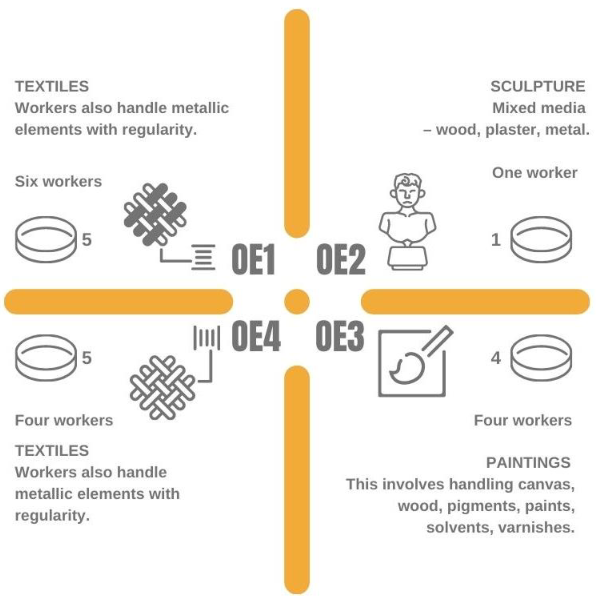
2.1. Working Settings Assessed

This study sampling campaign was conducted between May and June 2021 in four locations, three in the city of Lisbon and another one in a smaller coastal town. This last one was a private home-based studio, where a single conservator handles historic and artistic objects that can be made from a variety of substrates, some organic, some inorganic and most a composition of both. The several objects in line to be treated are kept at this location and the work is seldom initiated and maintained in different objects at the same time to make up for the obligatory intervals some treatments demand. The other private studio is dedicated to the restoration of paintings. At the time of the evaluation it was being used by three conservators and a master’s student. It is a relatively small studio, with two different stories and uses natural ventilation only. As happened in the previous case, there is also an accumulation of other paintings besides the ones that are being treated at the moment, either because the finalized work has not been collected, is between treatment phases or has not been initiated yet. The remaining two settings analyzed are both public run studios and both tend to the restoration of textiles. These are larger rooms and the only pieces that are on display are being treated by the conservators. At the time of the evaluation, one of the studios had 6 workers (this number can shift to 4 in case there is a low demand for conservation work) and the other had 4 workers in the premises (Figure 1).

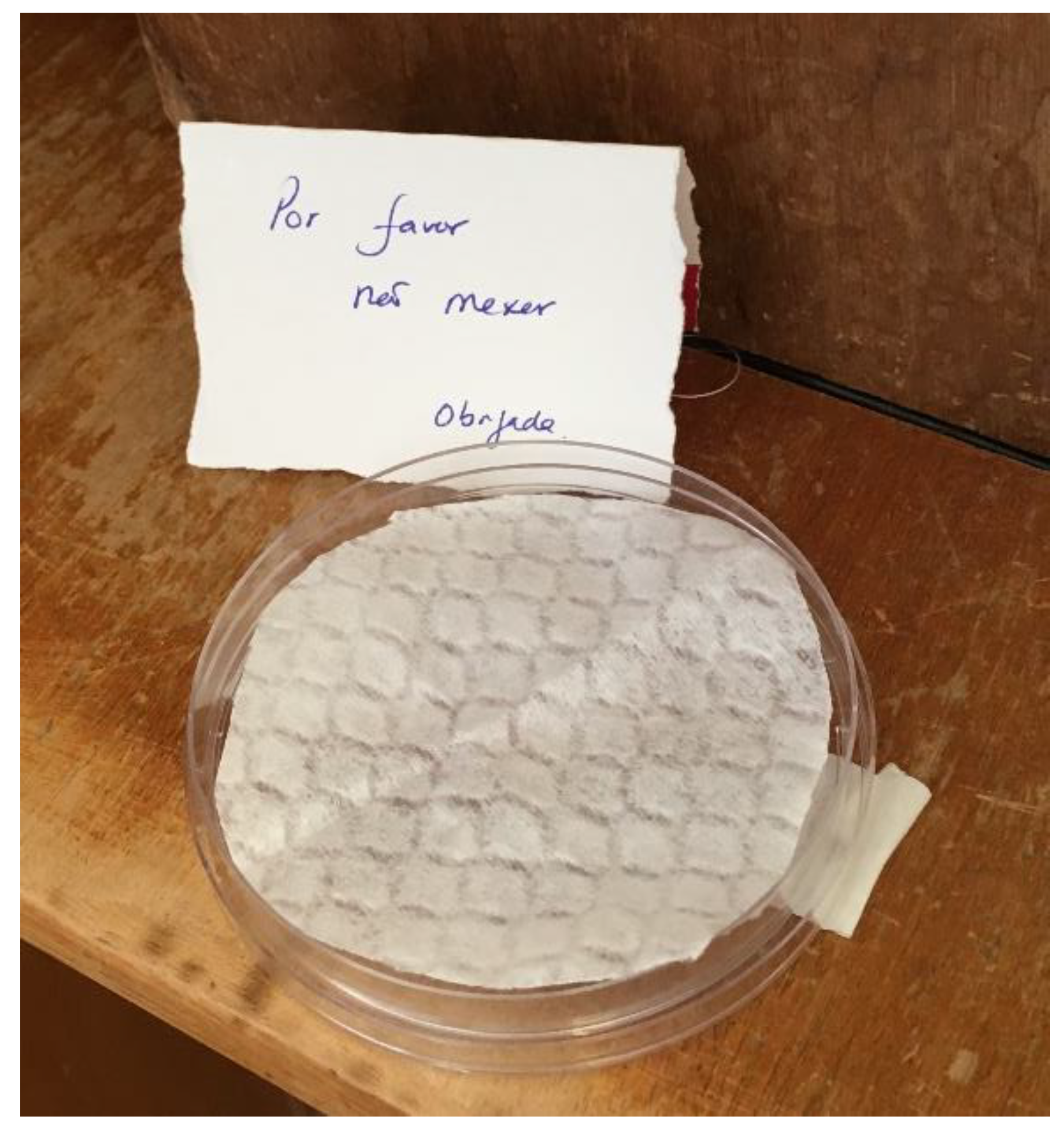
2.2. Sampling Approach Characterization through Culture-Dependent Methods

The EDCs were placed in each sampling site (Figure 2) for 30 days and transported under refrigeration (0–4 °C) to the laboratory for further analyses [26]. EDCs were weighted and processed with 20 mL of 0.1% Tween 80 saline (0.9% NaCl). For fungal assessment malt extract agar (MEA) supplemented with chloramphenicol (0.05%), and dichloran-glycerol agar (DG18) were used. EDC samples were incubated at 27 °C for 5–7 days. For bacteria assessment, tryptic soy agar (TSA, 30 °C, 7 days) and violet-red bile agar (VRBA 35 °C, 7 days) were used for mesophilic bacteria and coliforms (Gram negative bacteria), respectively. Microbial contamination densities (colony-forming units, CFU·g−1, CFU·m−2, CFU·m−2·day−1) were calculated as previously reported [27,28]. Fungal species were preliminary identified microscopically following procedures previous published [29]. Negative controls were employed to ensure the inexistence of background contamination, namely culture media (all samples) and extracts of control samples (EDC) without prior use were submitted to the same assays.

2.3. Azole Resistance Screenin

Sabouraud dextrose agar (SDA) (Frilabo, Maia, Portugal), either alone or supplemented with 4 µg/mL itraconazole (ITZ), 2 µg/mL voriconazole (VCZ), or 0.5 µg/mL posaconazole (PSZ), were used to screen fungal resistance to medical azoles (adapted from [30,31]). The A. fumigatus ATCC 204305 reference strain, and a pan-azole-resistant A. fumigatus were used as controls (both strains provided by National Health Institute Doutor Ricardo Jorge, IP). Briefly, SDA media plates inoculated with samples’ extracts from all the EDC were incubated at 27 °C (to enable optimal conditions for fungal growth) for three to four days. After incubation, fungal colonies were counted and identified by microscopy, as previously described [32].

2.4. Molecular Detection of Aspergillus Sections

The extracts (8.8 mL) from the EDCs were used for molecular detection of Aspergillus sections [26]. Fungal DNA was extracted using the ZR Fungal/Bacterial DNA MiniPrep Kit (Zymo Research, Irvine, CA, USA) and molecular identification was performed by Real Time PCR (qPCR) using the CFX-Connect PCR System (Bio-Rad, Amadora, Portugal). Reactions included 1× iQ Supermix (Bio-Rad, Amadora, Portugal), 0.5 μM of each primer, and 0.375 μM of TaqMan probe in a total volume of 20 μL. Amplification followed a three-step PCR: 40 cycles with denaturation at 95 °C for 30 s, annealing at 52 °C for 30 s, and extension at 72 °C for 30 s.
A non-template control and a positive control consisting of DNA obtained from a reference that belonged to the culture collection of the Reference Unit for Parasitic and Fungal Infections, Department of Infectious Diseases of the National Institute of Health, from Dr. Ricardo Jorge were used. These strains have been sequenced for ITS, B-tubulin, and Calmodulin.

2.5. Mycotoxins Analysis

Nineteen samples were screened for mycotoxins presence. In all samples, 38 mycotoxins were analyzed by HPL-MS (HPLC) Nexera (Shimadzu, Tokyo, Japan) with a mass spectrometry detector API 4000 (Sciex, Foster City, CA, USA) following the same laboratory procedures described in previous papers [27,28]. The mycotoxin concentration was calculated using external calibration. The limits of detection (LOD) obtained for each mycotoxin with the analytical method used are presented in Table 1.

2.6. Assessment of Cytotoxicity

The cell viability of human lung epithelial (A549), human liver carcinoma (HepG2) and swine kidney (SK) cells, exposed to EDC samples for 48 h at 5% CO2, 37 °C, and humid atmosphere, were determined by the 3-(4,5-dimethylthiazol-2-yl)-2,5-diphenyltetrazolium bromide (MTT) assay at 510 nm, as previously described [33]. Briefly, cells were at first maintained in Eagle’s minimum essential medium (MEM) supplemented with 10,000 units penicillin and 10 mg/mL streptomycin in 0.9% NaCl and fetal bovine serum (Sigma-Aldrich, St. Louis, MO, USA). After cell detachment (with 0.25% (w/v) Trypsin 0.53 mM EDTA), 100 µL cell suspension with densities of 2.0 × 10⁵ to 4.5 × 10⁵ cells/mL (Scepter™ 2.0 Cell Counter, Merck, NJ, USA) was transferred to a 96-well plate. Cells were then exposed to the EDC samples and cell viability was measured (ELISA LEDETECT 96, biomed Dr. Wieser GmbH; MikroWin 2013SC software). The threshold toxicity level was considered the lowest concentration dropping absorption to <50% of cell metabolic activity (IC50).

2.7. Statistical Analysis

Data were analyzed using SPSS statistical software for windows, version 27.0. The results were considered significant at the 5% significance level. To test the normality of the data, the Shapiro–Wilk test was used. For the comparison of bacterial contamination, fungal contamination and fungal resistance, the Kruskal–Wallis test was used, since the assumption of normality was not verified and given the small size of the sample. To study the relationship between bacterial contamination, fungal contamination and fungal resistance, Spearman’s correlation coefficient was used, since the assumption of normality was not verified. To assess species diversity, Simpson and Shannon indices, given by Shannon   Index   ( H ) = i = 1 s p i ln ( p i ) and Simpson   Index   ( D ) = 1 i = 1 s p i 2 , were used, where pi is the proportion (ni/n) of individuals of one particular species found (ni) divided by the total number of individuals found (n).

3. Results

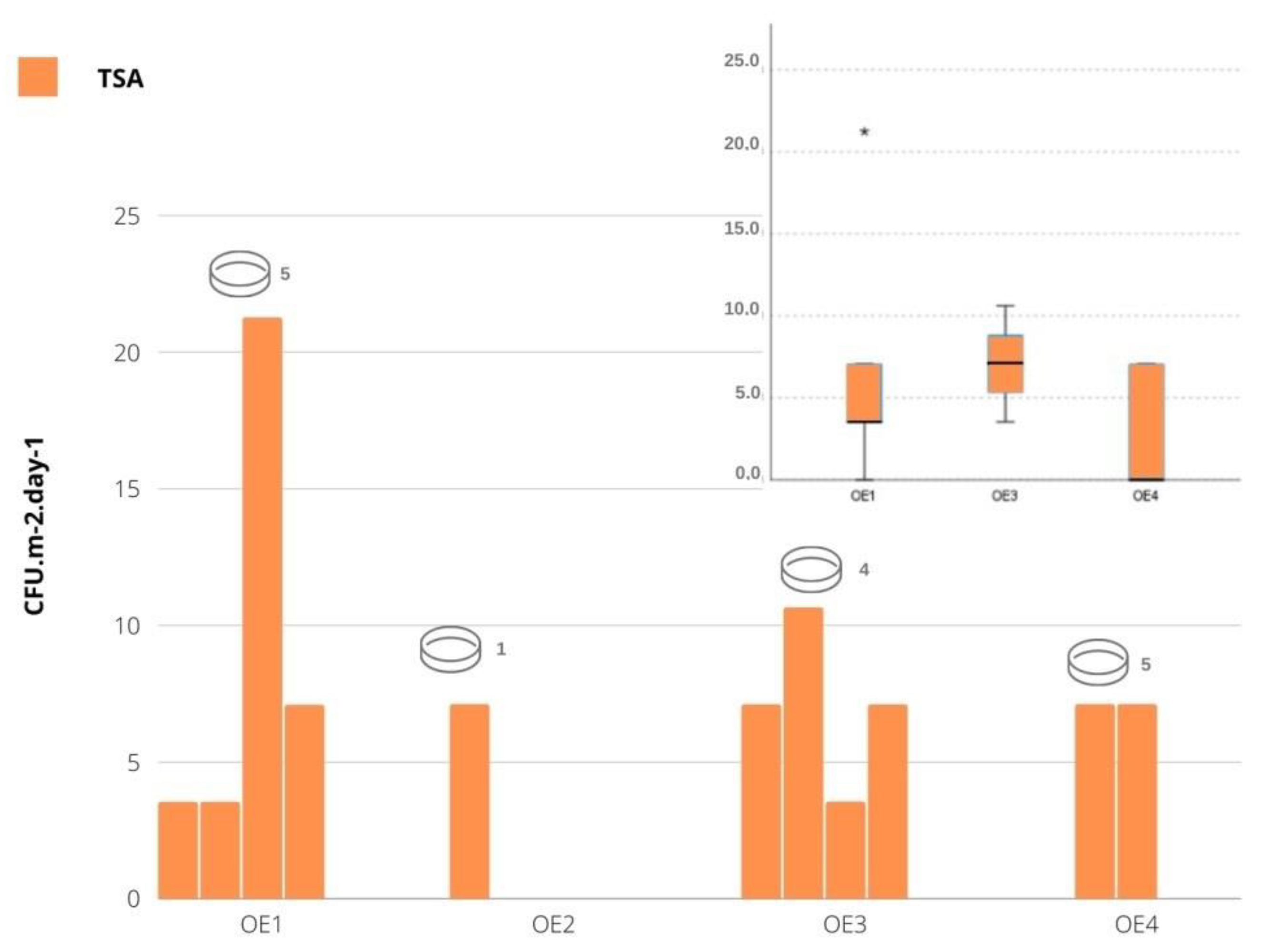
3.1. Viable Bacterial Contamination

Total bacteria contamination ranged from 0 to 21.23 CFU·m−2 in one of the textile’s working area (OE1) and from 0 to 7.08 CFU·m−2 in the other one (OE4). It ranged from 3.54 to 10.62 CFU·m−2 in the paintings area (OE3) and in OE2 the counts were 7.08 CFU·m−2. Among the sampled areas, no statistically significant differences were detected ( χ K W 2 ( 2 ) = 2.498 ,     p = 0.287 ), with the sculpture area excluded, since it only had one observation. From the analysis of Figure 3 (boxplot), it can be seen that OE3 displays the highest contamination in TSA and the OE1 is the one presenting higher variability. No gram-negative bacteria (VRBA) were detected in any of the areas sampled.

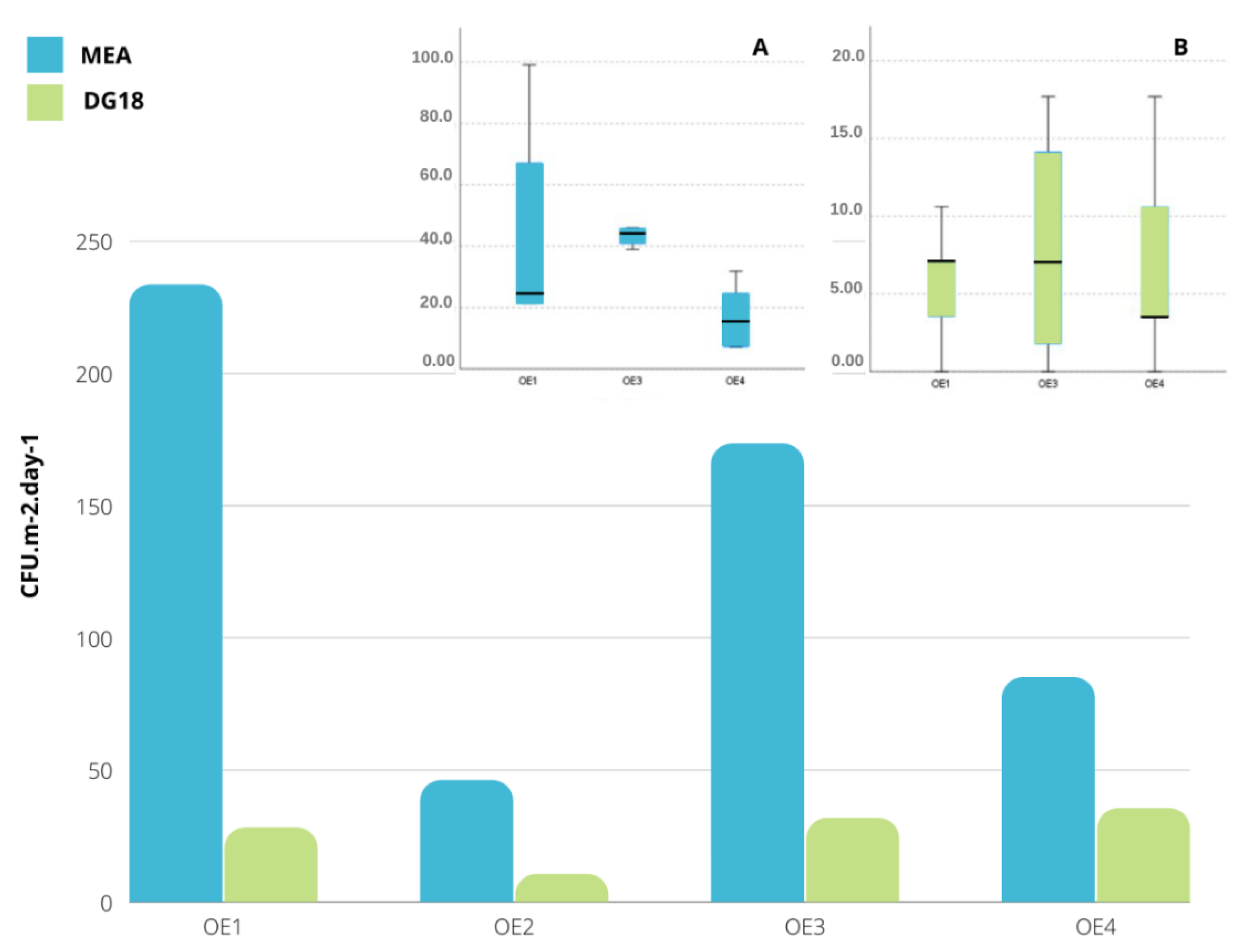
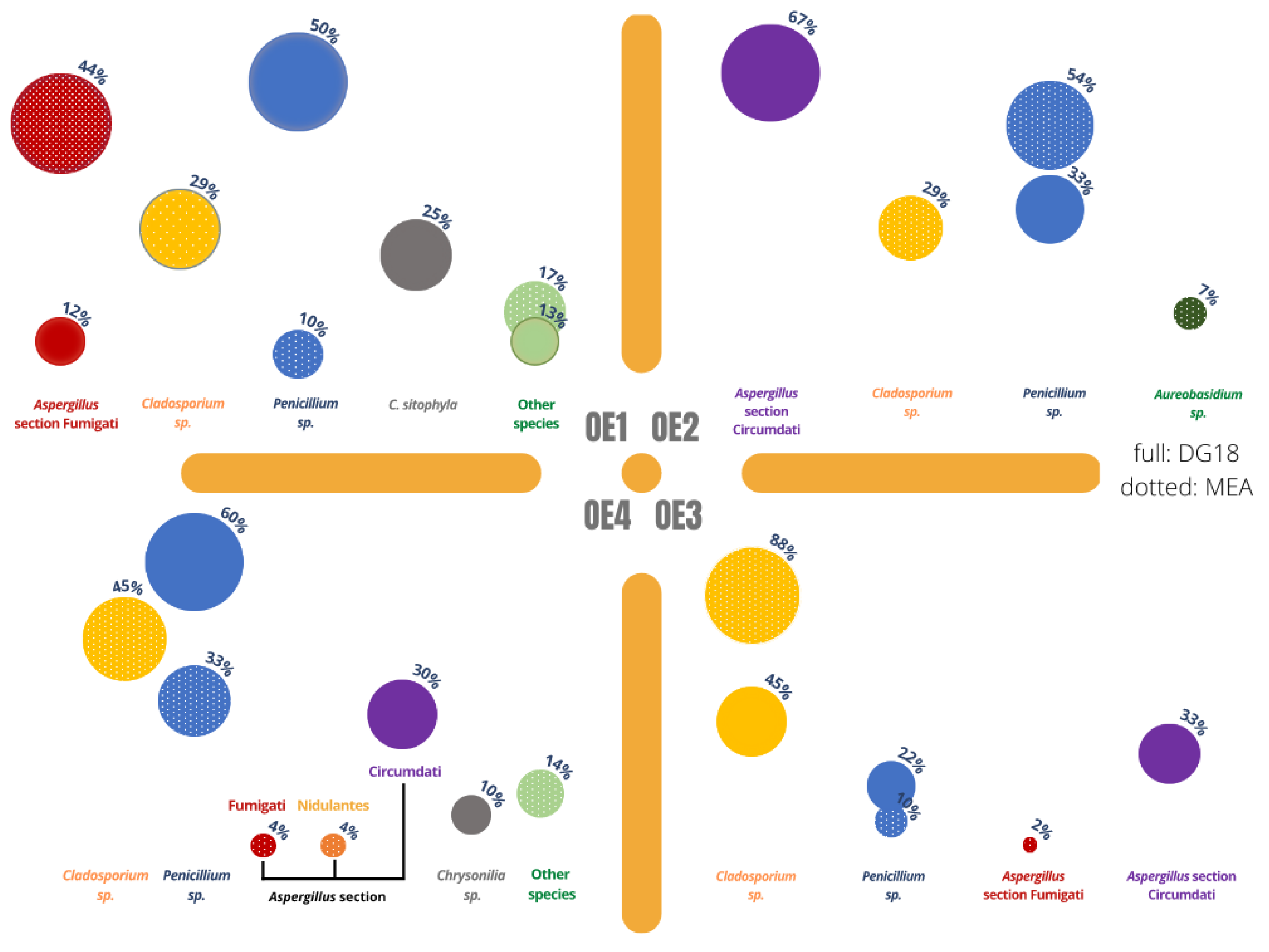
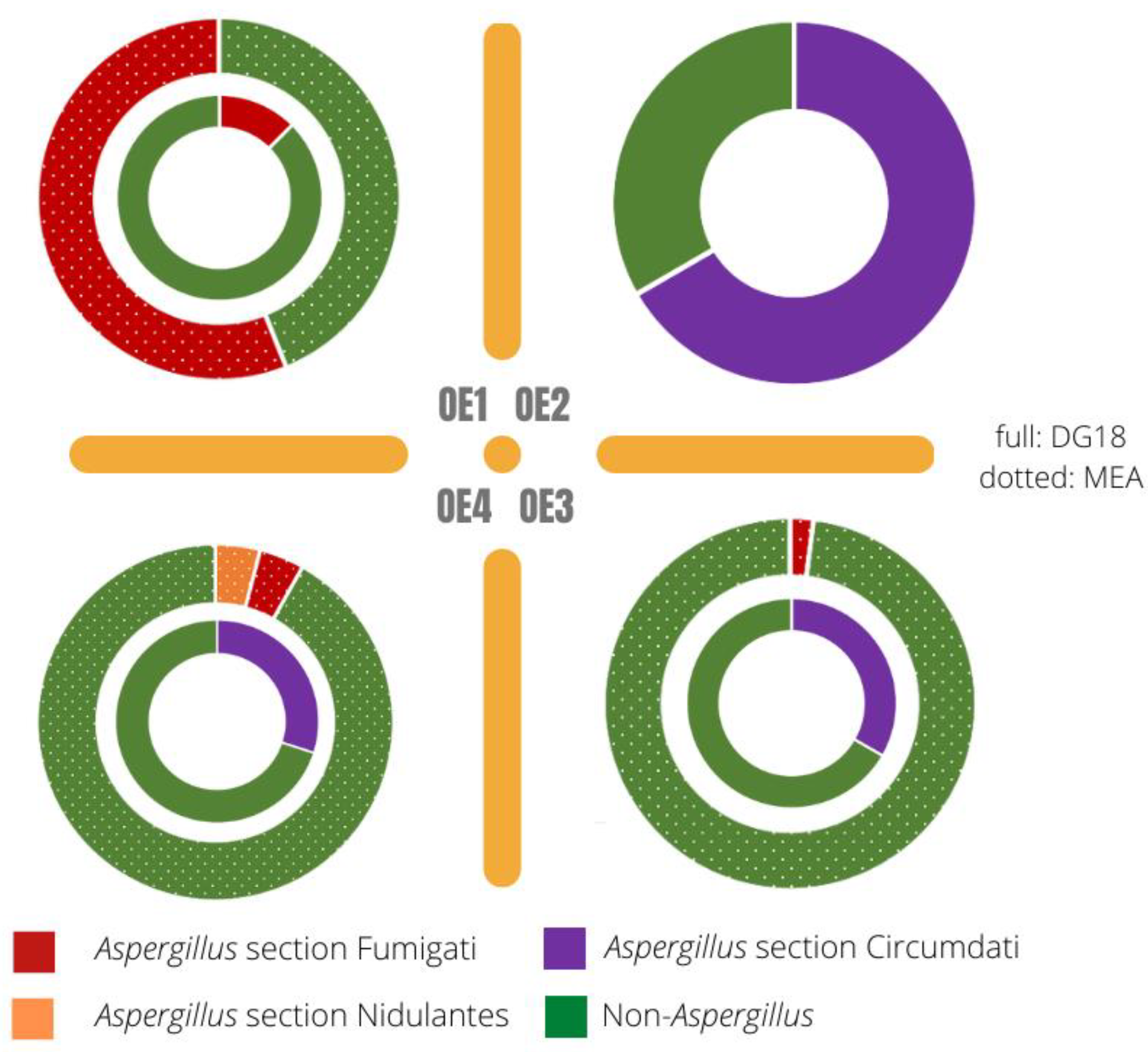
3.2. Viable Fungal Contamination

Total fungal contamination in indoor sites was 233.5 CFU·m−2·day−1 on MEA and 28.3 CFU·m−2·day−1 on DG18 in the OE1 (textiles); 46 CFU·m−2·day−1 on MEA and 10.6 CFU·m−2·day−1 on DG18 in OE2 (sculpture); 173 CFU·m−2·day−1 in MEA and 31.8 CFU·m−2·day−1 in DG18 in the paintings area (OE3). The highest fungal counts were found in the OE1 textiles area (233.5 CFU·m−2·day−1 on MEA; 28.3 CFU·m−2·day−1 on DG18) (Figure 4).
In both MEA and DG18 medium, no statistically significant differences were detected between the sampled areas ( χ K W 2 ( 2 )   = 5.696, p = 0.058 and χ K W 2 ( 2 )   = 0.177, p = 0.915, respectively). However, from the analysis of Figure 4 (boxplot), it can be seen that on MEA, OE3 was the one that presented the greatest fungal contamination and OE1 the one with the greatest variability. In DG18, one can see that OE1 and OE3 were the ones with the highest contamination, with the OE3 showing greater variability (sculpture area excluded as before).
Concerning fungal distribution per sampling location, Figure 4 presents the quantitative results and Figure 5 presents the qualitative results.
The highest number of fungal species was obtained on the OE1 (8 species MEA; 4 species DG18, more details on all identified genera are on Table 2), closely followed by OE4 (6 species MEA; 3 species DG18). Aspergillus section Fumigati was the most common species obtained in OE1 in MEA (43.94%) while Penicillium sp. was the most common species obtained on DG18 (50%) in this same location; in the sculpture area—OE2—the most common species were Aspergillus section Circumdati on DG18 (66.66%) and Penicillium sp. on MEA (53.85%), and in OE3 (paintings) the most common species observed was Cladosporium sp. both in MEA (87.76%) and in DG18 (44.44%). In OE4 Cladosporium sp. accounted for 44.84% in MEA, while Penicillium sp. was the prevalent genera on DG18 (60%).
Regarding Aspergillus sp., they were present in all the assessed environments. The highest value obtained in MEA (43.94%) was found in OE1 and in OE2 in DG18 (67%). On MEA, the areas with the highest values of the genera were the OE1 (43.94%), followed by the OE4 (8.16%). OE3 displays a lower percentage in MEA (2.04%). The genus was not identified in the MEA media in OE2 but accounts for 67% of the CFUs identified in this location when using DG18.
On DG18, two Aspergillus sections were identified, namely Circumdati (96.67%) and Fumigati (12.50%), also on MEA, two sections were reported, as follows: Fumigati (50.06%) and Nidulantes (4.08%). As for sections identification in OE1, one Aspergillus section was detected both on MEA (43.94% Fumigati) and DG18 (12.50% Fumigati). In OE2, no sections were detected on MEA and one section was detected on DG18 (66.66% Circumdati). In the painting area, OE3, two sections were identified, namely section Fumigati on MEA (2.04%) and section Circumdati on DG18 (33.33%). In the second textiles area, OE4, two sections were detected on MEA (4.08% Fumigati and 4.08% Nidulantes) and one section was detected on DG18 (30.00% Circumdati) (Figure 6).
Regarding species diversity on MEA, OE1 was the one with higher diversity (Shannon index (H) = 1.514, Simpson index (D) = 3.408) (Table 2).

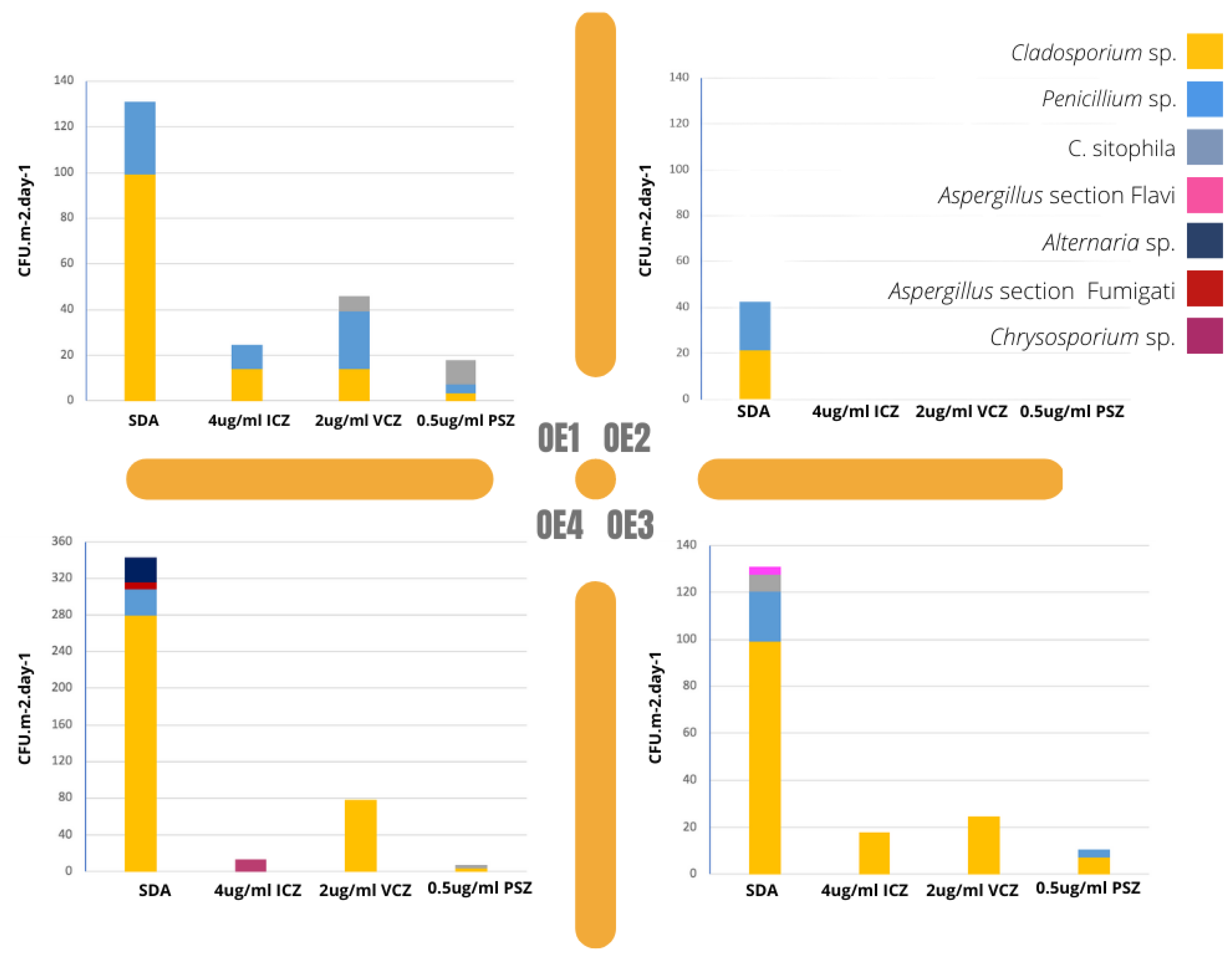
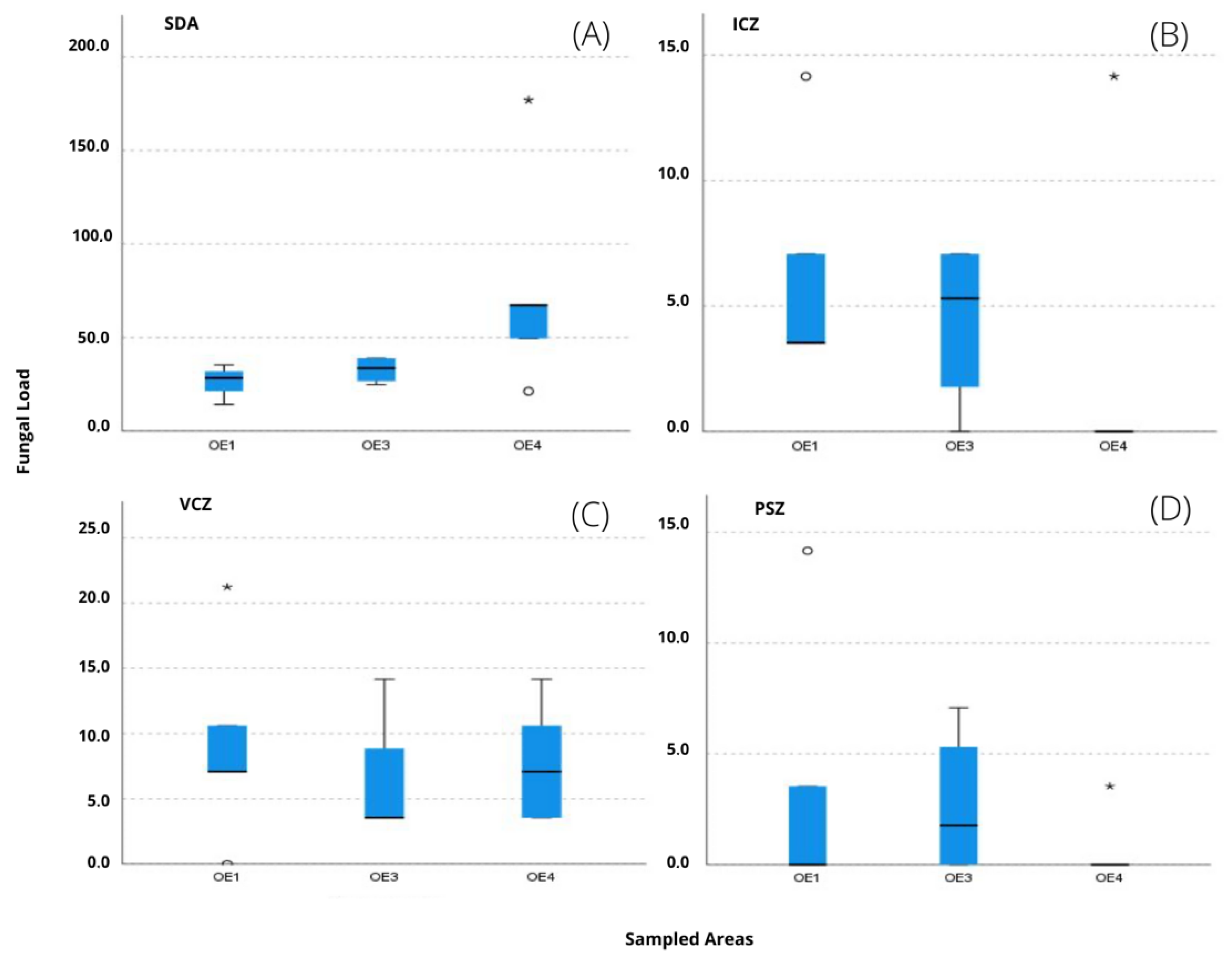
3.3. Fungal Growth in Azole-Supplemented Media

Regarding fungal contamination in azole-supplemented media, the results are presented in Figure 7. The most contaminated local was OE4 and the most frequent fungi in SDA (Saboraud Dextrose Agar) was Cladosporium sp. (5.0 × 10² CFU·m−2·day−1), followed by Penicillium sp. (1.0 × 10² CFU·m−2·day−1). Looking into each azole supplement individually, the most frequent fungi was Cladosporium sp. in voriconazole (VCZ) (7.8 × 101 CFU·m−2·day−1) and itraconazole (ICZ) (3.2 × 101 CFU·m−2·day−1), followed by Penicillium sp. in voriconazole (2.5 × 101 CFU·m−2·day−1). Aspergillus sections Flavi (3.5 CFU·m−2·day−1) and Fumigati (7.0 CFU·m−2·day−1) were observed in SDA but not in any of the azole added media. The media with PSZ recorded the lowest contamination rate in all settings.
Among the sampled areas, no statistically significant differences were detected in any of the media (p’s > 0.05) regarding fungal contamination. However, from the analysis of Figure 8A, it can be seen that, for the SDA, the textile area OE4 was the one with the highest values. The ICZ supplemented media (B) with the highest contamination was recorded in OE3. The textile working areas, both OE1 and OE4 presented similar results in in VCZ (C). Lastly, OE3 registered the highest contamination in PSZ (D).

3.4. Contamination of EDCs by Mycotoxins and Cytotoxicity Assessment

EDC sampling did not reveal the presence of any of the 38 mycotoxins evaluated at the sampled sites. Regarding the assessment of cell viability of the three distinct cell lines exposed to EDC, the results showed a majority of non-cytotoxic EDC, with only two samples exhibiting an IC50 value of 10 mm2/mL (one in A549 lung epithelial cells and another in SK cells).

3.5. Correlation Analysis

Only a significant positive correlation of moderate intensity was detected between bacterial contamination in TSA and fungal contamination in MEA (rS = 0.621, p = 0.013), revealing that greater bacterial contamination in TSA is related to greater fungal contamination in MEA (Table 3).

4. Discussion

Conservators–restorers are a professional class that has yet to see some of its occupational hazards being correctly addressed [11,34,35,36]. It is not difficult to imagine experiencing allergic respiratory or dermatological symptoms when handling old documentation or textiles that have been exposed to dust or pesticides in the past [12,34,37,38]. As far as health issues are concerned, allergic symptoms (eye and throat pruritus, nasal congestion) and traumatic disorders from the adoption of awkward and stressful body positions, are the top cause of absenteeism or even the abandonment of the profession (personal inquiry). Performing a comprehensive microbiological analysis is, therefore, an essential step to understand the environments where these workers perform their activities. The assessed locations, as mentioned earlier, are mostly dedicated to the treatment of organic-based art pieces (in this case textiles, canvas and wood-based sculptures).
In what concerns fungal contamination assessment, different results were obtained with the two different culture media applied (MEA and DG18), following the trend also found in other occupational environments already assessed [27,39]. In fact, a greater number of fungal counts was obtained in MEA; what is expected since DG18 favors the presence of xerophilic fungi and restricts some fungi with fast growing rates, such as the Mucorales order [27,40]. There appears to be no correlation to be made between the type of materials being handled and the results obtained. The differences in results, for both bacterial and fungal contamination, can be related to the accumulation of dust and the movement due to the performed activities indoors [41,42,43] that may cause its displacement and deposition on the EDC. In addition, outdoor air flow, besides human activities, was reported to be the leading factor responsible for the fungal contamination indoors [44]. The two locations where the fungal counts were higher had other artworks that were not being handled at the moment, which adds to the dust deposition, although this was more evident in OE3. The fungal counts follow the same tendency as bacteria results, which also deem OE1 as the more contaminated. Because conservators spend long hours in the same position, devoted to one task, future studies might find it useful to include the collection of dust from each particular art piece for analysis, as well as the surrounding dust, as performed in this study.
In terms of variability—for fungi only—Figure 5 and Figure 6 confirm a higher variability in the two locations where textiles are handled. This variability is particularly noticeable in OE4, where three sections of Aspergillus were identified. Thus, the workstations OE1 and OE4 can be identified as hotspots for widespread Aspergillus and seen as a priority for risk management intervention. Previously, this genus was found to be the most prevalent on historical textiles and also reported that even with maintenance of recommended conditions, the growth of xerophilic fungi cannot be prevented [45]. In addition, several Aspergillus sections (Circumdati, Flavi, Fumigati and Nidulantes) considered as indicators of harmful fungal contamination were observed, indicating the need for the implementation of corrective measures [46,47]. For this particular case, and because it is organic materials that we are discussing, it is important also to address the impact these fungal and bacterial contaminations can have on the artwork itself. In fact, the textiles’ microbial colonization can promote conservators–restorers occupational exposure to these microbiologic risks but also the biodeterioration of historical textiles. Fungi can promote the biodeterioration of cellulosic and proteinaceous archaeological textiles, whereas bacteria are the main players for silk biodegradation [45,48]. Bacteria, however, do need higher water availability, and these art objects are normally kept within safe intervals of relative humidity and temperature [49].
The screening of azole resistance revealed one textile handling environment (OE4) as the one with the highest fungal load in Sabouraud media. Although a limited fungal diversity was found, with predominant Cladosporium and Penicillium sp., two important Aspergillus sections with toxigenic potential—Flavi and Fumigati—were present in the assessed environments. The results come to add another Aspergillus genus to the ones already identified (Figure 6). These results are in accordance with previous studies assessing textile specimens contaminated by fungi in Slovene and Jordanian museums, in which the dominant contaminant fungal species also belonged to the genus Penicillium, Aspergillus and Cladosporium [45].
The fact that Cladosporium sp. and Penicillium sp. were able to grow at tested concentrations of voriconazole and itraconazole must be further investigated in order to determine fungal susceptibility to other commonly used medical azoles for the treatment of fungal infections in humans. Moreover, although not determined during this assessment, azole-resistant Fumigati isolates have been increasingly reported in different environments [50] and described as a potential health menace, especially for immunocompromised individuals [18,51]. A deeper knowledge of fungal susceptibilities to azoles or other biocides is also relevant to guide the adoption of better fungal control strategies in restoration environments and suitable policies on cultural heritage conservation, while ensuring the maintenance of the effectiveness of antifungals in the treatment of infections in humans and animals [52,53].
As reported, none of the mycotoxins analyzed were detected. This might be related with many factors, such as the occupational environment characteristics (e.g., humidity, temperature, availability of fungal nutrients), and the materials being used and handled [54,55]. However, this does not mean that exposure might not happen in this occupational environment since the environmental conditions are constantly changing. It might also depend of the previous contamination of the materials and pieces to be handled and their storage conditions. Further studies are warranted to confirm these scenarios.
No relevant cytotoxicity was observed in EDC samples and the reduced number of samples does not allow further conclusions. Nevertheless, the use of relevant cell lines to assess biological effects and estimate health risks is a valid strategy for risk assessment [56,57,58].

5. Conclusions

This work presents important findings concerning microbial contamination in an occupational setting not commonly studied. In this particular study, the workplace environment where textiles were handled revealed itself as more prone to a diverse fungal contamination and, more specifically, to Aspergillus sp. contamination. The results, obtained with the techniques identified above, show low contaminated environments overall, considering and comparing with other settings. Thus, after the results are analyzed and compared with different studies using the same methodology, the behavior and particularities of these particular professionals and settings may warrant a conjugation of different approaches. Conservation and restoration is a task that is developed slowly and with care, and this means the workers do not engage in activities that contribute to the aerosolization of particles and, therefore, of fungi and bacteria. This is good news because it reduces the exposure to air contaminants, and good cleaning practices may render the working place safe. However, future studies must accommodate not only the environmental approach, with the EDC placed strategically on the workplace, but also the analysis of the painting, textile, etc., being treated because the conservator works in close proximity to the artefact and shares a micro-atmosphere with the piece itself. Comparing the EDC results with the results obtained by vacuum cleaning, the artefact will possibly increase our knowledge on the particularities of these settings.
As such, in future studies an innovative approach (One Health approach)—simultaneously targeting workplaces, workers (and users) and the cultural heritage—should be implemented to allow researchers to map the potential risk of microorganism’s dissemination and then, if needed, define an appropriate remediation strategy to simultaneously protect the health of workers and users and prevent further biodeterioration on the cultural heritage artefacts.

Author Contributions

Conceptualization, C.V. and A.C.P.; methodology, C.V. and A.C.P.; formal analysis, C.V., R.C., M.D., B.G., P.P., E.C., M.T., R.K., E.S., S.V. and L.A.C.; investigation, C.V. and A.C.P.; resources, C.V. and A.C.P.; writing—original draft preparation, C.V. and A.C.P.; writing—review and editing, C.V. and A.C.P.; supervision, C.V. and A.C.P.; project administration, C.V. and A.C.P.; funding acquisition, C.V. and A.C.P. All authors have read and agreed to the published version of the manuscript.

Funding

This work was supported by FCT—Fundação para a Ciência e a Tecnologia, I.P. (Portugal), within the scope of the PhD Grant UI/BD/151431/2021 and by the Polish Minister of Science and Higher Education, under the program “Regional Initiative of Excellence” in 2019–2022 (Grant No. 008/RID/2018/19). Catarina Pinheiro is under a researcher contract with the HERCULES Laboratory, Évora University (CEECIND/02598/2017).

Institutional Review Board Statement

Not applicable.

Informed Consent Statement

Not applicable.

Data Availability Statement

Through the UIDB/05608/2020 and UIDP/05608/2020.

Acknowledgments

H&TRC authors gratefully acknowledge the FCT/MCTES national support through the UIDB/05608/2020 and UIDP/05608/2020. Catarina Pinheiro gratefully acknowledges the FCT/MCTES national support through the UIDB/04449/2020 and UIDP/04449/2020.

Conflicts of Interest

The authors declare no conflict of interest.

References

  1. Sterflinger, K. Fungi: Their role in deterioration of cultural heritage. Fungal Biol. Rev. 2010, 24, 47–55. [Google Scholar] [CrossRef]
  2. Pinheiro, A.C.; Sequeira, S.O.; Macedo, M.F. Fungi in archives, libraries, and museums: A review on paper conservation and human health. Crit. Rev. Microbiol. 2019, 45, 686–700. [Google Scholar] [CrossRef] [PubMed]
  3. Directive 89/391/EEC, on the Introduction of Measures to Encourage Improvements in the Safety and Health of Workers at Work. Off. J. Eur. Commun. 1989. Available online: https://osha.europa.eu/pt/legislation/directives/the-osh-framework-directive/1 (accessed on 2 February 2022).
  4. Lugauskas, A.; Krikštaponis, A.; Šveistyté, L. Airborne fungi in industrial environments—Potential agents of respiratory diseases. Ann. Agric. Environ. Med. 2004, 11, 19–25. [Google Scholar] [PubMed]
  5. Skorge, T.D.; Eagan, T.; Eide, G.E.; Gulsvik, A.; Bakke, P.S. Indoor exposures and respiratory symptoms in a Norwegian community sample. Thorax 2005, 60, 937–942. [Google Scholar] [CrossRef] [PubMed] [Green Version]
  6. Rusca, S.; Charrière, N.; Droz, P.O.; Oppliger, A. Effects of bioaerosol exposure on work-related symptoms among Swiss sawmill workers. Int. Arch. Occup. Environ. Health. 2008, 81, 415–421. [Google Scholar] [CrossRef] [PubMed]
  7. Domingo, J.L.; Nadal, M. Domestic waste composting facilities: A review of human health risks. Environ. Int. 2009, 35, 382–389. [Google Scholar] [CrossRef]
  8. Pinzari, F.; Fanelli, C.; Canhoto, O.; Magan, N. Electronic Nose for the Early Detection of Moulds in Libraries and Archives. Indoor Built Environ. 2004, 13, 387–395. [Google Scholar] [CrossRef]
  9. Bembibre, C.; Strlič, M. Smell of heritage: A framework for the identification, analysis and archival of historic odours. Heritage Sci. 2017, 5, 2. [Google Scholar] [CrossRef] [Green Version]
  10. Zielinska-Jankiewicz, K.; Kozajda, A.; Piotrowska, M.; SzadkowskaStanczyk, I. Microbiological contamination with moulds in workenvironment in libraries and archive storage facilities. Ann. Agric. Environ. Med. 2008, 15, 71–78. [Google Scholar] [PubMed]
  11. Varnai, V.M.; Macan, J.; Ćalušić, A.L.; Prester, L.; Macan, B.K. Upper Respiratory Impairment in Restorers of Cultural Heritage. Occup. Med. 2011, 61, 45–52. [Google Scholar] [CrossRef] [PubMed] [Green Version]
  12. Roussel, S.; Reboux, G.; Millon, L.; Parchas, M.-D.; Boudih, S.; Skana, F.; Delaforge, M.; Rakotonirainy, M.S. Microbiological evaluation of ten French archives and link to occupational symptoms. Indoor Air 2012, 22, 514–522. [Google Scholar] [CrossRef] [PubMed]
  13. Mesquita, N.; Portugal, A.; Videira, S.; Rodríguez-Echeverría, S.; Bandeira, A.; Santos, M.; Freitas, H. Fungal diversity in ancient documents. A case study on the Archive of the University of Coimbra. Int. Biodeterior. Biodegrad. 2009, 63, 626–629. [Google Scholar] [CrossRef]
  14. Karbowska-Berent, J.; Górny, R.L.; Strzelczyk, A.B.; Wlazło, A. Airborne and dust borne microorganisms in selected Polish libraries and archives. Build. Environ. 2011, 46, 1872–1879. [Google Scholar] [CrossRef]
  15. Skóra, J.; Zduniak, K.; Gutarowska, B.; Rembisz, D. Harmful biological agents at museum workposts. Med. Pracy 2012, 63, 153–165. [Google Scholar]
  16. Michaelsen, A.; Pinzari, F.; Barbabietola, N.; Piñar, G. Monitoring the effects of different conservation treatments on paper-infecting fungi. Int. Biodeterior. Biodegrad. 2013, 84, 333–341. [Google Scholar] [CrossRef] [Green Version]
  17. Snelders, E.; Camps, S.M.T.; Karawajczyk, A.; Schaftenaar, G.; Kema, G.H.; Van Der Lee, H.A.; Klaassen, C.H.; Melchers, W.J.G.; Verweij, P.E. Triazole Fungicides Can Induce Cross-Resistance to Medical Triazoles in Aspergillus fumigatus. PLoS ONE 2012, 7, e31801. [Google Scholar] [CrossRef] [Green Version]
  18. Verweij, P.E.; Chowdhary, A.; Melchers, W.J.; Meis, J.F. Azole Resistance in Aspergillus fumigatus: Can We Retain the Clinical Use of Mold-Active Antifungal Azoles? Clin. Infect Dis. 2016, 62, 362–368. [Google Scholar] [CrossRef] [Green Version]
  19. Schoustra, S.E.; Debets, A.J.; Rijs, A.J.; Zhang, J.; Snelders, E.; Leendertse, P.C.; Melchers, W.J.; Rietveld, A.G.; Zwaan, B.J.; Verweij, P.E. Environmental Hotspots for Azole Resistance Selection of Aspergillus fumigatus, the Netherlands. Emerg. Infect. Dis. 2019, 25, 1347–1353. [Google Scholar] [CrossRef] [Green Version]
  20. Bennett, J.W.; Klich, M. Mycotoxins. Clin. Microbiol. Rev. 2003, 16, 497–516. [Google Scholar] [CrossRef] [Green Version]
  21. Marin, S.; Ramos, A.J.; Cano-Sancho, G.; Sanchis, V. Mycotoxins: Occurrence, toxicology, and exposure assessment. Food Chem. Toxicol. 2013, 60, 218–237. [Google Scholar] [CrossRef]
  22. IARC. International Agency for Research on Cancer. Improving Public Health through Mycotoxin Control. IARC Scientific Publication No. 158. 2012. Available online: https://publications.iarc.fr/Book-And-Report-Series/Iarc-Scientific-Publications/Improving-Public-Health-Through-Mycotoxin-Control-2012 (accessed on 2 June 2022).
  23. Assunção, R.; Martins, C.; Viegas, S.; Viegas, C.; Jakobsen, L.S.; Pires, S.; Alvito, P. Climate change and the health impact of aflatoxins exposure in Portugal—an overview. Food Addit. Contam. Part A 2018, 35, 1610–1621. [Google Scholar] [CrossRef] [PubMed] [Green Version]
  24. Halstensen, A.S. Species-specific Fungal DNA in Airborne Dust as Surrogate for Occupational Mycotoxin Exposure? Int. J. Mol. Sci. 2008, 9, 2543–2558. [Google Scholar] [CrossRef] [PubMed]
  25. Viegas, S.; Martins, C. The Usefulness of Human Biomonitoring in the Case of Mycotoxins Exposure Assessment. In Reference Module in Life Sciences; Elsevier: Amsterdam, The Netherlands, 2020; pp. 1–6. [Google Scholar] [CrossRef]
  26. Viegas, C.; Sousa, P.; Dias, M.; Caetano, L.A.; Ribeiro, E.; Carolino, E.; Twarużek, M.; Kosicki, R.; Viegas, S. Bioburden contamination and Staphylococcus aureus colonization associated with firefighter's ambulances. Environ. Res. 2021, 197, 111125. [Google Scholar] [CrossRef] [PubMed]
  27. Viegas, C.; Gomes, B.; Pimenta, R.; Dias, M.; Cervantes, R.; Caetano, L.A.; Carolino, E.; Twarużek, M.; Soszczyńska, E.; Kosicki, R.; et al. Microbial contamination in firefighter Headquarters’: A neglected occupational exposure scenario. Build. Environ. 2022, 213, 108862. [Google Scholar] [CrossRef]
  28. Viegas, C.; Gomes, B.; Dias, M.; Carolino, E.; Aranha Caetano, L. Aspergillus Section Fumigati in Firefighter Headquarters. Microorganisms 2021, 9, 2112. [Google Scholar] [CrossRef]
  29. Hoog, D.; Guarro, J.; Gene, G.; Figueras, M. Atlas of Clinical Fungi—The Ultimate Benchtool for Diagnosis; Version 4.1.4; Utr. Centraalbureau voor Schimmelcultures: Utrecht, The Netherlands, 2016. [Google Scholar]
  30. Arendrup, M.C.; Rodriguez-Tudela, J.L.; Lass-Flörl, C.; Cuenca-Estrella, M.; Donnelly, J.P.; Hope, W. EUCAST technical note on anidulafungin. Clin. Microbiol. Infect. 2013, 19, 278–280. [Google Scholar] [CrossRef] [Green Version]
  31. European Committee on Antimicrobial Susceptibility Testing (EUCAST). Routine and Extended Internal Quality Control for MIC Determination and Agar Dilution for Yeasts, Moulds and Dermatophytes as Recommended by EUCAST. Version 5.0; EUCAST: 2020 Online Platform. Available online: http://www.eucast.org (accessed on 2 February 2022).
  32. Viegas, C.; Almeida, B.; Caetano, L.A.; Afanou, A.; Straumfors, A.; Veríssimo, C.; Gonçalves, P.; Sabino, R. Algorithm to assess the presence of Aspergillus fumigatus resistant strains: The case of Norwegian sawmills. Int. J. Environ. Health Res. 2020, 32, 963–971. [Google Scholar] [CrossRef]
  33. Hanelt, M.; Gareis, M.; Kollarczik, B. Cytotoxicity of mycotoxins evaluated by the MTT-cell culture assay. Mycopathologia 1994, 128, 167–174. [Google Scholar] [CrossRef]
  34. Santos, M.; Almeida, A. Principais Riscos e Fatores de Risco Ocupacionais dos Conservadores- Restauradores de Obras de Arte, bem como Doenças Profissionais associadas e medidas de Proteção recomendadas. Rev. Port. Saúde Ocup. Online 2019, 8, S108–S155. [Google Scholar] [CrossRef]
  35. Santos, M.; Almeida. A. Danos Ocupacionais associados ao Cádmio, com ênfase no setor da Conservação e Restauro de Obras de Arte. Rev. Port. Saúde Ocup. Online 2020, 9, S59–S73. [Google Scholar] [CrossRef]
  36. Pinheiro, A.C.; Ramos, A. Heritage Keepers: The Perils in Textile Conservation. Heritage 2021, 4, 4716–4725. [Google Scholar] [CrossRef]
  37. Blaser, L.; Peckham, S. Archives Conservators Discussion Group 2005: Hazardous holdings. Book Pap. Group Annu. 2005, 24, 73–83. [Google Scholar]
  38. Žuskin, E.; Schachter, E.N.; Mustajbegović, J.; Pucarin-Cvetković, J.; Lipozenčić, J. Occupational health hazards of artists. Acta Dermatovenerol. Croatica. 2007, 15, 167–177. [Google Scholar]
  39. Viegas, C.; Twarużek, M.; Dias, M.; Carolino, E.; Soszczyńska, E.; Caetano, L.A. Cytotoxicity of Aspergillus Section Fumigati Isolates Recovered from Protection Devices Used on Waste Sorting Industry. Toxins 2022, 14, 70. [Google Scholar] [CrossRef] [PubMed]
  40. Viegas, C.; Pena, P.; Dias, M.; Gomes, B.; Cervantes, R.; Carolino, E.; Twarużek, M.; Soszczyńska, E.; Kosicki, R.; Caetano, L.A.; et al. Microbial contamination in waste collection: Unveiling this Portuguese occupational exposure scenario. J. Environ. Manag. 2022, 314, 115086. [Google Scholar] [CrossRef] [PubMed]
  41. Buttner, M.P.; Stetzenbach, L.D. Monitoring airborne fungal spores in an experimental indoor environment to evaluate sampling methods and the effects of human activity on air sampling. Appl. Environ. Microbiol. 1993, 59, 219–226. [Google Scholar] [CrossRef] [PubMed] [Green Version]
  42. Chen, Q.; Hildemann, L.M. The Effects of Human Activities on Exposure to Particulate Matter and Bioaerosols in Residential Homes. Environ. Sci. Technol. 2009, 43, 4641–4646. [Google Scholar] [CrossRef] [PubMed]
  43. Heo, K.J.; Lim, C.E.; Kim, H.B.; Lee, B.U. Effects of human activities on concentrations of culturable bioaerosols in indoor air environments. J. Aerosol Sci. 2017, 104, 58–65. [Google Scholar] [CrossRef]
  44. Adams, R.I.; Miletto, M.; Taylor, J.W.; Bruns, T.D. Dispersal in microbes: Fungi in indoor air are dominated by outdoor air and show dispersal limitation at short distances. ISME J. 2013, 7, 1262–1273. [Google Scholar] [CrossRef] [Green Version]
  45. Kavkler, K.; Gunde-Cimerman, N.; Zalar, P.; Demšar, A. Fungal contamination of textile objects preserved in Slovene museums and religious institutions. Int. Biodeterior. Biodegrad. 2015, 97, 51–59. [Google Scholar] [CrossRef]
  46. AIHA. American Industrial Hygiene Association. In Field Guide for the Determination of Biological Contaminants in Environmental Samples; Fairfax: Toronto, ON, Canada, 1996. [Google Scholar]
  47. Viegas, C.; Santos, P.; Almeida, B.; Monteiro, A.; Carolino, E.; Gomes, A.Q.; Viegas, S. Electrostatic dust collector: A passive screening method to assess occupational exposure to organic dust in primary health care centers. Air Qual. Atmos. Health 2019, 12, 573–583. [Google Scholar] [CrossRef]
  48. Gutarowska, B.; Pietrzak, K.; Machnowski, W.; Milczarek, J. Historical textiles—A review of microbial deterioration analysis and disinfection methods. Text. Res. J. 2017, 87, 2388–2406. [Google Scholar] [CrossRef]
  49. Canadian Conservation Institute. CCI Notes 13/1: Textiles and the Environment. Canadian Conservation Institute (CCI), 2013. Available online: https://www.canada.ca/en/conservation-institute/services/conservation-preservation-publications/canadian-conservation-institute-notes/textiles-environment.html (accessed on 24 July 2022).
  50. Gonçalves, P.; Melo, A.; Dias, M.; Almeida, B.; Caetano, L.A.; Veríssimo, C.; Viegas, C.; Sabino, R. Azole-Resistant Aspergillus fumigatus Harboring the TR34/L98H Mutation: First Report in Portugal in Environmental Samples. Microorganisms 2021, 9, 57. [Google Scholar] [CrossRef] [PubMed]
  51. Zahar, J.R.; Jolivet, S.; Adam, H.; Dananché, C.; Lizon, J.; Alfandari, S.; Boulestreau, H.; Baghdadi, N.; Bay, J.O.; Bénéteau, A.M.; et al. French recommendations on control measures to reduce the infectious risk in immunocompromised patients. J. Mycol. Med. 2017, 27, 449–456. [Google Scholar] [CrossRef]
  52. Fisher, M.C.; Hawkins, N.J.; Sanglard, D.; Gurr, S.J. Worldwide emergence of resistance to antifungal drugs challenges human health and food security. Science 2018, 360, 739–742. [Google Scholar] [CrossRef] [Green Version]
  53. Verweij, P.E.; Lucas, J.A.; Arendrup, M.C.; Bowyer, P.; Brinkmann, A.J.; Denning, D.W.; Dyer, P.S.; Fisher, M.C.; Geenen, P.L.; Gisi, U.; et al. The one health problem of azole resistance in Aspergillus fumigatus: Current insights and future research agenda. Fungal Biol. Rev. 2020, 34, 202–214. [Google Scholar] [CrossRef]
  54. Viegas, C.; Monteiro, A.; dos Santos, M.; Faria, T.; Caetano, L.A.; Carolino, E.; Gomes, A.Q.; Marchand, G.; Lacombe, N.; Viegas, S. Filters from taxis air conditioning system: A tool to characterize driver's occupational exposure to bioburden? Environ. Res. 2018, 164, 522–529. [Google Scholar] [CrossRef]
  55. Viegas, S.; Viegas, C.; Oppliger, A. Occupational Exposure to Mycotoxins: Current Knowledge and Prospects. Ann. Work. Expo. Health 2018, 62, 923–941. [Google Scholar] [CrossRef]
  56. Swain, R.J.; Kemp, S.J.; Goldstraw, P.; Tetley, T.D.; Stevens, M.M. Assessment of Cell Line Models of Primary Human Cells by Raman Spectral Phenotyping. Biophys. J. 2010, 98, 1703–1711. [Google Scholar] [CrossRef] [Green Version]
  57. Heussner, A.H.; Dietrich, D.R. Primary porcine proximal tubular cells as an alternative to human primary renal cells in vitro: An initial characterization. BMC Cell Biol. 2013, 14, 55. [Google Scholar] [CrossRef] [Green Version]
  58. Viegas, C.; Twarużek, M.; Dias, M.; Almeida, B.; Carolino, E.; Soszczyńska, E.; Viegas, S.; Caetano, L.A. Cytotoxicity of filtering respiratory protective devices from the waste sorting industry: A comparative study between interior layer and exhalation valve. Environ. Int. 2021, 155, 106603. [Google Scholar] [CrossRef] [PubMed]
Figure 1. Sampling locations, OE1 and OE4 for textiles, OE2 for sculpture and OE3 for the painting’s restoration studio.
Microorganisms 10 01595 g001
Figure 2. Example of one of the EDCs placed at the assessed locations. A request for non-disturbance accompanies the EDC.
Microorganisms 10 01595 g002
Figure 3. Bacterial contamination in the four sampled locations observed in TSA. On the top right is the boxplot for comparison of bacterial contamination in TSA medium between sampled areas (excluding OE2, since it has a single record). * severe outlier.
Microorganisms 10 01595 g003
Figure 4. Total fungal counts on MEA and DG18 in samples from different areas (CFU·m−2·day−1). Boxplot for comparison of fungal contamination in MEA (A) and DG18 (B) media between sampled areas (excluding the sculpture area (OE2), since it has a single record).
Microorganisms 10 01595 g004
Figure 5. Fungal qualitative distribution in both MEA (dotted circles) and DG18 (full circles). Assigned to each circle is the percentage each genus occupies within the total results.
Microorganisms 10 01595 g005
Figure 6. Aspergillus sections distribution by media and sampling place.
Microorganisms 10 01595 g006
Figure 7. Fungal contamination (by azole screening method) per local (CFU·m−2·day−1). SDA = Saboraud dextrose agar; ICZ = itraconazole; VCZ = voriconazole; PSZ = posaconazole. Please mind the different scale used in OE4.
Microorganisms 10 01595 g007
Figure 8. Boxplot for comparison of fungal load in SDA (A), ICZ (B), VCZ (C) and PSZ (D) between sampled areas (excluding the sculpture area (OE2), since it has a single record). Please note the different scales used. * severe outlier; ° moderate outlier.
Microorganisms 10 01595 g008
Table 1. LOD values for the analyzed samples.
MycotoxinsLOD
(ng/g)
15-Acetyldeoxynivalenol8
3-Acetyldeoxynivalenol Aflatoxin B14
Aflatoxin B21
Aflatoxin G11
Aflatoxin G21
Aflatoxin M11
Deepoxydeoxynivalenol5
Deoxynivalenol8
Deoxynivalenol-3-glucoside5
Diacetoxyscirpenol2
Fumonisin B14
Fumonisin B23
Fusarenon X10
Griseofulvin2
HT-2 toxin4
Mevinolin7
Monoacetoxyscirpenol2
Mycophenolic acid3
Neosolaniol3
Nivalenol4
Ochratoxin A2
Ochratoxin B2
Patulin8
Roquefortine C2
Sterigmatocystin1
T-2 tetraol2
T-2 toxin2
T-2 triol5
Zearalanone2
Zearalenone1
α-Zearalanol2
α-Zearalenol2
β-Zearalanol2
β-Zearalenol3
Table 2. Shannon and Simpson indexes to assess species diversity.
Sampled AreasSpeciesCulture Media MEA (CFU/g−1·day−1)Shannon Index (H)Simpson Index (D)
OE1Alternaria sp.3.5391.5143.408
Aspergillus section Fumigati102.619
Aureobasidium sp.10.616
Chrysosporium sp.7.077
Cladosporium sp.67.233
Fusarium verticilloides7.077
Penicillium sp. 24.770
Rhizopus sp.10.616
Totals8233.546
OE2Aureobasidium sp.3.5390.2271.199
Cladosporium sp.17.693
Penicillium sp.24.770
Totals346.001
OE3Aspergillus section Fumigati3.5390.4160.425
Cladosporium sp. 152.159
Penicillium sp. 17.693
Totals3173.390
OE4Aspergillus section Fumigati3.5391.9770.371
Aspergillus section Nidulantes3.539
Cladosporium sp.38.924
Mucor sp.3.539
Penicillium sp. 28.309
Trichoderma sp.7.077
Totals684.926
Table 3. Study of the relationship between bacterial and fungal contamination and fungal resistance: Results of Spearman’s correlation coefficient.
BacteriaFungiFungal Resistance
VRBAMEADG18SDAITZVCZPSZ
BacteriaTSA-0.621 *−0.0030.191−0.149−0.4030.480
VRBA ------
FungiMEA 0.209−0.329−0.072−0.1770.239
DG18 −0.0160.139−0.4820.073
Fungal resistanceSDA −0.269−0.5050.157
ICZ −0.0980.018
VCZ −0.108
*. Correlation is significant at the 0.05 level (2-tailed).
Publisher’s Note: MDPI stays neutral with regard to jurisdictional claims in published maps and institutional affiliations.

