Article

Biochemical Diversity, Pathogenicity and Phylogenetic Analysis of Pseudomonas viridiflava from Bean and Weeds in Northern Spain

by
Ana M. Fernández-Sanz
1,
M. Rosario Rodicio
2 and
Ana J. González
1,*
1
Programa de Patología Vegetal, Servicio Regional de Investigación y Desarrollo Agroalimentario (SERIDA), Ctra AS-267, PK 19, 33300 Villaviciosa, Spain
2
Área de Microbiología, Departamento de Biología Funcional, Universidad de Oviedo, Julián Clavería 6, 33006 Oviedo, Spain
*
Author to whom correspondence should be addressed.
Microorganisms 2022, 10(8), 1542; https://doi.org/10.3390/microorganisms10081542
Submission received: 5 July 2022 / Revised: 26 July 2022 / Accepted: 27 July 2022 / Published: 29 July 2022
(This article belongs to the Special Issue Plant Pathogenic Microorganisms: State-of-the-Art Research in Spain)

Abstract

:
Pseudomonas viridiflava was originally reported as a bean pathogen, and subsequently as a wide-host range pathogen affecting numerous plants species. In addition, several authors have reported the epiphytic presence of this bacterium in “non-host plants”, which may act as reservoir of P. viridiflava and source of inoculum for crops. A new biotype of this bacterium, showing an atypical LOPAT profile, was found in Asturias, a Northern region of Spain, causing significant damage in beans, kiwifruit, lettuce, and Hebe. In order to investigate the involvement of weeds in bean disease, samples were collected from beans and weeds growing in the same fields. A total of 48 isolates of P. viridiflava were obtained, 39 from weeds and 9 from beans. 48% and 52% of them showed typical (L− O− P+ A− T+) and atypical (L+ O− P v A− T+) LOPAT profiles, and they displayed high biochemical diversity. Regarding virulence factors, the T-PAI and S-PAI pathogenicity islands were found in 29% and 70.8% of the isolates, 81.2% displayed pectinolytic activity on potato slices, and 59% of the weed isolates produced symptoms after inoculation on bean pods. A phylogenetic tree based on concatenated rpoD, gyrB, and gltA sequences separated the strains carrying S-PAI and T-PAI into different clusters, both containing isolates from beans and weeds, and pathogenic as well as non-pathogenic strains. Closely related strains were found in the two hosts, and more than half of the weed isolates proved to be pathogenic in beans. This is consistent with the role of weeds as a reservoir and source of inoculum for bean infection. Detection of P. viridiflava in weeds throughout the year further supports these roles.

1. Introduction

Pseudomonas viridiflava (Burkholder) Dowson was first described by Burkholder in 1930 as a bean (Phaseolus vulgaris L.) pathogen and subsequently reported to cause disease in many other crop plants. These bacteria produce several symptoms, including necrosis, spots, and rots, affecting different parts of the plant (stems, leaves, blossoms, and roots). For instance, it has been shown to cause bacterial blight in kiwifruit, necrosis in tomato, bacterial blight in pea, rots in alfalfa, carrot and Cucumis sativus, bacterial shoot blight in sweet crab apple, bacterial canker in stone fruit trees, and bacterial leaf spot in lettuce [1,2,3,4,5,6,7,8,9,10]. According to this, P. viridiflava is regarded as a generalist pathogen, able to attack multiple host species [11,12]. In Asturias, a Northern region of Spain, a new emerging biotype of P. viridiflava (termed BT2) was described [13]. This biotype differs from the typical biotype by the production of yellowish mucoid material in the sucrose medium used for the levan test, and by a variable pectinolytic activity on different potato varieties. First detected in 1999, BT2 has caused significant damage in several crops such as beans, in which symptoms ranged from red spots mainly in the petioles and pods; to plant death due to systemic infection associated with the destruction of the medulla; kiwifruit, showing dark brown spots in floral buds that developed into extensive rot, leading to the collapse of floral buds or the production of small or distorted fruits; and lettuce, generating soft rot of intermediate leaves, which could also progress and result in plant death [13]. Subsequently, this biotype was also shown to cause defoliation in Hebe [14], and has been isolated from rapeseed in South Korea [15].
P. viridiflava is a close relative of P. syringae. Based on phylogenetic analysis, it is located within the P. syringae species complex [16,17,18]. However, unlike P. syringae, the knowledge about the mechanisms of virulence in P. viridiflava is still limited. In this species, two alternative pathogenicity islands, T-PAI and S-PAI, have been detected [19]. Some isolates harbor a tripartite PAI, equivalent to that found in P. syringae (T-PAI), while in others the PAI has a single component (S-PAI). T-PAI and S-PAI occupy different chromosomal locations, but only one of them is present in each individual isolate [19]. T-PAI consists of a central hrp/hrc (hypersensitivity reaction and pathogenicity/hypersensitivity reaction and conserved) cluster, flanked by CEL (conserved effector locus) and EEL (exchangeable effector locus) regions. The hrp/hrc cluster encodes a Type III secretion system (TTSS) responsible for the formation of a syringe-like structure used to translocate effectors into the host cell. These effectors are encoded by the CEL and EEL loci. S-PAI is also composed by a hrp/hrc cluster, but it is not flanked by the CEL and EEL regions. Instead, the cluster is interrupted by an insertion that harbors genes encoding effector proteins [19]. Other virulence factors detected in P. viridiflava are the enzyme pectate lyase and extracellular proteases related to plant tissue maceration [20,21].
Apart from acting as a crop pathogen, P. viridiflava also exists as an epiphyte on weeds. Gitaitis et al. [22] found this bacterium on weeds associated with onion crops in the USA and observed that weed control is necessary to avoid plant disease. The weed species from which P. viridiflava was isolated included Oenothera laciniata Hill, Taraxacum officinale Weber, Fumaria officinalis L., Gnaphalium purpureum L., Sonchus asper (L.) Hill, Lepidium virginicum L., and Raphanus raphanistrum L. Similarly, Basavand & Khodaygan [23] detected P. viridiflava in Alisma plantago-aquatica, a perennial weed in rice fields in Northern Iran.
In the present study, we performed a comparative analysis of P. viridiflava isolated from the common bean type “granja asturiana”, and from weeds growing in the bean fields. “Granja asturiana” beans are one of the most popular food products in our region, have great culinary value within the Asturian gastronomy, and are an important economic resource for small farmers. Therefore, the possible role of weeds as a reservoir of P. viridiflava and their involvement in transmission of the pathogen to bean crops are worthy of investigation. In this study, strains isolated from the two hosts were characterized, their phylogenetic relationships established, and weed isolates were tested for pathogenicity on bean.

2. Materials and Methods

2.1. Bacterial Isolates, Culture Conditions and Phenotypic Characterization

Forty-eight isolates of P. viridiflava from weeds and beans were recovered (39 and 9, respectively) in 10 fields located in four councils of Asturias: Navia (Anleo), Valdés (Busto 1 and 2, Constancios, Ronda and Pontigón), Tineo (Carbajal, Bárcena and Yerbo), and Siero (Argüelles).
Bacteria were identified by means of the KOH test, to determine their Gram stain [24], and by standard biochemical tests. These included oxidation-fermentation of glucose [25], the LOPAT determinative tests [26], utilization of mannitol, m-inositol, erythritol, and sorbitol in Hellmers broth [27], utilization of homoserine, D-tartrate, sucrose, L-lactate, trigonelline, quinate, betaine and adonitol in Ayer’s solid medium [28], and hydrolysis of esculin, gelatin [29], casein, and Tween 80 [30]. Isolates showing identical features and collected from the same field site and host at the same period of the year, were considered as a single strain. In this way, a total of 39 strains were identified, 30 and 9 from weeds and bean, respectively.

2.2. 16S rDNA Sequencing and ARDRA (Amplified Ribosomal DNA Restriction Analysis

16S rDNA was amplified by PCR, with primers described by Edwards et al. [31], and ARDRA carried out with the SacI and HinfI restriction enzymes [13].

2.3. Detection of Pathogenicity Islands

Presence of T-PAI was detected with primers hoppsyAr1 (CYGGCTATGATTGATAAACGCATCG) and shcAf1 (GGCGCACTTAACCCTCTGKTCAA TGA) [19], while the presence of S-PAI and absence of T-PAI was revealed with primers R1-orf41f1 (GCCTTGCCTCTGATCTCATTC) and R1-dsorf78r1 (GTAGCAT TCGGCATATCCC) (M. San José, personal communication). The fragments expected are of 1113 bp for T-PAI and 889 bp for S-PAI.

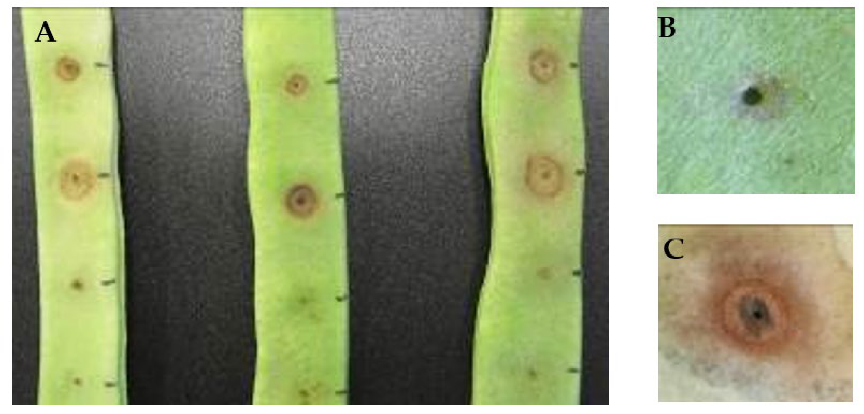
2.4. Pathogenicity Assays

Pathogenicity was tested by inoculation of isolates grown on King B medium on pods of beans cv. Helda with a sterile toothpick [32]. The assays were repeated with three replicates each time.

2.5. Phylogenetic Analysis

The phylogenetic analysis was performed for the 39 detected strains (see above), using the rpoD (RNA polymerase sigma D factor), gltA (citrate synthase), and gyrB (DNA gyrase subunit B) genes [33]. Amplification was conducted as in Hwang et al. [34]. The obtained fragments were sequenced by Secugen S.L. (Spain) or Eurofins (Germany). Sequences were submitted to GenBank (accession numbers MT683625-MT683672, MT709110-MT709148, and ON838894-ON838932, for gyrB, rpoD and gltA, respectively). Concatenated sequences of the three genes were aligned using Clustal W [35], and phylogenetic trees were constructed using Maximum-Likelihood with the Tamura–Nei model [36]. Their topological robustness was evaluated by bootstrap analysis based on 1000 replicates using Mega 6 software [37]. Sequences from P. viridiflava DSM 6694T, P. asturiensis LPPA 221T and P. protegens ChaoT were included as references.
In addition, the number of segregating sites (S) and the mean of the nucleotide diversity (π), defined as the average number of nucleotide differences by site between sequences of the whole population [38], were calculated both for the individual genes and the concatenated sequences, also using the Kimura two parameters model [37].

3. Results and Discussion

3.1. Identification and Biochemical Characterization of the Isolates

Forty eight isolates identified as P. viridiflava by ARDRA, and recovered during the period 2007–2009, were included in the present study. Thirty nine of them were isolated from weeds and nine from beans (Table 1). All isolates shared the ARDRA profile characteristically associated with P. viridiflava (not shown) and were Gram-negative bacilli.
Regarding the LOPAT scheme, presence of the two previously described biotypes was revealed, with 48% of the isolates (20 from weeds and 3 from beans) showing the typical (L−, O−, P+, A−, T+), and 52% (19 from weeds and 6 from beans) showing the atypical (L+, O−, Pv, A−, T+) biotype. The latter profile has persisted in beans in Asturias, at least since 2003 when it was first reported. In addition, it has also been described in South Korea in rapeseed [15]. Other biochemical features of the isolates are compiled in Table 2.
Results of the biochemical tests were highly variable, distributing the 48 isolates into 33 biochemical profiles (BP1 to BP33; Table 1 and Table S1). A correlation between profile and host plant or sample site was not found. Thus, several profiles were associated with the same host or field, and the same profile was shared by isolates from different hosts and sites. This wide variability makes phenotypic identification rather difficult. Consistent results were only obtained for the sucrose and lactose tests, both negative, and for the tobacco, esculin, quinate, and xylose tests, all positive, in 100% of the isolates.
Following the proposal of Billing [39], Wilkie et al. [40] found that the use of sucrose and tartrate could help in the initial identification of members of the species. Our results coincided in the case of sucrose utilization but not of D-tartrate, a test in which 29% of the isolates were positive. Nor do they agree with the results of Sarris et al. [41] who studied 18 isolates of P. viridiflava obtained from different hosts in Crete (Greece) and found no variability in the biochemical tests performed, except for the L-tartrate test. Regarding the latter, isolates obtained from tomato were positive, while the type strain was negative, as well as isolates from other hosts.

3.2. Occurrence of the Bacterium in Weeds and Bean Samples

In this study, P. viridiflava was isolated from twelve genera/species of weeds: Capsella bursa-pastoris, Chenopodium album, Cyperus rotundus, Fumaria sp, Galinsoga parviflora, Hypochaeris radicata, Malva sylvestris, Senecio vulgaris, Solanum nigrum, Sonchus oleraceus, Stellaria media, and Trifolium sp., and from three unidentified weeds which were different to each other (Table 1). The bacterium was most frequently found in Fumaria sp. (nine isolates from eight samples), followed by S. olereaceus and S. nigrum (four isolates from four samples), and C. rotundus (four isolates from three samples). This further expands the already wide host range of P. viridiflava which, as far as we know, has not been previously reported in 10 out of the 12 weed species/genera mentioned before. However, the bacterium has already been described by Gitaitis et al. [22] in Sonchus sp and Fumaria sp. associated with an onion crop. It is important to note that none of the weeds showed disease symptoms, consistent with an epiphytic existence of P. viridiflava in weeds.
Unlike Gitaitis et al. [22], who only isolated the bacteria from weeds during the onion growing season, we verified their presence throughout the year, i.e., before, during, and after the crop season. The survival of P. viridiflava in five species of weeds had already been described by Aysan and Uygur [42] before and after the tomato crop, and by Mariano and McCarter [43] also on tomato. In the latter study, persistence of the bacterium on the surface of the leaves of two weed species was observed by electron microscopy, for at least 16 weeks.
Pseudomonas viridiflava was less frequently detected in bean samples than in weeds. This species was isolated as a sole pathogen in 8% of the bean samples tested and together with P. syringae pv. phaseolicola in 1.1%. In contrast, a previous study in our region revealed P. viridiflava as the only pathogen in 28% of the samples, and together with P. syringae pv. phaseolicola and pv. syringae in a small percentage [13]. These differences could be due to the fact that the bean fields sampled in the present study had a significant presence of halo blight caused by P. syringae pv. phaseolicola, which could have displaced the less aggressive P. viridiflava. In any case, the simultaneous presence of P. viridiflava with other pathogens like P. syringae pv. syringae or pv. phaseolicola in the same sample highlights the epiphytic and opportunistic nature of the former species. Moreover, the relatively frequent detection of P. viridiflava in weeds, suggests that they could be an important reservoir and source of inoculum for crops. This is particularly true in Asturias, where the climatic conditions: mild temperatures, frequent rainfall, and high relative humidity values, are favorable both for growing of the weeds and for the development of the disease.

3.3. PAI Distribution, Pectinolysis Activity, and Pathogenicity Tests

Each isolate carried one of the pathogenicity islands (T-PAI or S-PAI) previously reported in P. viridiflava, thus confirming the polymorphism in terms of the presence/absence of these islands [19]. S-PAI was the most frequent, found in 26 and 8 of the isolates from weeds and beans, respectively (Table 1).
An important virulence factor in P. viridiflava is the enzyme pectate lyase, which causes maceration of plant tissues [20; 44]. In our study, 81.2% of the strains produced pectinolysis on potato slices, and 58.3% on bean pods (Table 1). Pectinolysis on potato slices was observed for isolates carrying both T-PAI (92.8%) and S-PAI (79.4%), although Jakob et al. [44] have shown that isolates with S-PAI had higher enzyme activity than those carrying T-PAI.
When pathogenicity tests were performed, different kinds of symptoms were observed (Figure 1). Some isolates caused only a small brown spot in the pods, around the inoculation point. Others produced a reddish or ferrous halo 24–48 h post-inoculation, which could be followed or not by maceration of the tissues, observed after 48–72 h. This coincides with results reported by Wilkie et al. [40] who, using the same method of inoculation, found different responses depending on the inoculated strain.
Twenty-seven strains of the 48 studied were pathogenic on bean pods, and one gave a variable response. Five of the pathogenic isolates came from bean samples, while the remaining 23 were from weeds belonging to the species C. bursa-pastoris, C. rotundus, C. album, Fumaria sp., G. parviflora, M. sylvestris, S. vulgaris, S. nigrum, S. media, and Trifolium sp. In total, 59% of the weed isolates were pathogenic to bean, which is highly relevant with respect to the epidemiology of the disease. This coincides with previous studies performed on tomato and onion, where P. viridiflava cause serious diseases [22,42,43] and a high percentage of weed isolates were pathogenic.
It is finally of note that the ability to produce maceration in bean pods did not correlate with biotype (since it was observed for 47.8% and 68% of the isolates with BT1 and BT2, respectively), nor with the type of PAI (soft rot was produce by 57% and 58.8% of the isolates with T-PAI and S-PAI, respectively). The latter observation is in line with results obtained by Bartoli et al. [45], who also found that the presence of S-PAI and T-PAI was not correlated with the ability to produce soft rot and with pathogenicity.

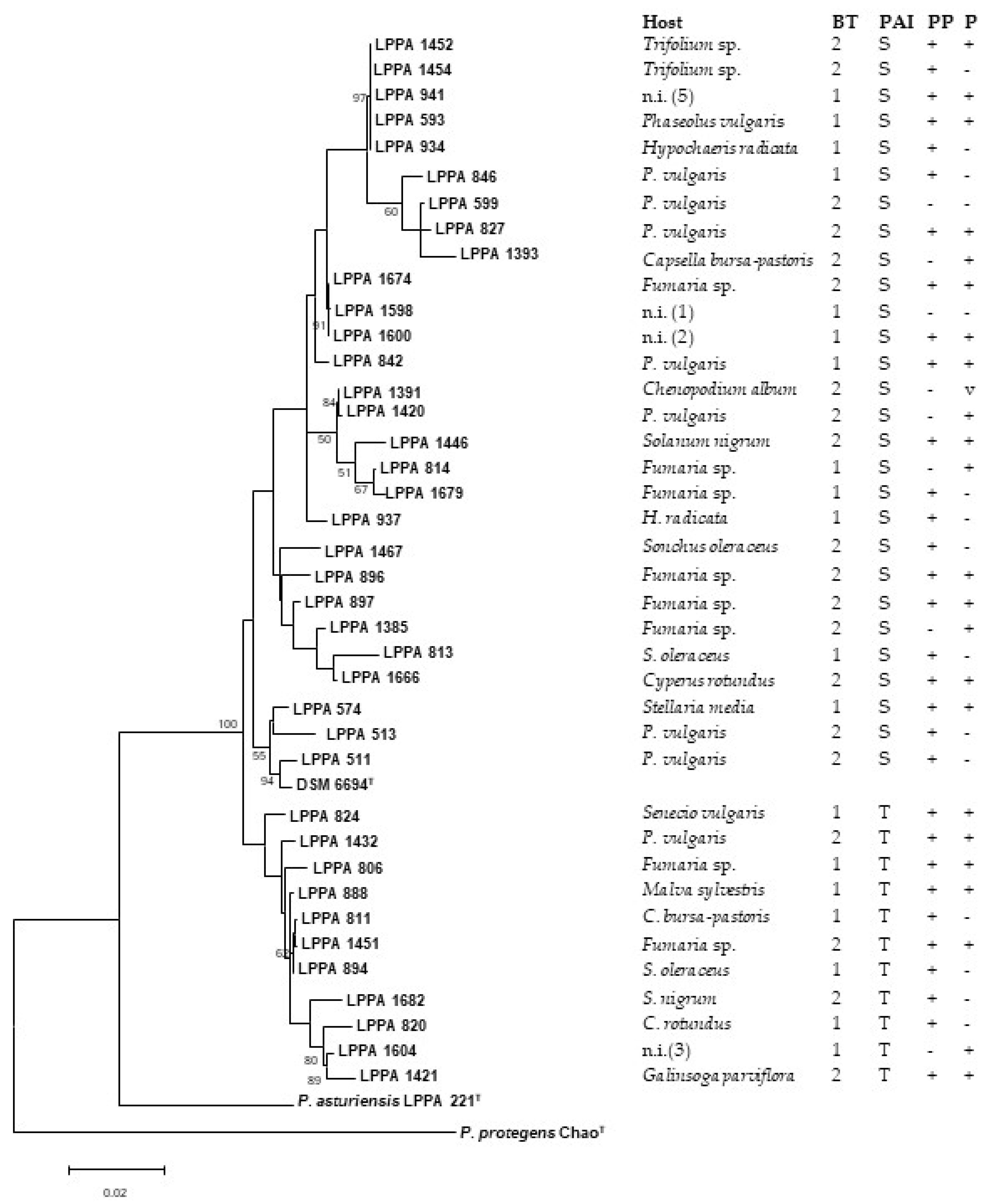
3.4. Phylogenetic Analysis

To establish the phylogenetic relationships of the isolates under study, the gyrB, rpoD and gltA genes from the 39 identified strains were sequenced. The 16S rDNA was not included because, being a highly conserved gene, it does not provide intraspecies variability [12]. The gyrB and rpoD genes were used for the investigation of populations of P. viridiflava in two previous studies [12,41].
The concatenated sequences of the three loci had a total length of 2450 bp (610 bp gyrB, 882 bp rpoD, and 958 bp gltA). By means of the Tajima’s test of neutrality, we have been able to verify that the concatenated sequence had 168 segregating sites and a nucleotide diversity (π) of 0.019. Tajima’s D-statistic test distinguishes between DNA sequences that evolve randomly (“neutrally”) from those that evolve under a non-random process. In our case, the D value was >0 so there are more haplotypes than number of segregating sites (Table 3). The gene that most contributed to nucleotide diversity was gyrB, a result already obtained by Yin et al. [46]. However, rpoD was the gene that provided nucleotide diversity (0.019617) closer to that obtained with the three concatenated genes (0.019432).
Figure 2 shows the phylogenetic tree based on the three concatenated gyrB, rpoD, and gltA genes of the 39 strains. They were distributed into two clades, A and B, which contain the S-PAI- and T-PAI-positive strains, respectively. In contrast, the strains were not separated according to biotype, soft rot activity, and pathogenicity, since isolates with these properties appear in the two clusters. Strains from weeds and beans were represented in both clusters, and closely related strains were obtained from the two hosts.

4. Conclusions

P. viridiflava was isolated not only from beans but also from fifteen different weeds growing in the same fields. The bacterium is reported for the first time in ten of the twelve identified weed genera/species (with the remaining three weeds, which could not be identified, but were all different than each other). Regardless of their origin, the isolates displayed wide biochemical diversity, hindering identification by traditional methods. Consistent results were only obtained for the sucrose and lactose tests (negative), and for the tobacco, esculin, quinate, and xylose tests (positive). Phylogenetic analysis with concatenated gyrB, rpoD, and gltA sequences separated the strains carrying S-PAI and T-PAI into two different clusters, with no correlation observed for other characteristics, such as plant host, LOPAT profile, pectinolytic activity, or pathogenicity. Detection of P. viridiflava before, during, and after the crop season shows survival of the bacteria in weeds throughout the year, hence supporting the role of weeds as reservoir of P. viridiflava, and as a source of inoculum for bean infection. The fact that 59% of the weed isolates behave as bean pathogens, and that some strains recovered from beans and weeds were closely related, further highlights the role of weeds on the epidemiology of the disease.

Supplementary Materials

The following supporting information can be downloaded at: https://www.mdpi.com/article/10.3390/microorganisms10081542/s1, Table S1: Biochemical profiles of the isolates under study; Table S2: Accession numbers of the gyrB, rpoD and gltA sequences used for phylogenetic analysis; Table S3: Pairwise distance matrix used to construct the phylogenetic tree.

Author Contributions

Conceptualization, A.J.G. and M.R.R.; methodology, A.M.F.-S. and A.J.G.; formal analysis, A.M.F.-S., M.R.R., and A.J.G.; investigation, A.M.F.-S. and A.J.G.; resources, A.J.G. and M.R.R.; data curation, A.M.F.-S.; writing—original draft preparation, A.J.G. and M.R.R.; writing—review and editing, A.M.F.-S., M.R.R., and A.J.G.; supervision, M.R.R. and A.J.G.; project administration, A.J.G.; funding acquisition, A.J.G. All authors have read and agreed to the published version of the manuscript.

Funding

This research was funded by Instituto Nacional de Investigación y Tecnología Agraria y Alimentaria, grant number RTA2005-00076 and RTA2008-00019, and Ana M Fernández-Sanz is recipient of a grant from the “Instituto Nacional de Investigación y Tecnología Agraria y Alimentaria”, both co-funded by the European Social Fund. The APC was funded by Consejería de Medio Rural y Cohesión Territorial, Government of the Principality of Asturias.

Institutional Review Board Statement

Not applicable.

Informed Consent Statement

Not applicable.

Data Availability Statement

The gyrB, rpoD and gltA sequences have been deposited in GenBank under accession numbers shown in Table S2.

Conflicts of Interest

The authors declare no conflict of interest.

References

  1. Young, J.M.; Cheesmur, G.J.; Welham, F.V.; Henshall, W.R. Bacterial blight of kiwifruit. Ann. Appl. Biol. 1988, 112, 91–105. [Google Scholar] [CrossRef]
  2. Yildiz, H.N.; Aysan, Y.; Sahin, F.; Cinar, O. Potential inoculum sources of tomato stem and pith necrosis caused by Pseudomonas viridiflava in the Eastern Mediterranean Region of Turkey. Z. Pflanzenkrankh. Pflanzenschutz 2004, 111, 380–387. [Google Scholar]
  3. Popović, T.; Ivanović, Ž.; Ignjatov, M. First report of Pseudomonas viridiflava causing pith necrosis of tomato (Solanum lycopersicum) in Serbia. Plant Dis. 2015, 99, 1033. [Google Scholar] [CrossRef]
  4. Martín-Sanz, A.; Palomo, J.L.; Pérez de la Vega, M.; Caminero, C. First report of bacterial blight caused by Pseudomonas viridiflava on pea in Spain. Plant Dis. 2010, 94, 128. [Google Scholar] [CrossRef]
  5. Heydari, A.; Khodakaramian, G.; Zafari, D. Characterization of Pseudomonas viridiflava causing alfalfa root rot disease in Hamedan Province of Iran. J. Plant Pathol. Microbiol. 2012, 3, 135. [Google Scholar] [CrossRef] [Green Version]
  6. Almeida, I.M.G.; Maciel, K.W.; Rodrigues Neto, J.; Beriam, L.O.S. Pseudomonas viridiflava in imported carrot seeds. Australas. Plant Dis. Notes 2013, 8, 17–19. [Google Scholar] [CrossRef] [Green Version]
  7. Al-Karablieh, N.; Mutlak, I.; Al-Dokh, A. Isolation and identification of Pseudomonas viridiflava, the causal agent of fruit rotting of Cucumis sativus. J. J. Agri. Sci. 2017, 13, 79–91. [Google Scholar]
  8. Choi, O.; Lee, Y.; Kang, B.; Kim, S.; Bae, J.; Kim, J. Bacterial shoot blight of sweet crab apple caused by Pseudomonas viridiflava. For. Pathol. 2020, 50, e12603. [Google Scholar] [CrossRef]
  9. Bophela, K.N.; Petersen, Y.; Bull, C.T.; Coutinho, T.A. Identification of Pseudomonas isolates associated with bacterial canker of stone fruit trees in the Western Cape, South Africa. Plant Dis. 2020, 104, 882–892. [Google Scholar] [CrossRef]
  10. Canik Orel, D. Biocontrol of bacterial diseases with beneficial bacteria in lettuce. Intern. J. Agric. Nat. Sci. 2020, 13, 108–117. [Google Scholar]
  11. Goumans, D.E.; Chatzaki, A.K. Characterization and host range evaluation of Pseudomonas viridiflava from melon, blite, tomato, chrysanthemum and eggplant. Eur. J. Plant Pathol. 1998, 104, 181–188. [Google Scholar] [CrossRef]
  12. Goss, E.M.; Kreitman, M.; Bergelson, J. Genetic diversity, recombination and cryptic clades in Pseudomonas viridiflava infecting natural populations of Arabidopsis thaliana. Genetics 2005, 169, 21–35. [Google Scholar] [CrossRef] [Green Version]
  13. González, A.J.; Rodicio, M.R.; Mendoza, M.C. Identification of an emergent and atypical Pseudomonas viridiflava lineage causing bacteriosis in plants of agronomic importance in a Spanish region. Appl. Environ. Microbiol. 2003, 69, 2936–2941. [Google Scholar] [CrossRef] [Green Version]
  14. González, A.J.; Rodicio, M.R. Pseudomonas viridiflava causing necrotic leaf spots and defoliation on Hebe spp. in Northern Spain. Plant Dis. 2006, 90, 830. [Google Scholar] [CrossRef]
  15. Myung, I.S.; Lee, Y.K.; Lee, S.W.; Kim, W.G.; Shim, H.S.; Ra, D.S. A new disease, bacterial leaf spot of rape, caused by atypical Pseudomonas viridiflava in South Korea. Plant Dis. 2010, 94, 1164. [Google Scholar] [CrossRef]
  16. Anzai, Y.; Kim, H.; Park, J.; Wakabayashi, H.; Oyaizu, H. Phylogenetic affiliation of the Pseudomonads based on 16S rRNA sequence. Int. J. Syst. Evol. Microbiol. 2000, 50, 1563–1589. [Google Scholar] [CrossRef] [Green Version]
  17. Yamamoto, S.; Kasai, H.; Arnold, D.L.; Jackson, R.W.; Vivian, A.; Harayama, S. Phylogeny of the genus Pseudomonas: Intrageneric structure reconstructed from the nucleotide sequences of gyrB and rpoD genes. Microbiology 2000, 146, 2385–2394. [Google Scholar] [CrossRef] [Green Version]
  18. Mulet, M.; Lalucat, J.; García-Valdés, E. DNA sequence-based analysis of the Pseudomonas species. Environ. Microbiol. 2010, 12, 1513–1530. [Google Scholar]
  19. Araki, H.; Tian, D.; Goss, E.M.; Jakob, K.; Halldorsdottir, S.S.; Kreitman, M.; Bergelson, J. Presence/absence polymorphism for alternative pathogenicity islands in Pseudomonas viridiflava, a pathogen of Arabidopsis. Proc. Natl. Acad. Sci. USA 2006, 103, 5887–5892. [Google Scholar] [CrossRef] [Green Version]
  20. Liao, C.H.; Hung, H.Y.; Chatterjee, A.K. An extracellular pectate lyase is the pathogenicity factor of the soft-rotting bacterium Pseudomonas viridiflava. Mol. Plant-Microbe Interact. 1988, 1, 199–206. [Google Scholar] [CrossRef]
  21. Liao, C.H.; McCallus, D.E.; Fett, W.F. Molecular characterization of two gene loci required for production of the key pathogenicity factor pectate lyase in Pseudomonas viridiflava. Mol. Plant-Microbe Interact. 1994, 7, 391–400. [Google Scholar] [CrossRef]
  22. Gitaitis, R.; Macdonald, G.; Torrance, R.; Hartley, R.; Sumner, D.R.; Gay, J.D.; Johnson, W.C., III. Bacterial streak and bulb rot of sweet onion: II. Epiphytic survival of Pseudomonas viridiflava in association with multiple weed hosts. Plant Dis. 1998, 82, 935–938. [Google Scholar] [CrossRef] [Green Version]
  23. Basavand, E.; Khodaygan, P. Common water-plantain, a new host of Pseudomonas viridiflava in rice fields in Iran. J. Plant Pathol. 2020, 102, 913. [Google Scholar] [CrossRef] [Green Version]
  24. Ryu, E. A simple method of differentiation between gram-positive and gram-negative organisms without staining. Kitasato Arch. Exp. Med. 1940, 17, 58–63. [Google Scholar]
  25. Hugh, R.; Leifson, E. The taxonomic significance of fermentative versus oxidative metabolism of carbohydrates by various Gram negative bacteria. J. Bacteriol. 1953, 66, 24–26. [Google Scholar] [CrossRef] [Green Version]
  26. Lelliott, R.A.; Billing, E.; Hayward, A.C. A determinative scheme for fluorescent plant pathogenic bacteria. J. Appl. Bacteriol. 1966, 29, 470–478. [Google Scholar] [CrossRef]
  27. Jansing, H.; Rudolph, K. A sensitive and quick test for determination of bean seed infestation by Pseudomonas syringae pv. phaseolicola. Z. Pflanzenkrankh. Pflanzenschutz 1990, 97, 42–55. [Google Scholar]
  28. Schaad, N.W.; Jones, J.B.; Chun, W. Laboratory Guide for Identification of Plant-Pathogenic Bacteria, 3rd ed.; CPL APS Press: St. Paul, MN, USA, 2001; p. 398. [Google Scholar]
  29. Noval, C. Medios de cultivo y pruebas de diagnóstico. In Manual de Laboratorio. Diagnóstico de Hongos, Bacterias y Nematodos Fitopatógenos; MAPA: Madrid, Spain, 1991; pp. 379–410. [Google Scholar]
  30. Goszczynska, T.; Serfortein, J. Milk-Tween agar, a semiselective medium for isolation and differentiation of Pseudomonas syringae pv. syringae, Pseudomonas syringae pv. phaseolicola and Xanthomonas axonopodis pv. phaseoli. J. Microbiol. Methods 1998, 32, 65–72. [Google Scholar] [CrossRef]
  31. Edwards, U.; Rogall, T.; Blöcker, H.; Emde, M.; Böttger, E.C. Isolation and direct complete nucleotide determination of entire genes. Characterization of a gene coding for 16S ribosomal RNA. Nucleic Acids Res. 1989, 17, 7843–7853. [Google Scholar] [CrossRef] [PubMed] [Green Version]
  32. Cheng, G.Y.; Legard, D.E.; Hunter, J.E.; Burr, T.J. Modified bean pod assay to detect strains of Pseudomonas syringae pv. syringae that cause bacterial brown spot of snap bean. Plant Dis. 1989, 73, 149–423. [Google Scholar] [CrossRef]
  33. Sarkar, S.F.; Guttman, D.S. Evolution of the core genome of Pseudomonas syringae, a highly clonal, endemic plant pathogen. Appl. Environ. Microbiol. 2004, 70, 1999–2012. [Google Scholar] [CrossRef] [Green Version]
  34. Hwang, M.S.H.; Morgan, R.L.; Sarkar, S.F.; Wang, P.W.; Guttman, D.S. Phylogenetic characterization of virulence and resistence phenotypes of Pseudomonas syringae. Appl. Environ. Microbiol. 2005, 71, 5182–5191. [Google Scholar] [CrossRef] [Green Version]
  35. Thompson, J.D.; Higgins, D.G.; Gibson, T.J. CLUSTAL W: Improving the sensitivity of progressive multiple sequence alignment through sequence weighting, position-specific gap penalties and weight matrix choice. Nucleic Acids Res. 1994, 22, 4673–4680. [Google Scholar] [CrossRef] [Green Version]
  36. Saitou, N.; Nei, M. The Neighbor-Joining method: A new method for reconstructing phylogenetic trees. Mol. Biol. Evol. 1987, 4, 406–425. [Google Scholar]
  37. Tamura, K.; Stecher, G.; Peterson, D.; Filipski, A.; Kumar, S. MEGA6: Molecular Evolutionary Genetics Analysis version 6. Mol. Biol. Evol. 2013, 30, 2725–2729. [Google Scholar] [CrossRef] [Green Version]
  38. Nei, M.; Kumar, S. Molecular Evolution and Phylogenetics; Oxford University Press: New York, NY, USA, 2000; p. 333. [Google Scholar]
  39. Billing, E. Pseudomonas viridiflava (Burkholder, 1930; Clara, 1934). J. Appl. Bacteriol. 1970, 33, 492–500. [Google Scholar] [CrossRef]
  40. Wilkie, J.P.; Dye, D.W.; Watson, D.R.W. Further hosts of Pseudomonas viridiflava. N. Z. J. Agric. Res. 1973, 16, 315–323. [Google Scholar] [CrossRef]
  41. Sarris, P.F.; Trantas, E.A.; Mpalantinaki, E.; Ververidis, F.; Goumas, D.E. Pseudomonas viridiflava, a multi host plant pathogen with significant genetic variation at the molecular level. PLoS ONE 2012, 7, e36090. [Google Scholar] [CrossRef]
  42. Aysan, Y.; Uygur, S. Epiphytic survival of Pseudomonas viridiflava, causal agent of pith necrosis of tomato, on weeds in Turkey. J. Plant Pathol. 2005, 87, 135–139. [Google Scholar]
  43. Mariano, R.D.; McCarter, S.M. Epiphytic survival of Pseudomonas viridiflava on tomato and selected weed species. Microb. Ecol. 2004, 26, 47–58. [Google Scholar] [CrossRef]
  44. Jakob, K.; Kniskern, J.M.; Bergelson, J. The role of pectate lyase and the jasmonic acid defense response in Pseudomonas viridiflava virulence. Mol. Plant-Microbe Interact. 2007, 20, 146–158. [Google Scholar] [CrossRef] [Green Version]
  45. Bartoli, C.; Berge, O.; Monteil, C.L.; Guilbaud, C.; Balestra, G.M.; Varvaro, L.; Jones, C.; Dangl, J.L.; Baltrus, D.A.; Sands, D.C.; et al. The Pseudomonas viridiflava phylogroups in the P. syringae species complex are characterized by genetic variability and phenotypic plasticity of pathogenicity-related traits. Environ. Microbiol. 2014, 16, 2301–2315. [Google Scholar] [CrossRef]
  46. Yin, H.; Cao, L.; Xie, M.; Chen, Q.; Qiu, G.; Zhou, J.; Wu, L.; Wang, D.; Liu, X. Bacterial diversity based on 16S rRNA and gyrB genes at Yinshan mine, China. Syst. Appl. Microbiol. 2008, 31, 302–311. [Google Scholar] [CrossRef]
Figure 1. Inoculation of bean pods cv. Helda with representative isolates of Pseudomonas viridiflava (A) and enlarged details without (B) and with symptoms (C).
Microorganisms 10 01542 g001
Figure 2. Phylogenetic tree based on concatenated partial sequences of the gyrB, rpoD, and gltA genes, inferred with the Maximum Likelihood method. The evolutionary distances were computed by the Tamura–Nei model. Bootstrap values ≥50% (based on 1000 replicates) are indicated at branch points. P. viridiflava DSM 6694T was used as control, P. asturiensis LPPA 221T as a member of the closest-related species, and P. protegens strain ChaoT as outgroup. Bar scale, substitutions per site. Relevant features related to the strains are shown at the right of the figure. BT, Biotype; PAI, pathogenicity island; PP, pectinolysis on potato; P, pathogenicity on bean pods. Accession numbers of the sequences and the pairwise distance matrix used to construct the phylogenetic tree are shown in Tables S2 and S3, respectively.
Microorganisms 10 01542 g002
Table 1. Origin and general features of Pseudomonas viridiflava isolates used in this study.
YearSiteIsolateHostBTBPPAIPPP
2007CarbajalLPPA 511Phaseolus vulgaris229S+
LPPA 574Stellaria media12S++
2007BárcenaLPPA 513P. vulgaris225S+
2007PontigonLPPA 1598 an.i.113S
LPPA 1600 an.i.12S++
LPPA 1604n.i.112T+
2008CarbajalLPPA 593P. vulgaris13S++
LPPA 842P. vulgaris13S++
2008AnleoLPPA 599P. vulgaris230S+
2008Busto 1LPPA 820Cyperus rotundus15T+
2008ConstanciosLPPA 806Fumaria sp.18T++
LPPA 824Senecio vulgaris14T++
2008RondaLPPA 811Capsella bursa-pastoris110T+
LPPA 813Sonchus oleraceus14S+
LPPA 814Fumaria sp.112S+
2008ArgüellesLPPA 827P. vulgaris223S++
2008YerboLPPA 846P. vulgaris111S+
2009Busto 1LPPA 1420P. vulgaris233S+
LPPA 888Malva sylvestris16T++
LPPA 891S. oleraceus12T+
LPPA 894S. oleraceus11T+
LPPA 896 bFumaria sp.224S++
LPPA 897 bFumaria sp.217S++
LPPA 1674Fumaria sp.227S++
LPPA 1676Fumaria sp.215S++
LPPA 1679Fumaria sp.17S+
LPPA 934 cHypochaeris radicata11S+
LPPA 935 cH. radicata12S+
LPPA 937 cH. radicata11S+
LPPA 1421Galinsoga parviflora219T++
LPPA1665 dC. rotundus228S+
LPPA 1666 dC. rotundus215S++
LPPA 1671C. rotundus19S+
LPPA 1417Solanum nigrum214S++
LPPA 1680S. nigrum215T+
LPPA 1682S. nigrum214T+
LPPA 939 en.i.11S++
LPPA 941 en.i.11S++
2009Busto 2LPPA 1385Fumaria sp.216S+
LPPA 1391Chenopodium album231S+v
LPPA 1393 fC. bursa-pastoris232S+
LPPA 1394 fC. bursa-pastoris216S+
2009YerboLPPA 1432P. vulgaris226T++
2009RondaLPPA 1446S. nigrum218S++
LPPA 1451Fumaria sp.221T++
LPPA 1452 gTrifolium sp.222S++
LPPA 1454 gTrifolium sp.214S+
2009ConstanciosLPPA 1467S. oleraceus220S+
LPPA, Laboratory of Phytopathology of the Principality of Asturias; a–g, isolated from the same sample; ni, not identified, but all were different; BT, biotype; 2, atypical profile; 1, typical profile; BP, biochemical profile according to Table S1; PAI, pathogenicity island; T, T-PAI; S, S-PAI; PP, pectinolysis on potato; P, pathogenicity; +, positive; −, negative; v, variable.
Table 2. Biochemical features of the isolates under study.
TestTotal (N = 48)BT1 (N = 23)BT2 (N = 25)
Levan58.30100
Oxidase000
Potato rot81.28776
Arginine000
Tobacco100100100
Oxidative100100100
Esculin100100100
Sucrose000
Casein93.7587100
Tween805060.840
Gelatin91.682.6100
Mannitol97.910096
Erythritol89.591.388
Sorbitol97.910096
M-inositol95.810092
Adonitol24.30
D-Tartrate29.121.736
L-Lactate79.182.676
Trigonelline97.995.6100
Betaine87.595.680
Homoserine24.30
Quinate100100100
Xylose100100100
Lactose000
The numbers correspond to the percentage of isolates positive for a given test.
Table 3. Results from Tajima’s neutrality test.
GenemnSπD
gyrB40610540.0255420.812835
rpoD40882630.0196170.605882
gltA40958510.0153710.813580
gyrB + rpoD + gltA4024501680.0194320.760686
m = number of sequences, n = number of positions, S = number of segregating sites, π = nucleotide diversity, D = Tajima Test statistic.
Publisher’s Note: MDPI stays neutral with regard to jurisdictional claims in published maps and institutional affiliations.

