Article

Antiplanktonic and Antibiofilm Activity of Rheum palmatum against Streptococcus oralis and Porphyromonas gingivalis

1
Department of Prosthetic Dentistry and Biomedical Materials Science, Hannover Medical School, Carl-Neuberg-Straße 1, 30625 Hannover, Germany
2
Department of Chemistry, Johannes Gutenberg-University, Duesbergweg 10–14, 55128 Mainz, Germany
3
Department of Oral- and Maxillofacial Surgery, University Medical Center Mainz, Augustusplatz 2, 55131 Mainz, Germany
*
Author to whom correspondence should be addressed.
Microorganisms 2022, 10(5), 965; https://doi.org/10.3390/microorganisms10050965
Submission received: 7 April 2022 / Revised: 24 April 2022 / Accepted: 1 May 2022 / Published: 3 May 2022
(This article belongs to the Special Issue Advance in Antimicrobial Activity of Natural Products)

Abstract

:
Periodontitis and peri-implantitis are inflammatory conditions with a high global prevalence. Oral pathogens such as Porphyromonas gingivalis play a crucial role in the development of dysbiotic biofilms associated with both diseases. The aim of our study was to identify plant-derived substances which mainly inhibit the growth of “disease promoting bacteria”, by comparing the effect of Rheum palmatum root extract against P. gingivalis and the commensal species Streptococcus oralis. Antiplanktonic activity was determined by measuring optical density and metabolic activity. Antibiofilm activity was quantified using metabolic activity assays and live/dead fluorescence staining combined with confocal laser scanning microscopy. At concentrations of 3.9 mg/L, R. palmatum root extract selectively inhibited planktonic growth of the oral pathogen P. gingivalis, while not inhibiting growth of S. oralis. Selective effects also occurred in mature biofilms, as P. gingivalis was significantly more stressed and inhibited than S. oralis. Our studies show that low concentrations of R. palmatum root extract specifically inhibit P. gingivalis growth, and offer a promising approach for the development of a potential topical agent to prevent alterations in the microbiome due to overgrowth of pathogenic P. gingivalis.

1. Introduction

Periodontitis and peri-implantitis remain global health problems with high prevalence rates across many countries [1,2]. According to analyses of the 2019 Global Burden of Disease Study, the global burden of severe periodontitis has substantially increased in recent decades [3]. Periodontitis is a disease of the gums surrounding the teeth, and is influenced by many interacting factors, including the host immune response, the inflammatory response and the subgingival microbes [4]. Patients with periodontitis are at increased risk of swollen or bleeding gums, bad breath, tooth loss and edentulism, which adversely impact oral related quality of life [5]. Furthermore, periodontitis is known to have a mutual impact on systemic health and may negatively influence diseases such as diabetes, the overall cancer risk or cardiovascular disease [6,7] and is a possible risk factor for the development of peri-implantitis. Peri-implantitis is defined as a biofilm-associated “pathological condition occurring in tissues around dental implants, characterized by inflammation in the peri-implant mucosa and subsequent progressive loss of supporting bone” [2]. The prevalence of peri-implantitis is relatively high, at 26% [8]. Oral pathogens and their activity in dysbiotic biofilms play a crucial role in the development of both diseases. A shift from subgingival Gram-positive bacteria to mainly Gram-negative anaerobic bacteria results in a dysbiotic oral biofilm [9,10]. Periodontitis and peri-implantitis pathogens involve different bacteria. Bacteria highly associated with periodontitis include Porphyromonas gingivalis, Tannerella forsythia, Fusobacterium nucleatum, Treponema denticola and Aggregatibacter actinomycetemcomitans [11,12,13]. In general, periodontal pathogens are found in lower concentrations in peri-implantitis, but P. gingivalis, T. forsythia and F. nucleatum are also abundant in peri-implantitis [14].
Current therapeutic strategies for periodontitis and peri-implantitis involve nonsurgical procedures, such as mechanical removal of supra- and subgingival biofilms, surgical procedures and the use of adjuvant antiseptics or antibiotics to reduce overall bacterial load [15,16]. These treatments change ecological conditions in the subgingival pockets [17], reduce inflammation and thus tend to suppress the ecological niche for the growth and re-growth of pathogenic bacteria [18]. If therapeutic strategies for these two diseases are to be effective, it is essential that they ultimately lead to the recovery of microbial homeostasis. Thus, antiseptics such as chlorhexidine (CHX) or povidone iodine (PVP-Iod) are commonly used as adjuvants in dentistry. Both CHX and PVP have broad spectra of antimicrobial activity against Gram-positive and Gram-negative bacteria [19,20]. Thus, CHX keeps the overall bacterial count low by unselective targeting of bacteria and therefore decreases the bacterial biomass. Nevertheless, unselective targeting of oral bacteria may facilitate re-infection by pathogens, since the resident microflora is then less diverse and there is then less competition for pathogens [21]. In short-term use, clinical studies have demonstrated a reduction in microbial diversity and a shift in the microbiome, leading to more acidic conditions and lower nitrite availability [22,23]. Therefore, the use of CHX may lead to less bacterial diversity. However, a systematic review could not be completed since more clinical studies are needed to conclude long-term positive or negative effects on oral microbiota [24]. Well known common side effects of oral CHX use are burning sensations in the mouth, mucosal irritation, increased tooth staining, increased calculus formation and altered taste perception [25,26].
For the above-mentioned reasons, novel therapeutic strategies such as the use of pre- or probiotics have been evaluated in order to rectify bacterial imbalance in periodontal or peri-implantitis therapy. Pre-clinical and in vitro studies have shown promising results for a positive shift in oral microbiota, but long-term effects are not yet fully elucidated [27,28]. To complement novel concepts addressing bacterial imbalance, there is a need for novel treatment options, which specifically target pathogens in oral biofilms without targeting commensal bacteria. Rheum palmatum root extract showed promising antibacterial results against planktonic P. gingivalis. A minimum inhibitory concentration (MIC) of 4 mg/L was reported using broth microdilution assay [29]. Kampo is a traditional Japanese herbal formulation containing R. palmatum rhizomes and Glycyrrhiza roots; a study by Liao et al. has shown that this inhibits on P. gingivalis growth and virulence properties, such as bacterial adherence to epithelial cells [30].
In order to identify plant-derived substances for long-term topical use in oral hygiene, which inhibit mainly the growth of “disease promoting bacteria”, we have now compared the effects of R. palmatum root extract on different oral bacteria, such as P. gingivalis and Streptococcus oralis [11]. P. gingivalis is a major periodontal pathogen and is associated with periodontitis and peri-implantitis. A healthy composition of the oral commensal microbiota can be changed and disturbed by P. gingivalis, even at very low colonisation levels [31], leading to disruption of the host-microbial homeostasis [32]. Streptococci make up over 80% of early commensal oral biofilms [33,34,35]. S. oralis is an oral commensal that is highly abundant in healthy dental implant sites [36] and has been shown to be capable of actively protecting oral tissue homeostasis in vitro [37].
Therefore, our study aimed to examine the effect of R. palmatum root extract on oral mature biofilms of these two bacterial species, since it is well known that bacteria in biofilms exhibit greater resistance to antiseptics than planktonic bacteria do [38].

2. Materials and Methods

2.1. Microorganisms and Culture Conditions

S. oralis strain ATCC 9811 was obtained from the American Type Culture Collection (ATCC) and P. gingivalis strain ATCC 33277/DSM 20709 from the German Collection of Microorganisms and Cell Cultures (DSMZ). The strains were grown under anaerobic conditions (80% N2, 10% H2, 10% CO2; anaerobic workbench Concept 400-M, Ruskinn Technology Ltd., Pencoed, UK) at 37 °C for 24 h in 50 mL screw cap tube (Sarstedt, Nümbrecht, Germany) with Brain Heart Infusion Medium (BHI; Oxoid, Wesel, Germany) containing 10 µg/mL vitamin K (Roth, Karlsruhe, Germany; BHI/VitK) to obtain 24-h-old precultures for further processing.

2.2. Preparation of Rheum Palmatum Extract Solutions

R. palmatum root extract was obtained from Paninkret (Paninkret, Pinneberg, Germany). The autoclaved substances were dissolved in sterile filtered, deionised and autoclaved water. By serial dilutions with water (for planktonic experiments) or BHI/VitK (for biofilm experiments), the solutions were adjusted to concentration of 1.95–1000 mg/L. Solutions were freshly prepared for each experiment. The identity of the R. palmatum root extract used in this study was identified according to the European Pharmacopeia and the European Union herbal monograph on Rheum palmatum L. and Rheum officinale Baillon, radix (EMEA/HMPC/189624/2007).

2.3. HPLC (High-Performance Liquid Chromatography) Analysis

HPLC of R. palmatum root extract (Paninkret, Pinneberg, Germany) was performed on an Agilent Infinity II 1260 system with a diode array detector and a quadrupol ESI mass spectrometer. The results of the HPLC analysis containing the UV chromatogram at 254 nm and the corresponding peak list have been published in a previous study [29]. The UV chromatogram at 254 nm and the extracted ion current chromatograms (EIC) of the five most abundant anthraquinones and their glycosides in negative ionization mode (ESI–) aredepicted in Figure S1.

2.4. Investigation of the Planktonic Bacterial Growth and Metabolic Activity of S. oralis and P. gingivalis under the Influence of R. palmatum Root Extract

In order to determine the influence of R. palmatum root extract on planktonic S. oralis and P. gingivalis, the 24-h precultures were pelleted by centrifugation at 4000× g for 15 min at 4 °C, the supernatant was discarded and the bacterial pellets were resuspended in fresh doubly concentrated BHI/VitK medium (2× BH/VitK). The optical density was measured at 600 nm (OD600; BioPhotometer, Eppendorf, Hamburg, Germany), adjusted to 0.2 and diluted 1:10 with 2× BHI/VitK to 0.02. The freshly prepared R. palmatum root extract solutions were mixed equally with the bacterial cultures (OD600 = 0.02) to a final OD600 = 0.01, with the following final concentrations as depicted in Table 1.
As positive controls, bacterial cultures (OD600 = 0.02) were mixed 1:2 with sterile filtered, deionised and autoclaved water (growth controls). As negative controls, 2× BHI/VitK was mixed equally with sterile filtered, deionised and autoclaved water (medium control) and R. palmatum root extract solutions were mixed 1:2 with 2× BHI/VitK (R. palmatum solution control). Finally, 150 µL of each suspension was transferred into each well of a 96-well plate (Nucleon 96 Flat Bottom Transparent Polystyrene; Thermo Fisher Scientific, Waltham, MA, USA) and cultured for 24 h at 37 °C, with rotation (180 rpm; Shaking Incubator Typ 3032, GFL, Burgwedel, DE) under anaerobic conditions (Anaerobic Jar and AnaeroGen; Oxoid, Wesel, Germany). All experiments were carried out in three biological and two technical replicates.
To evaluate the effect of R. palmatum root extract on the growth of planktonic S. oralis and P. gingivalis cultures, the 24-h bacterial cultures were mixed and the optical density (OD600) was measured with a plate reader (Tecan, Mennedorf, Switzerland). Metabolic activity was measured using the BacTiter-GloTM Microbial Viability Assay (Promega, Mannheim, Germany). 50 µL BacTiter-GloTM reagent and 50 µL of the well mixed bacterial cultures were added to opaque 96-well plates (Nucleon 96 Flat Bottom Black Polystyrene; Thermo Fisher Scientific, Waltham, MA, USA). After 5 min incubation at room temperature under light protection, the samples were mixed again, and the amount of adenosine triphosphate (ATP) was determined by measuring the luminescence using the plate reader. All results were normalised to the medium or to the control R. palmatum root extract solution.

2.5. Investigation of R. palmatum Root Extract Effects on S. oralis and P. gingivalis Biofilms

To evaluate the effect of R. palmatum root extract on biofilms of S. oralis and P. gingivalis, 24-h-old precultures were processed as described for the planktonic experiments, except that 1× BHI/VitK medium was used and that OD600 was adjusted to 0.1. 2 mL of each bacterial culture (OD600 = 0.1) were added to each well of a 6-well plate (Cellstar; Greiner Bio-One, Frickenhausen, Germany) and cultured for 24 h at 37 °C under anaerobic conditions. All experiments were carried out in three biological and two technical replicates.
After 24 h of growth, supernatants were removed, 2 mL fresh BHI/VitK was added to the controls, and 2 mL of R. palmatum root extract solutions were added to the biofilms that were be treated with final concentrations of 1.95 mg/L, 31.25 mg/L, 500 mg/L.
After two hours of incubation under anaerobic conditions at 37 °C, the metabolic activity was determined using the BacTiter-Glo Microbial Viability Assay (Promega, Mannheim, Germany). Biofilms were rinsed twice with Phosphate Buffered Saline (PBS; Biochrom GmbH, Berlin, Germany) and 1 mL BacTiter-Glo reagent was added to each biofilm. The biofilms were rinsed with the reagent by pipetting up and down several times. After 5 min of incubation under rotation (180 rpm) and light protection, 100 µL of the solutions were added to opaque 96-well plates. Samples were mixed again and the luminescence was measured with the plate reader. In order to analyse the effect R. palmatum root extract on biofilm volume and live/dead distribution, the LIVE/DEAD BacLight Bacterial Viability Kit (Life Technologies, Carlsbad, CA, USA) was used to stain the biofilms with the green fluorescent nucleic acid stain SYTO 9 and the red fluorescent nucleic acid stain propidium iodide according to the manufacturer’s recommendations. Subsequently, the biofilms were rinsed twice with PBS and fixed with 2.5% glutardialdehyde (Carl Roth, Karlsruhe, Germany) in PBS for 30 min. After fixation, the biofilms were washed twice and covered with 3 mL PBS. They were then microscopically analysed using a confocal laser scanning microscope (CLSM; Leica TCS SP8, Leica Microsystems, Mannheim, Germany) with excitation and emission wavelengths for SYTO 9 of 488 nm and 500–550 nm and for propidium iodide of 552 nm and 675–750 nm. Three images were taken from each biofilm, using a z-step of 1 μm. Three-dimensional image processing and analysis of the biofilm volumes and proportions of viable (green; SYTO 9), dead (red; propidium iodide) and colocalized (orange; SYTO 9 + propidium iodide) bacteria was performed using Imaris x64 software (version 8.4.1, Bitplane AG, Zurich, Switzerland). Colocalized cells were defined as dead because the propidium iodide was able to penetrate the membrane.

2.6. Statistical Analysis

Graphic processing and statistical analysis were performed using the GraphPad Prism software 8.4 (GraphPad Software Inc., La Jolla, CA, USA). To determine whether the data are normally distributed, the Kolmogorov–Smirnov normality test was applied. If data were normally distributed, Ordinary One-Way ANOVA with Dunnett’s correction for multiple comparisons was used for to determine statistically significant differences of treated samples compared to the controls. If data failed the normal distribution assumption, the Kruskal–Wallis test with Dunn’s correction for multiple comparisons was used. The significance level was set to p ≤ 0.05 for all comparisons.

3. Results

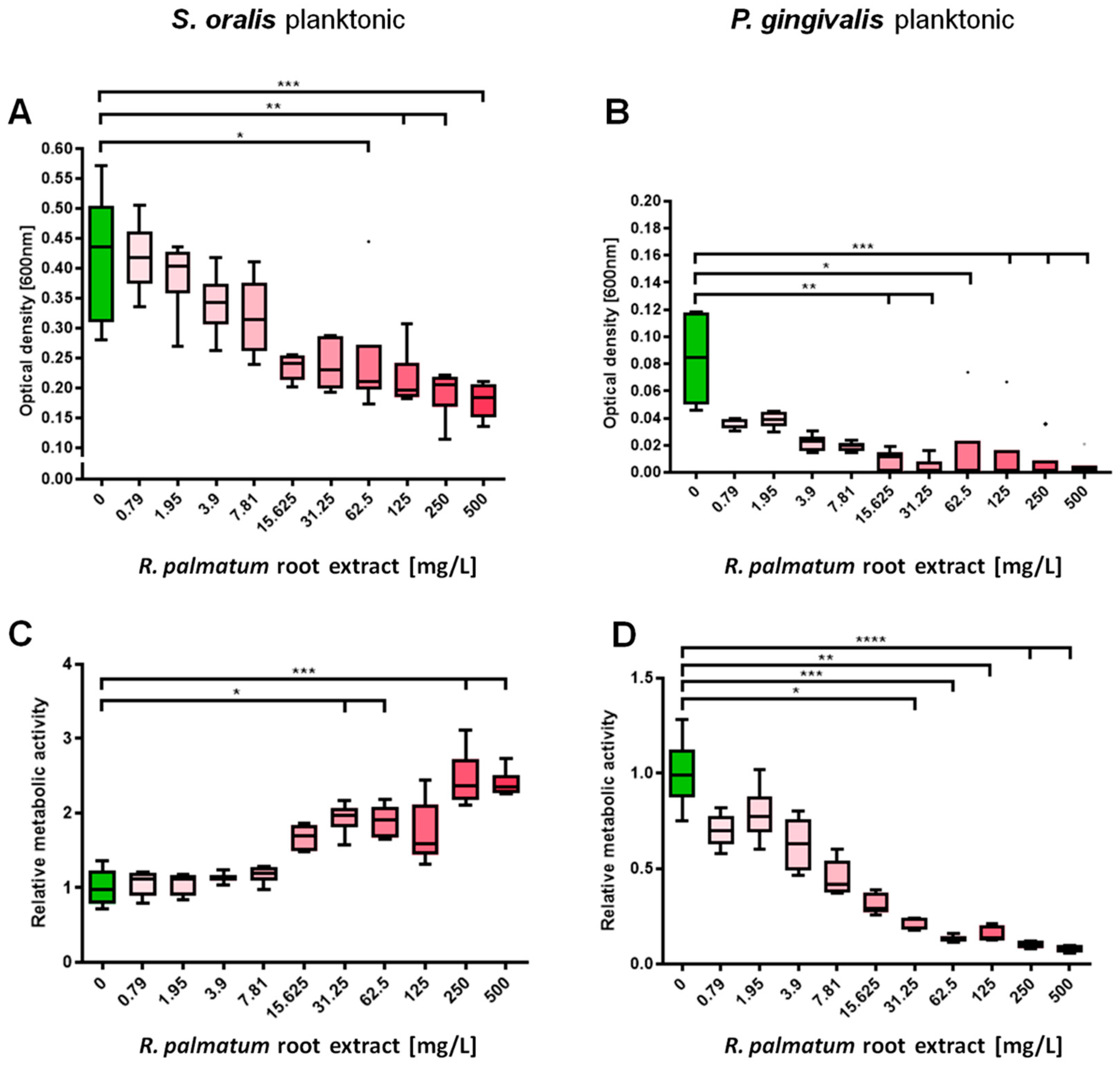
3.1. Selective Antimicrobial Effects of R. palmatum Root Extract on Planktonic S. oralis and P. gingivalis

R. palmatum root extract significantly inhibited planktonic growth of S. oralis, starting at a concentration of 62.5 mg/L (Figure 1A). The metabolic activity of S. oralis increased significantly from 31.25 mg/L R. palmatum extract (except for 125 mg/L) (Figure 1C). Planktonic growth of P. gingivalis was already severely reduced at a concentration of 3.9 mg/L, and significant reduction started at 15.625 mg/L R. palmatum root extract (Figure 1C). The metabolic activity of P. gingivalis was likewise significantly reduced-starting at a concentration of 31.25 mg/L (Figure 1D).

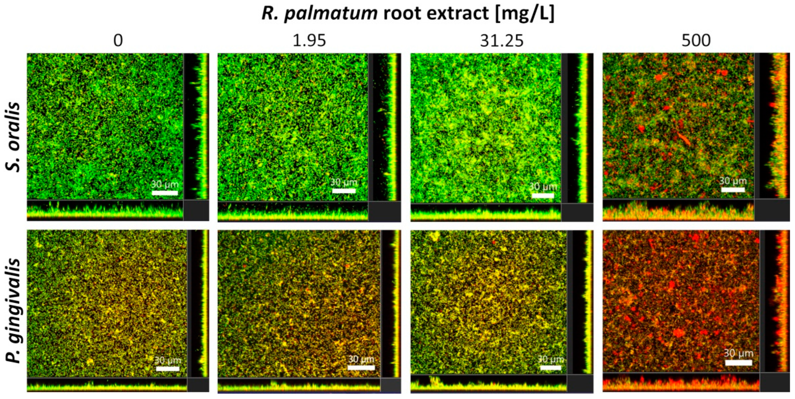
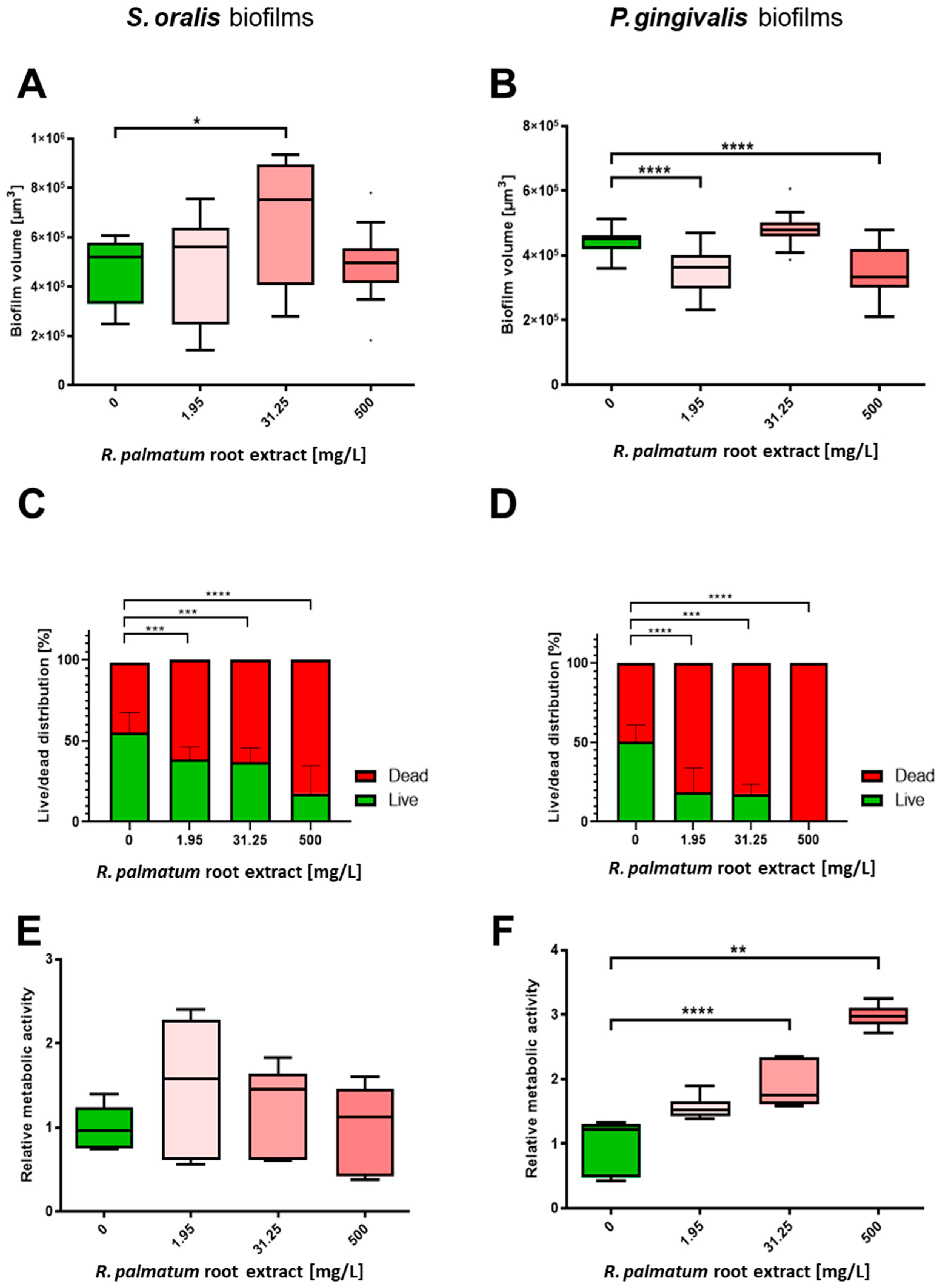
3.2. Selective Antibiofilm Activity of R. palmatum Root Extract on S. oralis and P. gingivalis

One representative CLSM image for each experiment of S. oralis and P. gingivalis biofilms after 24 h of cultivation followed by 2 h treatment with R. palmatum root extract is shown in Figure 2. With increasing concentration of R. palmatum root extract, the proportion of dead bacteria within the biofilms increased visually for both species; however, the antibacterial effect seemed to be much stronger for P. gingivalis than for S. oralis. For both bacterial species, the vertical sections showed a considerable increase in biofilm thickness and the biofilm structures became more porous and holey, as the concentration of R. palmatum increased, in particular at 500 mg/L.
The biofilm volumes of S. oralis increased slightly under the influence of R. palmatum root extract compared to the control; at a concentration of 31.25 mg/L, the increase was significant (Figure 3A). The proportion of viable bacteria was significantly lower under the influence of R. palmatum extract than for the control biofilms (Figure 2 and Figure 3C). The metabolic activity of S. oralis increased moderately at 1.95 mg/L R. palmatum extract compared to control biofilms, but decreased with increasing concentrations, to approximately the level of the control biofilms (Figure 3E).
Significantly decreased biofilm volumes were observed for P. gingivalis under the influence of 1.95 and 500 mg/L R. palmatum root extract (Figure 3B). As detectable with the Bacterial Viability Kit, the number of viable bacteria in the biofilm decreased significantly down to 0% at a concentration of 500 mg/L R. palmatum (Figure 2 and Figure 3D). Metabolic activity of P. gingivalis biofilms increased consistently with increasing concentrations of R. palmatum extract; at 31.25 and 500 mg/L R. palmatum, metabolic activity increased significantly compared to control (Figure 3F).

4. Discussion

Treatment of periodontitis and peri-implantitis is not straightforward: Bacteria embedded in biofilms are less susceptible for host defense mechanisms, or to treatment with antiseptics or antibiotics [39,40]. Multiple properties specific to the biofilm lead to specific resistance mechanisms, one of which is the failure of molecules to penetrate deeply into biofilm due to diffusion problems [41]. Furthermore, bacteria embedded in biofilms may develop antimicrobial drug resistance due to repeated antibiotic therapy. Substances such as CHX, which unselectively target bacteria, keep the total bacterial count low and may therefore lead to easier reinfection by pathogenic bacteria [21].
As antiseptics or antibiotics may have negative side effects, and bearing in mind the spread of antibiotic resistance, there is great interest in developing novel therapeutics against bacterial infections. Plants have shown antimicrobial activities and therefore may be a source for the development of novel antimicrobial therapeutics or may be used as co-therapeutics in combination with antibiotics in order to overcome antibiotic resistance [42]. Plant extracts may possess different biologically active compounds, which act in different target sites of microorganisms [42,43]. This may contribute to the overall activity of the plant extract and thus may slow down the development of resistance, since not only one single mechanism is involved. In order to investigate natural alternatives, our study aimed to test R. palmatum root extract against one commensal and one oral pathogenic species-S. oralis and P. gingivalis-initially planktonic and subsequently in biofilms.
R. palmatum root extract significantly inhibited planktonic growth of S. oralis starting at a concentration of 62.5 mg/L, whereas planktonic growth of P. gingivalis was significantly inhibited at a concentration of 15.625 mg/L. Intense suppression of P. gingivalis planktonic growth even started at a concentration of 3.9 mg/L R. palmatum root extract, which is consistent with the results of the previous study [29]. These results were surprising, since plant secondary metabolites often show a higher level of activity against Gram-positive than Gram-negative bacteria [44,45]. Trans-envelope multidrug efflux pumps (EP) span the two membranes of Gram-negative bacteria [46] and are capable of extruding toxins across the membrane. P. gingivalis ATCC 33277 has gene clusters encoding for such efflux pumps [47]. It is known that a deactivation of efflux pumps increases antimicrobial activity in Gram-negative bacteria. Depending on the species, a 100–2000-fold antimicrobial potentiation was found for the anthraquinone rhein, a major component of R. palmatum, if efflux pump inhibitors were used at the same time [45]. In a previous study by the authors, HPLC analysis was performed and a comparison of the UV signals of R. palmatum root extract with the UV signals of the anthraquinone rhein revealed a concentration of 5% rhein in the extract [29]. Besides rhein, R. palmatum root extract contains phenolic components such as resveratrol [48] which is known to block efflux pumps in Gram-negative bacteria [49]. This additional antimicrobial effect may enhance antimicrobial activity against the Gram-negative P. gingivalis.
The metabolic activity of S. oralis increased significantly from a R. palmatum extract concentration of 31.25 mg/L, whereas the metabolic activity of P. gingivalis was significantly reduced at concentration at or above of 31.25 mg/L. In general, bacterial growth and metabolism are mutually related. In growing cells, the metabolic activity is high whereas it is low in non-growing or growth-inhibited cells [50]. The results for P. gingivalis reflect these facts. However, under the influence of higher concentrations of R. palmatum root extract, S. oralis shows increasing metabolic activity, with at the same time decreasing optical density. Antibiotics or antiseptics are known to cause bacterial stress responses and metabolic adaptions [51,52,53]. The increase in metabolic activity as a stress response is a well-known finding in previous studies [54,55]. Bacteria can stop their cell division under stress. This phenomenon, in which bacteria are metabolically active, but do not multiply, is called “quiescence” [56]. Thus, S. oralis seems to be slightly stressed but not completely inhibited in growth by the R. palmatum root extract.
R. palmatum root extract concentrations of 1.95, 31.25 and 500 mg/L were chosen for the subsequent biofilm experiments, in which 24-h-old biofilms were exposed to the substances for two hours. The concentration of 1.95 mg/L was selected because there were no significant effects on optical density or metabolic activity for any of the species in the planktonic experiments. A concentration of 31.25 mg/L was chosen because this gave significant differences in the bacterial growth and metabolic activity in planktonic P. gingivalis but not in S. oralis, and 500 mg/L was picked as this was the highest tested concentration in the planktonic experiments that showed significant efficacy in both species.
Evaluation of the CLMS images of S. oralis and P. gingivalis biofilms demonstrated that with an increase in R. palmatum root extract concentration, the biofilms of both bacterial species became more porous and holey, and the biofilm thickness increased considerably. This indicates that the R. palmatum root extract seems to disrupting the biofilms. A disrupting effect on biofilm structures was already detected for anthraquinone-2-carboxlic acid and rhein [57], both components of R. palmatum.
Biofilm volumes were analysed to determine the potential of R. palmatum root extract for biofilm eradication. The biofilm volumes of S. oralis increased slightly and the P. gingivalis biofilm volumes decreased slightly under the influence of R. palmatum root extract. There was no clear dependence on the applied concentrations of R. palmatum root extract. Based on these results, 1.95, 31.25 and 500 mg/L R. palmatum root extract appear to only slightly influence the biofilm volumes of S. oralis and P. gingivalis. Since the calculation of the biofilm volume is based on the volume of the fluorescence stained bacteria within the biofilms, the fluctuations in the biofilm volumes are not correlated to the increase in biofilm thickness. These effects could be associated with stress effects and the volumes of single cells [58], but this would have to be clarified by further experiments with single bacterial cells.
Live/dead staining was performed to evaluate the effect of R. palmatum root extract on bacterial viability within the biofilms in relation to membrane integrity. A concentration of 1.95 mg/L inhibited the number of viable cells in the 24-h old biofilms of S. oralis and P. gingivalis significantly after two hours of incubation. A concentration of 1.95 mg/L significantly reduced viable P. gingivalis from 50.38% (±10.32) to 18.59% (±14.92), whereas viable S. oralis was reduced from 56.82% (±9.75), to 31.72% (±7.38). At a concentration of 500 mg/L R. palmatum root extract, the number of viable P. gingivalis decreased significantly down to 0%. Similar results were found for CHX, where the MIC and minimum bactericidal concentration in P. gingivalis biofilms were both 4 μg/mL [59]. The results demonstrate that R. palmatum root extract has a strong effect on cell membrane integrity, probably due to cell membrane destruction, and thus on the vitality of bacteria in the biofilm. P. gingivalis was much more strongly affected than S. oralis.
In a study with methicillin-resistant Staphylococcus aureus, antibacterial and antibiofilm activity of rhein has been investigated. Rhein showed bactericidal activity (MIC < 200 µg/mL) and antibiofilm activity after 24 h of exposure, a slight biomass reduction has been observed and viable cells within the biofilm have been significantly dispersed [57]. These results are in line with our findings.
Aqueous rhubarb extract with a rhein content of 2.6% among other anthraquinones, has been reported to inhibit the biofilm formation of Streptococcus suis not by bacterial cell death but by downregulating transductions systems and affecting levels of transcriptional regulating factors and DNA binding proteins [60]. This may represent an additional antibiofilm mechanism of action.
The metabolic activity in S. oralis biofilms increased moderately at 1.95 mg/L R. palmatum root extract compared to control biofilms but decreased with increasing concentration, approximately to the level of the control biofilms. In contrast, metabolic activity of P. gingivalis biofilms increased consistently with increasing R. palmatum root extract concentration. Therefore, R. palmatum root extract had an ambiguous effect on the metabolic activity of S. oralis, whereas P. gingivalis responded with severe stress.
In summary, R. palmatum root extract strongly inhibited growth of planktonic P. gingivalis, while not inhibiting growth of the commensal S. oralis at identical concentrations. Furthermore, it exhibited stronger antimicrobial activity on biofilms of P. gingivalis than on S. oralis. These results indicate that R. palmatum root extract has the potential to selectively inhibit the oral pathogen P. gingivalis.
R. palmatum root extract slightly broke up the biofilms and made them porous. This porosity effect could potentially be useful for better penetration of active substances into biofilms. S. oralis showed a slight increase in metabolic activity, both in planktonic samples and in biofilms. This may be beneficial, since S. oralis is not known to cause cellular damage to host tissue, but might instead positively influence soft tissue cells [37]. Besides analysing the underlying mechanism of the different antibacterial efficacy of R. palmatum, future studies could also address the question of whether the slightly increased metabolic activity of S. oralis is also beneficial for host tissue homeostasis.
We are aware that clinical isolates may exhibit increased virulence and resistance mechanisms [61,62] and thus, antimicrobial results may vary among different strains. Furthermore, the effect of R. palmatum root extract on a wide variety of commensal and pathogen oral bacteria and polymicrobial biofilms remains to be elucidated. This specific therapeutic strategy follows the idea of the novel antibiotic amixicile, which only targets anaerobic bacteria, including P. gingivalis, T. forsythia, F. nucleatum and Prevotella intermedia [21] and thus promotes restoration of microbial homeostasis.
R. palmatum was not cytotoxic to oral epithelial cells in concentrations up to 500 mg/mL [30]. Thus, R. palmatum root extract could be used as mouthwash or toothpaste ingredient to prevent overgrowth of pathogenic bacteria, in turn, to prevent onset or progression of periodontitis. The specific and selective antibacterial mode of action of R. palmatum root extract would be advantageous to restore microbial homeostasis.

5. Conclusions

Our studies showed that R. palmatum root extract specifically inhibits P. gingivalis growth of planktonic bacteria and in biofilms in low concentrations, while growth of the oral commensal S. oralis was not inhibited at the same low concentrations. R. palmatum root extract could therefore be used topically to prevent microbiome alterations due to overgrowth of pathogenic bacteria and thus, to prevent the onset or progression of periodontitis.

Supplementary Materials

The following supporting information can be downloaded at: https://www.mdpi.com/article/10.3390/microorganisms10050965/s1, Figure S1: Extract from HPLC-MS analysis of R. palmatum.

Author Contributions

N.K., L.K.M.-H., N.V., J.G. and T.O. conceived and designed the experiments; N.K., N.V. and J.G. performed the experiments; N.K., N.V., J.G., T.O., B.A.-N. and L.K.M.-H. analysed the data; N.K., T.O. and L.K.M.-H. contributed reagents/materials/analysis tools; N.K. and L.K.M.-H. wrote the original draft of the paper. N.K. and L.K.M.-H. prepared the draft of the paper. N.K., B.A.-N., N.V., J.G., T.O. and L.K.M.-H. reviewed and edited the paper. All authors have read and agreed to the published version of the manuscript.

Funding

This research received no external funding.

Institutional Review Board Statement

Not applicable.

Informed Consent Statement

Not applicable.

Data Availability Statement

Data are contained in this manuscript or Supplementary Materials.

Acknowledgments

We thank Marly Felicia Dalton for her technical support.

Conflicts of Interest

L.K.M.-H. is holder of the “startup innovativ” grant of the ministry of economics, Rhineland-Palatinate, and a mouth rinse containing R. palmatum root extract has been launched in 06/2021. The other authors declare no conflict of interest. The funding sponsors had no role in the design of the study; in the collection, analyses, or interpretation of data; in the writing of the manuscript, and in the decision to publish the results.

References

  1. Tonetti, M.S.; Jepsen, S.; Jin, L.; Otomo-Corgel, J. Impact of the global burden of periodontal diseases on health, nutrition and wellbeing of mankind: A call for global action. J. Clin. Periodontol. 2017, 44, 456–462. [Google Scholar] [CrossRef] [PubMed] [Green Version]
  2. Berglundh, T.; Armitage, G.; Araujo, M.G.; Avila-Ortiz, G.; Blanco, J.; Camargo, P.M.; Chen, S.; Cochran, D.; Derks, J.; Figuero, E.; et al. Peri-implant diseases and conditions: Consensus report of workgroup 4 of the 2017 World Workshop on the Classification of Periodontal and Peri-Implant Diseases and Conditions. J. Clin. Periodontol. 2018, 89, S286–S291. [Google Scholar] [CrossRef] [PubMed] [Green Version]
  3. Chen, M.X.; Zhong, Y.J.; Dong, Q.Q.; Wong, H.M.; Wen, Y.F. Global, regional, and national burden of severe periodontitis, 1990–2019: An analysis of the Global Burden of Disease Study 2019. J. Clin. Periodontol. 2021, 48, 1165–1188. [Google Scholar] [CrossRef] [PubMed]
  4. Lang, N.P.; Bartold, P.M. Periodontal health. J. Periodontol. 2018, 89, S9–S16. [Google Scholar] [CrossRef] [Green Version]
  5. Rawlinson, A.; Vettore, M.V.; Baker, S.R.; Robinson, P.G. Periodontal treatment, psychological factors and oral health-related quality of life. J. Clin. Periodontol. 2021, 48, 226–236. [Google Scholar] [CrossRef]
  6. Liccardo, D.; Cannavo, A.; Spagnuolo, G.; Ferrara, N.; Cittadini, A.; Rengo, C.; Rengo, G. Periodontal disease: A risk factor for diabetes and cardiovascular disease. Int. J. Mol. Sci. 2019, 20, 1414. [Google Scholar] [CrossRef] [Green Version]
  7. Michaud, D.S.; Fu, Z.; Shi, J.; Chung, M. Periodontal disease, tooth loss, and cancer risk. Epidemiol. Rev. 2017, 39, 49–58. [Google Scholar] [CrossRef] [Green Version]
  8. Dreyer, H.; Grischke, J.; Tiede, C.; Eberhard, J.; Schweitzer, A.; Toikkanen, S.E.; Glöckner, S.; Krause, G.; Stiesch, M. Epidemiology and risk factors of peri-implantitis: A systematic review. J. Periodontal. Res. 2018, 53, 657–681. [Google Scholar] [CrossRef]
  9. Mohanty, R.; Asopa, S.J.; Joseph, M.D.; Singh, B.; Rajguru, J.P.; Saidath, K.; Sharma, U. Red complex: Polymicrobial conglomerate in oral flora: A review. J. Fam. Med. Prim. Care 2019, 8, 3480–3486. [Google Scholar] [CrossRef]
  10. Al-Ahmad, A.; Muzafferiy, F.; Anderson, A.C.; Wölber, J.P.; Ratka-Krüger, P.; Fretwurst, T.; Nelson, K.; Vach, K.; Hellwig, E. Shift of microbial composition of peri-implantitis-associated oral biofilm as revealed by 16S rRNA gene cloning. J. Med. Microbiol. 2018, 67, 332–340. [Google Scholar] [CrossRef]
  11. Duran-Pinedo, A.E.; Frias-Lopez, J. Beyond microbial community composition: Functional activities of the oral microbiome in health and disease. Microbes Infect. 2015, 17, 505–516. [Google Scholar] [CrossRef] [PubMed] [Green Version]
  12. Sahrmann, P.; Gilli, F.; Wiedemeier, D.B.; Attin, T.; Schmidlin, P.R.; Karygianni, L. The microbiome of peri-implantitis: A systematic review and meta-analysis. Microorganisms 2020, 8, 661. [Google Scholar] [CrossRef]
  13. Fine, D.H.; Patil, A.G.; Velusamy, S.K. Aggregatibacter actinomycetemcomitans (Aa) Under the Radar: Myths and Misunderstandings of Aa and Its Role in Aggressive Periodontitis. Front. Immunol. 2019, 10, 728. [Google Scholar] [CrossRef] [PubMed]
  14. Waal, Y.C.D.; Eijsbouts, H.V.; Winkel, E.G.; Winkelhoff, A.J.V. Microbial Characteristics of Peri-Implantitis: A Case-Control Study. J. Periodontol. 2017, 88, 209–217. [Google Scholar] [CrossRef]
  15. Quirynen, M.; Teughels, W.; Soete, M.D.; Steenberghe, D.V. Topical antiseptics and antibiotics in the initial therapy of chronic adult periodontitis: Microbiological aspects. Periodontology 2000 2002, 28, 72–90. [Google Scholar] [CrossRef]
  16. Smeets, R.; Henningsen, A.; Jung, O.; Heiland, M.; Hammächer, C.; Stein, J.M. Definition, etiology, prevention and treatment of peri-implantitis—A review. Head Face Med. 2014, 10, 34. [Google Scholar] [CrossRef] [Green Version]
  17. Rhemrev, G.E.; Timmerman, M.F.; Veldkamp, I.; Winkelhoff, A.J.V.; Velden, U.V.D. Immediate effect of instrumentation on the subgingival microflora in deep inflamed pockets under strict plaque control. J. Clin. Periodontol. 2006, 33, 42–48. [Google Scholar] [CrossRef]
  18. Takahashi, N. Oral microbiome metabolism:from “who are they?” to “what are they doing?”. J. Dent. Res. 2015, 94, 1628–1637. [Google Scholar] [CrossRef]
  19. Russell, A.D. Chlorhexidine: Antibacterial action and bacterial resistance. Infection 1986, 14, 212–215. [Google Scholar] [CrossRef]
  20. Lachapelle, J.-M.; Castel, O.; Casado, A.; Leroy, B.; Micali, G.; Tennstedt, D.; Lambert, J. Antiseptics in the era of bacterial resistance: A focus on povidone iodine. Clin. Pract. 2013, 10, 579–592. [Google Scholar] [CrossRef]
  21. Hutcherson, J.A.; Sinclair, K.M.; Belvin, B.R.; Gui, Q.; Hoffman, P.S.; Lewis, J.P. Amixicile, a novel strategy for targeting oral anaerobic pathogens. Sci. Rep. 2017, 7, 10474. [Google Scholar] [CrossRef] [PubMed]
  22. Al-Kamel, A.; Baraniya, D.; Al-Hajj, W.A.; Halboub, E.; Abdulrab, S.; Chen, T.; Al-Hebshi, N.N. Subgingival microbiome of experimental gingivitis: Shifts associated with the use of chlorhexidine and N-acetyl cysteine mouthwashes. J. Oral Microbiol. 2019, 11, 1608141. [Google Scholar] [CrossRef] [PubMed] [Green Version]
  23. Bescos, R.; Ashworth, A.; Cutler, C.; Brookes, Z.L.; Belfield, L.; Rodiles, A.; Casas-Agustench, P.; Farnham, G.; Liddle, L.; Burleigh, M.; et al. Effects of Chlorhexidine mouthwash on the oral microbiome. Sci. Rep. 2020, 10, 5254. [Google Scholar] [CrossRef] [PubMed] [Green Version]
  24. Brookes, Z.L.S.; Belfield, L.A.; Ashworth, A.; Casas-Agustench, P.; Raja, M.; Pollard, A.J.; Bescos, R. Effects of chlorhexidine mouthwash on the oral microbiome. J. Dent. 2021, 113, 103768. [Google Scholar] [CrossRef]
  25. Gürgan, C.A.; Zaim, E.; Bakirsoy, I.; Soykan, E. Short-term side effects of 0.2% alcohol-free chlorhexidine mouthrinse used as an adjunct to non-surgical periodontal treatment: A double-blind clinical study. J. Periodontol. 2006, 77, 370–384. [Google Scholar] [CrossRef]
  26. Rath, S.K.; Singh, M. Comparative clinical and microbiological efficacy of mouthwashes containing 0.2% and 0.12% chlorhexidine. Dent. Res. J. 2013, 10, 364–369. [Google Scholar]
  27. Nguyen, T.; Brody, H.; Radaic, A.; Kapila, Y. Probiotics for periodontal health-Current molecular findings. Periodontology 2000 2021, 87, 254–267. [Google Scholar] [CrossRef]
  28. Ng, E.; Tay, J.R.H.; Ong, M.M.A.; Bostanci, N.; Belibasakis, G.N.; Seneviratne, C.J. Probiotic therapy for periodontal and peri-implant health—Silver bullet or sham? Benef. Microbes 2021, 12, 215–230. [Google Scholar] [CrossRef]
  29. Müller-Heupt, L.K.; Vierengel, N.; Groß, J.; Opatz, T.; Deschner, J.; Loewenich, F.D.V. Antimicrobial Activity of Eucalyptus globulus, Azadirachta indica, Glycyrrhiza glabra, Rheum palmatum Extracts and Rhein against Porphyromonas gingivalis. Antibiotics 2022, 11, 186. [Google Scholar] [CrossRef]
  30. Liao, J.; Zhao, L.; Yoshioka, M.; Hinode, D.; Grenier, D. Effects of Japanese traditional herbal medicines (Kampo) on growth and virulence properties of Porphyromonas gingivalis and viability of oral epithelial cells. Pharm. Biol. 2013, 51, 1538–1544. [Google Scholar] [CrossRef] [Green Version]
  31. Hajishengallis, G.; Liang, S.; Payne, M.A.; Hashim, A.; Jotwani, R.; Eskan, M.A.; McIntosh, M.L.; Alsam, A.; Kirkwood, K.L.; Lambris, J.D.; et al. Low-abundance biofilm species orchestrates inflammatory periodontal disease through the commensal microbiota and complement. Cell Host Microbe 2011, 10, 497–506. [Google Scholar] [CrossRef] [Green Version]
  32. Olsen, I.; Lambris, J.D.; Hajishengallis, G. Porphyromonas gingivalis disturbs host-commensal homeostasis by changing complement function. J. Oral Microbiol. 2017, 9, 1340085. [Google Scholar] [CrossRef] [Green Version]
  33. Kolenbrander, P.E.; Palmer, R.J., Jr.; Periasamy, S.; Jakubovics, N.S. Oral multispecies biofilm development and the key role of cell-cell distance. Nat. Rev. Microbiol. 2010, 8, 471–480. [Google Scholar] [CrossRef]
  34. Nyvad, B.; Kilian, M. Comparison of the Initial Streptococcal Microflora on Dental Enamel in Caries-Active and in Caries-Inactive Individuals. Caries Res. 1990, 24, 267–272. [Google Scholar] [CrossRef]
  35. Listgarten, M.A.; Helldén, L. Relative distribution of bacteria at clinically healthy and periodontally diseased sites in humans. J. Clin. Periodontol. 1978, 5, 115–132. [Google Scholar] [CrossRef]
  36. Belibasakis, G.N.; Charalampakis, G.; Bostanci, N.; Stadlinger, B. Peri-implant infections of oral biofilm etiology. Adv. Exp. Med. Biol. 2015, 830, 69–84. [Google Scholar] [CrossRef]
  37. Ingendoh-Tsakmakidis, A.; Eberhard, J.; Falk, C.S.; Stiesch, M.; Winkel, A. In Vitro Effects of Streptococcus oralis Biofilm on Peri-Implant Soft Tissue Cells. Cells 2020, 9, 1226. [Google Scholar] [CrossRef]
  38. Gerits, E.; Verstraeten, N.; Michiels, J. New approaches to combat Porphyromonas gingivalis biofilms. J. Oral. Microbiol. 2017, 9, 1300366. [Google Scholar] [CrossRef] [Green Version]
  39. Khan, J.; Tarar, S.M.; Gul, I.; Nawaz, U.; Arshad, M. Challenges of antibiotic resistance biofilms and potential combating strategies: A review. 3 Biotech 2021, 11, 169. [Google Scholar] [CrossRef]
  40. Costerton, J.W.; Stewart, P.S.; Greenberg, E.P. Bacterial biofilms: A common cause of persistent infections. Science 1999, 284, 1318–1322. [Google Scholar] [CrossRef] [Green Version]
  41. Stewart, P.S. Theoretical aspects of antibiotic diffusion into microbial biofilms. Antimicrob. Agents Chemother. 1996, 40, 2517–2522. [Google Scholar] [CrossRef] [PubMed] [Green Version]
  42. Khameneh, B.; Iranshahy, M.; Soheili, V.; Bazzaz, B.S.F. Review on plant antimicrobials: A mechanistic viewpoint. Antimicrob. Resist. Infect. Control 2019, 8, 118. [Google Scholar] [CrossRef] [PubMed] [Green Version]
  43. Gupta, P.D.; Birdi, T.J. Development of botanicals to combat antibiotic resistance. J. Ayurveda Integr. Med. 2017, 8, 266–275. [Google Scholar] [CrossRef] [PubMed]
  44. Lewis, K.; Salyers, A.; Taber, H. Bacterial Resistance to Antimicrobials: Mechanisms, Genetics, Medical Practice and Public Health; Wax, R., Ed.; Marcel Dekker, Inc.: New York, NY, USA, 2001. [Google Scholar]
  45. Tegos, G.; Stermitz, F.R.; Lomovskaya, O.; Lewis, K. Multidrug pump inhibitors uncover remarkable activity of plant antimicrobials. Antimicrob. Agents Chemother. 2002, 46, 3133–3141. [Google Scholar] [CrossRef] [Green Version]
  46. Zgurskaya, H.I.; Rybenkov, V.V.; Krishnamoorthy, G.; Leus, I.V. Trans-envelope multidrug efflux pumps of Gram-negative bacteria and their synergism with the outer membrane barrier. Res. Microbiol. 2018, 169, 351–356. [Google Scholar] [CrossRef]
  47. Inoue, T.; Nakayama, M.; Taguchi, Y.; Kano, K.; Ono, M.; Shimizu, Y.; Kuroda, T.; Ohara, N. Characterization of the tripartite drug efflux pumps of Porphyromonas gingivalis aTCC 33277. New Microbiol. 2015, 38, 101–108. [Google Scholar]
  48. Zhang, Q.; Liu, J.; Li, R.; Zhao, R.; Zhang, M.; Wei, S.; Ran, D.; Jin, W.; Wu, C. A Network Pharmacology Approach to Investigate the Anticancer Mechanism and Potential Active Ingredients of Rheum palmatum L. against Lung Cancer via Induction of Apoptosis. Front. Pharmacol. 2020, 11, 528308. [Google Scholar] [CrossRef]
  49. Seukep, A.J.; Kuete, V.; Nahar, L.; Sarker, S.D.; Guo, M. Plant-derived secondary metabolites as the main source of efflux pump inhibitors and methods for identification. J. Pharm. Anal. 2020, 10, 277–290. [Google Scholar] [CrossRef]
  50. Lempp, M.; Lubrano, P.; Bange, G.; Link, H. Metabolism of non-growing bacteria. Biol. Chem. 2020, 401, 1479–1485. [Google Scholar] [CrossRef]
  51. Czech, L.; Mais, C.-N.; Kratzat, H.; Sarmah, P.; Giammarinaro, P.; Freibert, S.-A.; Esser, H.F.; Musial, J.; Berninghausen, O.; Steinchen, W.; et al. Inhibition of SRP-dependent protein secretion by the bacterial alarmone (p)ppGpp. Nat. Commun. 2022, 13, 1069. [Google Scholar] [CrossRef]
  52. Stokes, J.M.; Lopatkin, A.J.; Lobritz, M.A.; Collins, J.J. Bacterial metabolism and antibiotic efficacy. Cell Metab. 2019, 30, 251–259. [Google Scholar] [CrossRef] [PubMed]
  53. Muehler, D.; Mao, X.; Czemmel, S.; Geißert, J.; Engesser, C.; Hiller, K.-A.; Widbiller, M.; Maisch, T.; Buchalla, W.; Al-Ahmad, A.; et al. Transcriptomic stress response in Streptococcus mutans following treatment with a sublethal concentration of chlorhexidine digluconate. Microorganisms 2022, 10, 561. [Google Scholar] [CrossRef] [PubMed]
  54. Kommerein, N.; Weigel, A.J.; Stiesch, M.; Doll, K. Plant-based oral care product exhibits antibacterial effects on different stages of oral multispecies biofilm development in vitro. BMC Oral Health 2021, 21, 170. [Google Scholar] [CrossRef] [PubMed]
  55. Guan, N.; Li, J.; Shin, H.D.; Du, G.; Chen, J.; Liu, L. Microbial response to environmental stresses: From fundamental mechanisms to practical applications. Appl. Microbiol. Biotechnol. 2017, 101, 3991–4008. [Google Scholar] [CrossRef] [PubMed]
  56. Rittershaus, E.S.; Baek, S.H.; Sassetti, C.M. The normalcy of dormancy: Common themes in microbial quiescence. Cell Host Microbe 2013, 13, 643–651. [Google Scholar] [CrossRef] [Green Version]
  57. Song, Z.M.; Zhang, J.L.; Zhou, K.; Yue, L.M.; Zhang, Y.; Wang, C.Y.; Wang, K.L.; Xu, Y. Anthraquinones as Potential Antibiofilm Agents against Methicillin-Resistant Staphylococcus aureus. Front. Microbiol. 2021, 12, 709826. [Google Scholar] [CrossRef]
  58. Rode, D.K.H.; Singh, P.K.; Drescher, K. Multicellular and unicellular responses of microbial biofilms to stress. Biol. Chem. 2020, 401, 1365–1374. [Google Scholar] [CrossRef]
  59. Noiri, Y.; Okami, Y.; Narimatsu, M.; Takahashi, Y.; Kawahara, T.; Ebisu, S. Effects of chlorhexidine, minocycline, and metronidazole on Porphyromonas gingivalis strain 381 in biofilms. J. Periodontol. 2003, 74, 1647–1651. [Google Scholar] [CrossRef]
  60. Ding, W.-Y.; Li, Y.-H.; Lian, H.; Ai, X.-Y.; Zhao, Y.-L.; Yang, Y.-B.; Han, Q.; Liu, X.; Chen, X.-Y.; He, Z. Sub-minimum inhibitory concentrations of rhubarb water extracts inhibit Streptococcus suis biofilm formation. Front. Pharmacol. 2017, 8, 425. [Google Scholar] [CrossRef] [Green Version]
  61. Seers, C.A.; Mahmud, A.S.M.; Huq, N.L.; Cross, K.J.; Reynolds, E.C. Porphyromonas gingivalis laboratory strains and clinical isolates exhibit different distribution of cell surface and secreted gingipains. J. Oral Microbiol. 2021, 13, 1858001. [Google Scholar] [CrossRef]
  62. Kulik, E.M.; Thurnheer, T.; Karygianni, L.; Walter, C.; Sculean, A.; Eick, S. Antibiotic susceptibility patterns of Aggregatibacter actinomycetemcomitans and Porphyromonas gingivalis strains from different decades. Antibiotics 2019, 8, 253. [Google Scholar] [CrossRef] [PubMed] [Green Version]
Figure 1. Effects of R. palmatum root extract on planktonic S. oralis and P. gingivalis. After 24 h of incubation. planktonic growth of P. gingivalis was significantly inhibited by lower concentrations of R. palmatum root extract than the growth of S. oralis was. (A) Treatment with 62.5 mg/L R. palmatum root extract or more resulted in significant reductions in the optical density of S. oralis. (B) Treatment with 15.625 mg/L R. palmatum root extract or more resulted in a significant reduction in the optical density of P. gingivalis. (C) Treatment with 31.25 mg/L R. palmatum root extract or more resulted in a significant increase in the relative metabolic activity of S. oralis. (D) Treatment with 31.25 mg/L R. palmatum root extract or more resulted in a significant reduction in the relative metabolic activity of P. gingivalis. Kruskal–Wallis test with Dunn’s correction for multiple comparisons, N = 6 with S. oralis strain ATCC 9811 and P. gingivalis strain ATCC 33277/DSM 20709. * p < 0.05, ** p < 0.01, *** p < 0.001, **** p < 0.0001.
Microorganisms 10 00965 g001
Figure 2. Representative CLSM images of biofilms formed by S. oralis and P. gingivalis after 24 h of cultivation followed by two hours treatment with different concentrations of R. palmatum root extract. The large panels show the horizontal sections (x-y planes) of the biofilms and the bottom/side panels display the vertical sections (x-z and y-z planes) through the biofilms. Biofilms were stained with SYTO 9 and propidium iodide (viable bacteria = green; dead bacteria = red or orange). The scale bars indicate 30 µm.
Microorganisms 10 00965 g002
Figure 3. Effects of R. palmatum root extract on S. oralis and P. gingivalis biofilms. After two hours of incubation, viability of S. oralis and P. gingivalis in 24-h biofilms was significantly inhibited by low concentrations of R. palmatum root extract of at least 1.95 mg/L-with a greater decrease in the viability of P. gingivalis. (A) Treatment with 1.95 mg/L and 31.25 mg/L R. palmatum root extract increased biofilm volume of S. oralis, whereas biofilm volume was decreased by 500 mg/L R. palmatum root extract. (B) Treatment with 1.95 mg/L and 500 mg/L R. palmatum root extract significantly increased biofilm volume of P. gingivalis. (C) Treatment with 1.95 mg/L or more of R. palmatum root extract significantly decreased viability of S. oralis in biofilms. (D) Treatment with 1.95 mg/L or more of R. palmatum root extract significantly decreased viability of P. gingivalis in biofilms. (E) Treatment with 1.95 mg/L and above of R. palmatum root extract increased metabolic activity of S. oralis in biofilms. (F) Treatment with 1.95 mg/L or more of R. palmatum root extract increased metabolic activity of P. gingivalis in biofilms. N = 6 with S. oralis strain ATCC 9811 and P. gingivalis strain ATCC 33277/DSM 20709. * p < 0.05, ** p < 0.01, *** p < 0.001, **** p < 0.0001.
Microorganisms 10 00965 g003
Table 1. Final concentrations of R. palmatum root extract.
R. palmatum Root Extract
Concentration (mg/L) 0.791.953.907.812515.62531.2562.5125250500
Publisher’s Note: MDPI stays neutral with regard to jurisdictional claims in published maps and institutional affiliations.

