Review

Campylobacter jejuni in Poultry: Pathogenesis and Control Strategies

by
Walid Ghazi Al Hakeem
1,
Shahna Fathima
1,
Revathi Shanmugasundaram
2,* and
Ramesh K. Selvaraj
1
1
Department of Poultry Science, The University of Georgia, Athens, GA 30602, USA
2
Toxicology and Mycotoxin Research Unit, US National Poultry Research Center, Agricultural Research Service, U.S. Department of Agriculture, Athens, GA 30605, USA
*
Author to whom correspondence should be addressed.
Microorganisms 2022, 10(11), 2134; https://doi.org/10.3390/microorganisms10112134
Submission received: 5 October 2022 / Revised: 22 October 2022 / Accepted: 25 October 2022 / Published: 28 October 2022
(This article belongs to the Special Issue Foodborne Pathogens: Infections and Pathogenesis)

Abstract

:
C. jejuni is the leading cause of human foodborne illness associated with poultry, beef, and pork consumption. C. jejuni is highly prevalent in commercial poultry farms, where horizontal transmission from the environment is considered to be the primary source of C. jejuni. As an enteric pathogen, C. jejuni expresses virulence factors regulated by a two-component system that mediates C. jejuni’s ability to survive in the host. C. jejuni survives and reproduces in the avian intestinal mucus. The avian intestinal mucus is highly sulfated and sialylated compared with the human mucus modulating C. jejuni pathogenicity into a near commensal bacteria in poultry. Birds are usually infected from two to four weeks of age and remain colonized until they reach market age. A small dose of C. jejuni (around 35 CFU/mL) is sufficient for successful bird colonization. In the U.S., where chickens are raised under antibiotic-free environments, additional strategies are required to reduce C. jejuni prevalence on broilers farms. Strict biosecurity measures can decrease C. jejuni prevalence by more than 50% in broilers at market age. Vaccination and probiotics, prebiotics, synbiotics, organic acids, bacteriophages, bacteriocins, and quorum sensing inhibitors supplementation can improve gut health and competitively exclude C. jejuni load in broilers. Most of the mentioned strategies showed promising results; however, they are not fully implemented in poultry production. Current knowledge on C. jejuni’s morphology, source of transmission, pathogenesis in poultry, and available preharvest strategies to decrease C. jejuni colonization in broilers are addressed in this review.

1. Introduction

C. jejuni was first recognized in 1886 by Escherich as he described the C. jejuni as a spiral bacteria isolated from the colon of dead children [1]. Escherich also identified the C. jejuni microscopically in stool specimens of children who suffered from diarrhea without being able to culture it on solid agar [1]. In 1909, a Vibrio-like bacterium was frequently isolated from aborted fetuses [2], later named Vibrio fetus [3]. Similar reports linked Vibrio-like organisms to sterility in cows [4], and dysentery in pigs, and later named Vibrio jejuni [5].
Similarly, several reports noted the presence of Vibrio fetus in the blood of pregnant women [6] and the blood of people associated with outbreaks related to consumption of milk contaminated with Vibrio fetus [7]. The absence of a proper isolation method for Vibrio fetus (C. jejuni) from feces resulted in fewer case reports despite the high prevalence of this pathogen. However, C. jejuni was successfully isolated from the stool of a patient suffering from acute enteritis [8]. The development of simpler isolation techniques for culturing C. jejuni led to the rapid isolation of this pathogen. In the mid-1980s, C. jejuni was recognized as one of the major causes of enterocolitis in humans [9].
C. jejuni is the leading cause of human foodborne illness associated with poultry, beef, and pork consumption [10]. C. jejuni is found in the gut of warm-blooded animals, with poultry species being the major reservoirs [11]. C. jejuni colonizes the ceca of chicken between 2 and 3 weeks of age and reaches around 1 × 10⁹ CFU/g in the ceca at market age [12]. Furthermore, poultry carcass is cross-contaminated at the processing facility due to spillage of intestinal contents. Handling and consuming improperly cooked poultry products account for the majority of C. jejuni infections [13]. With the spread of antibiotic resistance across C. jejuni isolates, the burden of Campylobacteroisis has increased [14].
The poultry industry is facing several challenges with legislative restrictions on the subtherapeutic use of antibiotics, in addition to the shift in consumers’ preference for “zero” use of antibiotics in poultry production. Therefore, finding an antimicrobial alternative to control C. jejuni in poultry production is the need of the hour. Different antibiotic alternatives include prebiotics, probiotics, synbiotic, bacteriocins, bacteriophages, vaccines, and organic acids [15].
This review article focuses on the C. jejuni’s morphology, source of transmission, pathogenesis in poultry, and available preharvest strategies to decrease C. jejuni colonization in broilers.

2. Campylobacter jejuni Cellular Structure and Morphology

C. jejuni is a gram-negative, corkscrew-shaped, and motile bacteria that belongs to the family of Campylobacteraceae. C. jejuni is characterized by a spiral/helical morphology with an amphitrichous sheathed flagella responsible for C. jejuni’s corkscrew motility [16]. The corkscrew motility or darting motility is a key advantage of C. jejuni movement in a highly viscous environment such as the mucus [16]. The enzyme C. jejuni’s peptidoglycan peptidase ensures the formation of the helical form, as mutations in peptidoglycan peptidase result in straight body formation [17]. The loss of C. jejuni’s helical form results in lower colonization capacity in chickens [17] and lower infectivity in mouse models [17]. C. jejuni produces a capsular polysaccharide (CPS) that helps the bacteria evade the immune system and contributes significantly to C. jejuni virulence [18]. Intra-strain variation in the capsular polysaccharide results in the formation of 47 serotypes [19]. C. jejuni outer membrane is comprised of Lipo-oligosaccharides (LOS) that lack the O-antigen found in lipopolysaccharides (LPS) of many gram-negative bacteria [20]. In some C. jejuni strains, the LOS binds with sialic acid resulting in a modified structure that mimics the gangliosides in human neurons [21]. This molecular mimicry plays a central role in developing Guillain–Barré syndrome (GBS) in humans [21].
The presence of phase-variable loci in C. jejuni bacteria contributes to its inherent ability to generate different phenotypes and genotypes. These phase-variable loci are mainly located in the CPS, flagella, and LOS, forming new structures to evade the immune system and help C. jejuni survive different environmental factors. Different stress conditions lead to morphological changes in C. jejuni, for example, oxygen-rich compounds that change from spiral shape to coccoid form [22]. Oxidative stress leads to forming the viable but non-culturable (VPNC) form of C. jejuni [23].

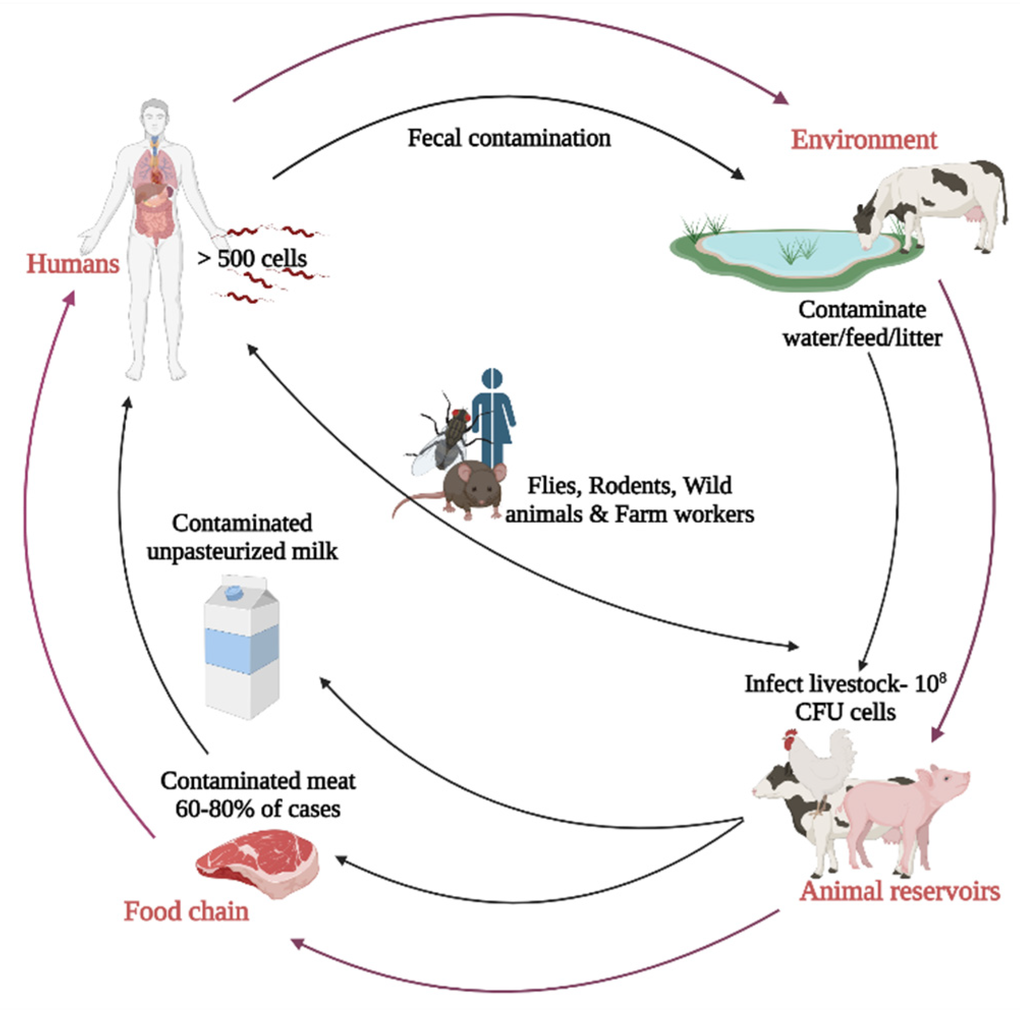
3. Source and Transmission of Campylobacter jejuni in Poultry

C. jejuni is a versatile bacterium that occupies different niches and hosts [20]. C. jejuni can be found in water and is part of the commensal microbiota of many animals, including poultry [20]. Poultry species are considered the major reservoirs for thermophile Campylobacter species, including C. jejuni, C. coli, and C. lari. C. jejuni accounts for the majority of campylobacteriosis in humans [20].
C. jejuni is highly prevalent in commercial poultry farms, where horizontal transmission from the environment is considered the primary source of C. jejuni [24]. Following the infection, broilers rapidly show a high load of C. jejuni in the cecal content [25]. Fecal shedding of C. jejuni and fecal ingestion is the main source of bird-to-bird transmission in broiler farms [26]. Vertical transmission of C. jejuni in broiler farms is controversial as C. jejuni is detected in broilers at 2–3 weeks of age, irrespective of C. jejuni positive parent flocks [26]. Furthermore, the isolation of C. jejuni from eggs in commercial and experimental layer flocks has been unsuccessful [27]. C. jejuni led to embryonic mortality in experimentally infected eggs [28], and C. jejuni did not survive more than 3–6 h following egg penetration [29,30]. Broiler flocks are usually infected with strains different than the strains detected in the breeder flocks [31], suggesting the negligible role of vertical transmission in C. jejuni in broiler flocks. Despite such observations, bacteriological and molecular methods confirmed the [32,33] presence of C. jejuni in eggshells. Furthermore, the survival of C. jejuni in a viable but not culturable form might be a critical factor behind the unsuccessful isolation of C. jejuni from infected eggs, and young hatchlings [34]. Therefore, future studies are needed to elucidate the role of vertical transmission in introducing C. jejuni to commercial broiler flocks.
The dry nature of chicken feed and wood shavings decreases the presence of C. jejuni, as C. jejuni’s viability is hindered by high O2 and low moisture levels [35]. Nevertheless, feed and bedding material can be a source of C. jejuni as it becomes contaminated by other sources such as fecal material and insects [35]. Reused litter can act as a source of C. jejuni infection. However, common litter management practices can limit the spread of C. jejuni to the next flock [35]. Unchlorinated water has been suggested as a potential source of C. jejuni in broiler farms. Water can act as a vehicle to transmit C. jejuni [36] as it requires microaerophilic conditions and cannot grow at a temperature less than 31 °C. C. jejuni was found in water lines only after the flock was colonized; however, the strains found in the water lines were not fully present in infected broilers, indicating that water is not the original source of contamination [37,38].
Flies and insects can act as a vector for several pathogens on broiler farms. Flies [39] and beetles [40] may introduce C. jejuni into chicken farms from multiple sources such as animal feces and lakes contaminated with C. jejuni [41]. The presence of livestock animals on the farm is associated with an increased risk of C. jejuni transmission through flies to broiler flocks [41]. C. jejuni colonization pattern peaks during summer [42], correlating directly with the insect populations. Therefore, Insects might be an important factor in C. jejuni seasonality incidences. Rodents are vectors for pathogens, including C. jejuni [43,44]. However, C. jejuni strains circulating in humans and livestock differ from those carried by rodents [43]. In some instances, rodents living close to humans and farms may carry the same C. jejuni strains [44]. Hence, rodents may not be the original source of C. jejuni, yet they remain an important vector that can transmit C. jejuni in broiler farms.
C. jejuni has been isolated from wild animals. Due to their migratory behaviors, wild animals can spread C. jejuni at far distances from the source of infection [45]. The proximity of wild animals in agricultural settings increases the transmission of zoonotic diseases [46]. Furthermore, a wide array of wild animals is hunted for human consumption and can potentially involve in the zoonotic transfer of C. jejuni [47]. C. jejuni has been isolated from waterfowls [48], songbirds [49], raccoons [50], raptors [51], wild boars [52], and deer [53]. C. jejuni isolated from wild animals carries a different lineage from C. jejuni isolated from broilers farm [54]. However, C. jejuni isolated from wild animals living near broilers farm shows a similar lineage with C. jejuni strains found in the broiler farms [54]. The role of wild animals in introducing C. jejuni to chicken farms is not fully understood and requires additional studies. Furthermore, farm workers and equipment can have a role in introducing C. jejuni to broiler flocks [55]. The movement of contaminated equipment between different farms can potentially transmit C. jejuni. Evidence from C. jejuni isolation from crates [55], and farmers’ boots [55] proved that contaminated transport crates transmit C. jejuni to the slaughterhouse [55]. Reservoirs and routes of transmission of C. jejuni are summarized in Figure 1.

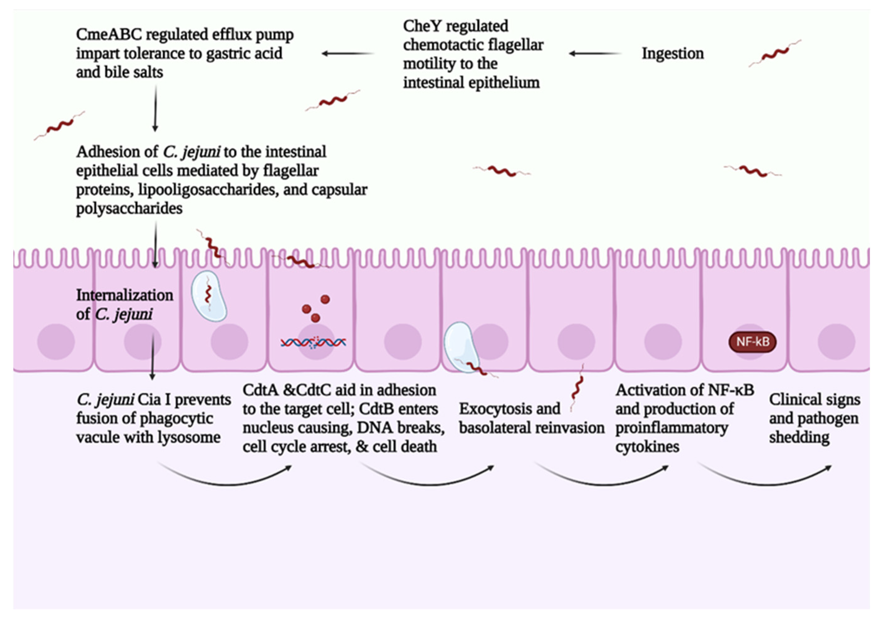
4. Pathogenesis of C. jejuni in Broilers

C. jejuni pathogenesis consists of four main steps: (1) ingestion, (2) acid tolerance and bile resistance, (3) reproduction in mucus, and (4) invasion of epithelial cells [12]. C. jejuni infection is transmitted between birds via the fecal–oral route [56]. A small dose of C. jejuni (around 35 CFU/mL) is sufficient for successful bird colonization [57]. As an enteric pathogen, C. jejuni expresses virulence factors regulated by a two-component system that mediates C. jejuni’s ability to survive the gut’s harsh conditions [58]. Campylobacter multidrug efflux pump (CmeABC) helps C. jejuni in eliminating toxic compounds such as antimicrobials, bile salts, and heavy metals. CmeABC comprises three proteins, a periplasmic protein, an inner membrane protein, and an outer membrane protein [59]. CmeABC gene encodes the multidrug efflux pump in C. jejuni and it is regulated through Cme repressor (CmeR) [60]. The presence of bile compounds stimulates the expression of CmeABC, increasing C. jejuni’s resistance to bile salts [60]. Mutations in regulator genes related to bile resistance block C. jejuni’s colonization ability [61].
C. jejuni depends on the two-component system consisting of CheY (cytoplasmic response regulator protein) and CheA (membrane-associated histidine auto kinase sensor) in responding to different chemoattractant/chemorepellents found in different environments [62]. In response to a stimulus, CheA is autophosphorylated, and a phosphate group is transferred to activate CheY. CheY interacts with the flagellar motor switch proteins leading to a clockwise rotation of the flagella [63]. The flagella play a central role in C. jejuni motility, adhesion, and invasion of the intestinal epithelial cells [64]. The flagellum consists of seven protofilaments of FlaA and FlaB subunits [64] and is attached to the basal structure through FlgE, which serves as a hook [64]. FlaA is the major Flagellin in C. jejuni and is regulated by σ28 promotor [20]. On the other hand, FlaB is the minor flagellin in C. jejuni and is regulated by the σ58 promoter [20]. Chemotaxis such as aspartate, glutamate, citrate, and L-fucose upregulates the σ58 gene [65]. FlaA plays a significant role in C. jejuni’s initial colonization of the chicken GIT [66]. The FlaA mutant has a ability to decrease the C. jejuni colonization in chicken [67]. Furthermore, the flagella include the type III secretion system (T3SS), which is responsible for delivering effector proteins needed for cellular invasion [20]. Thus, mutations in the flagellum lead to a decreased ability in colonization and invasion of intestinal epithelial cells.
The absence of immortalized chicken intestinal cell line hinders the capacity to characterize the mechanism of C. jejuni invasion of epithelial cells. In vitro, C. jejuni was capable of invading primary avian cells [68,69]. The presence of avian mucus protected the human cell line against the C. jejuni invasion [70]. It is well-known that C. jejuni survives and reproduces in avian mucus [71]. However, several factors interfere with C. jejuni’s capacity to invade the avian epithelial cells in vivo and might explain the near-commensal relationship of C. jejuni in avian species. Several differences are observed between humans and avian species in terms of body temperature (37 °C vs. 42 °C), mucus pH (the avian mucus is more acidic), and difference in mucus structure. C. jejuni upregulates genes related to metabolism and regulatory systems and downregulates genes related to periplasmic proteins at 37 °C in comparison with 42 °C [72]. This difference in gene expression may explain the C. jejuni adaptability and pathogenicity in humans’ intestinal tract. C. jejuni upregulates the CadF gene, which is responsible for cell adhesion at 37 °C and 42 °C, indicating the ability of C. jejuni to adhere to intestinal cells in humans and avian species [72]. On the other hand, C. jejuni isolates showed different gene expressions at 37 °C vs. 42 °C [73]. The difference in gene expression might explain some of the differences in C. jejuni’s pathogenicity. However, it might not be enough to justify the complete picture of C. jejuni pathogenicity in humans vs. the near-commensal relationship in avian species. It was hypothesized that the pH of the avian mucus confers protection for avian species against C. jejuni. However, through in vitro studies, the neutralization of the avian mucus did not diminish its anti-Campylobacter jejuni properties [68].
Purified chicken mucin inhibited the adherence and internalization of C. jejuni to a human intestinal cell line without affecting C. jejuni viability [70]. The oxidation of purified chicken mucin with sodium metaperiodate enabled C. jejuni to invade the intestinal cell line [70]. The results indicate the protecting role of o-glycosylated mucin structure in the intestinal cell line against C. jejuni.
The avian mucus is highly sulphated and sialylated compared with the human mucus [74]. The comparison between chicken and human mucin structures revealed thirty-three unique structures in chicken mucin [75]. The large intestine in chicken contains the highest sulphated structures, followed by the small intestine and cecum [75]. In chicken, C. jejuni colonizes mainly the ceca and, to a lesser extent, in the small and large intestines [76]. Evidence from in vitro studies shows that the purified chicken mucin from the large intestine had a higher inhibition ability against C. jejuni compared with the purified chicken mucin from the small intestine and cecum [75]. These results highlighted that presence of sulphated O-glycans is inversely correlated with the concentration of C. jejuni in the host. Furthermore, the increased sulfation and sialyation increase the anionic charge in the chicken mucin, creating a charge repulsion effect against C. jejuni [74]. These results indicate the role of chicken mucus in modulating C. jejuni virulence in avian species. C. jejuni pathogenesis is summarized in Figure 2.

5. Control of C. jejuni in Broilers: (Preharvest)

C. jejuni establishes colonization in the lower intestinal tract of the chicken, particularly in the ceca, within 24 h [65]. C. jejuni concentration can reach up to 1 × 10⁹ CFU/g in infected birds [20]. Birds are usually infected from two to four weeks of age and remain colonized until they reach market age [65]. Therefore, control methods are needed to reduce C. jejuni prevalence across broiler farms.

5.1. Biosecurity

Strict biosecurity measures are the key role in preventing the transmission of C. jejuni in broilers’ houses [35]. Identifying the potential sources and methods to detect C. jejuni at the farm level are the major steps needed for a successful biosecurity measures. Restricting access to poultry houses is key to maintaining a C. jejuni-free flock and following strict biosecurity measures can decrease C. jejuni prevalence by more than 50% in broilers at market age [38]. Cleaning and disinfecting poultry houses between cycles can reduce C. jejuni prevalence [38]. Furthermore, strict hygiene practices such as boot covers, hand washing, and footbaths can decrease C. jejuni transmission [35]. Standard litter management practices are also critical to decrease the presence of C. jejuni in the litter, and transmission is reduced with enough downtime between flocks [77].
Partial depopulation (thinning) of broiler flocks can increase the risk of C. jejuni transmission on the farm [78]. A strict biosecurity measure during the thinning process is required to ensure a low transmission rate of C. jejuni [78]. Furthermore, C. jejuni prevalence peaks during summer and early autumn time [79]. This seasonality of C. jejuni also correlates with the peak of insect populations [80]. Therefore, strict biosecurity measures are needed during summer, early autumn, and thinning to ensure a low prevalence of C. jejuni within the farm.

5.2. Probiotics, Prebiotics, and Synbiotic

Probiotics have the ability to improve gut health and prevent enteric diseases in poultry. Several mechanisms of probiotics include (1) antagonism and competitive exclusion of enteric pathogenic bacteria, (2) pH reduction by producing organic acids, (3) bacteriocin production, (4) stimulation/modulation of host immune response, (5) and alteration of virulence factors of enteric pathogens [81]. On the other hand, prebiotics are non-digestible feed ingredients that confer a beneficial effect on the host by promoting the proliferation of beneficial bacteria in the gut [81]. The combination of probiotics and prebiotics is known as synbiotics [81].
The ability of probiotics, prebiotics, and synbiotics to combat C. jejuni has been demonstrated in vitro, in vivo, and in field studies. In vitro, the anti-Campylobacter activity of probiotics has been carried out in agar-plate diffusion assays [82], co-cultures assays [83], and adhesion and colonization assays using cell lines [82].
Probiotics secrete organic acids that exhibit antimicrobial activity against gram-negative bacteria [84]. In vitro, E. faecium, P. acidilactic, L. salivarius, and L. reuteri supernatant inhibited C. jejuni growth [84]. Similarly, the supernatant of L. crispatus significantly decreased C. jejuni growth [85]. L. crispatus antimicrobial activity was mediated through the production of organic acids, namely: lactic acid [85]. In ceca, E. faecalis strain inoculation decreased C. jejuni load to 1 log CFU/g reduction after 6 h post-inoculation [86]. The anti-Campylobacter ability of lactic acid-producing bacteria was mediated through the production of organic acids [85].
Probiotic species can disrupt the expression of virulence factors in enteric pathogens [87,88]. The cell-free supernatant media of L. acidophilus strain La-5 and Bifidobacterium longum strain NCC2705 downregulated ciaB (invasion) and FlaA (motility) [87]. Similarly, the cell-free supernatant of L. salivarius, L. johnsonii, L. crispatus, and L. gasseri downregulated flaA, flab, flhA (motility), ciaB (invasion), and AI-2 (quorum sensing molecule autoinducer-2) [89]. These studies demonstrate the ability of probiotics species to attenuate the C. jejuni virulence factors. The downregulation of C. jejuni motility and invasion genes results in a lower ability to colonize the GIT of broilers and invade human and chicken primary cell lines [89]. Lactobacillus spp. Supplementation can modulate the host immune system [89]. L. salivarius, L. johnsonii, L. crispatus, and L. gasseri supplementation increased nitric oxide production and phagocytic ability of chicken macrophages, leading to a decrease in the C. jejuni load [89]. Furthermore, a mixture of Lactobacillus spp. Increased the expression of costimulatory molecules, namely: CD40, CD80, and CD86, in macrophages [89]. The costimulatory molecules are essential to initiate an adaptive and humoral immune response; hence probiotic supplementation can initiate the innate and adaptive immune response against C. jejuni [90].
Probiotic bacteria adhere to and occupy gut mucosal surfaces and competitively exclude enteric pathogens. In vitro studies are fundamental to investigating the probiotics’ mechanism of action. On the other hand, in vivo studies provide a comprehensive assessment of probiotics’ ability to benefit the host. Not all promising results in vitro are replicated in vivo. For example, in vitro studies with E. faecalis strain decreased the C. jejuni load by two log CFU/g. However, the E. faecalis strain could not decrease C. jejuni’s load in vivo [86]. In vitro studies with L. plantarum N8, N9, ZL5, and L. casei ZL4 adhered to the HT-29 cell line and competitively disrupted the adhesion and invasion of C. jejuni to the HT-29 cell line [91]. Similarly, L. paracasei JR, L. rhamnosus 15b, Y L. lactis, and L. lactis FOA decreased C. jejuni adhesion and invasion of the primary chicken cell line [92]. Probiotics enhance the integrity of the intestinal barrier by upregulating the expression of tight junction genes [93]. In a study, the supplementation of E.coli Nissle 1917 to the Ht-29 cell line upregulated tight junction genes expression resulting in lower C. jejuni intracellular invasion [93].
The supplementation of Poultry Star®, (Overland Park, KS, USA) (E. faecium, P. acidilactic, L. salivarius, and L. reuteri) via drinking water from the day of hatch to slaughtering decreased C. jejuni cecal load by six log CFU/g at 35 days of age [84]. However, studies with Poultry Star® supplementation only decreased C. jejuni cecal load by 2 log CFU/g at day 35, and no reduction in C. jejuni cecal load at 42 days of age [94]. The variability in probiotic efficacy between the two studies can be due to the difference in experimental design, challenge timing, and cecal microbiota of the broiler birds.
Isolated B. subtilis exhibited an anti-Campylobacter activity in vitro [95]. The supplementation of B. subtilis isolates reduced C. jejuni cecal load by one log CFU/g [95]. Motile probiotic bacteria can migrate towards the ceca where C. jejuni resides, thus having more chances to eliminate C. jejuni [95]. Therefore, the same B. subtilis isolates were propagated ten times to increase their motility. The propagated B. subtilis supplementation reduced C. jejuni cecal load by 2.5 log CFU/g at 21 days of age [95]. Similarly, the supplementation of B. longum subsp. longum PCB133 + galactooligosaccharide decreased C. jejuni cecal load by 1 log CFU/g at 56 days of age [96]. The oral gavage of L. salivarius SMXD51, every 2–3 days from the hatch until day 35, resulted in a 2.5 log CFU/g reduction in C. jejuni cecal load [97]. Furthermore, the supplementation of L. paracasei J. R, L. rhamnosus 15b, L. lactis Y, and L. lactis FOA combination used seven days before slaughter decreased C. jejuni load by five log CFU/g [92]. These data may suggest that multispecies probiotics may be better than single species probiotics in decreasing C. jejuni load in the poultry.
Contradictory results were obtained when evaluating the efficacy of probiotics on C. jejuni load in broilers. The probiotic bacterial strain, the supplementation dose of probiotics, the route of administration, C. jejuni challenge strain, age, sex, and breed of birds used for the study should be considered when evaluating the efficacy of probiotics. Moreover, C. jejuni survival in the host depends on the host’s microbiota [98]. Therefore, an interaction between C. jejuni and residing microbiota can influence the efficacy of the supplemented probiotic either positively or negatively.

5.3. Organic Acids

Organic acids are organic compounds that have acidic properties [99]. Organic acids are differentiated from other acids by having a carboxyl acid -COOH to which hydrogen or an organic compound might be attached [99]. Organic acids can be (1) short-chain fatty acids (SCFAs) (≤C6) such as acetic, lactic, butyric, fumaric, and propanoic acid, (2) medium-chain fatty acids (C7:C10) such as capric, caprylic acid, and (3) long-chain fatty acids (≥C11) such as lauric acid [99]. The gastrointestinal tract of avian species harbors millions of bacteria that produce different metabolites, including organic acids [99]. The antimicrobial mechanism in probiotics is mediated through the production of organic acids [81]. Therefore, supplementing organic acid is expected to impact the bird’s health positively. Organic acids supplementation leads to a decrease in the gut pH, enhancing the proteolytic enzymes and nutrient digestibility [99]. Furthermore, organic acids can act as bacteriostatic and/or bactericidal against gram-negative pathogenic bacteria, making them a suitable antibiotic alternative [83].
The supplementation of 2% formic acid in combination with 0.1% sorbate prevented the colonization of C. jejuni [100]. However, supplementing 2% formic acid alone was insufficient to prevent C. jejuni colonization [100]. Formic acid lowers the pH of the gut affecting the acid-sensitive bacteria present in the environment. On the other hand, sorbate targets the bacteria by diffusing through the cell membrane and lowering the pH of C. jejuni.
In vitro, butyrate supplementation demonstrated a bactericidal effect against C. jejuni [101]. However, the supplementation of butyrate-coated micro-beads did not decrease C. jejuni colonization [101]. The ineffectiveness of in vivo butyrate supplementation can be attributed to the fast absorption of butyrate by enterocytes. Similarly, the feed acidification with 5.7% lactic acid and 0.7% acetic acid decreased the presence of C. jejuni in the feed [102]. However, the limited effect of organic acids on C. jejuni colonization in broilers might be attributed to the possibility of being absorbed by the gut microbes before it reaches the ceca [102].
Another study tested different combinations of organic acids, prebiotics, and probiotics against C. jejuni infection. Only Adimix® Precision, (Dendermonde, Belgium) (sodium salt butyrate) decreased the cecal load of C. jejuni by 2 log CFU/g at 42 days of age [94]. Other compounds containing organic compounds such as lactic acid had a limited ability to decrease C. jejuni cecal load. The efficacy of organic acids in controlling enteric pathogens relies heavily on (1) type of SCFAs, (2) dose of SCFAs in feed, (3) buffering capacity of the feed and (4) complex microbiota in the host.
When choosing the type of organic acid for supplementation, the pathogen’s metabolism also should be considered. C. jejuni cannot ferment carbohydrates and depends mainly on amino acids and some SCFAs to proliferate in the avian gut [103]. C. jejuni utilizes acetate, lactate, fumarate, succinate, and malate as part of its citric acid cycle to satisfy its energy needs [103]. The ability of C. jejuni to use SCFAs might explain the ineffectiveness of organic acid supplementation such as lactic acid, formic acid, and acetic acid in the presented studies. Butyrate supplementation might be the organic acid of choice to control C. jejuni. Yet, the fact that butyrate is the primary energy source for enterocytes [104] limits its presence in the gut and decreases its ability to fight pathogens.

5.4. Bacteriophages

Bacteriophages are viruses ubiquitously found in nature and infect bacterial and archaeal cells [105]. The world contains approximately 1032, which is almost 10 times more than the number of bacterial cells on earth [106]. Bacteriophages were discovered in 1915 by Frederick Twort and Félix d’Hérelle in 1917 [107]. Bacteriophages are considered non-pathogenic to humans as they are frequently isolated from human saliva [108] and feces [109]. Humans are frequently exposed to bacteriophages in food and drinking water without any adverse reaction to their consumption [110]. Moreover, bacteriophages dominate the human gut virome [110]. However, the chicken virome is yet to be characterized.
Bacteriophages are suggested as an antibiotic alternative in controlling foodborne pathogens, as they are easy to isolate, have narrow specific, and do not alter the microbiome of the treated host [111]. Though more than 170 C. jejuni phages have been documented, the majority of these phages have a narrow spectrum [105] to control foodborne pathogens. C. jejuni phages are divided into two categories: lytic and lysogenic. Lytic bacteriophages are preferred as they can lyse the targeted cell immediately [111]. In contrast, the lysogenic bacteriophages are not used as they incorporate into the bacterial genome and transfer virulence factors between bacteria [111].
Lytic Campylobacter phages are categorized based on their size into three categories [105]. The first category includes large phages, ranging between 320 and425 kbp, whereas the second category comprises phages that range between 175 and183 and show a high affinity towards C. jejuni and C. coli [105]. The third category includes Campylobacter phages with the smallest size and the greatest affinity and lytic ability toward C. jejuni [105]. Campylobacter phages are versatile tools that can be incorporated into the preharvest and postharvest control of foodborne pathogens.
The efficacy of two Campylobacter phages, CP8 and CP34, was tested for five days post-C. jejuni infection, resulting in a 0.5–5 log CFU/g reduction in C. jejuni based on the intestinal site and phage dose [112]. The best results in C. jejuni load reduction was obtained 24–48 h post bacteriophage supplementation [112]. Similarly, the efficacy was tested with two administration routes (drinking water vs. feed) of a phage cocktail (phiCcoIBB35, phiCcoIBB37, and phiCcoIBB12) [113]. The highest reduction (2 log CFU/g) of C. jejuni load was recorded when the cocktail phage was supplemented in the feed [113]. These results indicated the ability of bacteriophages to control C. jejuni and highlighted the need to determine the dose and route of administration to achieve the best results.
A comparison between a single phage and a cocktail phage to reduce C. jejuni colonization resulted in a maximum of 2.8 log CFU/g cecal C. jejuni load in both groups [114]. The single phage resulted in 43% phage resistance, whereas the cocktail phage led to 24% phage resistance [114]. The development of phage resistance limits the using Campylobacter phages in controlling C. jejuni prevalence. Though the use of phage cocktails might delay the C. jejuni development, the effectiveness of C. jejuni phages is yet to be determined at the farm scale [114].

5.5. Bacteriocins

Bacteriocins are ribosomal synthesized antimicrobial peptides secreted by bacteria. Bacteriocins can act as bacteriostatic and bactericidal against related bacterial species [115]. The secretion of bacteriocins confers the destruction of targeted bacteria without damaging the host. Bacteriocin mode of action is mediated through membrane permeabilization followed by cell lysis [116]. The supplementation of bacteriocins in C. jejuni infected broilers efficiently reduces C. jejuni’s load and contamination in the food chain [117,118]. Seven-day-old broilers treated with purified encapsulated OR7 bacteriocins produced by L. acidophilus NRRL B-30514 significantly reduced C. jejuni load [118].
Similarly, supplementing two purified forms, L. salivarius NRRL B-30514 and P. polymyxa NRRL B-30509, decreased the C. jejuni cecal load by three log CFU/g [117].
Recently, reuterin emerged as a promising bacteriocin in controlling C. jejuni colonization in broilers. Reuterin is an antimicrobial compound produced during the anaerobic formation of glycerol by L. reuteri [119]. Reuterin exhibits a wide antimicrobial spectrum against gram-negative and gram-positive bacteria, yeast, and mold [119]. The mechanism of action of reuterin is mediated through the reaction of acrolein with the thiol groups of glutathione, inhibiting the redox-base defenses and leading to oxidative stress in the targeted bacteria [120]. The genome analysis of C. jejuni revealed the absence of glutathione biosynthesis protein, suggesting that C. jejuni lacks the ability to detoxify acrolein [121]. The absence of glutathione biosynthesis protein might explain the susceptibility of C. jejuni during in vitro studies to reuterin [122].
Bacteriocins production has a high metabolic cost; hence probiotic species will not overproduce it. Supplementing encapsulated bacteriocins with probiotic species might play a prominent role in competitively excluding C. jejuni from the avian gut.

5.6. Vaccines

Vaccination remains a potentially effective strategy to mitigate the prevalence of foodborne pathogens (C. jejuni and Salmonella) in poultry production [123]. Vaccination aims to stimulate a mucosal anti-Campylobacter jejuni immune response and reduce the C. jejuni load at market age. Several vaccine strategies have been developed to control C. jejuni in broilers:

5.6.1. Whole Cell Vaccine and Live Attenuated Vaccine

A formalin-killed C. jejuni whole cell vaccine containing 2.7 × 10⁸ CFU/mL C. jejuni combined with an oil adjuvant or aluminum hydroxide gel adjuvant was inoculated subcutaneously in Japanese Jordi chicken at 37 days of age [124]. The aluminum hydroxide gel adjuvant group received a booster at 58 days of age. The birds were challenged with C. jejuni on 72 days of age. Both vaccine groups induced high anti-C. jejuni IgG levels [124]. Similarly, a formalin-killed C. jejuni whole-cell vaccine was formulated with or without an E. coli heat-labile toxin as an adjuvant. The vaccine administration enhanced the anti-C. jejuni levels and reduced C. jejuni colonization from 16% to 93% in the vaccinated group compared with the non-vaccinated one [125]. However, the E. coli heat liable toxin did not increase the immunogenicity of the vaccine [125].
Oxidative stress response plays a significant role in C. jejuni’s enteric lifestyle. C. jejuni oxidative stress defense mutant shows a low ability to persist in the avian gut. In this study, birds were orally gavaged with 0.5 mL of C. jejuni ΔahpC mutant at 3 and 7 days of age, followed by a challenge (WT C. jejuni) at 14 days of age [126]. The pre-colonization of broilers with the C. jejuni ΔahpC mutant decreased the C. jejuni by three log CFU/g reduction at 42 days of age [126]. These results suggest that the C. jejuni ΔahpC mutant has the potential to be used at the farm level to control C. jejuni at the preharvest stage; however, more safety studies are required at a farm level.

5.6.2. Crude Cell Lysate

The efficacy of a nanoparticle vaccine composed of poly lactide-co-glycolide nanoparticle (NP) and encapsulated 25, 125, or 250 μg outer membrane of C. jejuni was evaluated against C. jejuni [127]. The subcutaneous route induced the highest immune response in vaccinated broilers and decreased the C. jejuni load by 5.7 logCFU/g in the ceca [127]. Similarly, the oral delivery of C. jejuni oral lysate reduced the C. jejuni load in layer and broiler chickens by 2.24 log CFU/g and 2.14 log CFU/g, respectively, at 22 days post-infection [128].

5.6.3. Subunit Vaccine

Type VI secretion system (T6SS) enables bacteria to infect neighboring cells and plays a vital role in inter-bacterial competition and bacterial communication with the host’s cells [129]. In C. jejuni, the type VI secretion system (T6SS) plays a role in evading the immune system and bacterial survival [130]. A subunit vaccine was formulated from a purified 50 µg of recombinant hemolysin co-regulated protein (RHCP) entrapped in chitosan sodium tripolyphosphate nanoparticles [131]. The broilers were orally gavaged with subunit vaccine at 7 days of age and then boosted at 14 and 21 days of age [131]. The vaccinated broilers were then challenged with C. jejuni at 28 days of age. The vaccinated group had one log CFU/g reduction of C. jejuni load in the ceca [131].

5.6.4. Bacterial Vector-Based Vaccine

Live or genetically engineered bacterial strains emerge as potential vaccine candidates against enteric pathogens. Bacteria that are avirulent to chickens and elicit an immune response are considered suitable vectors. These vectors can present C. jejuni virulent antigens to the birds’ immune system. C. jejuni mutants show a transient colonization pattern in the chicken gut and do not persist enough to activate an immune response [132]. Avirulent Salmonella and Lactobacillus strains are the bacterial vectors of choice for creating a bacterial vector-based vaccine against C. jejuni. An avirulent Salmonella Typhimurium χ3987 strain expressing CjaA was orally gavaged in broilers at 1 and 14 days of age (booster) [133]. At 28 days of age, the broiler was challenged by C. jejuni. The vaccine inoculation led to 6.0 log CFU/g reductions in the C. jejuni cecal load [133].
Finally, phase variation and strain differences in C. jejuni, C. coli, and C. lari complicate the development of a potential vaccine that can decrease campylobacteriosis around the globe.

5.7. Quorum Sensing Inhibitors

In broilers, C. jejuni cecal load can reach up to 1 × 10⁹ CFU/g [134]. C. jejuni can detect and respond to rapid changes in bacterial densities using quorum sensing [135]. Quorum sensing is a cell-to-cell communication in which bacteria produce, detect, and respond to signaling molecules known as autoinducers [136]. The accumulation of autoinducers happens in a density-dependent manner [135]. When the autoinducer concentration reaches a certain threshold, it leads to the activation of a signal cascade [137]. The signal cascade alters gene expression, resulting in morphological changes in the bacterium that aids its survival in the environment [137].
At first, quorum sensing studies in C. jejuni identified a gene that encodes an orthologue of the LuxS system that mediates the production of autoinducer-2 (AI-2) [138]. In the same study, the C. jejuni luxS mutants showed a decreased motility in semisolid media, indicating a key role of luxS in regulating darting motility in C. jejuni [138]. Furthermore, the role of luxS in host colonization was evaluated in a study testing the colonization ability of the C. jejuni luxS mutant strain vs. the C. jejuni wild-type strain [139]. The luxS mutant showed decreased colonization capacity in chicken seven days post-inoculation; however, some birds inoculated with the luxS mutant strains maintained a similar level of colonization compared with groups inoculated with the wild-type strain [139]. Furthermore, a competitive fitness experiment between the wild-type and luxS mutant showed a decrease in the recovery of the mutant in comparison with the wild-type, indicating an important role of luxS in C. jejuni fitness [140].
The imminent role of luxS in C. jejuni’s adaption to environmental conditions [141], expression of virulence factors [140], and biofilm formation [142] make it a potential target for controlling C. jejuni infection. In vitro, (-)-α-pinene showed an anti-quorum sensing activity against C. jejuni by decreasing the C. jejuni quorum signaling by more than 80% [143]. The supplementation of 250 mg/L of (-)-α-pinene in C. jejuni-challenged broilers resulted in 0.8 log CFU/g reduction in the cecal load [143]. (-)-α-pinene inhibitory activity against C. jejuni is attributed to (-)-α-pinene ability to inhibit efflux pump activity and quorum sensing, which play a crucial role in colonizing the host [143]. Thus, (-)-α-pinene can potentially contribute to the control of C. jejuni in broilers.
Citrus extracts decreased motility and biofilm formation in E. coli O157:H7, S. typhimurium, and P. aeruginosa. Similarly, citrus extracts inhibited C. jejuni autoinducer-2 quorum sensing, resulting in lower motility and lower biofilm formation [144]. Similarly, Sedum rosea (roseroot) extract decreased C. jejuni quorum signaling by more than 90% and decreased C. jejuni invasion of INT407 cells by 80% [145]. These results demonstrate the ability of natural phenolic compounds to alter the quorum sensing in C. jejuni, resulting in a lower fitness in C. jejuni. Quorum inhibitory compounds are a promising tool to control C. jejuni in poultry. However, additional studies are needed to determine the required dose and treatment period to decrease the load of C. jejuni in poultry. The control strategies of C. jejuni are summarized in Figure 3.

6. Conclusions

Chicken around the globe remains the main reservoir for campylobacteriosis in humans. With the increase in campylobacteriosis worldwide, antibiotic resistance in C. jejuni and increased post-C. jejuni infection complications (such as GBS and MRS), looking for a suitable control strategy becomes the need of the hour. A multi-hurdle approach is needed to ensure the control of foodborne pathogens from farm to fork. Strict biosecurity combined with feed additives and a suitable vaccine (if developed) might be the method of choice to control C. jejuni in broiler production.

Author Contributions

W.G.A.H.: Writing—original draft preparation; S.F.: writing, R.S.: review, editing, and funding acquisition, R.K.S.: editing and funding acquisition. All authors have read and agreed to the published version of the manuscript.

Funding

This work was funded by USDA ARS project number 6040-42000-046-000D to RS.

Institutional Review Board Statement

Not applicable.

Informed Consent Statement

Not applicable.

Data Availability Statement

No new data were created or analyzed in this study. Data sharing is not applicable to this article.

Conflicts of Interest

The authors declare no conflict of interest.

References

  1. Escherich, T. Beitrage zur Kenntniss der Darmbacterien. III. Ueber das Vorkommen von Vibrionen im Darmcanal und den Stuhlgangen der Sauglinge (Articles adding to the knowledge of intestinal bacteria. III. On the existence of vibrios in the intestines and feces of babies). Münchener Med. Wochenschrift 1886, 33, 815–817. [Google Scholar]
  2. McFadyean, J.; Stockman, S. Report of the Deparmental Committee Appointed by the Board of Agriculture and Fisheries Toenquire into Epizootic Abortion, Part III 1–64; Hermajesty’s Stationary office: London, UK, 1913. [Google Scholar]
  3. Smith, T. Spirilla associated with disease of the fetal membranes in cattle (infectious abortion). J. Exp. Med. 1918, 28, 701–719. [Google Scholar] [CrossRef] [Green Version]
  4. Stegenga, T.; Terpstra, J. Over Vibrio fetus infecties bij het rund en enzootishe steriliteit. Tijdschr. Diergeneeskd. 1949, 74, 293–296. [Google Scholar]
  5. Doyle, L. A vibrio associated with swine dysentery. Am. J. Vet. Res. 1944, 5, 3–5. [Google Scholar]
  6. Vinzent; Dumas; Picard; Lemierre. Septicemie Grave au Cours de la Grossesse, Due a un Vibrion-Avortement Consecutif. In Proceedings of the Semaine des Hopitaux, Paris, France, June 1947; p. 709. [Google Scholar]
  7. Levy, A. A gastro-enteritis outbreak probably due to a bovine strain of vibrio. Yale J. Biol. Med. 1946, 18, 243. [Google Scholar] [PubMed]
  8. Dekeyser, P.; Gossuin-Detrain, M.; Butzler, J.-P.; Sternon, J. Acute enteritis due to related vibrio: First positive stool cultures. J. Infect. Dis. 1972, 125, 390–392. [Google Scholar] [CrossRef] [PubMed]
  9. Butzler, J.P. Campylobacter, from obscurity to celebrity. Clin. Microbiol. Infect. 2004, 10, 868–876. [Google Scholar] [CrossRef] [Green Version]
  10. Epps, S.V.; Harvey, R.B.; Hume, M.E.; Phillips, T.D.; Anderson, R.C.; Nisbet, D.J. Foodborne Campylobacter: Infections, metabolism, pathogenesis and reservoirs. Int. J. Environ. Res. Public Health 2013, 10, 6292–6304. [Google Scholar] [CrossRef] [PubMed] [Green Version]
  11. Skarp, C.P.A.; Hänninen, M.L.; Rautelin, H.I.K. Campylobacteriosis: The role of poultry meat. Clin. Microbiol. Infect. 2016, 22, 103–109. [Google Scholar] [CrossRef] [PubMed] [Green Version]
  12. Pielsticker, C.; Glünder, G.; Rautenschlein, S. Colonization properties of Campylobacter jejuni in chickens. Eur. J. Microbiol. Immunol. 2012, 2, 61–65. [Google Scholar] [CrossRef] [PubMed] [Green Version]
  13. Kozu Clarke, A.; Ajlouni, S. Recommended Practices to Eliminate Campylobacter from Live Birds and Chicken Meat in Japan. Food Saf. 2021, 9, 57–74. [Google Scholar] [CrossRef] [PubMed]
  14. Ruiz-Palacios, G.M. The Health Burden of Campylobacter Infection and the Impact of Antimicrobial Resistance: Playing Chicken. Clin. Infect. Dis. 2007, 44, 701–703. [Google Scholar] [CrossRef] [PubMed]
  15. Fathima, S.; Shanmugasundaram, R.; Adams, D.; Selvaraj, R. Gastrointestinal Microbiota and Their Manipulation for Improved Growth and Performance in Chickens. Foods 2022, 11, 1401. [Google Scholar] [CrossRef] [PubMed]
  16. Cohen, E.J.; Nakane, D.; Kabata, Y.; Hendrixson, D.R.; Nishizaka, T.; Beeby, M. Campylobacter jejuni motility integrates specialized cell shape, flagellar filament, and motor, to coordinate action of its opposed flagella. PLoS Pathog. 2020, 16, e1008620. [Google Scholar] [CrossRef] [PubMed]
  17. Frirdich, E.; Biboy, J.; Adams, C.; Lee, J.; Ellermeier, J.; Gielda, L.D.; Dirita, V.J.; Girardin, S.E.; Vollmer, W.; Gaynor, E.C. Peptidoglycan-modifying enzyme Pgp1 is required for helical cell shape and pathogenicity traits in Campylobacter jejuni. PLoS Pathog. 2012, 8, e1002602. [Google Scholar] [CrossRef]
  18. Maue, A.C.; Mohawk, K.L.; Giles, D.K.; Poly, F.; Ewing, C.P.; Jiao, Y.; Lee, G.; Ma, Z.; Monteiro, M.A.; Hill, C.L.; et al. The polysaccharide capsule of Campylobacter jejuni modulates the host immune response. Infect. Immun. 2013, 81, 665–672. [Google Scholar] [CrossRef] [Green Version]
  19. Pike, B.L.; Guerry, P.; Poly, F. Global Distribution of Campylobacter jejuni Penner Serotypes: A Systematic Review. PLoS ONE 2013, 8, e67375. [Google Scholar] [CrossRef] [Green Version]
  20. Burnham, P.M.; Hendrixson, D.R. Campylobacter jejuni: Collective components promoting a successful enteric lifestyle. Nat. Rev. Microbiol. 2018, 16, 551–565. [Google Scholar] [CrossRef] [PubMed]
  21. Houliston, R.S.; Vinogradov, E.; Dzieciatkowska, M.; Li, J.; St Michael, F.; Karwaski, M.F.; Brochu, D.; Jarrell, H.C.; Parker, C.T.; Yuki, N.; et al. Lipooligosaccharide of Campylobacter jejuni: Similarity with multiple types of mammalian glycans beyond gangliosides. J. Biol. Chem. 2011, 286, 12361–12370. [Google Scholar] [CrossRef] [PubMed] [Green Version]
  22. Kim, J.-C.; Oh, E.; Hwang, S.; Ryu, S.; Jeon, B. Non-selective regulation of peroxide and superoxide resistance genes by PerR in Campylobacter jejuni. Front. Microbiol. 2015, 6, 126. [Google Scholar] [CrossRef] [PubMed] [Green Version]
  23. Oh, E.; McMullen, L.; Jeon, B. Impact of oxidative stress defense on bacterial survival and morphological change in Campylobacter jejuni under aerobic conditions. Front. Microbiol. 2015, 6, 295. [Google Scholar] [CrossRef]
  24. Sahin, O.; Morishita, T.Y.; Zhang, Q. Campylobacter colonization in poultry: Sources of infection and modes of transmission. Anim. Health Res. Rev. 2002, 3, 95–105. [Google Scholar] [CrossRef]
  25. Shanker, S.; Lee, A.; Sorrell, T. Horizontal transmission of Campylobacter jejuni amongst broiler chicks: Experimental studies. Epidemiol. Infect. 1990, 104, 101–110. [Google Scholar] [CrossRef] [PubMed]
  26. Conlan, A.J.; Coward, C.; Grant, A.J.; Maskell, D.J.; Gog, J.R. Campylobacter jejuni colonization and transmission in broiler chickens: A modelling perspective. J. R. Soc. Interface 2007, 4, 819–829. [Google Scholar] [CrossRef] [PubMed] [Green Version]
  27. Doyle, M.P. Association of Campylobacter jejuni with laying hens and eggs. Appl. Environ. Microbiol. 1984, 47, 533–536. [Google Scholar] [CrossRef] [PubMed] [Green Version]
  28. Lam, K.; DaMassa, A.; Morishita, T.; Shivaprasad, H.L.; Bickford, A. Pathogenicity of Campylobacter jejuni for turkeys and chickens. Avian Dis. 1992, 36, 359–363. [Google Scholar] [CrossRef]
  29. Fonseca, B.B.; Beletti, M.E.; Melo, R.T.d.; Mendonça, E.P.; Coelho, L.R.; Nalevaiko, P.C.; Rossi, D.A. Campylobacter jejuni in commercial eggs. Braz. J. Microbiol. 2014, 45, 76–79. [Google Scholar] [CrossRef] [PubMed] [Green Version]
  30. Neill, S.; Campbell, J.; O’brien, J. Egg penetration by Campylobacter jejuni. Avian Pathol. 1985, 14, 313–320. [Google Scholar] [CrossRef] [Green Version]
  31. Prachantasena, S.; Charununtakorn, P.; Muangnoicharoen, S.; Hankla, L.; Techawal, N.; Chaveerach, P.; Tuitemwong, P.; Chokesajjawatee, N.; Williams, N.; Humphrey, T.; et al. Distribution and Genetic Profiles of Campylobacter in Commercial Broiler Production from Breeder to Slaughter in Thailand. PLoS ONE 2016, 11, e0149585. [Google Scholar] [CrossRef] [PubMed] [Green Version]
  32. Jonaidi-Jafari, N.; Khamesipour, F.; Ranjbar, R.; Kheiri, R. Prevalence and antimicrobial resistance of Campylobacter species isolated from the avian eggs. Food Control 2016, 70, 35–40. [Google Scholar] [CrossRef]
  33. Ghoneim, N.H.; Abdel-Moein, K.A.-A.; Barakat, A.M.A.K.; Hegazi, A.G.; Abd El-Razik, K.A.E.-H.; Sadek, S.A.S. Isolation and molecular characterization of Campylobacter jejuni from chicken and human stool samples in Egypt. Food Sci. Technol. 2020, 41, 195–202. [Google Scholar] [CrossRef]
  34. Cappelier, J.M.; Minet, J.; Magras, C.; Colwell, R.R.; Federighi, M. Recovery in embryonated eggs of viable but nonculturable Campylobacter jejuni cells and maintenance of ability to adhere to HeLa cells after resuscitation. Appl. Environ. Microbiol. 1999, 65, 5154–5157. [Google Scholar] [CrossRef] [PubMed] [Green Version]
  35. Sibanda, N.; McKenna, A.; Richmond, A.; Ricke, S.C.; Callaway, T.; Stratakos, A.C.; Gundogdu, O.; Corcionivoschi, N. A Review of the Effect of Management Practices on Campylobacter Prevalence in Poultry Farms. Front. Microbiol. 2018, 9, 2002. [Google Scholar] [CrossRef] [PubMed]
  36. Hazeleger, W.C.; Wouters, J.A.; Rombouts, F.M.; Abee, T. Physiological activity of Campylobacter jejuni far below the minimal growth temperature. Appl. Environ. Microbiol. 1998, 64, 3917–3922. [Google Scholar] [CrossRef] [Green Version]
  37. Herman, L.; Heyndrickx, M.; Grijspeerdt, K.; Vandekerchove, D.; Rollier, I.; De Zutter, L. Routes for Campylobacter contamination of poultry meat: Epidemiological study from hatchery to slaughterhouse. Epidemiol. Infect. 2003, 131, 1169–1180. [Google Scholar] [CrossRef]
  38. Newell, D.G.; Elvers, K.T.; Dopfer, D.; Hansson, I.; Jones, P.; James, S.; Gittins, J.; Stern, N.J.; Davies, R.; Connerton, I.; et al. Biosecurity-based interventions and strategies to reduce Campylobacter spp. on poultry farms. Appl. Environ. Microbiol. 2011, 77, 8605–8614. [Google Scholar] [CrossRef] [Green Version]
  39. Wright, E. The isolation of Campylobacter jejuni from flies. Epidemiol. Infect. 1983, 91, 223–226. [Google Scholar]
  40. Royden, A.; Wedley, A.; Merga, J.Y.; Rushton, S.; Hald, B.; Humphrey, T.; Williams, N.J. A role for flies (Diptera) in the transmission of Campylobacter to broilers? Epidemiol. Infect. 2016, 144, 3326–3334. [Google Scholar] [CrossRef] [Green Version]
  41. Hald, B.; Skovgård, H.; Bang, D.D.; Pedersen, K.; Dybdahl, J.; Jespersen, J.B.; Madsen, M. Flies and Campylobacter infection of broiler flocks. Emerg. Infect. Dis. 2004, 10, 1490–1492. [Google Scholar] [CrossRef]
  42. Friedrich, A.; Marshall, J.C.; Biggs, P.J.; Midwinter, A.C.; French, N.P. Seasonality of Campylobacter jejuni isolates associated with human campylobacteriosis in the Manawatu region, New Zealand. Epidemiol. Infect. 2016, 144, 820–828. [Google Scholar] [CrossRef]
  43. Olkkola, S.; Rossi, M.; Jaakkonen, A.; Simola, M.; Tikkanen, J.; Hakkinen, M.; Tuominen, P.; Huitu, O.; Niemimaa, J.; Henttonen, H.; et al. Host-Dependent Clustering of Campylobacter Strains From Small Mammals in Finland. Front. Microbiol. 2021, 11, 621490. [Google Scholar] [CrossRef] [PubMed]
  44. Kim, J.; Guk, J.-H.; Mun, S.-H.; An, J.-U.; Kim, W.; Lee, S.; Song, H.; Seong, J.K.; Suh, J.G.; Cho, S. The Wild Mouse (Micromys minutus): Reservoir of a Novel Campylobacter jejuni Strain. Front. Microbiol. 2020, 10, 3066. [Google Scholar] [CrossRef] [PubMed]
  45. Reed, K.D.; Meece, J.K.; Henkel, J.S.; Shukla, S.K. Birds, migration and emerging zoonoses: West Nile virus, Lyme disease, influenza A and enteropathogens. Clin. Med. Res. 2003, 1, 5–12. [Google Scholar] [CrossRef] [PubMed] [Green Version]
  46. White, R.J.; Razgour, O. Emerging zoonotic diseases originating in mammals: A systematic review of effects of anthropogenic land-use change. Mammal Rev. 2020, 50, 336–352. [Google Scholar] [CrossRef]
  47. Keatts, L.O.; Robards, M.; Olson, S.H.; Hueffer, K.; Insley, S.J.; Joly, D.O.; Kutz, S.; Lee, D.S.; Chetkiewicz, C.-L.B.; Lair, S.; et al. Implications of Zoonoses From Hunting and Use of Wildlife in North American Arctic and Boreal Biomes: Pandemic Potential, Monitoring, and Mitigation. Front. Public Health 2021, 9, 627654. [Google Scholar] [CrossRef]
  48. Wysok, B.; Sołtysiuk, M.; Stenzel, T. Wildlife Waterfowl as a Source of Pathogenic Campylobacter Strains. Pathogens 2022, 11, 113. [Google Scholar] [CrossRef] [PubMed]
  49. Waldenström, J.; Axelsson-Olsson, D.; Olsen, B.; Hasselquist, D.; Griekspoor, P.; Jansson, L.; Teneberg, S.; Svensson, L.; Ellström, P. Campylobacter jejuni colonization in wild birds: Results from an infection experiment. PLoS ONE 2010, 5, e9082. [Google Scholar] [CrossRef] [Green Version]
  50. Mutschall, S.K.; Hetman, B.M.; Bondo, K.J.; Gannon, V.P.J.; Jardine, C.M.; Taboada, E.N. Campylobacter jejuni Strain Dynamics in a Raccoon (Procyon lotor) Population in Southern Ontario, Canada: High Prevalence and Rapid Subtype Turnover. Front. Vet. Sci. 2020, 7, 27. [Google Scholar] [CrossRef]
  51. Indykiewicz, P.; Andrzejewska, M.; Minias, P.; Śpica, D.; Kowalski, J. Prevalence and Antibiotic Resistance of Campylobacter spp. in Urban and Rural Black-Headed Gulls Chroicocephalus ridibundus. EcoHealth 2021, 18, 147–156. [Google Scholar] [CrossRef]
  52. Cecchi, F.; Fabbri, M.C.; Tinacci, L.; Nuvoloni, R.; Marotta, F.; Di Marcantonio, L.; Cilia, G.; Macchioni, F.; Armani, A.; Fratini, F.; et al. Genetic resistance to Campylobacter coli and Campylobacter jejuni in wild boar (Sus scrofa L.). Rendiconti Lincei Scienze Fisiche Naturali 2022, 33, 407–417. [Google Scholar] [CrossRef]
  53. Morita, S.; Sato, S.; Maruyama, S.; Miyagawa, A.; Nakamura, K.; Nakamura, M.; Asakura, H.; Sugiyama, H.; Takai, S.; Maeda, K.; et al. Prevalence and whole-genome sequence analysis of Campylobacter spp. strains isolated from wild deer and boar in Japan. Comp. Immunol. Microbiol. Infect. Dis. 2022, 82, 101766. [Google Scholar] [CrossRef] [PubMed]
  54. Hald, B.; Skov, M.N.; Nielsen, E.M.; Rahbek, C.; Madsen, J.J.; Wainø, M.; Chriél, M.; Nordentoft, S.; Baggesen, D.L.; Madsen, M. Campylobacter jejuni and Campylobacter coli in wild birds on Danish livestock farms. Acta Vet. Scand. 2016, 58, 11. [Google Scholar] [CrossRef] [PubMed] [Green Version]
  55. Ramabu, S.; Boxall, N.; Madie, P.; Fenwick, S. Some potential sources for transmission of Campylobacter jejuni to broiler chickens. Lett. Appl. Microbiol. 2004, 39, 252–256. [Google Scholar] [CrossRef] [PubMed]
  56. Ondrašovičová, S.; Pipová, M.; Dvořák, P.; Hričínová, M.; Hromada, R.; Kremeň, J. Passive and active immunity of broiler chickens against Campylobacter jejuni and ways of disease transmission. Acta Vet. Brno 2012, 81, 103–106. [Google Scholar] [CrossRef]
  57. Cawthraw, S.A.; Wassenaar, T.M.; Ayling, R.; Newell, D.G. Increased colonization potential of Campylobacter jejuni strain 81116 after passage through chickens and its implication on the rate of transmission within flocks. Epidemiol. Infect. 1996, 117, 213–215. [Google Scholar] [CrossRef] [Green Version]
  58. Sharifi, S.; Bakhshi, B.; Najar-Peerayeh, S. Significant contribution of the CmeABC Efflux pump in high-level resistance to ciprofloxacin and tetracycline in Campylobacter jejuni and Campylobacter coli clinical isolates. Ann. Clin. Microbiol. Antimicrob. 2021, 20, 1–9. [Google Scholar] [CrossRef]
  59. Alav, I.; Kobylka, J.; Kuth, M.S.; Pos, K.M.; Picard, M.; Blair, J.M.; Bavro, V.N. Structure, assembly, and function of tripartite efflux and type 1 secretion systems in gram-negative bacteria. Chem. Rev. 2021, 121, 5479–5596. [Google Scholar] [CrossRef]
  60. Lin, J.; Cagliero, C.; Guo, B.; Barton, Y.W.; Maurel, M.C.; Payot, S.; Zhang, Q. Bile salts modulate expression of the CmeABC multidrug efflux pump in Campylobacter jejuni. J. Bacteriol. 2005, 187, 7417–7424. [Google Scholar] [CrossRef] [Green Version]
  61. Guo, B.; Wang, Y.; Shi, F.; Barton, Y.W.; Plummer, P.; Reynolds, D.L.; Nettleton, D.; Grinnage-Pulley, T.; Lin, J.; Zhang, Q. CmeR functions as a pleiotropic regulator and is required for optimal colonization of Campylobacter jejuni in vivo. J. Bacteriol. 2008, 190, 1879–1890. [Google Scholar] [CrossRef] [Green Version]
  62. Chandrashekhar, K.; Kassem, I.I.; Rajashekara, G. Campylobacter jejuni transducer like proteins: Chemotaxis and beyond. Gut Microbes 2017, 8, 323–334. [Google Scholar] [CrossRef] [Green Version]
  63. Tram, G.; Klare, W.P.; Cain, J.A.; Mourad, B.; Cordwell, S.J.; Day, C.J.; Korolik, V. Assigning a role for chemosensory signal transduction in Campylobacter jejuni biofilms using a combined omics approach. Sci. Rep. 2020, 10, 6829. [Google Scholar] [CrossRef] [PubMed] [Green Version]
  64. Guerry, P. Campylobacter flagella: Not just for motility. Trends Microbiol. 2007, 15, 456–461. [Google Scholar] [CrossRef] [PubMed]
  65. Hermans, D.; Van Deun, K.; Martel, A.; Van Immerseel, F.; Messens, W.; Heyndrickx, M.; Haesebrouck, F.; Pasmans, F. Colonization factors of Campylobacter jejuni in the chicken gut. Vet. Res. 2011, 42, 82. [Google Scholar] [CrossRef] [PubMed] [Green Version]
  66. Jones, M.A.; Marston, K.L.; Woodall, C.A.; Maskell, D.J.; Linton, D.; Karlyshev, A.V.; Dorrell, N.; Wren, B.W.; Barrow, P.A. Adaptation of Campylobacter jejuni NCTC11168 to high-level colonization of the avian gastrointestinal tract. Infect. Immun. 2004, 72, 3769–3776. [Google Scholar] [CrossRef]
  67. Fernando, U.; Biswas, D.; Allan, B.; Willson, P.; Potter, A.A. Influence of Campylobacter jejuni fliA, rpoN and flgK genes on colonization of the chicken gut. Int. J. Food Microbiol. 2007, 118, 194–200. [Google Scholar] [CrossRef]
  68. Byrne, C.M.; Clyne, M.; Bourke, B. Campylobacter jejuni adhere to and invade chicken intestinal epithelial cells in vitro. Microbiology 2007, 153, 561–569. [Google Scholar] [CrossRef] [Green Version]
  69. John, D.A.; Williams, L.K.; Kanamarlapudi, V.; Humphrey, T.J.; Wilkinson, T.S. The bacterial species Campylobacter jejuni induce diverse innate immune responses in human and avian intestinal epithelial cells. Front. Microbiol. 2017, 8, 1840. [Google Scholar] [CrossRef] [Green Version]
  70. Alemka, A.; Whelan, S.; Gough, R.; Clyne, M.; Gallagher, M.E.; Carrington, S.D.; Bourke, B. Purified chicken intestinal mucin attenuates Campylobacter jejuni pathogenicity in vitro. J. Med. Microbiol. 2010, 59, 898–903. [Google Scholar] [CrossRef]
  71. Van Deun, K.; Pasmans, F.; Ducatelle, R.; Flahou, B.; Vissenberg, K.; Martel, A.; Van den Broeck, W.; Van Immerseel, F.; Haesebrouck, F. Colonization strategy of Campylobacter jejuni results in persistent infection of the chicken gut. Vet. Microbiol. 2008, 130, 285–297. [Google Scholar] [CrossRef]
  72. Zhang, M.-J.; Xiao, D.; Zhao, F.; Gu, Y.-X.; Meng, F.-L.; He, L.-H.; Ma, G.-Y.; Zhang, J.-Z. Comparative proteomic analysis of Campylobacter jejuni cultured at 37 °C and 42 °C. Jpn J. Infect. Dis. 2009, 62, 356–361. [Google Scholar]
  73. Oliveira, M.G.d.; Rizzi, C.; Galli, V.; Lopes, G.V.; Haubert, L.; Dellagostin, O.A.; Silva, W.P.D. Presence of genes associated with adhesion, invasion, and toxin production in Campylobacter jejuni isolates and effect of temperature on their expression. Can. J. Microbiol. 2019, 65, 253–260. [Google Scholar] [CrossRef] [PubMed]
  74. Duangnumsawang, Y.; Zentek, J.; Goodarzi Boroojeni, F. Development and Functional Properties of Intestinal Mucus Layer in Poultry. Front. Immunol. 2021, 12, 745849. [Google Scholar] [CrossRef] [PubMed]
  75. Struwe, W.B.; Gough, R.; Gallagher, M.E.; Kenny, D.T.; Carrington, S.D.; Karlsson, N.G.; Rudd, P.M. Identification of O-glycan Structures from Chicken Intestinal Mucins Provides Insight into Campylobactor jejuni Pathogenicity. Mol. Cell. Proteom. 2015, 14, 1464–1477. [Google Scholar] [CrossRef] [PubMed] [Green Version]
  76. Mortada, M.; Cosby, D.E.; Akerele, G.; Ramadan, N.; Oxford, J.; Shanmugasundaram, R.; Ng, T.T.; Selvaraj, R.K. Characterizing the immune response of chickens to Campylobacter jejuni (Strain A74C). PLoS ONE 2021, 16, e0247080. [Google Scholar] [CrossRef] [PubMed]
  77. Bailey, M.A.; Bourassa, D.V.; Krehling, J.T.; Munoz, L.; Chasteen, K.S.; Escobar, C.; Macklin, K.S. Effects of Common Litter Management Practices on the Prevalence of Campylobacter jejuni in Broilers. Animals 2022, 12, 858. [Google Scholar] [CrossRef] [PubMed]
  78. Smith, S.; Messam, L.L.; Meade, J.; Gibbons, J.; McGill, K.; Bolton, D.; Whyte, P. The impact of biosecurity and partial depopulation on Campylobacter prevalence in Irish broiler flocks with differing levels of hygiene and economic performance. Infect. Ecol. Epidemiol. 2016, 6, 31454. [Google Scholar] [CrossRef] [Green Version]
  79. Jorgensen, F.; Ellis-Iversen, J.; Rushton, S.; Bull, S.A.; Harris, S.A.; Bryan, S.J.; Gonzalez, A.; Humphrey, T.J. Influence of season and geography on Campylobacter jejuni and C. coli subtypes in housed broiler flocks reared in Great Britain. Appl. Environ. Microbiol. 2011, 77, 3741–3748. [Google Scholar] [CrossRef] [Green Version]
  80. Ekdahl, K.; Normann, B.; Andersson, Y. Could flies explain the elusive epidemiology of campylobacteriosis? BMC Infect. Dis. 2005, 5, 11. [Google Scholar] [CrossRef] [Green Version]
  81. Shehata, A.A.; Yalçın, S.; Latorre, J.D.; Basiouni, S.; Attia, Y.A.; Abd El-Wahab, A.; Visscher, C.; El-Seedi, H.R.; Huber, C.; Hafez, H.M. Probiotics, prebiotics, and phytogenic substances for optimizing gut health in poultry. Microorganisms 2022, 10, 395. [Google Scholar] [CrossRef]
  82. Saint-Cyr, M.J.; Guyard-Nicodème, M.; Messaoudi, S.; Chemaly, M.; Cappelier, J.M.; Dousset, X.; Haddad, N. Recent Advances in Screening of Anti-Campylobacter Activity in Probiotics for Use in Poultry. Front. Microbiol. 2016, 7, 553. [Google Scholar] [CrossRef] [Green Version]
  83. Mortada, M.; Cosby, D.E.; Shanmugasundaram, R.; Selvaraj, R.K. In vivo and in vitro assessment of commercial probiotic and organic acid feed additives in broilers challenged with Campylobacter coli. J. Appl. Poult. Res. 2020, 29, 435–446. [Google Scholar] [CrossRef]
  84. Ghareeb, K.; Awad, W.A.; Mohnl, M.; Porta, R.; Biarnés, M.; Böhm, J.; Schatzmayr, G. Evaluating the efficacy of an avian-specific probiotic to reduce the colonization of Campylobacter jejuni in broiler chickens. Poult. Sci. 2012, 91, 1825–1832. [Google Scholar] [CrossRef] [PubMed]
  85. Neal-McKinney, J.M.; Lu, X.; Duong, T.; Larson, C.L.; Call, D.R.; Shah, D.H.; Konkel, M.E. Production of Organic Acids by Probiotic Lactobacilli Can Be Used to Reduce Pathogen Load in Poultry. PLoS ONE 2012, 7, e43928. [Google Scholar] [CrossRef] [PubMed]
  86. Robyn, J.; Rasschaert, G.; Messens, W.; Pasmans, F.; Heyndrickx, M. Screening for lactic acid bacteria capable of inhibiting Campylobacter jejuni in in vitro simulations of the broiler chicken caecal environment. Benef. Microbes 2012, 3, 299–308. [Google Scholar] [CrossRef] [PubMed]
  87. Mundi, A.; Delcenserie, V.; Amiri-Jami, M.; Moorhead, S.; Griffiths, M.W. Cell-free preparations of Lactobacillus acidophilus strain La-5 and Bifidobacterium longum strain NCC2705 affect virulence gene expression in Campylobacter jejuni. J. Food Prot. 2013, 76, 1740–1746. [Google Scholar] [CrossRef]
  88. Mohan, V. The role of probiotics in the inhibition of Campylobacter jejuni colonization and virulence attenuation. Eur. J. Clin. Microbiol. Infect. Dis. 2015, 34, 1503–1513. [Google Scholar] [CrossRef]
  89. Taha-Abdelaziz, K.; Astill, J.; Kulkarni, R.R.; Read, L.R.; Najarian, A.; Farber, J.M.; Sharif, S. In vitro assessment of immunomodulatory and anti-Campylobacter activities of probiotic lactobacilli. Sci. Rep. 2019, 9, 17903. [Google Scholar] [CrossRef] [Green Version]
  90. Śmiałek, M.; Kowalczyk, J.; Koncicki, A. The Use of Probiotics in the Reduction of Campylobacter spp. Prevalence in Poultry. Animals 2021, 11, 1355. [Google Scholar] [CrossRef]
  91. Wang, G.; Zhao, Y.; Tian, F.; Jin, X.; Chen, H.; Liu, X.; Zhang, Q.; Zhao, J.; Chen, Y.; Zhang, H.; et al. Screening of adhesive lactobacilli with antagonistic activity against Campylobacter jejuni. Food Control 2014, 44, 49–57. [Google Scholar] [CrossRef]
  92. Ştef, L.; Dumitrescu, G.; Simiz, E.; Cean, A.; Julean, C.; Ştef, D.; Pet, E.; Peţ, I.; Gherasim, V.; Corcionivoschi, N. The Effect of Probiotics on Broiler Growth and Intestinal Morphology when Used to Prevent Campylobacter jejuni Colonization. Sci. Pap. Anim. Sci. Biotechnol. 2015, 48, 43–50. [Google Scholar]
  93. Helmy, Y.A.; Kassem, I.I.; Kumar, A.; Rajashekara, G. In vitro evaluation of the impact of the probiotic E. coli Nissle 1917 on Campylobacter jejuni’s invasion and intracellular survival in human colonic cells. Front. Microbiol. 2017, 8, 1588. [Google Scholar] [CrossRef] [PubMed] [Green Version]
  94. Guyard-Nicodème, M.; Keita, A.; Quesne, S.; Amelot, M.; Poezevara, T.; Le Berre, B.; Sánchez, J.; Vesseur, P.; Martín, Á.; Medel, P.; et al. Efficacy of feed additives against Campylobacter in live broilers during the entire rearing period. Poult. Sci. 2016, 95, 298–305. [Google Scholar] [CrossRef] [PubMed]
  95. Aguiar, V.F.; Donoghue, A.M.; Arsi, K.; Reyes-Herrera, I.; Metcalf, J.H.; de los Santos, F.S.; Blore, P.J.; Donoghue, D.J. Targeting motility properties of bacteria in the development of probiotic cultures against Campylobacter jejuni in broiler chickens. Foodborne Pathog. Dis. 2013, 10, 435–441. [Google Scholar] [CrossRef] [PubMed]
  96. Baffoni, L.; Gaggìa, F.; Di Gioia, D.; Santini, C.; Mogna, L.; Biavati, B. A Bifidobacterium-based synbiotic product to reduce the transmission of C. jejuni along the poultry food chain. Int. J. Food Microbiol. 2012, 157, 156–161. [Google Scholar] [CrossRef] [PubMed]
  97. Saint-Cyr, M.J.; Haddad, N.; Taminiau, B.; Poezevara, T.; Quesne, S.; Amelot, M.; Daube, G.; Chemaly, M.; Dousset, X.; Guyard-Nicodème, M. Use of the potential probiotic strain Lactobacillus salivarius SMXD51 to control Campylobacter jejuni in broilers. Int. J. Food Microbiol. 2017, 247, 9–17. [Google Scholar] [CrossRef]
  98. Indikova, I.; Humphrey, T.J.; Hilbert, F. Survival with a Helping Hand: Campylobacter and Microbiota. Front. Microbiol. 2015, 6, 1266. [Google Scholar] [CrossRef]
  99. Dittoe, D.K.; Ricke, S.C.; Kiess, A.S. Organic Acids and Potential for Modifying the Avian Gastrointestinal Tract and Reducing Pathogens and Disease. Front. Vet. Sci. 2018, 5, 216. [Google Scholar] [CrossRef] [Green Version]
  100. Skånseng, B.; Kaldhusdal, M.; Moen, B.; Gjevre, A.G.; Johannessen, G.; Sekelja, M.; Trosvik, P.; Rudi, K. Prevention of intestinal Campylobacter jejuni colonization in broilers by combinations of in-feed organic acids. J. Appl. Microbiol. 2010, 109, 1265–1273. [Google Scholar] [CrossRef]
  101. Van Deun, K.; Haesebrouck, F.; Van Immerseel, F.; Ducatelle, R.; Pasmans, F. Short-chain fatty acids and l-lactate as feed additives to control Campylobacter jejuni infections in broilers. Avian Pathol. 2008, 37, 379–383. [Google Scholar] [CrossRef]
  102. Heres, L.; Engel, B.; Urlings, H.A.; Wagenaar, J.A.; van Knapen, F. Effect of acidified feed on susceptibility of broiler chickens to intestinal infection by Campylobacter and Salmonella. Vet. Microbiol. 2004, 99, 259–267. [Google Scholar] [CrossRef]
  103. Stahl, M.; Butcher, J.; Stintzi, A. Nutrient acquisition and metabolism by Campylobacter jejuni. Front. Cell. Infect. Microbiol. 2012, 2, 5. [Google Scholar] [CrossRef] [PubMed] [Green Version]
  104. Louis, P.; Young, P.; Holtrop, G.; Flint, H.J. Diversity of human colonic butyrate-producing bacteria revealed by analysis of the butyryl-CoA: Acetate CoA-transferase gene. Environ. Microbiol. 2010, 12, 304–314. [Google Scholar] [CrossRef] [PubMed]
  105. Ushanov, L.; Lasareishvili, B.; Janashia, I.; Zautner, A.E. Application of Campylobacter jejuni Phages: Challenges and Perspectives. Animals 2020, 10, 279. [Google Scholar] [CrossRef] [Green Version]
  106. Keen, E.C. A century of phage research: Bacteriophages and the shaping of modern biology. Bioessays 2015, 37, 6–9. [Google Scholar] [CrossRef] [PubMed] [Green Version]
  107. Wernicki, A.; Nowaczek, A.; Urban-Chmiel, R. Bacteriophage therapy to combat bacterial infections in poultry. Virol. J. 2017, 14, 179. [Google Scholar] [CrossRef] [PubMed] [Green Version]
  108. Bachrach, G.; Leizerovici-Zigmond, M.; Zlotkin, A.; Naor, R.; Steinberg, D. Bacteriophage isolation from human saliva. Lett. Appl. Microbiol. 2003, 36, 50–53. [Google Scholar] [CrossRef]
  109. Mukhopadhya, I.; Segal, J.P.; Carding, S.R.; Hart, A.L.; Hold, G.L. The gut virome: The ‘missing link’ between gut bacteria and host immunity? Ther. Adv. Gastroenterol. 2019, 12, 1756284819836620. [Google Scholar] [CrossRef] [Green Version]
  110. Cao, Z.; Sugimura, N.; Burgermeister, E.; Ebert, M.P.; Zuo, T.; Lan, P. The gut virome: A new microbiome component in health and disease. eBioMedicine 2022, 81, 104113. [Google Scholar] [CrossRef]
  111. Jagannathan, B.V.; Dakoske, M.; Vijayakumar, P.P. Bacteriophage-mediated control of pre-and post-harvest produce quality and safety. LWT 2022, 169, 113912. [Google Scholar] [CrossRef]
  112. Carrillo, C.L.; Atterbury, R.J.; El-Shibiny, A.; Connerton, P.L.; Dillon, E.; Scott, A.; Connerton, I.F. Bacteriophage Therapy To Reduce Campylobacter jejuni Colonization of Broiler Chickens. Appl. Environ. Microbiol. 2005, 71, 6554–6563. [Google Scholar] [CrossRef] [Green Version]
  113. Carvalho, C.M.; Gannon, B.W.; Halfhide, D.E.; Santos, S.B.; Hayes, C.M.; Roe, J.M.; Azeredo, J. The in vivo efficacy of two administration routes of a phage cocktail to reduce numbers of Campylobacter coli and Campylobacter jejuni in chickens. BMC Microbiol. 2010, 10, 232. [Google Scholar] [CrossRef] [PubMed] [Green Version]
  114. Fischer, S.; Kittler, S.; Klein, G.; Glünder, G. Impact of a Single Phage and a Phage Cocktail Application in Broilers on Reduction of Campylobacter jejuni and Development of Resistance. PLoS ONE 2013, 8, e78543. [Google Scholar] [CrossRef] [PubMed] [Green Version]
  115. Darbandi, A.; Asadi, A.; Mahdizade Ari, M.; Ohadi, E.; Talebi, M.; Halaj Zadeh, M.; Darb Emamie, A.; Ghanavati, R.; Kakanj, M. Bacteriocins: Properties and potential use as antimicrobials. J. Clin. Lab. Anal. 2022, 36, e24093. [Google Scholar] [CrossRef] [PubMed]
  116. Yi, Y.; Li, P.; Zhao, F.; Zhang, T.; Shan, Y.; Wang, X.; Liu, B.; Chen, Y.; Zhao, X.; Lü, X. Current status and potentiality of class II bacteriocins from lactic acid bacteria: Structure, mode of action and applications in the food industry. Trends Food Sci. Technol. 2022, 120, 387–401. [Google Scholar] [CrossRef]
  117. Stern, N.J.; Eruslanov, B.V.; Pokhilenko, V.D.; Kovalev, Y.N.; Volodina, L.L.; Perelygin, V.V.; Mitsevich, E.V.; Mitsevich, I.P.; Borzenkov, V.N.; Levchuk, V.P.; et al. Bacteriocins reduce Campylobacter jejuni colonization while bacteria producing bacteriocins are ineffective. Microb. Ecol. Health Dis. 2008, 20, 74–79. [Google Scholar] [CrossRef]
  118. Stern, N.J.; Svetoch, E.A.; Eruslanov, B.V.; Perelygin, V.V.; Mitsevich, E.V.; Mitsevich, I.P.; Pokhilenko, V.D.; Levchuk, V.P.; Svetoch, O.E.; Seal, B.S. Isolation of a Lactobacillus salivarius strain and purification of its bacteriocin, which is inhibitory to Campylobacter jejuni in the chicken gastrointestinal system. Antimicrob. Agents Chemother. 2006, 50, 3111–3116. [Google Scholar] [CrossRef] [PubMed]
  119. Axelsson, L.; Chung, T.; Dobrogosz, W.; Lindgren, S. Production of a broad spectrum antimicrobial substance by Lactobacillus reuteri. Microb. Ecol. Health Dis. 1989, 2, 131–136. [Google Scholar]
  120. Schaefer, L.; Auchtung, T.A.; Hermans, K.E.; Whitehead, D.; Borhan, B.; Britton, R.A. The antimicrobial compound reuterin (3-hydroxypropionaldehyde) induces oxidative stress via interaction with thiol groups. Microbiology 2010, 156, 1589. [Google Scholar] [CrossRef] [Green Version]
  121. Fouts, D.E.; Mongodin, E.F.; Mandrell, R.E.; Miller, W.G.; Rasko, D.A.; Ravel, J.; Brinkac, L.M.; DeBoy, R.T.; Parker, C.T.; Daugherty, S.C. Major structural differences and novel potential virulence mechanisms from the genomes of multiple Campylobacter species. PLoS Biol. 2005, 3, e15. [Google Scholar] [CrossRef]
  122. Asare, P.T.; Zurfluh, K.; Greppi, A.; Lynch, D.; Schwab, C.; Stephan, R.; Lacroix, C. Reuterin demonstrates potent antimicrobial activity against a broad panel of human and poultry meat Campylobacter spp. isolates. Microorganisms 2020, 8, 78. [Google Scholar] [CrossRef] [Green Version]
  123. Acevedo-Villanueva, K.Y.; Akerele, G.O.; Al Hakeem, W.G.; Renu, S.; Shanmugasundaram, R.; Selvaraj, R.K. A Novel Approach against Salmonella: A Review of Polymeric Nanoparticle Vaccines for Broilers and Layers. Vaccines 2021, 9, 1041. [Google Scholar] [CrossRef]
  124. Okamura, M.; Tominaga, A.; Ueda, M.; Ohshima, R.; Kobayashi, M.; Tsukada, M.; Yokoyama, E.; Takehara, K.; Deguchi, K.; Honda, T.; et al. Irrelevance between the induction of anti-Campylobacter humoral response by a bacterin and the lack of protection against homologous challenge in Japanese Jidori chickens. J. Vet. Med. Sci. 2012, 74, 75–78. [Google Scholar] [CrossRef] [Green Version]
  125. Rice, B.E.; Rollins, D.M.; Mallinson, E.T.; Carr, L.; Joseph, S.W. Campylobacter jejuni in broiler chickens: Colonization and humoral immunity following oral vaccination and experimental infection. Vaccine 1997, 15, 1922–1932. [Google Scholar] [CrossRef]
  126. Jeon, B.; Saisom, T.; Sasipreeyajan, J.; Luangtongkum, T. Live-Attenuated Oral Vaccines to Reduce Campylobacter Colonization in Poultry. Vaccines 2022, 10, 685. [Google Scholar] [CrossRef]
  127. Annamalai, T.; Pina-Mimbela, R.; Kumar, A.; Binjawadagi, B.; Liu, Z.; Renukaradhya, G.J.; Rajashekara, G. Evaluation of nanoparticle-encapsulated outer membrane proteins for the control of Campylobacter jejuni colonization in chickens. Poult. Sci. 2013, 92, 2201–2211. [Google Scholar] [CrossRef]
  128. Taha-Abdelaziz, K.; Hodgins, D.C.; Alkie, T.N.; Quinteiro-Filho, W.; Yitbarek, A.; Astill, J.; Sharif, S. Oral administration of PLGA-encapsulated CpG ODN and Campylobacter jejuni lysate reduces cecal colonization by Campylobacter jejuni in chickens. Vaccine 2018, 36, 388–394. [Google Scholar] [CrossRef]
  129. Coulthurst, S. The Type VI secretion system: A versatile bacterial weapon. Microbiology 2019, 165, 503–515. [Google Scholar] [CrossRef]
  130. Lien, Y.-W.; Lai, E.-M. Type VI secretion effectors: Methodologies and biology. Front. Cell. Infect. Microbiol. 2017, 7, 254. [Google Scholar] [CrossRef] [Green Version]
  131. Singh, A.; Nisaa, K.; Bhattacharyya, S.; Mallick, A.I. Immunogenicity and protective efficacy of mucosal delivery of recombinant hcp of Campylobacter jejuni Type VI secretion system (T6SS) in chickens. Mol. Immunol. 2019, 111, 182–197. [Google Scholar] [CrossRef]
  132. Ziprin, R.L.; Young, C.R.; Stanker, L.H.; Hume, M.E.; Konkel, M.E. The absence of cecal colonization of chicks by a mutant of Campylobacter jejuni not expressing bacterial fibronectin-binding protein. Avian Dis. 1999, 43, 586–589. [Google Scholar] [CrossRef]
  133. Wyszyńska, A.; Raczko, A.; Lis, M.; Jagusztyn-Krynicka, E.K. Oral immunization of chickens with avirulent Salmonella vaccine strain carrying C. jejuni 72Dz/92 cjaA gene elicits specific humoral immune response associated with protection against challenge with wild-type Campylobacter. Vaccine 2004, 22, 1379–1389. [Google Scholar] [CrossRef]
  134. Beery, J.; Hugdahl, M.; Doyle, M. Colonization of gastrointestinal tracts of chicks by Campylobacter jejuni. Appl. Environ. Microbiol. 1988, 54, 2365–2370. [Google Scholar] [CrossRef] [Green Version]
  135. Plummer, P. LuxS and quorum-sensing in Campylobacter. Front. Cell. Infect. Microbiol. 2012, 2, 22. [Google Scholar] [CrossRef] [Green Version]
  136. Engebrecht, J.; Silverman, M. Identification of genes and gene products necessary for bacterial bioluminescence. Proc. Natl. Acad. Sci. USA 1984, 81, 4154–4158. [Google Scholar] [CrossRef] [Green Version]
  137. Bassler, B.L. How bacteria talk to each other: Regulation of gene expression by quorum sensing. Curr. Opin. Microbiol. 1999, 2, 582–587. [Google Scholar] [CrossRef]
  138. Elvers, K.T.; Park, S.F. Quorum sensing in Campylobacter jejuni: Detection of a luxS encoded signalling molecule. Microbiology 2002, 148, 1475–1481. [Google Scholar] [CrossRef] [Green Version]
  139. Quiñones, B.; Miller, W.G.; Bates, A.H.; Mandrell, R.E. Autoinducer-2 Production in Campylobacter jejuni Contributes to Chicken Colonization. Appl. Environ. Microbiol. 2009, 75, 281–285. [Google Scholar] [CrossRef]
  140. Plummer, P.; Sahin, O.; Burrough, E.; Sippy, R.; Mou, K.; Rabenold, J.; Yaeger, M.; Zhang, Q. Critical Role of LuxS in the Virulence of Campylobacter jejuni in a Guinea Pig Model of Abortion. Infect. Immun. 2012, 80, 585–593. [Google Scholar] [CrossRef] [Green Version]
  141. Ligowska, M.; Cohn, M.T.; Stabler, R.A.; Wren, B.W.; Brøndsted, L. Effect of chicken meat environment on gene expression of Campylobacter jejuni and its relevance to survival in food. Int. J. Food Microbiol. 2011, 145 (Suppl. 1), S111–S115. [Google Scholar] [CrossRef]
  142. Shagieva, E.; Teren, M.; Michova, H.; Strakova, N.; Karpiskova, R.; Demnerova, K. Adhesion, Biofilm Formation, and luxS Sequencing of Campylobacter jejuni Isolated From Water in the Czech Republic. Front. Cell. Infect. Microbiol. 2020, 10, 596613. [Google Scholar] [CrossRef]
  143. Šimunović, K.; Sahin, O.; Kovač, J.; Shen, Z.; Klančnik, A.; Zhang, Q.; Smole Možina, S. (-)-α-Pinene reduces quorum sensing and Campylobacter jejuni colonization in broiler chickens. PLoS ONE 2020, 15, e0230423. [Google Scholar] [CrossRef] [Green Version]
  144. Castillo, S.; Heredia, N.; Arechiga-Carvajal, E.; García, S. Citrus Extracts as Inhibitors of Quorum Sensing, Biofilm Formation and Motility of Campylobacter jejuni. Food Biotechnol. 2014, 28, 106–122. [Google Scholar] [CrossRef]
  145. Šimunović, K.; Ramić, D.; Xu, C.; Smole Možina, S. Modulation of Campylobacter jejuni Motility, Adhesion to Polystyrene Surfaces, and Invasion of INT407 Cells by Quorum-Sensing Inhibition. Microorganisms 2020, 8, 104. [Google Scholar] [CrossRef] [PubMed]
Figure 1. Reservoirs and routes of transmission of C. jejuni. Created with Biorender.com (accessed on 20 October 2022).
Microorganisms 10 02134 g001
Figure 2. Overview of C. jejuni pathogenesis. Created with Biorender.com (accessed on 20 September 2022).
Microorganisms 10 02134 g002
Figure 3. Control strategies of C. jejuni in poultry. Created with Biorender.com (accessed on 20 October 2022).
Microorganisms 10 02134 g003
Publisher’s Note: MDPI stays neutral with regard to jurisdictional claims in published maps and institutional affiliations.

