Article

Cases of Acute Flaccid Paralysis Associated with Coxsackievirus A2: Findings of a 20-Year Surveillance in the Russian Federation

by
Olga E. Ivanova
1,2,*,
Armen K. Shakaryan
1,3,
Nadezhda S. Morozova
4,
Yulia A. Vakulenko
5,
Tatyana P. Eremeeva
1,
Liubov I. Kozlovskaya
1,2,
Olga Y. Baykova
1,
Elena Y. Shustova
1,
Yulia M. Mikhailova
4,
Natalia I. Romanenkova
6,
Nadezhda R. Rozaeva
6,
Natela I. Dzhaparidze
7,
Nadezhda A. Novikova
8,
Vladimir V. Zverev
8,
Lyudmila N. Golitsyna
8 and
Alexander N. Lukashev
5,*
1
Federal State Autonomous Scientific Institution “Chumakov Federal Center for Research and Development of Immune-and-Biological Products of the Russian Academy of Sciences” (Institute of Poliomyelitis) (FSASI “Chumakov FSC R&D IBP RAS”), 108819 Moscow, Russia
2
Department of Organization and Technology of Production of Immunobiological Preparations, Institute for Translational Medicine and Biotechnology, First Moscow State Medical University (Sechenov University), 119991 Moscow, Russia
3
Pirogov Russian National Research Medical University, 119121 Moscow, Russia
4
Federal Budget Institution of Healthcare of Rospotrebnadzor “Center for Hygiene and Epidemiology in Moscow”, 129626 Moscow, Russia
5
Martsinovsky Institute of Meidcal Parasitology, Tropical and Vector-Borne Diseases, First Moscow State Medical University (Sechenov University), 119991 Moscow, Russia
6
Saint-Petersburg Pasteur Institute, 197101 Saint-Petersburg, Russia
7
Federal Budgetary Institution of Healthcare of Rospotrebnadzor “Center for Hygiene and Epidemiology in the Vladimir Region”, 600005 Vladimir, Russia
8
Academician I.N. Blokhina Nizhny Novgorod Scientific Research Institute of Epidemiology and Microbiology, 603950 Nizhny Novgorod, Russia
*
Authors to whom correspondence should be addressed.
Microorganisms 2022, 10(1), 112; https://doi.org/10.3390/microorganisms10010112
Submission received: 23 November 2021 / Revised: 22 December 2021 / Accepted: 29 December 2021 / Published: 6 January 2022
(This article belongs to the Special Issue Epidemiology and Pathogenesis of Human Enteroviruses)

Abstract

:
Surveillance for acute flaccid paralysis syndrome (AFP) in children under 15 is the backbone of the Global Polio Eradication Initiative. Laboratory examination of stool samples from AFP cases allows the detection of, along with polioviruses, a variety of non-polio enteroviruses (NPEV). The etiological significance of these viruses in the occurrence of AFP cases has been definitively established only for enteroviruses A71 and D68. Enterovirus Coxsackie A2 (CVA2) is most often associated with vesicular pharyngitis and hand, foot and mouth disease. Among 7280 AFP cases registered in Russia over 20 years (2001–2020), CVA2 was isolated only from five cases. However, these included three children aged 3 to 4 years, without overt immune deficiency, immunized with 4–5 doses of poliovirus vaccine in accordance with the National Vaccination Schedule. The disease resulted in persistent residual paralysis. Clinical and laboratory data corresponded to poliomyelitis developing during poliovirus infection. These findings are compatible with CVA2 being the cause of AFP. Molecular analysis of CVA2 from these patients and a number of AFP cases in other countries did not reveal association with a specific phylogenetic group, suggesting that virus genetics is unlikely to explain the pathogenic profile. The overall results highlight the value of AFP surveillance not just for polio control but for studies of uncommon AFP agents.

1. Introduction

Coxsackievirus A2 (CVA2) belongs to the genus Enterovirus of the Picornaviridae family and is a representative of the species Enterovirus A [1]. CVA2 is widespread throughout the world, causing outbreaks [2,3,4] or sporadic cases of disease [5]. It is most commonly associated with vesicular pharyngitis, hand, foot and mouth disease (HFMD) [4,6] and pleurodynia [7]. Rare events of CVA2 infection in the form of myocarditis [8,9] and a case of fulminant type 1 diabetes mellitus associated with CVA2 [10] have been described. CVA2 has also been reported to cause infectious diseases of the nervous system, such as meningitis, encephalitis, and myelitis [7]. The latter can manifest as acute flaccid paralysis syndrome (AFP), accompanied by persistent movement disorders [7], and is clinically (without laboratory tests) indistinguishable from paralytic poliomyelitis caused by poliovirus [11].
Acute flaccid paralysis (AFP) is a clinical syndrome characterized by rapid onset of weakness of the limbs, progressing to maximum severity within several days to weeks [11]. The term “flaccid” indicates the weakness of muscles and decreased tone and tendon reflexes. AFP can be a manifestation of many diseases of various etiologies—both infectious (viral, bacterial) and non-infectious (neurological lesions, oncological diseases, trauma, exposure to toxins, and a number of others) [11]. More than 50% of AFP cases are due to Guillain-Barrée syndrome—an autoimmune disease of the peripheral nervous system, which can be caused by many factors [11,12]. AFP syndrome is a specific feature of paralytic poliomyelitis, and global AFP surveillance became one of the cornerstones of the Global Polio Eradication Program initiated by the World Health Organization (WHO) in 1988 [13]. Each AFP syndrome case is investigated to establish its etiology and any potential link to polio infection, and poliovirus isolation is attempted for each patient. A small but significant fraction of AFP patients yield non-polio enteroviruses, represented by over 100 types comprising four species.
The proportion of AFP cases from which non-polio enteroviruses (NPEV) were isolated varies from country to country: 4.3% in Tunisia [14], 6.12% in Poland [15], 19.4% in Italy [16], and 20.8% in the Philippines [17]. The etiological role of NPEV is not always obvious, since the WHO-recommended AFP case investigation algorithm consists of examining stool samples [18], and the isolation of ubiquitously present NPEV from a non-sterile specimen may be an accidental finding. The small numbers of AFP cases associated with a particular NPEV type and the presence of the same viruses in healthy humans make it difficult to prove their etiological role. One argument may come from a meta-analysis by Suresh et al. [19], reporting that enterovirus A71 (EV-A71) was most frequently associated with AFP cases, followed by echovirus 13 and echovirus 11. These types are widespread but not the most common [20,21], thus supporting their role as AFP causative agents.
The detection of CVA2 virus from AFP cases is apparently a rare event [14,22,23], but in a study carried out in Brazil, CVA2 was one of the leading types of NPEVs isolated from AFP cases [24]. Among the 7280 AFP cases registered in the Russian Federation over 20 years (2001–2020) [25], CVA2 was isolated from only five cases. Here, we present the epidemiological, virological and clinical characteristics of these AFP cases.

2. Materials and Methods

2.1. Case Identification

AFP cases have been identified in the framework of the Russian Federation’s national polio and AFP surveillance program, which was initiated in 1996 as a part of the WHO Global Polio Eradication Program [26]. According to routine, AFP cases were registered in the epidemiological database. Two fecal samples (obligatory, at 24–48 h intervals) were taken from each patient. In our investigation, an oropharyngeal swab was additionally taken from one patient.

2.2. Epidemiological and Clinical Data

Epidemiological information on the AFP cases was obtained from the cards of the epidemiological investigation. Data on the clinical presentations were obtained from the medical documentation during hospitalization or personal observations.

2.3. Virological Investigation

Fecal samples were examined in virological laboratories of the Russian polio surveillance network in accordance with the WHO recommendations [18]. To isolate viruses, we used cell cultures RD, Hep-2 and L20B obtained from sources approved by the WHO. Virus identification was performed using a neutralization assay according to the standard WHO protocol [18] with polyclonal sera (RIVM, Bilthoven, the Netherlands) for the identification of poliovirus type 1–3, pools A–G for the identification of 20 EV-B types and one parechovirus and pools H–R for the identification of 30 non-polio enteroviruses of different types (11 EV-A, 12 EV-B, 5 EV-C, 1 EV-D and one parechovirus). Virus isolates from all five patients were sequenced in the partial VP1 genome region as described previously [27].

2.4. Phylogenetic Studies

All available CVA2 sequences (as of 12 January 2021) were extracted from Genbank (n = 538). In addition, we used 15 CVA2 sequences identified from viruses collected in Russia as a part of enterovirus monitoring [28] and AFP surveillance [26]. Sequences identified from archive samples for this study were deposited to Genbank with accession numbers JQ518448-JQ518450, JQ518465-JQ518470, JX139774-JX139777 and OL381925-OL381989. The fragment of VP1 gene referred to as the typing fragment routinely used for enterovirus identification [29] was most common in this dataset and was used for further studies. The alignment of the typing fragment was automatically generated using in-house Python scripts [30]. The preparation of alignment included the following steps:
(1)
Collection date and place of sequences extracted from the Genbank database were retrieved from Genbank annotation. Sequences with missing metadata were omitted.
(2)
Too-short (250 nt) and too-long sequences (over 8000 nt) were removed from the dataset.
(3)
Both sequences downloaded from Genbank and 15 sequences of isolates collected in Russia were aligned using MAFFT v.7.304 [31].
(4)
Typing VP1 sequence was excised from an alignment automatically according to a reference sequence.
(5)
Sequences with multiple ambiguous nucleotides and obvious errors were omitted.
The resulting alignment contained 248 sequences of VP1 typing fragment and was further used for phylogenetic analysis.
A maximum likelihood (ML) phylogenetic tree was reconstructed using IQ-TREE v.1.6.12 [32] with 1000 ultrafast bootstrap replicates (UFBoot) [33]. The best-fit nucleotide substitution model (TIM2e + I + G4) was chosen according to the Bayesian information criterion yielded by ModelFinder [34] implemented in IQ-TREE. To evaluate the temporal signal of the sequence dataset, an ML tree rooted by the midpoint was further analyzed using TempEst (formerly Path-O-Gene) software [35].
Bayesian phylogenetic analysis was performed using BEAST v1.10.4 [36]. The best-fit partitioning scheme (1, 2 + 3) and substitution models (K80 + I + G, SYM + I + G) for Bayesian analysis were chosen according to the Bayesian Information Criterion using the PartitionFinder 2 program [37]. Then, we compared different combinations of coalescent tree priors (coalescent constant size, coalescent exponential growth, Bayesian skygrid) and molecular clock models (strict, relaxed log-normal, relaxed exponential) by the Bayes factor (BF) test. Marginal likelihoods were calculated using the path sampling/stepping stone procedure implemented in BEAST [38]. The combination of the coalescent exponential tree prior and relaxed log-normal molecular clock model was strongly favoured (log BF > 13). Three independent analyses were run for 50 million generations, and trees and parameters were sampled every 5000 generations. Convergence was inspected using Tracer 1.6 [39]. The maximum clade credibility (MCC) tree was annotated with TreeAnnotator v1.8.2 using a burn-in of 5 million generations. The tree was visualized with FigTree v1.4.2 [40].

2.5. Ethical Statement

Samples were collected as a part of the Russian state program for polio surveillance. According to the national regulations, the use of anonymous samples and data from state epidemiological surveillance does not require informed consent. Clinical data were extracted from case histories, and no interventions were undertaken specifically for this study.

3. Results

3.1. Clinical and Epidemiological Characteristics of Cases

The main descriptive characteristics of AFP cases are shown in the Table 1.
AFP cases with CVA2 isolation were detected in five regions of the central part of the Russian Federation in 2008 (two cases), 2015 (one case) and 2019 (two cases) in two girls and three boys. The age of the children ranged from 1 to 4 years (2.6 ± 1.14). All cases developed in autumn—four cases in September and one in October. Before disease onset, children had completed vaccination against poliomyelitis in accordance with the contemporary national schedule of preventive vaccinations of the Russian Federation (except for patient H.A., who received two doses of OPV out of the three required at his age). Between 5 and to 24 months (12.4 ± 7.23) had passed from the last vaccination to AFP onset.
In all children except H.A., the disease began with fever (37.8–39.0 °C). None of the children had skin lesions (hand, foot and mouth disease, HFMD). Motor disorders appeared 3.6 ± 3.78 days after disease onset (interval from 1 to 10 days). The localization of paresis varied: tetraparesis (one case, K.O.), upper paraparesis (one case, N.T.), lower paraparesis (one case, H.A.) and lower monoparesis (two cases, K.G., D.A.). In all cases, the paralysis was asymmetric, being more pronounced in the proximal and peripheral parts, with decreased muscle tone and absence of tendon reflexes. There were no signs of sensitive disorders. In one case (K.G), a short-term episode of encopresis and enuresis occurred. Cerebrospinal fluid (CSF) was examined in three cases. Increased cell level with normal albumin level on the first examination of CSF was replaced with albuminocytological dissociation over 7 days (increased protein concentration with normal cell count) in patient K.O. Albuminocytological dissociation was also detected in K.G during the study in the acute period. In N.T., no pathological CSF changes were found on day 90. During the 60-day follow-up, stable motor disorders in the affected limbs persisted in three cases (N.T., D.A., and K.O.). An MRI (magnetic resonance imaging) examination of the spinal cord was performed in two patients (K.O. and N.T.). No pathological changes were found in either case on days 7 and 20 after the paralysis onset, respectively. Re-examination on day 90 in patient N.T. detected focal changes in the cervical part of the spinal cord. Patients D.A., N.T. and K.O. had an electromyographic study with signs of damage of the anterior horns of the spinal cord. Thus, in at least three cases (D.A., N.T. and K.O.), we observed an acute onset with fever; the development of acute flaccid asymmetry, predominantly proximal paresis of the extremities, with an increase period of less than 4 days; residual paralysis after 60 days and changes in electromyography corresponding to damage to the anterior horns of the spinal cord. A CSF study of patients N.T. and K.O revealed inflammatory changes. These clinical, laboratory and instrumental data meet the diagnostic criteria for acute infection-related lesions of the anterior horns of the spinal cord, very similar to the clinical characteristics of poliovirus infection. According to the currently accepted regulations [41,42], such patients may be considered as cases of paralytic poliomyelitis.
Clinical features of these three patients were also poorly compatible with other causes of AFP. The most common cause of AFP is Guillain-Barré syndrome (GBS)—a condition that develops 2–4 weeks after a nonspecific infection or an infection caused by Campylobacter jejuni, against a background of normal temperature, accompanied by the symmetrical, predominantly distal paresis of all extremities, with sensory disorders. The duration of the increase in paresis in GBS is usually 7–14 days, with albuminocitologycal dissociation in the CSF, and EMG signs of neural involvement [11].
In patients D.A., N.T. and K.O., AFP coincided with fever, pareses were asymmetric, mostly proximal, without sensory disturbances, abd the motor dysfunction increase period was less than 4 days with residual movement disorders that persisted after a 60 day follow-up. Electromyography in patients D.A., N.T. and K.O. revealed signs of damage of the anterior horns of the spinal cord. Thus, the clinical features in patients N.T., D.A. and K.O. were clinically and instrumentally not compatible with GBS, traumatic neuritis, ischemic damage of nerves and other neurological disorders.
The disease of patient H.A. was characterized by a febrile onset with a period of increase in symmetrical flaccid paralysis of the lower extremities for 10 days, without residual effects at 60 day follow-up. The disease of patient K.G. proceeded with the clinical characteristics of transverse myelitis (a combination of fever with acute flaccid paralysis of the extremities, dysfunction of the pelvic organs, albuminocytological dissociation in the CSF at the onset), but this was not accompanied by the formation of the persistent residual paralysis that is one of the key features of poliomyelitis. Although CVA2 was isolated from the latter two cases, the clinical features were not compatible with paralytic poliomyelitis.

3.2. Virological Investigation

CVA2 viruses were isolated in RD cell culture from fecal samples of all children. For patient N.T., the virus was also isolated from an oropharyngeal swab. The patients were tested negative for other pathogens potentially causing the AFP syndrome: tick-borne borreliosis (Lyme disease), herpesvirus infection and tick-borne encephalitis.

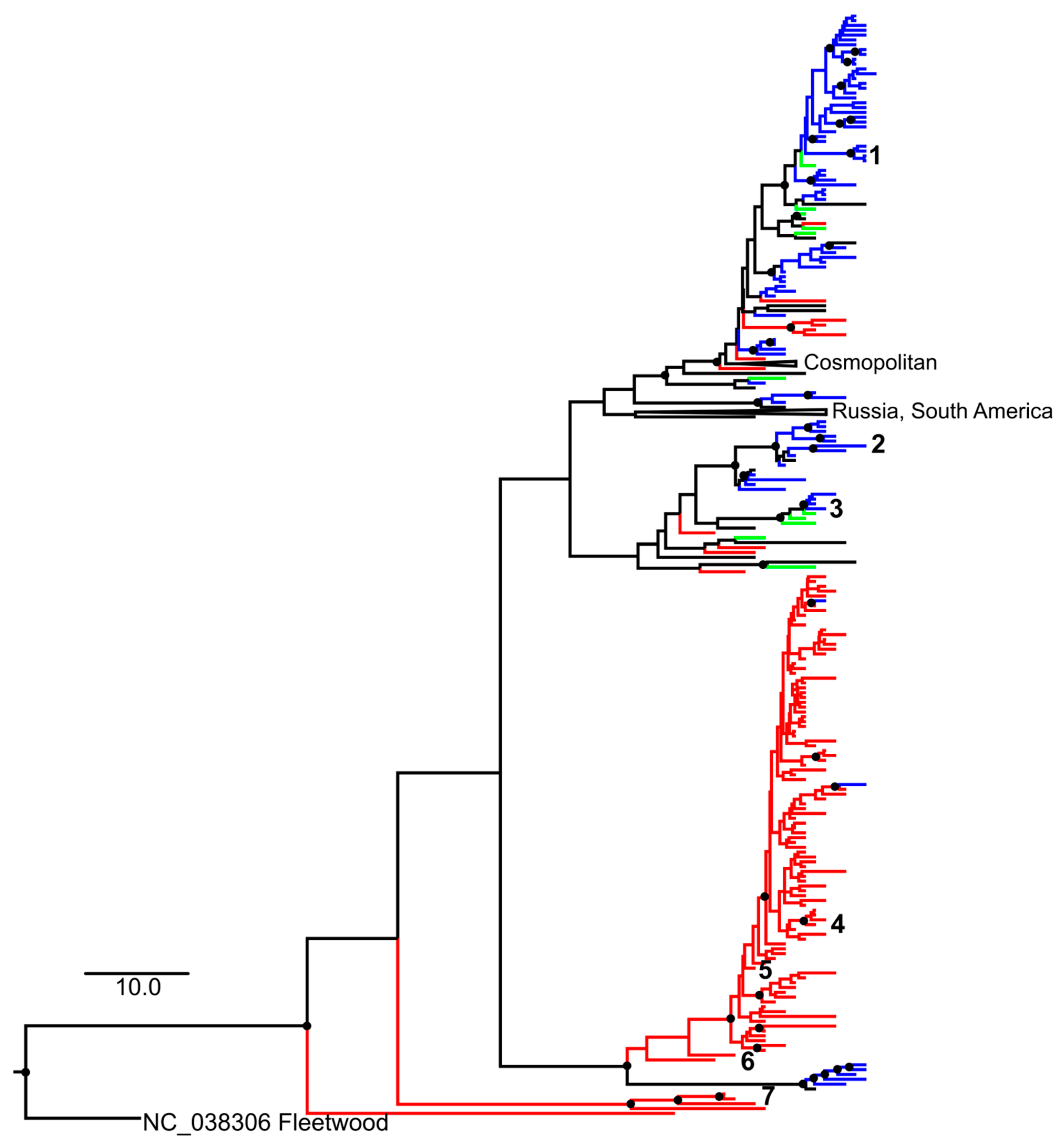
3.3. Phylogenetic Analysis

The global distribution of CVA2 genetic variants was, in general, associated with geographic location (Figure 1). There was a large and distinct group of Chinese CVA2 isolates, and relatively few transfers between Europe and Asia. CVA2 isolates associated with AFP in this and other studies were scattered among other CVA2 sequences on the phylogenetic tree. There was no association of AFP with any specific phylogenetic group. Viruses that were genetically identical or very similar to those associated with AFP in this study were isolated in different regions of Russia and were associated with acute respiratory diseases, enteroviral vesicular pharyngitis, HFMD and rarely aseptic virus meningitis.

4. Discussion

The main goal of AFP surveillance is the investigation of cases of a clinical syndrome that can be caused by multiple factors with the aim of identifying those cases that are associated with poliovirus—wild-type or derived from Sabin oral polio vaccine (OPV) strains (vaccine-related or vaccine-like). At the same time, the methodology of AFP surveillance and subsequent laboratory investigation allows the detection of NPEV in materials from AFP cases. NPEVs are ubiquitous and usually do not cause overt disease. Thus, the frequency of NPEV isolation from stool samples allows the assessment of the quality of surveillance for poliomyelitis and AFP. The frequency fluctuates in different countries depending on climatic and socio-economic conditions [14,15,16,17]. According to our data, in the Russian Federation, the proportion of AFP cases positive for NPEV was 3.2% in the period 2000–2019 (238 out of 7414). NPEV isolated from a non-sterile material (fecal sample, oropharyngeal swabs) may be an accidental finding that reflects the natural circulation of NPEV in the population and does not prove its etiological role in AFP. The link between an NPEV and AFP is well established for the types EV-A71 and EV-D68 [19]. The possibility of a large outbreak of paralytic disease, clinically indistinguishable from poliomyelitis, with fatal outcomes, but caused by non-polio EV-A71, was reported as early as 1975 by M.P. Chumakov and colleagues [44]. The capacity of this virus to cause AFP has been further confirmed [11]. EV-D68 has established itself as a human pathogen causing seasonal outbreaks with a large number of neurological manifestations in the form of acute flaccid myelitis in the United States and Europe since 2014 [45,46,47], necessitating the systematic monitoring of its circulation in some countries [48,49,50]. The etiological relationship between other NPEV types and AFP has not been definitively established. Despite the wide global circulation of the CVA2 virus, it is not commonly isolated from AFP cases, and available publications do not allow the determination of the role of CVA2 in the development of this syndrome. Over a 20-year period of AFP surveillance in the Russian Federation, we found only five AFP cases yielding CVA2. In three of five children, the disease resulted in the development of persistent residual paralysis, and the complex of clinical and laboratory data corresponded to poliomyelitis developing during poliovirus infection. It should be emphasized that all children were repeatedly (4 to 5 doses) immunized against poliomyelitis and did not have immunodeficiency disorders. No poliovirus or other enterovirus was isolated, and patients did not receive OPV recently (the last OPV dose being given at least two months prior to AFP onset). This allows the association of the AFP in these three patients with the CVA2 virus with a significant degree of confidence. The isolation of CVA in two other patients with clinical manifestations not compatible with poliomyelitis is a mundane finding that is not related in any way to the hypothetical ability of CVA2 to cause poliomyelitis. Indeed, if up to 20% of healthy children excrete NPEV [51,52]), then a comparable fraction of children with unrelated neurological disorders will be positive as well.
NPEVs can cause a wide range of symptoms, and a very similar disease can be caused by diverse types [7]. On the other hand, there have been descriptions of types (most prominently, EV-A71) or even distinct subtypes or phylogenetic groups causing specific clinical manifestations or having variable epidemiological properties [53,54,55]. Phylogenetic analysis of CV-A2 isolated from AFP cases did not reveal an association with a specific phylogenetic group. Viruses from AFP patients from this and other studies around the world were scattered across the phylogenetic tree. Thus, the severity and clinical manifestations of the disease are most likely determined by the individual characteristics of the infected child rather than by virus genetics. In this regard, it is worth noting that, in two children with persistent residual paralysis (N.T. and K.O.), AFP was preceded by other diseases (Table 1).
Tracing rare manifestations of common infections is generally a challenging task. The AFP surveillance system, which includes epidemiological and laboratory (virological) components, is well established, with a more than 30-year history of the Global Polio Eradication Initiative implementation. This system has clear criteria for detecting AFP cases, sampling and delivering materials for laboratory research and an algorithm for studying these samples [56]. Based on the goals of the poliomyelitis eradication program, the main objective of the surveillance is to detect poliovirus, and the main test materials are fecal samples [18]. However, owing to large sampling volumes and standardized methodology, there have been many scientific by-products of AFP surveillance, such as data on NPEV [14,17] and adenovirus circulation [57,58,59,60]. The future of AFP surveillance is currently debated, and some favor a more simple and versatile environmental surveillance program to trace both poliovirus and NPEV circulation. We have suggested previously that sewage surveillance may lack the sensitivity needed to ensure early poliovirus detection by orders of magnitude [55] and cannot fully substitute for AFP surveillance. This study exemplifies the additional value of AFP surveillance for the investigation of AFP etiology and studies on enterovirus pathogenicity. The value of AFP surveillance for AFP etiology studies could be further expanded by the systematic collection of additional material (oropharyngeal swabs, CSF) and use of molecular methods (PCR and high-throughput sequencing) for virus detection and identification. With the expected global certification of polio eradication [61], the elongation of AFP surveillance in the countries that conduct it will thus be reasonable.

Author Contributions

Conceptualization, A.N.L., O.E.I., T.P.E.; virological investigation, L.I.K., N.I.D., N.I.R., N.R.R., O.E.I., O.Y.B.; molecular investigation, A.N.L., V.V.Z., E.Y.S., L.N.G., N.A.N., Y.A.V.; epidemiological data, N.S.M., Y.M.M.; clinical investigation, A.K.S.; data analysis, A.N.L., A.K.S., O.E.I.; writing—original draft preparation, A.N.L., O.E.I.; writing—review and editing, A.N.L., O.E.I.; visualization, A.N.L., O.E.I., Y.A.V. All the authors contributed to discussion of the article and reviewed the final version of the manuscript. All authors have read and agreed to the published version of the manuscript.

Funding

The research was carried out with support from the Federal Budget of the Russian Federation allocated for the implementation of the Polio Eradication Programme in the Russian Federation, the WHO Polio Eradication Programme, the WHO Regional Office for Europe. Sequencing and molecular analysis was supported by Russian Scientific Foundation (RSF) grant 19-15-00055.

Institutional Review Board Statement

Not applicable.

Informed Consent Statement

Not applicable.

Data Availability Statement

Not applicable.

Acknowledgments

We thank the Federal Service for Surveillance on Consumer Rights Protection and Human Wellbeing (Rospotrebnadzor), the Polio Laboratory Network of the Russian Federation for their assistance in the collection and delivery of the clinical specimens, providing epidemiological data and the initiation of the additional clinical investigation of AFP cases. We thank the physician who provided clinical information about the AFP patients. We express our gratitude to the members of the National Expert Classification Committee, including those who previously worked on the commission—O.P. Chernyavskaya, V.K. Tatochenko, O. Zinovieva—for their participation in the classification of AFP cases.

Conflicts of Interest

The authors declare no conflict of interest.

References

  1. International Committee on Taxonomy of Viruses (ICTV). Available online: https://ictv.global/taxonomy/ (accessed on 27 October 2021).
  2. Chiang, K.-L.; Wei, S.-H.; Fan, H.-C.; Chou, Y.-K.; Yang, J.-Y. Outbreak of recombinant coxsackievirus A2 infection and polio-like paralysis of children, Taiwan, 2014. Pediatr. Neonatol. 2019, 60, 95–99. [Google Scholar] [CrossRef] [Green Version]
  3. Li, W.; Gao, H.-H.; Zhang, O.; Liu, Y.-J.; Tao, R.; Cheng, Y.-P.; Shu, O.; Shang, S.-Q. Large outbreak of herpangina in children caused by enterovirus in summer of 2015 in Hangzhou, China. Sci. Rep. 2016, 6, 35388. [Google Scholar] [CrossRef] [PubMed] [Green Version]
  4. Yang, Q.; Zhang, Y.; Yan, D.; Zhu, S.; Wang, D.; Ji, T.; Li, X.; Song, Y.; Gu, X.; Xu, W. Two genotypes of coxsackievirus A2 associated with hand, foot, and mouth disease circulating in China since 2008. PLoS ONE 2016, 11, e0169021. [Google Scholar] [CrossRef] [PubMed] [Green Version]
  5. Ji, H.; Fan, H.; Lu, P.-X.; Zhang, X.-F.; Ai, J.; Shi, C.; Huo, X.; Bao, C.-J.; Shan, J.; Jin, Y. Surveillance for severe hand, foot, and mouth disease from 2009 to 2015 in Jiangsu province: Epidemiology, etiology, and disease burden. BMC Infect. Dis. 2019, 19, 79. Available online: https://bmcinfectdis.biomedcentral.com/articles/10.1186/s12879-018-3659-7 (accessed on 29 December 2021). [CrossRef] [PubMed] [Green Version]
  6. Chen, S.P.; Huang, Y.C.; Li, W.C.; Chiu, C.H.; Huang, C.G.; Tsao, K.C.; Lin, T.Y. Comparison of clinical features between Coxsackievirus A2 and Enterovirus 71 during the enterovirus outbreak in Taiwan, 2008: A children’s hospital experience. Microbiol. Immunol. Infect. 2010, 43, 99–104. [Google Scholar] [CrossRef] [Green Version]
  7. Tapparel, C.; Siegrist, F.; Petty, T.J.; Kaiser, L. Picornavirus and enterovirus diversity with associated human diseases. Infect. Genet. Evol. 2013, 14, 282–293. [Google Scholar] [CrossRef]
  8. Bendig, J.W.A.; O’Brien, P.S.; Muir, P.; Porter, H.J.; ECaul, E.O. Enterovirus sequences resembling Coxsackievirus A2 detected in stool and spleen from a girl with fatal myocarditis. J. Med. Virol. 2001, 64, 482–486. [Google Scholar] [CrossRef]
  9. Moleta, L.; Saloum, K.; Marque-Juillet, S.; Garbarg-Chenon, A.; Henquell, C.; Schuffenecker, I.; Peigue-Lafeuille, H.; Rozenberg, F.; Mirand, A. Enterovirus infections in hospitals of Ile de France region over 2013. J. Clin. Virol. 2016, 74, 37–42. [Google Scholar] [CrossRef]
  10. Ohara, N.; Kaneko, M.; Nishibori, T.; Sato, K.; Furukawa, T.; Koike, T.; Sone, H.; Kaneko, K.; Kamoi, K. Fulminant type 1 diabetes mellitus associated with Coxsackie virus type A2 infection: A case report and literature review. Intern. Med. 2016, 55, 643–646. [Google Scholar] [CrossRef] [Green Version]
  11. Marx, A.; Glass, J.D.; Sutter, R.W. Differential diagnosis of Acute Flaccid Paralysis and its role in poliomyelitis. Epidemiol. Rev. 2000, 22, 298–316. [Google Scholar] [CrossRef] [Green Version]
  12. Olivé, J.M.; Castillo, C.; Castro, R.G.; de Quadros, C.A. Epidemiologic study of Guillain-Barre syndrome in children <15 years of age in Latin America. J. Infect. Dis. 1997, 175 (Supple. 1), S160–S164. [Google Scholar] [CrossRef] [Green Version]
  13. World Health Assembly. Global Eradication of Poliomyelitis by the Year 2000; WHA Resolution No. WHA41.28; World Health Organization: Geneva, Switzerland, 1988; Available online: https://apps.who.int/iris/handle/10665/164531 (accessed on 29 December 2021).
  14. Chouikha, A.; Rezig, D.; Driss, N.; Abdelkhalek, I.; Ben Yahia, A.; Touzi, H.; Meddeb, Z.; Ben Farhat, E.; Yahyaoui, M.; Triki, H. Circulation and molecular epidemiology of enteroviruses in paralyzed, immunodeficient and healthy individuals in Tunisia, a country with a polio-free status for decades. Viruses 2021, 13, 380. [Google Scholar] [CrossRef] [PubMed]
  15. Wieczorek, M.; Krzysztoszek, A. Molecular characterization of enteroviruses isolated from acute flaccid paralysis cases in Poland, 1999–2014. Pol. J. Microbiol. 2016, 65, 443–450. [Google Scholar] [CrossRef] [PubMed] [Green Version]
  16. Pellegrinelli, L.; Galli, C.; Primache, V.; Bubba, L.; Buttinelli, G.; Stefanelli, P.; Pariani, E.; Binda, S. Emerging non-polio enteroviruses recognized in the framework of the acute flaccid paralyses (AFP) surveillance system in Northern Italy, 2016–2018. Int. J. Infect. Dis. 2021, 106, 36–40. [Google Scholar] [CrossRef] [PubMed]
  17. Jiao, M.M.A.; Apostol, L.N.; de Quiroz-Castro, M.; Jee, Y.; Roque, V.; Mapue, M.; Navarro, F.M.; Fe Tabada, C.; Tandoc, A. Non-polio enteroviruses among healthy children in the Philippines. BMC Public Health 2020, 20, 167. [Google Scholar] [CrossRef] [Green Version]
  18. World Health Organization (WHO). Manual for the Virological Investigation of Polio, 4th ed.; WHO: Geneva, Switzerland, 2004; Available online: http://whqlibdoc.who.int/hq/2004/WHO_IVB_04.10.pdf (accessed on 27 October 2021).
  19. Suresh, S.; Rawlinson, W.D.; Andrews, P.I.; Stelzer-Braid, S. Global epidemiology of nonpolio enteroviruses causing severe neurological complications: A systematic review and meta-analysis. Rev. Med. Virol. 2020, 30, e2082. [Google Scholar] [CrossRef]
  20. van der Sanden, S.M.G.; Koopmans, M.P.G.; van der Avoort, H.G.A.M. Detection of human enteroviruses and parechoviruses as part of the national enterovirus surveillance in the Netherlands, 1996–2011. Eur. J. Clin. Microbiol. Infect. Dis. 2013, 32, 1525–1531. [Google Scholar] [CrossRef] [PubMed]
  21. Khetsuriani, N.; LaMonte, A.; Oberste, M.S.; Pallansch, M. Neonatal enterovirus infections reported to the National Enterovirus Surveillance System in the United States, 1983–2003. Pediatr. Infect. Dis. J. 2006, 25, 889–893. [Google Scholar] [CrossRef] [Green Version]
  22. The National Committee for the Certification of Wild Poliovirus Eradication in Hong Kong (NCC). Fifteen years of acute flaccid paralysis surveillance in Hong Kong: Findings from 1997 to 2011. J. Paediatr. Child Health 2014, 50, 545–552. [Google Scholar] [CrossRef]
  23. Kim, H.; Kang, B.; Hwang, S.; Lee, S.W.; Cheon, D.-S.; Kim, K.; Jeong, Y.-S.; Hyeon, J.-Y. Clinical and enterovirus findings associated with acute flaccid paralysis in the Republic of Korea during the recent Decade. J. Med. Virol. 2014, 86, 1584–1589. [Google Scholar] [CrossRef]
  24. Sousa, I.P.; de Lourdes Aguiar Oliveira, M.; Burlandy, F.M.; Machado, R.S.; Oliveira, S.S.; Tavares, F.N.; Gomes-Neto, F.; da Costa, E.V.; da Silva, E.E. Molecular characterization and epidemiological aspects of non-polio enteroviruses isolated from acute flaccid paralysis in Brazil: A historical series (2005–2017). Emerg. Microbes Infect. 2020, 9, 2536–2546. [Google Scholar] [CrossRef]
  25. World Health Organization (WHO). Regional Office for Europe. Available online: http://data.euro.who.int/cisid/default.aspx?TabID=539511 (accessed on 27 October 2021).
  26. The Ministry of Health of the Russian Federation; The State Committee for Sanitary and Epidemiological Supervision of the Russian Federation. On Approval of the Poliomyelitis Eradication Program on the Territory of the Russian Federation. Available online: https://base.garant.ru/4120180/ (accessed on 29 December 2021).
  27. Oberste, M.S.; Maher, K.; Kilpatrick, D.R.; Flemister, M.R.; Brown, B.A.; Pallansch, M.A. Typing of human enteroviruses by partial sequencing of VP1. J. Clin. Microbiol. 1999, 37, 1288–1293. [Google Scholar] [CrossRef] [Green Version]
  28. Federal Service for Consumer Rights Protection and Human Wellbeing (Rospotrebnadzor). Program “Epidemiological Surveillance and Prevention of Enterovirus (Non-Polio) Infection for 2018–2022. Available online: https://fcgie.ru/download/koord_tsentr/%D0%AD%D0%92%D0%98%20%D0%9F%D0%A0%D0%9E%D0%93%D0%A0%D0%90%D0%9C%D0%9C%D0%90%2018-22.pdf (accessed on 29 December 2021).
  29. Nix, W.A.; Oberste, M.S.; Pallansch, M.A. Sensitive, seminested PCR amplification of VP1 sequences for direct identification of all enterovirus serotypes from original clinical specimens. J. Clin. Microbiol. 2006, 44, 2698–2704. [Google Scholar] [CrossRef] [Green Version]
  30. Available online: https://github.com/v-julia/GenAlignment (accessed on 29 December 2021).
  31. Katoh, K.; Standley, D.M. MAFFT multiple sequence alignment software version 7: Improvements in performance and usability. Mol. Biol. Evol. 2013, 30, 772–780. [Google Scholar] [CrossRef] [PubMed] [Green Version]
  32. Minh, B.Q.; Schmidt, H.A.; Chernomor, O.; Schrempf, D.; Woodhams, M.D.; von Haeseler, A.; Lanfear, R. 2 IQ-TREE 2: New models and efficient methods for phylogenetic inference in the genomic era. Mol. Biol. Evol. 2020, 37, 1530–1534. [Google Scholar] [CrossRef] [Green Version]
  33. Hoang, D.P.; Chernomor, O.; von Haeseler, A.; Minh, B.Q.; Vinh, L.S. UFBoot2: Improving the ultrafast bootstrap approximation. Mol. Biol. Evol. 2018, 35, 518–522. [Google Scholar] [CrossRef] [PubMed]
  34. Kalyaanamoorthy, S.; Minh, B.Q.; Wong, T.K.F.; von Haeseler, A.; Jermiin, L.S. ModelFinder: Fast model selection for accurate phylogenetic estimates. Nat. Methods 2017, 14, 587–589. [Google Scholar] [CrossRef] [PubMed] [Green Version]
  35. Rambaut, A.; Lam, T.T.; Carvalho, L.M.; Pybus, O.G. Exploring the temporal structure of heterochronous sequences using TempEst (formerly Path-O-Gen). Virus Evol. 2016, 2, vew007. [Google Scholar] [CrossRef] [PubMed] [Green Version]
  36. Suchard, M.A.; Lemey, P.; Baele, G.; Ayres, D.L.; Drummond, A.J.; Rambaut, A. Bayesian phylogenetic and phylodynamic data integration using BEAST 1.10. Virus Evol. 2018, 4, vew016. [Google Scholar] [CrossRef] [Green Version]
  37. Lanfear, R.; Frandsen, P.B.; Wright, A.M.; Senfeld, T.; Calcott, B. PartitionFinder 2: New Methods for Selecting Partitioned Models of Evolution for Molecular and Morphological Phylogenetic Analyses. Mol. Biol. Evol. 2016, msw260. [Google Scholar] [CrossRef] [Green Version]
  38. Baele, G.; Lemey, P.; Bedford, T.; Rambaut, A.; Suchard, M.A.; Alekseyenko, A.V. Improving the Accuracy of Demographic and Molecular Clock Model Comparison While Accommodating Phylogenetic Uncertainty. Mol. Biol. Evol. 2012, 29, 2157–2167. [Google Scholar] [CrossRef] [PubMed] [Green Version]
  39. Available online: http://tree.bio.ed.ac.uk/software/tracer (accessed on 29 December 2021).
  40. Available online: http://tree.bio.ed.ac.uk/software/figtree/ (accessed on 29 December 2021).
  41. World Health Organization (WHO). Report of the Second Meeting of the Technical Consultation Group for Global Eradication of Poliomyelitis; WHO/EPI/GEN/98/04; WHO: Geneva, Switzerland, 1998; Available online: https://apps.who.int/iris/bitstream/handle/10665/63994/WHO_EPI_GEN_98.04.pdf?sequence=1&isAllowed=y (accessed on 29 December 2021).
  42. Leschinskaya, E.V.; Latysheva, I.N. The Clinic, Diagnosis and Treatment of Acute Poliomyelitis. In Guidelines. Moscow: M.P. Chumakov Institute of Poliomyelitis and Viral Encephalitis Them; M.P. Chumakov Russian Academy of Medical Sciences: Moscow, Russian, 1998. [Google Scholar]
  43. Yen, T.-Y.; Huang, Y.-P.; Hsu, Y.-L.; Chang, Y.-T.; Lin, H.-C.; Wu, H.-S.; Hwang, K.-P. A case of recombinant coxsackievirus A2 infection with neurological complications in Taiwan. J. Microbiol. Immunol. Infect. 2017, 50, 928–930. [Google Scholar] [CrossRef] [PubMed]
  44. Chumakov, M.; Voroshilova, M.; Shindarov, L.; Lavrova, I.; Gracheva, L.; Koroleva, G.; Vasilenko, S.; Brodvarova, I.; Nikolova, M.; Gyurova, S.; et al. Enterovirus 71 isolated from cases of epidemic poliomyelitis-like disease in Bulgaria. Arch. Virol. 1979, 60, 329–340. [Google Scholar] [CrossRef] [PubMed]
  45. Midgley, C.M.; Watson, J.T.; Nix, W.A.; Curns, A.T.; Rogers, S.L.; Brown, B.A.; Conover, C.; Dominguez, S.R.; Feikin, D.R.; The EV-D68 Working Group; et al. Severe respiratory illness associated with a nationwide outbreak of enterovirus D68 in the USA (2014): A descriptive epidemiological investigation. Lancet Respir. Med. 2015, 3, 879–887. [Google Scholar] [CrossRef] [Green Version]
  46. Centers for Disease Control and Prevention. Clusters of acute respiratory illness associated with human enterovirus 68—Asia, Europe, and United States, 2008–2010. MMWR Morb. Mortal. Wkly. Rep. 2011, 60, 1301–1304. [Google Scholar]
  47. Poelman, R.; Schuenecker, I.; Van Leer-Buter, C.; Josset, L.; Niesters, H.G.M.; Lina, B. European surveillance for enterovirus D68 during the emerging North-American outbreak in 2014. J. Clin. Virol. 2015, 71, 1–9. [Google Scholar] [CrossRef] [PubMed] [Green Version]
  48. Centers for Disease Control and Prevention. Acute Flaccid Myelitis (AFM) 2020 Case Definition. Available online: https://wwwn.cdc.gov/nndss/conditions/acute-flaccid-myelitis/case-definition/2020/ (accessed on 27 October 2021).
  49. Kramer, R.; Sabatier, M.; Wirth, T.; Pichon, M.; Lina, B.; Schuffenecker, I.; Josset, L. Molecular diversity and biennial circulation of enterovirus D68: A systematic screening study in Lyon, France, 2010 to 2016. Eurosurveillance 2018, 23, 1700711. [Google Scholar] [CrossRef] [Green Version]
  50. Dickson, C.; Ho Mi Fane, B.; Squires, S.G. Acute flaccid myelitis in Canada, 2018 to 2019. Can. Commun. Dis. Rep. 2020, 46, 349–353. [Google Scholar] [CrossRef]
  51. Simonen-Tikka, M.L.; Hiekka, A.K.; Klemola, P.; Poussa, T.; Ludvigsson, J.; Korpela, R.; Vaarala, O.; Merja Roivainen, M. Early human entero- virus infec-tions in healthy Swedish children participating in the PRODIA pilot study. J. Med. Virol. 2012, 84, 923–930. [Google Scholar] [CrossRef]
  52. Witso, E.; Palacios, G.; Cinek, O.; Stene, L.C.; Grinde, B.; Janowitz, D.; Lipkin, W.I.; Rønningen, K.S. High prevalence of human entero- virus a infec-tions in natural circulation of human enteroviruses. J. Clin. Microbiol. 2006, 44, 4095–4100. [Google Scholar] [CrossRef] [Green Version]
  53. Solomon, T.; Lewthwaite, P.; Perera, D.; Cardosa, M.J.; McMinn, P.; Ooi, M.H. Virology, epidemiology, pathogenesis, and control of enterovirus 71. Lancet Infect. Dis. 2010, 10, 778–790. [Google Scholar] [CrossRef]
  54. Gaunt, E.; Harvala, H.; Österback, R.; Sreenu, V.B.; Thomson, E.; Waris, M.; Simmonds, P. Genetic characterization of human coxsackievirus A6 variants associated with atypical hand, foot and mouth disease: A potential role of recombination in emergence and pathogenicity. J. Gen. Virol. 2015, 96, 1067–1079. [Google Scholar] [CrossRef] [PubMed] [Green Version]
  55. Ivanova, O.E.; Yarmolskaya, M.S.; Eremeeva, T.P.; Babkina, G.M.; Baykova, O.Y.; Akhmadishina, L.V.; Krasota, A.Y.; Kozlovskaya, L.I.; Lukashev, A.N. Environmental Surveillance for Poliovirus and Other Enteroviruses: Long-Term Experience in Moscow, Russian Federation, 2004–2017. Viruses 2019, 11, 424. [Google Scholar] [CrossRef] [Green Version]
  56. World Health Organization (WHO). Manual for Virological Investigation of Poliomyelitis; WHO/EPI/GEN/97.1; WHO: Geneva, Switzerland, 1997; Available online: https://apps.who.int/iris/bitstream/handle/10665/62186/WHO_EPI_CDS_POLIO_90.1.pdf?sequence=1&isAllowed=y (accessed on 27 October 2021).
  57. Ivanova, O.E.; Yurashko, O.V.; Eremeeva, T.P.; Baikova, O.Y.; Morozova, N.S.; Lukashev, A.N. Adenovirus isolation rates in acute flaccid paralysis patients. J. Med. Virol. 2012, 84, 75–80. [Google Scholar] [CrossRef]
  58. de Azevedo, J.P.R.; Nascimento, L.R.; Cortinovis, M.C.; Oliveira, S.S.; da Costa, E.V.; da Silva, E.E. Characterization of species B adenoviruses isolated from fecal specimens taken from poliomyelitis-suspected cases. J. Clin. Virol. 2004, 31, 248–252. [Google Scholar] [CrossRef]
  59. Yousefi, M.; Nejati, A.; Zahraei, S.M.; Mahmoudi, S.; Parhizgari, N.; Farsani, S.M.J.; Mahmoodi, M.; Nategh, R.; Shahmahmoodi, S. Enteroviruses and Adenoviruses in stool specimens of paralytic children- can they be the cause of paralysis? Iran. J. Microbiol. 2018, 10, 194–201. Available online: http://www.ncbi.nlm.nih.gov/pmc/articles/pmc6087694/ (accessed on 29 December 2021). [PubMed]
  60. Haddad-Boubaker, S.; Joffret, M.-L.; Pérot, P.; Bessaud, M.; Meddeb, Z.; Touzi, H.; Delpeyroux, F.; Triki, H.; Eloit, M. Metagenomic analysis identifies human adenovirus 31 in children with acute flaccid paralysis in Tunisia. Arch. Virol. 2019, 164, 747–755. [Google Scholar] [CrossRef] [PubMed]
  61. World Health Organization (WHO). Polio Eradication Strategy 2022−2026: Executive Summary; 2021(WHO/Polio/21.04); Licence CC BY-NC-SA 3.0 IGO. World Health Organization: Geneva, Switzerland. Available online: https://polioeradication.org/wp-content/uploads/2021/10/9789240031937-eng.pdf (accessed on 27 October 2021).
Figure 1. MCC tree for CVA2 VP1 gene typing fragment sequences available in GenBank. The scale bar corresponds to 10 years. Nodes with posterior probabilities above 0.95 are indicated as filled circles. Branches leading to sequences collected in Russia, East Asia (China, Japan) and the European Union are colored in blue, red and green, respectively. Isolates associated with AFP are numbered as follows: (1)—patient N.T.; (2)—patient K.O.; (3)—patient D.A.; (4)—2014, Taiwan, sequence described earlier [43]; (5)—2008, China, no reference; (6), (7)—China, 2006, no reference. Scale bar indicates time in years. Groups that did not contain AFP-associated CVA2 were collapsed.
Microorganisms 10 00112 g001
Table 1. Descriptive characteristics of confirmed AFP cases in patients yielding CVA2, Russian Federation, in 2001–2020.
CharacteristicPatients
H.A.K.G.D.A.N.T.K.O.
Place of residenceBryanskSaratovSt. PetersburgVladimir regionNizhny Novgorod region
GenderFMMFM
Age, years12433
Number of doses and the type of polio vaccine2 OPV5 OPV4 IPV3 IPV, 2 OPV2 IPV, 3 OPV
Date of the paralysis onset 5 October 20089 September 200823 September 201510 September 201923 September 2019
Time from the last vaccination to the disease onset, months1352482
Time from the disease onset to the full manifestation of paralysis, days104112
Body temperature at the disease onset normal39.0 °Celevated, value unknown39.0 °C37.8 °C
Localization of paralysisleft leg, right legleft leg > right leg left legleft hand > right handquadriparesis (lower limbs > upper limbs)
Proximal/distalproximalproximal > dystalbothproximal > distalboth
Other neurological signs encopresis, enuresis
Virus source and type isolatedfeces, CVA2feces, CVA2feces, CVA2feces, CVA2
oropharyngeal swab, CVA2
feces, CVA2
CSF study resultsDay since disease onset unknown 09.12.1927 September 20194 October 2019
Cytosis, cells/mm3no data5no data36112
Protein, g/L 0.990.840.140.57
Glucose, mmol/Lno datano data1.812.0
Lymphocytes, %8084
Neutrophils, %2016
Health condition before AFPhealthyinfectious mononucleosis one month prior to paresishealthyconvalescence of bilateral focal pneumoniaaplasia of the left kidney, minor anomaly of heart development.
Residual paralysis after 60 days from the onsetnonoyesyesyes
Clinical diagnosis at discharge *Paraparesis of the lower limbs associated with CVA2. Organic lesion of central nervous system.Polyradiculo-neuritis associated with non-polio enterovirusMonoparesis of left lower limbEnterovirus infection, cervical myelitis, upper flaccid paraparesisAcute meningomyelitis
* As provided in the medical records, not updated according to this paper’s results. IPV—inactivated poliovirus vaccine; OPV—oral poliovirus vaccine; CSF—cerebrospinal fluid.
Publisher’s Note: MDPI stays neutral with regard to jurisdictional claims in published maps and institutional affiliations.

