Article

Organ Tropism of Angiostrongylus vasorum Larval Stages in Infected African Giant Snails (Lissachatina fulica)

1
Institute of Parasitology, Biomedical Research Center Seltersberg (BFS), Justus Liebig University Giessen, 35392 Giessen, Germany
2
Institute of Pathology, Justus Liebig University Giessen, 35392 Giessen, Germany
*
Author to whom correspondence should be addressed.
Pathogens 2024, 13(11), 946; https://doi.org/10.3390/pathogens13110946
Submission received: 26 September 2024 / Revised: 14 October 2024 / Accepted: 24 October 2024 / Published: 30 October 2024

Abstract

:
Angiostrongylus vasorum is a metastrongyloid lungworm causing severe cardiovascular disease in domestic and wild animals. During its heteroxenous life cycle, A. vasorum requires obligate gastropod intermediate hosts. Little is known about A. vasorum larval organ tropism and development in gastropod intermediate hosts. Thus, the aim of this study was to analyze in vivo development of A. vasorum larval stages in experimentally infected African giant snails (Lissachatina fulica). Adult L. fulica (n = 26) were orally infected with A. vasorum-L1 and thereafter continuously euthanized. Gastropod organs were artificially digested and microscopically analyzed for the presence of A. vasorum larvae. Moreover, paraffin-fixed organs were investigated histologically for snail-borne innate immune response. In the current study, the success of L. fulica oral infection was demonstrated, thereby reaching larval recovery rates of up to 49.7%. During snail infection, an organ tropism of A. vasorum larvae was detected for the lungs and the foot muscular tissue. Overall, A. vasorum-driven gastropod innate immune reactions against larvae varied greatly. In some specimens, larvae were found effectively ensnared by recruited hemocytes, resulting in granuloma formation, whilst in others, hemocyte-mediated reactions were barely observed. Nevertheless, these evidences demand more studies on hemocyte-derived effector mechanisms against A. vasorum.

1. Introduction

Angiostrongylus vasorum (Baillet, 1866) is a metastrongyloid lungworm that resides in the cardiovascular system of both domestic and wild canids, including dogs (Canis familiaris), wolves (Canis lupus) [1,2], golden jackals (Canis aureus) [3,4], and red foxes (Vulpes vulpes) [5,6,7,8,9], with the latter representing important wildlife reservoirs in Europe. Further definitive hosts include striped skunks (Mephitis mephitis), weasels (Mustela erminea), ferrets (Mustela putorius furo), badgers (Meles meles), neotropical otters (Lontra longicaudis), raccoon dogs (Nyctereutes procyonoides), and red pandas (Ailurus fulgens) [10,11,12,13,14]. Due to the rising urbanization of many of these wild animals, the risk of A. vasorum transmission to domestic dogs and vice versa is increasing [1,15]. This metastrongyloid parasite can be found on multiple continents and generally shows a patchy epizootiological distribution, with hyper- and hypoendemic foci in close proximity [16,17]. Clinical signs of A. vasorum infections in dogs may vary from mild respiratory symptoms to severe cardiopulmonary disorders but also include hemorrhage and neurological, ocular, and gastrointestinal symptoms [13,18,19,20,21,22].
The life cycle of A. vasorum is heteroxenous, with terrestrial gastropods acting as obligatory intermediate hosts. Thus, canine angiostrongylosis is considered a gastropod-borne parasitosis with a complex epizootiology, including various slug, semi-slug, and snail species as suitable intermediate hosts [15,23,24]. By feeding on the feces of an A. vasorum-infected definitive host, gastropod intermediate hosts become infected with first-stage larvae (L1). Another possibility is the active penetration of the gastropod epidermis by exogenous L1 [25,26]. While migrating in the mollusc intermediate hosts, A. vasorum L1 must molt into second-stage (L2) and third-stage larvae (L3). The canid definitive host will become infected by consuming either an A. vasorum-infected intermediate host carrying L3 or by a paratenic host (PH; e.g., amphibians, birds) hosting infectious L3. Alternatively, spontaneous shedding of A. vasorum L3 from dead intermediate hosts can occur, which are capable of surviving in the environment for some days or weeks, depending on the climatic condition [27]. As already stated, a wide spectrum of gastropod species may act as intermediate hosts, including the terrestrial African giant snail (Lissachatina fulica) (Bowdich, 1822) [28,29,30].
This gastropod species is considered one of the largest terrestrial snails, and its shell is cone-formed with an average length of 90 mm and contains a minimum of seven whorls in fully grown snails. The shell is medium brown and decorated with darker and lighter stripes, and the tip is usually lighter in color [31,32]. Originally native to East Africa, this terrestrial giant snail is now considered an invasive species (i.e., neozoa) in multiple tropical and subtropical countries, such as Colombia, the United States of America, Brazil, Ecuador, Indonesia, and India [11,33,34]. Furthermore, L. fulica has been identified as a natural intermediate host not only for A. vasorum but also for Aelurostrongylus abstrusus, Crenosoma vulpis, and Troglostrongylus brevior in the tropics of South America, thereby representing a risk factor for metastrongyloid transmission for both domestic and wild canids/felids [28]. Based on both the invasive nature of L. fulica and the emergence of canine angiostrongylosis as a cardiopulmonary disease in various continents, it is of great interest to gain further knowledge on A. vasorum L1 migration and organ tropism in L. fulica [35], which currently is still not fully understood. Several studies have shown that, after cutaneous or oral gastropod infection, A. vasorum L1 must migrate inside the mollusc to find adequate organs for fulfilling further development into L2 and, finally, infective L3 [36,37,38]. During this larval migration in vivo, A. vasorum larvae will be exposed to the gastropod host innate immune system, mainly composed of hemocytes present in the hemolymph, which might not only influence migration routes but also hamper larval development. As opposed to mammals, which own both an innate and an adaptive immune system, gastropods exclusively possess an innate immune system. Hematopoiesis in molluscs is complex and less studied when compared to other metazoan species [39]. Gastropod hematopoiesis leads to the generation of hemocytes (syn. amoebocytes) in hemolymph circulation, tissues, and organs [40,41]. Gastropod hemocytes constitute the main cellular component of hemolymph but, in addition, serve as resident cells at other sites, such as connective and vascular tissues [39,42,43]. Invertebrate hemocytes have multiple functions, such as the transport of molecules, wound repair, coagulation, and defense against invasive pathogens [41]. Hence, hemocytes show diverse effector mechanisms to fight invasive pathogens, such as phagocytosis [44], cell-mediated cytotoxicity, production of reactive oxygen species (ROS), and encapsulation [45]. More recently, also the release of gastropod invertebrate extracellular phagocyte traps (InEPT) against metastrongyloid larvae, including A. vasorum, has been reported, which is an effective immune response against invading L1 [40,41,46].
While several histological studies on hemocyte-derived innate immune reactions against nematode larvae exist, much less is known on metastrongyloid lungworm organ tropism [36,47,48], especially on gastropod intermediate hosts being experimentally infected under standardized laboratory conditions, including humidity, light, and temperatures mimicking natural field conditions [36,49]. The aim of this study was, therefore, to gain knowledge on A. vasorum larval migration in the gastropod intermediate host L. fulica, including organ tropism, with selected histological analyses to tackle hemocyte-mediated innate responses against invasive larvae in vivo.

2. Material and Methods

2.1. Gastropod Maintenance Under Standardized and Parasite-Free Conditions

German-bred African giant snails (L. fulica) were kept in a full-automatized climate chamber (ECP01E, Snijders Scientific B.V. Tilburg, the Netherlands) under standardized laboratory conditions with humidity and temperatures mimicking the gastropod’s natural habitat by temperatures ranging from 20 to 26 °C and 50% humidity, according to Penagos-Tabares et al. (2018). The light cycle consisted of 10 h of light and 10 h of darkness, in between 2 h for dawn and 2 h for dusk. On the ground of plastic boxes for gastropod maintenance, a layer of coconut soil (Kokosfaser–Humusziegel, TropicShop, Germany) of approximately 5 cm thickness was used. Exclusively parasite-free newly bred L. fulica (i.e., F1 and F2 generations) were used for A. vasorum infections. These F1/F2 L. fulica were fed with lettuce (Lactuca sativa), carrots (Daucus carota), cucumber (Cucumis sativus), zucchini (Cucurbita pepo), and commercial dog food (Premium Trockenfutter Romeo, Germany). In order to raise the pH of the soil, garden lime was mixed into the coconut soil. Furthermore, common cuttlefish (Sepia officinalis) internal shells (Sepiaschalen, TropicShop, Germany) were offered ad libitum to L. fulica as a natural source of calcium.

2.2. Isolation of Vital Angiostronglyus Vasorum L1 and Gastropod Oral Infection

Vital first-stage larvae (L1) were isolated from feces of naturally A. vasorum-infected dogs (kindly provided by the Institute of Parasitology at the University of Veterinary Medicine Hannover, Hannover, Germany) via the modified Baermann funnel technique, according to Conboy et al. [50]. After 24 h of incubation, 7 mL were obtained from each funnel apparatus, quantified microscopically (Olympus BH-2), and pelleted (200× g, 5 min) to collect vital A. vasorum-L1. L. fulica (n = 26) were orally infected with a dose of 1000 larvae each. In order to be as close as possible to field conditions, the following procedure was used for infection: since gastropods are coprophagic, the remaining fecal solution from the Baermann funnels was used as a suspension medium for the larvae. Thus, 50 mL of diluted canine fecal solution (DCFS) containing 1000 motile larvae were orally fed to the snails via a syringe until complete consumption (please refer to Video 1).

2.3. Gastropod Euthanasia and Organ Isolation

A. vasorum-infected L. fulica snails were cryo-euthanized in an ice bath at 4 °C, as reported previously by Lange et al. [51], at different time points post infection (p.i.; please refer to Supplementary Table S1). Following euthanasia, the snails were dissected, and the following 11 organs/tissues were isolated: crop/esophagus, stomach, intestine, hepatopancreas, reproductive tract, albumen gland, foot, kidney, lung, head, and heart. Four gastropods dissected at 5, 14, 29, and 47 days p.i. were used for histological analyses on the different tissues and organs. The organs of these four snails were fixed in 10% neutral-buffered formalin for further histological analyses.

2.4. Artificial Digestion and Identification of Larvae

A total of 22 of the dissected snails were further processed by artificial digestion. Snail single organs and tissues were artificially digested, according to Lange et al. [51]. Afterward, the remains were microscopically analyzed applying 40×, 200×, and/or 400× magnification (Olympus BH-2). To differentiate A. vasorum larvae (e.g., L1, L2, L3) from free-living or parasitic larvae of gastropods (genus Phasmarhaptidis), the type and length of the esophagus (e.g., non-rhabditiform, 1/2–1/3 of total larvae length), the tail form, and the length and width of the larvae were analyzed [52,53].

2.5. Histology

In total, 11 organs and tissues of four euthanized snails were used for histological analysis. As a first step, the snail’s shell was carefully removed. Thereafter, the inner organs were separated and fixed in 10% buffered formalin and embedded in paraffin wax by standard techniques. Tissue/organ sections of 5 µm thickness were cut from paraffin-embedded specimens and stained following the standard hematoxylin and eosin (HE) protocol for histological examination. HE staining was performed at the Institute of Pathology at the Faculty of Veterinary Medicine, Justus Liebig University Giessen, Giessen, Germany. The tissue slides were analyzed using an optical light microscope (Olympus BH-2) equipped with a digital camera (Olympus SC30) at the Institute of Parasitology of the Justus Liebig University Giessen, Germany.

3. Results

3.1. Angiostrongylus Vasorum Larval Development and Organ Tropism in Experimentally Infected L. fulica Snails

In order to investigate in detail A. vasorum larval organ tropism and development in vivo, single organs of L. fulica-infected snails were analyzed for the presence of different larval developmental stages. Overall, all 26 experimentally infected L. fulica proved positive for A. vasorum larvae. Even though all snails had obtained the same infection dose (1000 L1), different larval recovery rates per animal were observed when summing up all larvae found in one specimen. Thus, the overall larval recovery rates ranged from 1.8% to 49.7% (Table 1).
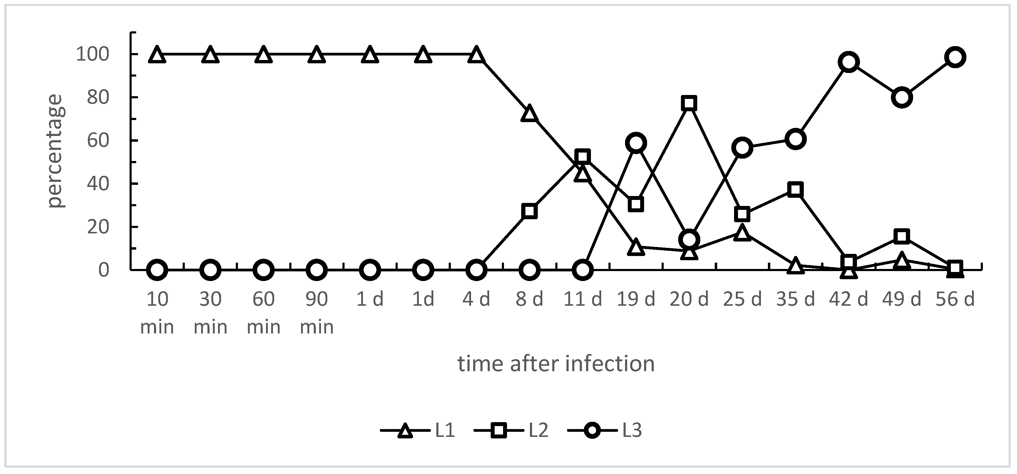
Referring to the larval development within L. fulica snails, L1 stages started to disappear with day 8 p.i. ongoing; in parallel, L2 stages were first detected at 8 days p.i. (Figure 1). The proportion of recovered L2 stages peaked at day 20 p.i. and thereafter declined until the end of the investigation period (56 days p.i.). The first L3 stages were found at 11 days p.i., and the proportion of L3 in snails increased from this time point onwards; consequently, at day 56 p.i., most of the larval stages had developed into L3 (see Figure 1). Of note is that the speed of larval development varied between individual L. fulica. Hence, even after 49 days p.i., some larvae had not developed into the infective L3 stage (please refer to Figure 1 and Table 1). However, when analyzing L. fulica long-term infections of two years p.i., vital A. vasorum L3 were still found in all four cryo-euthanized snails, even though recovery rates had considerably dropped to 0.1–4.4% after this long period (Table 2).
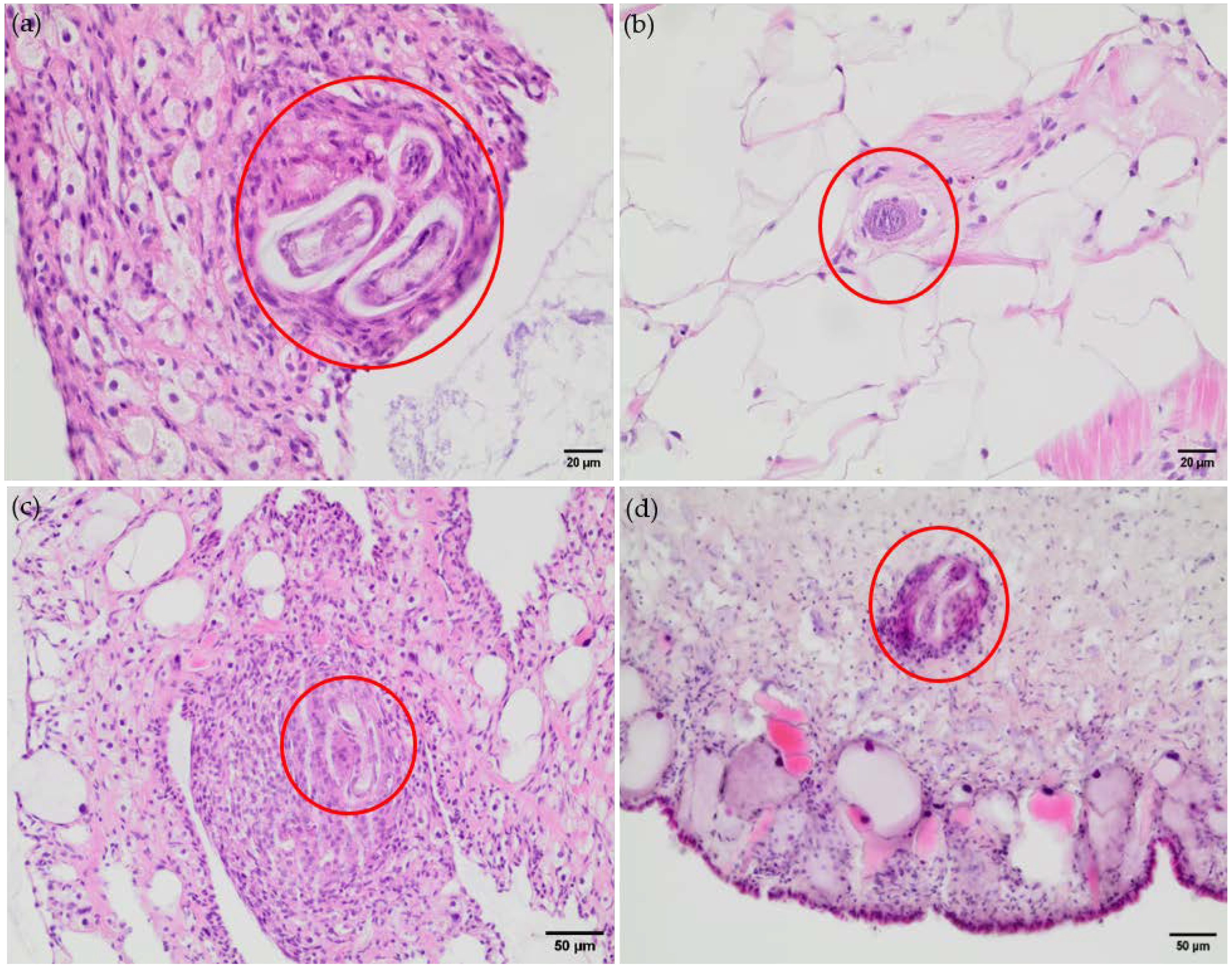
Analyses on parasite dispersion in L. fulica snails indeed revealed a marked tropism of A. vasorum larvae for specific organs and tissues, dependent on the time point of infection. Thus, the overall majority of larvae were found in lung and foot (muscles) tissues, while other organs such as the head, heart, albumen gland, crop-esophagus, intestine, kidney, and reproductive tract contained only a few larval specimens (Table 1). Referring to larval lung tropism, an interesting finding was observed here. The lungs of one A. vasorum-infected snail showed macroscopically visible, multiple calcified granulomas, most likely reflecting effective hemocyte-mediated innate immune reactions against invasive A. vasorum larvae (Figure 2).
Of note, A. vasorum L1 stages rapidly migrated in experimentally infected L. fulica, reaching distant organs already within 10 min after oral infection. In addition, the overall L1 migration highly differed between individual snails. As such, at 10 min p.i., 28% and 21% of L1 larvae were still located in the esophagus and stomach, respectively, while 24% were already found in the foot and 13% in the head. At 30 min p.i., most L1 larvae were localized in the foot (50%); 18.75% were found in the head and in the lungs. At 60 min p.i., a larger proportion of L1 was located in the hepatopancreas (36.07%), in the foot (22.4%), and in the head (15.85%). At 90 min p.i., 32%, 25%, and 12% L1 were located in the foot, head, and hepatopancreas, respectively. In one A. vasorum-infected snail, most L1 was detected in the foot (60.42%) at 1 day p.i., while only 19.72% of L1 larvae were located in the foot in another snail harboring 49.30% in the lung instead. Four days p.i., most larvae were observed in the lung (39.02%). Nonetheless, the head (17.07%), foot, and hepatopancreas (both with 12.20%) also contained relatively high proportions. At later time points, e.g., 8 days p.i., a high proportion of L1 was found in the hepatopancreas (41.77%), lung (27.85%), and stomach (17.72%). For the first time, L2 larvae were observed, but without any preference for specific organs. At 11 days p.i., most larvae were located in the lung (36.51%) and foot (17.06%). On this day, the first L3 larval stage was observed, being located in the heart. Of note, two-thirds of the observed larvae were still in the L2 stage. At 19 days p.i., 43.14% and 27.45% of the larvae were located in the lung and hepatopancreas, respectively. It can be noted that in the lung, the vast majority of larvae were L3 stages (40/44), while most larvae in the hepatopancreas were still L2 stages (18/28). At 20 days p.i., more than half of the larvae were found in the lung (55.26%), besides hepatopancreas (21.93%). In line, a high proportion of larvae was observed in the lung (34.09%) and foot (32.18%) at 25 days p.i. Here, the majority of larvae (86/136) had developed into L3 stages. Equally, at 35 days p.i., the lung (46.76%) and foot (29.90%) harbored most of the larvae, with more than half of the larvae (59.76%, 153/256) representing L3 stages. At 42 days p.i., larvae were mainly found in the lung (33.88%), hepatopancreas (17.13%), and stomach (17.13%). L1 larval stages were no longer observed, and almost all larvae were in the L3 stage (97.8%; 489/500). At 49 and 56 days p.i., mainly L3 stages (98.9%) were located in the lung (36.85% and 47.76%, respectively) and foot (45.70% and 25.52%, respectively).
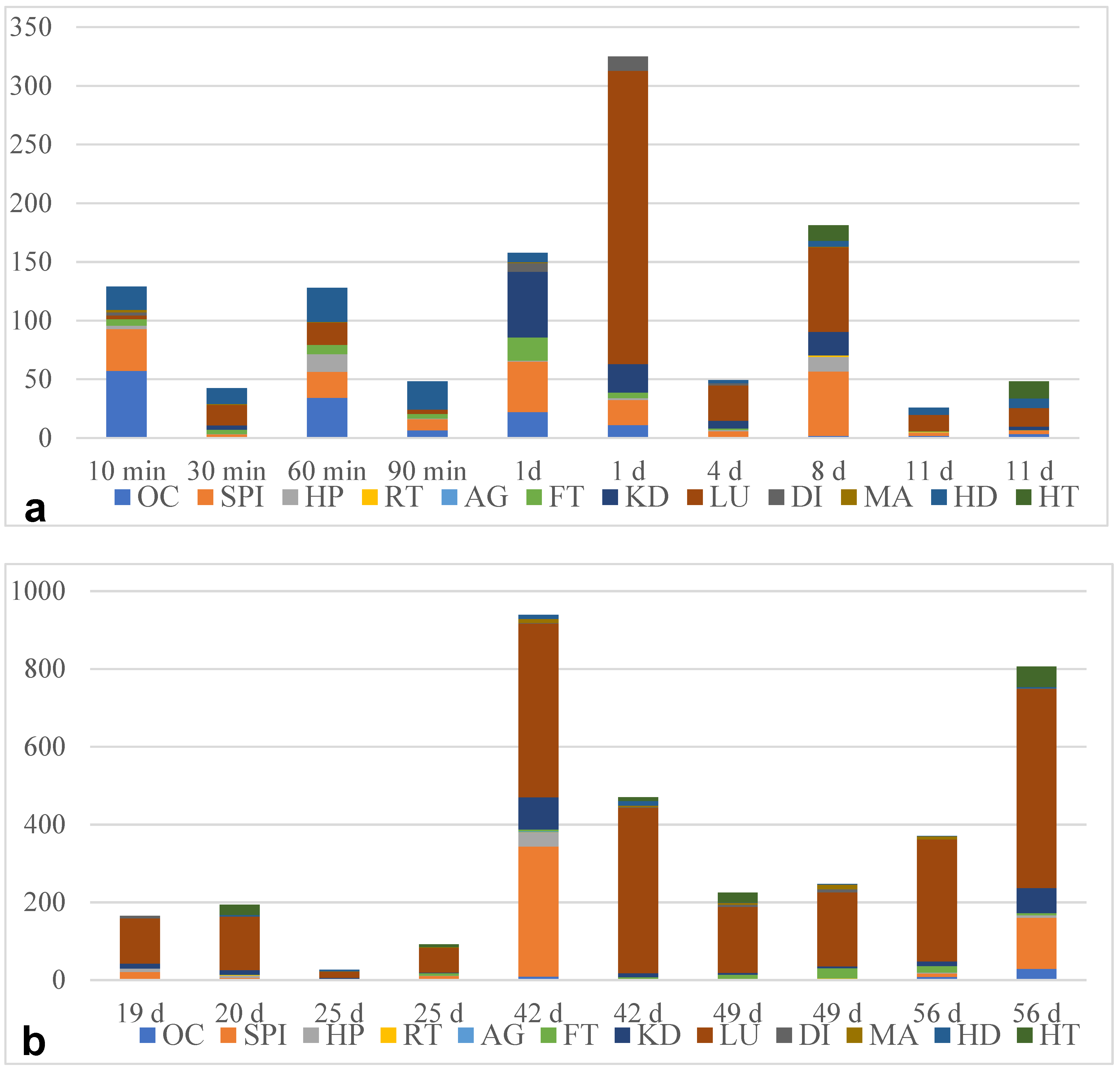
Considering larval burdens, clear differences were stated for the different organs (Figure 3). In general, the lung showed a high larval burden [up to 511 larvae per gram (L/g) of organ tissue] and, therefore, was the most infected organ, especially at days 42, 49, and 56 p.i. In contrast to this finding, the reproduction tract and albumin gland showed much lower larval burdens (0.83–1.75 L/g and 2.45 L/g, respectively). Of note, the different organs showed high interindividual variations in terms of larval burden (Table 2). Overall, the larval burden did not affect the larval developmental speed.

3.2. Histological Findings Indicate Hemocyte Responses to Angiostrongylus Vasorum Larval Stages

The analysis of histological samples confirmed the presence of A. vasorum-larvae in snail tissue, e.g., organs like lungs and feet (Figure 2). In the snail’s lungs, parasites were found separated by thin hemocyte-derived layers localized in the connective tissue surrounding blood vessels. As an exemplary finding in the connective tissue, we detected a single capsule enclosing three larvae at a time, while other larvae were found separated by thin hemocyte-derived layers (Figure 2). Due to the lack of karyolysis and/or foamy cytoplasm of these gastropod phagocytes, a clear hemocyte activation could not be confirmed for this microenvironment. Moreover, the larvae seemed still intact and showed no signs of cuticle destruction. In the thin gas exchange surface, no hemocyte accumulation was found in close vicinity to A. vasorum larvae, but a thin layer of connective tissue surrounded these pulmonary stages. In the snail foot, many A. vasorum larvae were located in the connective tissue. Numerous hemocytes accumulated around these parasitic stages, thereby forming a multicellular capsule. Herein, karyolysis was observed in some of these phagocytes, indicating hemocyte activation, while other hemocytes surrounding the larvae appeared to be flattened and potentially not activated.
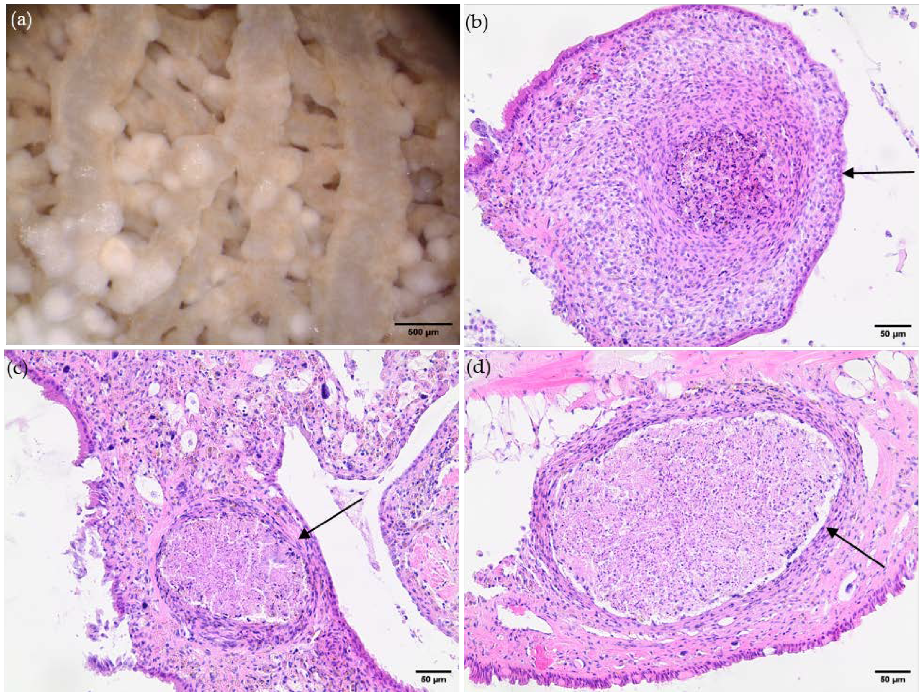
As an interesting finding, the macroscopical analysis of the lungs of a long-term infected L. fulica snail (euthanized two years p.i.) revealed the presence of multiple calcified granulomas (Figure 4). These pathologic structures were excised, stained, and examined by bright field microscopy. Here, the classical characteristics of granuloma were confirmed histologically, but no larvae were found in any of these pulmonary hemocyte-derived structures, most probably indicating successful larval degradation.

4. Discussion

Detailed investigations on A. vasorum larval migration, organ tropism, development, and hemocyte-derived innate immune reactions within gastropod intermediate hosts are still very limited [40,54,55,56]. The present study confirmed L. fulica as a suitable obligate intermediate host for A. vasorum, as previously reported in tropical biomes of Colombia [28]. Consequently, all A. vasorum developmental larval stages were found in the host, L1, L2, and infective L3. Interestingly, in some experimentally infected L. fulica, vital A. vasorum L3 was still present two years after infection, indicating long-term larval survival in these hosts. These long-living stages might be part of not only parasitic survival strategies apart from the canid definitive host but also be relevant for parasite dissemination and transmission. Of note, the neozoan L. fulica can live for up to 9 years and thereby migrate over longer distances when compared to short-living terrestrial gastropods [28,29]. As such, L. fulica has demonstrated an alarming capacity for rapid spread. In the US State of Florida, three abandoned snails led to massive multiplication and the discovery of more than 18,000 individuals only seven years later, and currently, 15 US states have been colonized by this neozoan species [57]. Even though the mechanisms of A. vasorum spreading into non-endemic areas are still under debate [55,56], geographic shifts in mollusc intermediate host populations are assumed as one underlying factor [58,59]. Meta-analyses have revealed that both global warming and anthropogenic pressure strongly affect the phenology of terrestrial gastropods, including slugs, semi-slugs, and snails [16,60].
Referring to experimental infections, a high variation in A. vasorum recovery rates was rather obvious in the current study, even though applying the same infection dose (1000 L1/snail). A reason for these deviations might be seen in individual innate immune reactions mediated by hemocytes, as already reported for Arion spp. slugs and L. fulica snails [30,38,52]. As recently demonstrated in vitro, exposed hemocytes not only firmly adhered to the cuticle of A. vasorum L1 and L3 but also reacted by forming invertebrate extracellular phagocyte traps [40,54]. In line, hemocytes circulating within the hemolymph system are considered multifunctional professional phagocytes in gastropods, thereby resembling leukocytes of vertebrates [39,40,61,62,63]. Hemocytes display diverse effector mechanisms against invasive pathogens, including phagocytosis, encapsulation, cell-mediated cytotoxicity, and release of invertebrate extracellular phagocyte traps (InEPTs) [40,42,54,64]. Some of these hemocyte-derived defense mechanisms were here confirmed in histological analyses of A. vasorum-infected L. fulica tissues. Hence, accumulation of hemocytes around larvae was found in addition to multicellular larval encapsulation, leading to the formation of large granuloma. Importantly, A. vasorum larvae, being surrounded by hemocytes, were detected in several tissues like the foot, distal intestine, lung, and the mantle of snails as early as 10 min p.i. These findings indicated that despite a fast A. vasorum larval migration, hemocytes are rapidly recruited to sites of parasite infection [40,54]. Rapid hemocyte recruitment might be a consequence of the open circulatory system of gastropods, with the hemolymph flowing through sinuses and thereby directly bathing organs. Thus, inner organs are surrounded by hemolymph containing circulating hemocytes, which may immediately react against pathogens, including A. vasorum larvae [65,66]. Interestingly, percutaneous A. vasorum L1 infection of molluscs resulted in InEPTs within gastropod mucous extrapallial space in vivo [30]. Accordingly, gastropod hemocytes spontaneously reacted against vital A. vasorum L1 within 30 min by forming InEPTs, resulting in firm entrapment of these motile larvae [54].
In the current study, A. vasorum L2 was found as early as 8 days p.i., and the first fully developed L3 stages were present at 11 days p.i. in snail tissues. Consequently, canids might become infected via L. fulica-derived L3 as early as 11 days p.i. Compared to a previous report stating a duration of 17 days for A. vasorum L3 development [36], the current data indicated a faster development using the same gastropod intermediate host species, which may be due to the climate chamber-based standardized experimental conditions (i.e., a circadian cycle of 10 h of light, 10 h of darkness, 20–26 °C, and 50% humidity) used in the current study. As well documented for other intermediate host species like mosquitoes [67,68], the larval development within intermediate hosts strongly depends on climatic conditions. In line with current observations, the individual developmental rate varied significantly under natural conditions [36]. Hence, the first L3 stages were detected at 11 days p.i., but some L1 stages were still found in foot tissues at 56 days p.i. in a few snail individuals, thereby verifying an asynchronous development of A. vasorum-larvae in intermediate hosts. This phenomenon might rely on different biological aspects, such as varying oxygen concentrations and biochemical, cellular, and immunological conditions in different organs or within the same organ, finally resulting in delayed individual larval development. Alternatively, a discontinued or asynchronous development could represent a kind of larval hypobiosis known from numerous other parasitic nematodes [69,70] to overcome adverse exogenous or even endogenous in vivo conditions.
Regarding organ tropism, clear differences were observed in the early (<1 d) and late (>1 d) stages of infection. Hence, the esophagus and stomach showed a higher larval concentration shortly after an oral infection due to the passive passage of the liquid-containing larvae through the proximal digestive tract. In the hepatopancreas, larval proportions strongly fluctuated, ranging from 0% (11 days p.i.) to 41.77% (8 days p.i.). The physiological functions of the hepatopancreas include resorption and storage of nutrients, as well as mucus and enzyme production [71], thereby representing a potential nutrient source for larval development. In addition, this organ also owns an immune function [71,72], which plays a role in fighting invading pathogens, eventually explaining the varying numbers of larvae in this organ.
Of note, several organs, such as the heart, mantle, and distal intestine, showed a low larval presence throughout the entire infection period. This may be linked to the fact that these inner organs have fewer metabolic activities and nutrient abundance, thus being less favorable for larval development. Conversely, a high larval concentration was observed in the lung in later phases of infection (up to 72.65%) as opposed to earlier time points (max. 18.75%). Here, anatomical restrictions may hamper the larvae from directly migrating to this location, most likely due to the passive flow of the digestive tract, the anatomical location of the lung, and the breathing mechanism [36].
Obviously, the number of A. vasorum larvae present in these tissues reflects the organ tropism; however, favorable organ conditions may also be linked to accelerated larval development into L3 stages. As specifically detected on days 20, 25, and 49, the relation between the L2 and L3 stages balanced in favor of the L3 stages in the lung when compared to the foot or hepatopancreas. An accelerated development might be associated with favorable organ-specific metabolic activities or high-rate hemolymph circulation, thereby delivering a more effective nutrient supply to fulfill the needs of A. vasorum larvae. The same holds true for gas exchange, including CO2 and O2, as previously postulated elsewhere [36]. As reported for other closely related metastrongyloid lungworm species, A. vasorum seems to favor hyperoxic conditions (like in the snail lung) since not only the late larval but also adult stages are allocated in the right heart and Arteria pulmonalis in the canid definitive host [20,73]. In contrast to lung tissues, very few A. vasorum larvae were found in the reproductive tract of hermaphrodite snails, consisting of the uterus, oviduct, vas deferens, penis, vagina, ovotestis and gonopore [36,74], but also of the albumen gland, which secretes the perivitelline fluid being deposited on the eggs of the snail. Both the gland’s size and activity are closely linked to the sexual cycle of snails, therefore not representing an ideal place for lungworm larval development. In agreement with current findings, a minor role of the L. fulica reproductive tract for larval migration was also described by Sauerländer et al. [36]. However, in contrast to A. vasorum, the gastropod reproductive tract may represent a target organ for other parasites, such as trematodes [71,75].
As expected, histological analyses of infected snail tissues confirmed that L. fulica indeed mounts an innate immune response against A. vasorum larvae. Consistently, the strongest hemocyte-mediated reactions were found in foot and lung tissues. In the latter, hemocytes effectively encapsulated A. vasorum larvae by pulmonary granuloma formation. So far, the current data do not allow any conclusion on whether encapsulated larvae were killed since their cuticle still seemed intact. Besides granuloma formation, the first in vivo evidence of InEPT extrusion by activated hemocytes was found since some hemocytes showed karyolysis and a foamy cytoplasm, i.e., cellular events being associated with InEPT formation in vitro [40,54]. In contrast to the above findings, A. vasorum larvae present in the thin gas exchange surface neither induced hemocyte accumulation nor granuloma formation, indicating niche-specific reactions. However, we can only speculate that this may result from a general lower abundance of hemocytes in gas exchange areas to guarantee undisturbed gas exchange.
A striking evidence of granuloma formation was found in long-term A. vasorum-infected L. fulica, i.e., as late as two years after infection. Here, a massive accumulation of macroscopically visible granuloma was found in the lungs, unveiling marked snail-derived encapsulation of migrating larvae, as previously reported [30,45]. Histological analyses of these pathological structures confirmed the classical characteristics of granuloma. Surprisingly, neither fragments nor complete A. vasorum larvae were found inside these cellular structures. Thus, they may represent long-term remnants of effective hemocyte-mediated innate immune reactions, finally leading to effective killing and dissolvement of larvae. Prominent calcification processes in these granulomas may underline this assumption. Another interesting finding in one of these long-term-infected snails was the presence of vital larvae, primarily in the hepatopancreas. It is tempting to hypothesize that the hepatopancreas serves as a safe haven for L3 long-term survival, eventually based on a low abundance of hemocytes in this digestive gland. Undoubtedly, this hypothesis awaits further experimentation.
In conclusion, our findings confirm L. fulica as a suitable intermediate host for A. vasorum and further demonstrate the prolonged survival of infective L3 stages. Given that L. fulica can reach a remarkable age of nine years [72], this long-term survival of A. vasorum in its intermediate host may, on the one hand, aid parasite geographic spread to formerly free areas and, on the other hand, pose a considerable long-term risk to susceptible definitive hosts in several regions, endemic for this invasive snail species, such as Colombia, Ecuador, Cuba, the USA, Indonesia, or India [17,28,33,34,76,77]. The preference of A. vasorum larvae for certain organs indicates a complex interaction between the parasite and its intermediate host, which warrants further investigation.

Supplementary Materials

The following supporting information can be downloaded at: https://www.mdpi.com/article/10.3390/pathogens13110946/s1.

Author Contributions

Conceptualization, C.H. and A.T.; methodology, C.H. and A.T.; software, A.D. and L.S.; validation, A.D. and L.S.; investigation, A.D. and L.S.; resources, M.H., C.H. and A.T.; data curation, L.S. and A.D.; writing—original draft preparation, A.D.; writing—review and editing, A.D., M.H., C.H., L.S. and A.T.; visualization, A.D. and L.S.; supervision, C.H. and A.T.; project administration, C.H. and A.T. All authors have read and agreed to the published version of the manuscript.

Funding

This research was funded by the Institute of Parasitology, Justus Liebig University Giessen, Germany.

Institutional Review Board Statement

Not applicable.

Informed Consent Statement

Not applicable.

Data Availability Statement

Data are contained in this article.

Acknowledgments

We are grateful to Katharina Raue (TiHo Hannover, Hannover, Germany) for providing feces containing L1 of A. vasorum.

Conflicts of Interest

The authors declare no conflicts of interest.

References

  1. Hermosilla, C.; Kleinertz, S.; Silva, L.M.R.; Hirzmann, J.; Huber, D.; Kusak, J.; Taubert, A. Protozoan and Helminth Parasite Fauna of Free-Living Croatian Wild Wolves (Canis lupus) Analyzed by Scat Collection. Vet. Parasitol. 2017, 233, 14–19. [Google Scholar] [CrossRef] [PubMed]
  2. De Liberato, C.; Grifoni, G.; Lorenzetti, R.; Meoli, R.; Cocumelli, C.; Mastromattei, A.; Scholl, F.; Rombolà, P.; Calderini, P.; Bruni, G.; et al. Angiostrongylus vasorum in Wolves in Italy: Prevalence and Pathological Findings. Parasites Vectors 2017, 10, 386. [Google Scholar] [CrossRef] [PubMed]
  3. Szabo, L.; Juhász, L.; Takács, A.; Lanszki, J.; Takács, P.; Heltai, M. Data on the Parasitological Status of Golden Jackal (Canis aureus L., 1758) in Hungary. Acta Vet. Hung. 2013, 62, 1–9. [Google Scholar] [CrossRef]
  4. Gavrilović, P.; Marinković, D.; Vidanović, D.; Dobrosavljević, I.; Gavrilović, A. Are Golden Jackals (Canis aureus) Definitive Hosts for Angiostrongylus vasorum? Transbound. Emerg. Dis. 2019, 66, 2305–2310. [Google Scholar] [CrossRef] [PubMed]
  5. Santoro, M.; D’Alessio, N.; Di Prisco, F.; Neola, B.; Restucci, B.; Pagano, T.B.; Veneziano, V. Angiostrongylus vasorum Infection in Red Foxes (Vulpes vulpes) in Southern Italy. Acta Parasitol. 2015, 60, 356–359. [Google Scholar] [CrossRef] [PubMed]
  6. Morgan, E.R.; Tomlinson, A.; Hunter, S.; Nichols, T.; Roberts, E.; Fox, M.T.; Taylor, M.A. Angiostrongylus vasorum and Eucoleus aerophilus in Foxes (Vulpes vulpes) in Great Britain. Vet. Parasitol. 2008, 154, 48–57. [Google Scholar] [CrossRef]
  7. Simpson, V.R. Angiostrongylus vasorum Infection in Foxes (Vulpes vulpes) in Cornwall. Vet. Rec. 1996, 139, 443–445. [Google Scholar] [CrossRef]
  8. Otranto, D.; Cantacessi, C.; Dantas-Torres, F.; Brianti, E.; Pfeffer, M.; Genchi, C.; Guberti, V.; Capelli, G.; Deplazes, P. The Role of Wild Canids and Felids in Spreading Parasites to Dogs and Cats in Europe. Part II: Helminths and Arthropods. Vet. Parasitol. 2015, 213, 24–37. [Google Scholar] [CrossRef]
  9. Germitsch, N.; KAPEL, C.; THAMSBORG, S.; Deplazes, P.; Schnyder, M. Host-Specific Serological Response to Angiostrongylus vasorum Infection in Red Foxes (Vulpes vulpes): Implications for Parasite Epidemiology. Parasitology 2017, 144, 1–10. [Google Scholar] [CrossRef]
  10. Magi, M.; Guardone, L.; Dell’Omodarme, M.; Prati, M.C.; Mignone, W.; Torracca, B.; Monni, G.; Macchioni, F. Angiostrongylus vasorum in Red Foxes (Vulpes vulpes) and Badgers (Meles meles) from Central and Northern Italy. Hystrix Ital. J. Mamm. 2009, 20, 121–126. [Google Scholar]
  11. Uribe, M.; Payán, E.; Brabec, J.; Vélez, J.; Taubert, A.; Chaparro-Gutiérrez, J.J.; Hermosilla, C. Intestinal Parasites of Neotropical Wild Jaguars, Pumas, Ocelots, and Jaguarundis in Colombia: Old Friends Brought Back from Oblivion and New Insights. Pathogens 2021, 10, 822. [Google Scholar] [CrossRef] [PubMed]
  12. Bružinskaitė-Schmidhalter, R.; Šarkūnas, M.; Malakauskas, A.; Mathis, A.; Torgerson, P.R.; Deplazes, P. Helminths of Red Foxes (Vulpes vulpes) and Raccoon Dogs (Nyctereutes procyonoides) in Lithuania. Parasitology 2012, 139, 120–127. [Google Scholar] [CrossRef] [PubMed]
  13. Patterson-Kane, J.C.; Gibbons, L.M.; Jefferies, R.; Morgan, E.R.; Wenzlow, N.; Redrobe, S.P. Pneumonia from Angiostrongylus vasorum Infection in a Red Panda (Ailurus fulgens fulgens). J. Vet. Diagn. Investig. 2009, 21, 270–273. [Google Scholar] [CrossRef] [PubMed]
  14. Koch, J.; Willesen, J.L. Canine Pulmonary Angiostrongylosis: An Update. Vet. J. 2009, 179, 348–359. [Google Scholar] [CrossRef] [PubMed]
  15. Segeritz, L.; Westhoff, K.M.; Schaper, R.; Hermosilla, C.; Taubert, A. Angiostrongylus vasorum, Aelurostrongylus abstrusus, Crenosoma vulpis and Troglostrongylus brevior Infections in Native Slug Populations of Bavaria and Baden-Wuerttemberg in Germany. Pathogens 2022, 11, 747. [Google Scholar] [CrossRef]
  16. Maksimov, P.; Hermosilla, C.; Taubert, A.; Staubach, C.; Sauter-Louis, C.; Conraths, F.J.; Vrhovec, M.G.; Pantchev, N. GIS-Supported Epidemiological Analysis on Canine Angiostrongylus vasorum and Crenosoma vulpis Infections in Germany. Parasites Vectors 2017, 10, 108. [Google Scholar] [CrossRef]
  17. Aziz, N.A.A.; Daly, E.; Allen, S.; Rowson, B.; Greig, C.; Forman, D.; Morgan, E.R. Distribution of Angiostrongylus vasorum and Its Gastropod Intermediate Hosts along the Rural–Urban Gradient in Two Cities in the United Kingdom, Using Real Time PCR. Parasites Vectors 2016, 9, 56. [Google Scholar] [CrossRef]
  18. Colombo, M.; Traversa, D.; Grillotti, E.; Pezzuto, C.; De Tommaso, C.; Pampurini, F.; Schaper, R.; Drake, J.; Crisi, P.E.; Russi, I.; et al. Highly Variable Clinical Pictures in Dogs Naturally Infected with Angiostrongylus vasorum. Pathogens 2021, 10, 1372. [Google Scholar] [CrossRef]
  19. Morgan, E.; Shaw, S. Angiostrongylus vasorum Infection in Dogs: Continuing Spread and Developments in Diagnosis and Treatment. J. Small Anim. Pract. 2010, 51, 616–621. [Google Scholar] [CrossRef]
  20. Traversa, D.; Di Cesare, A.; Meloni, S.; Frangipane di Regalbono, A.; Milillo, P.; Pampurini, F.; Venco, L. Canine Angiostrongylosis in Italy: Occurrence of Angiostrongylus vasorum in Dogs with Compatible Clinical Pictures. Parasitol. Res. 2013, 112, 2473–2480. [Google Scholar] [CrossRef]
  21. Schnyder, M.; Fahrion, A.; Riond, B.; Ossent, P.; Webster, P.; Kranjc, A.; Glaus, T.; Deplazes, P. Clinical, Laboratory and Pathological Findings in Dogs Experimentally Infected with Angiostrongylus vasorum. Parasitol. Res. 2010, 107, 1471–1480. [Google Scholar] [CrossRef] [PubMed]
  22. Elsheikha, H.M.; Holmes, S.A.; Wright, I.; Morgan, E.R.; Lacher, D.W. Recent Advances in the Epidemiology, Clinical and Diagnostic Features, and Control of Canine Cardio-Pulmonary Angiostrongylosis. Vet. Res. 2014, 45, 92. [Google Scholar] [CrossRef] [PubMed]
  23. Valente, R.; Robles, M.d.R.; Diaz, J.I. Gastropods as Intermediate Hosts of Angiostrongylus spp. in the Americas: Bioecological Characteristics and Geographical Distribution. Mem. Inst. Oswaldo Cruz 2020, 115, e200236. [Google Scholar] [CrossRef] [PubMed]
  24. Segeritz, L.; Cardona, A.; Taubert, A.; Hermosilla, C.; Ruiz, A. Autochthonous Angiostrongylus cantonensis, Angiostrongylus vasorum and Aelurostrongylus abstrusus Infections in Native Terrestrial Gastropods from the Macaronesian Archipelago of Spain. Parasitol. Res. 2021, 120, 2671–2680. [Google Scholar] [CrossRef]
  25. Guilhon, J.; Cens, B. Angiostrongylus vasorum (Baillet, 1866). Etude biologique et morphologique. Ann. Parasitol. Hum. Comp. 1973, 48, 567–596. [Google Scholar] [CrossRef]
  26. Ferdushy, T.; Kapel, C.M.O.; Webster, P.; Al-Sabi, M.N.S.; Grønvold, J.R. The Effect of Temperature and Host Age on the Infectivity and Development of Angiostrongylus vasorum in the Slug Arion lusitanicus. Parasitol. Res. 2010, 107, 147–151. [Google Scholar] [CrossRef]
  27. Robbins, W.; Conboy, G.; Greenwood, S.; Schaper, R. Infectivity of Gastropod-Shed Third-Stage Larvae of Angiostrongylus vasorum and Crenosoma vulpis to Dogs. Parasites Vectors 2021, 14, 307. [Google Scholar] [CrossRef]
  28. Penagos-Tabares, F.; Lange, M.K.; Vélez, J.; Hirzmann, J.; Gutierrez-Arboleda, J.; Taubert, A.; Hermosilla, C.; Chaparro Gutierrez, J.J. The Invasive Giant African Snail Lissachatina fulica as Natural Intermediate Host of Aelurostrongylus abstrusus, Angiostrongylus vasorum, Troglostrongylus brevior, and Crenosoma vulpis in Colombia. PLoS Negl. Trop. Dis. 2019, 13, e0007277. [Google Scholar] [CrossRef]
  29. Lange, M.K.; Penagos-Tabares, F.; Vélez, J.; Gutiérrez, J.; Hirzmann, J.; Chaparro-Gutiérrez, J.J.; Piedrahita, D.; Taubert, A.; Hermosilla, C. Regional Report on Angiostrongylus vasorum in Colombia: Genetic Similarity to European Lineage. Vet. Parasitol. Reg. Stud. Rep. 2018, 13, 21–23. [Google Scholar] [CrossRef]
  30. Thiengo, S.C.; Faraco, F.A.; Salgado, N.C.; Cowie, R.H.; Fernandez, M.A. Rapid Spread of an Invasive Snail in South America: The Giant African Snail, Achatina fulica, in Brasil. Biol. Invasions 2007, 9, 693–702. [Google Scholar] [CrossRef]
  31. Odaibo, A.B.; Olayinka, S.O. Shell Morphology, Radula and Genital Structures of New Invasive Giant African Land Snail Species, Achatina fulica (Bowdich, 1822), Achatina albopicta (E.A. Smith, 1878) and Achatina reticulata (Pfeiffer, 1845), (Gastropoda:Achatinidae) in Southwest Nigeria. bioRxiv 2019. [Google Scholar] [CrossRef]
  32. Sobrepeña, J.M.M.; Demayo, C.G. Banding Pattern and Shape Morphology Variations on Shells of the Invasive Giant African Land Snail Achatina fulica (Bowdic,h 1822) from the Philippines. Ann. Biol. Res. 2014, 5, 64–79. [Google Scholar]
  33. van Bruggen, A.C. Achatina fulica on the Island of Timor, Lesser Sunda Islands (Indonesia). Basteria 1981, 45, 90. [Google Scholar]
  34. Rekha Sarma, R.; Munsi, M.; Neelavara Ananthram, A. Effect of Climate Change on Invasion Risk of Giant African Snail (Achatina fulica, Férussac, 1821: Achatinidae) in India. PLoS ONE 2015, 10, e0143724. [Google Scholar] [CrossRef]
  35. Traversa, D.; Di Cesare, A.; Conboy, G. Canine and Feline Cardiopulmonary Parasitic Nematodes in Europe: Emerging and Underestimated. Parasit. Vectors 2010, 3, 62. [Google Scholar] [CrossRef]
  36. Sauerländer, R.; Eckert, J. Die Achatschnecke (Achatina fulica) als experimenteller Zwischenwirt für Angiostrongylus vasorum (Nematoda). Z. Parasitenk. 1974, 44, 59–72. [Google Scholar] [CrossRef]
  37. Sauerländer, R. Histologische Veränderungen bei experimentell mit Angiostrongylus vasorum oder Angiostrongylus cantonensis (Nematoda) infizierten Achatschnecken (Achatina fulica). Z. Parasitenk. 1976, 49, 263–280. [Google Scholar] [CrossRef]
  38. Giannelli, A.; Colella, V.; Abramo, F.; Ramos, R.A.d.N.; Falsone, L.; Brianti, E.; Varcasia, A.; Dantas-Torres, F.; Knaus, M.; Fox, M.T.; et al. Release of Lungworm Larvae from Snails in the Environment: Potential for Alternative Transmission Pathways. PLOS Negl. Trop.-Dis. 2015, 9, e0003722. [Google Scholar] [CrossRef]
  39. Pila, E.A.; Sullivan, J.T.; Wu, X.Z.; Fang, J.; Rudko, S.P.; Gordy, M.A.; Hanington, P.C. Haematopoiesis in Molluscs: A Review of Haemocyte Development and Function in Gastropods, Cephalopods and Bivalves. Dev. Comp. Immunol. 2016, 58, 119–128. [Google Scholar] [CrossRef]
  40. Lange, M.K.; Penagos-Tabares, F.; Muñoz-Caro, T.; Gärtner, U.; Mejer, H.; Schaper, R.; Hermosilla, C.; Taubert, A. Gastropod-Derived Haemocyte Extracellular Traps Entrap Metastrongyloid Larval Stages of Angiostrongylus vasorum, Aelurostrongylus abstrusus and Troglostrongylus brevior. Parasit. Vectors 2017, 10, 50. [Google Scholar] [CrossRef]
  41. Nakayama, K.; Nomoto, A.M.; Nishijima, M.; Maruyama, T. Morphological and Functional Characterization of Hemocytes in the Giant Clam Tridacna crocea. J. Invertebr. Pathol. 1997, 69, 105–111. [Google Scholar] [CrossRef] [PubMed]
  42. Loker, E.S. Gastropod Immunobiology. In Invertebrate Immunity; Söderhäll, K., Ed.; Springer: Berlin/Heidelberg, Germany, 2010; pp. 17–43. ISBN 978-1-4419-8059-5. [Google Scholar]
  43. Schoenberg, D.A.; Cheng, T.C. The Behavior of Biomphalaria glabrata (Gastropoda: Pulmonata) Hemocytes Following Exposure to Lectins. Trans. Am. Microsc. Soc. 1981, 100, 345–354. [Google Scholar] [CrossRef]
  44. Hanington, P.C.; Forys, M.A.; Dragoo, J.W.; Zhang, S.-M.; Adema, C.M.; Loker, E.S. Role for a Somatically Diversified Lectin in Resistance of an Invertebrate to Parasite Infection. Proc. Natl. Ac. Sci. USA 2010, 107, 21087–21092. [Google Scholar] [CrossRef] [PubMed]
  45. Loker, E.S.; Bayne, C.J.; Buckley, P.M.; Kruse, K.T. Ultrastructure of Encapsulation of Schistosoma mansoni Mother Sporocysts by Hemocytes of Juveniles of the 10-R2 Strain of Biomphalaria glabrata. J. Parasitol. 1982, 68, 84–94. [Google Scholar] [CrossRef] [PubMed]
  46. Hahn, U.; Bender, R.; Bayne, C. Production of Reactive Oxygen Species by Hemocytes of Biomphalaria glabrata. Dev. Comp. Immunol. 2000, 24, 531–541. [Google Scholar] [CrossRef] [PubMed]
  47. Kassai, T. Larvae of protostrongylins in snails. Acta Vet. 1958, 8, 223–236. [Google Scholar]
  48. Zmoray, I.; Svarc, R.; Lestan, P. Localization of Larvae of Muellerius capillaris in the Tissues of the Intermediate Host Cepaea vindobonensis (Fér.). (Study of the Development of Muellerius capillaris in the Intermediate Host, IV). Biologia 1969, 24, 113–128. [Google Scholar]
  49. Napoli, E.; Sfacteria, A.; Rifici, C.; Mazzullo, G.; Gaglio, G.; Brianti, E. Reaction of Cornu aspersum Immune System against Different Aelurostrongylus abstrusus Developmental Stages. Pathogens 2023, 12, 542. [Google Scholar] [CrossRef]
  50. Conboy, G.; Guselle, N.; Schaper, R. Spontaneous Shedding of Metastrongyloid Third-Stage Larvae by Experimentally Infected Limax maximus. Parasitol. Res. 2017, 116, 41–54. [Google Scholar] [CrossRef]
  51. Lange, M.K.; Penagos-Tabares, F.; Hirzmann, J.; Failing, K.; Schaper, R.; Van Bourgonie, Y.R.; Backeljau, T.; Hermosilla, C.; Taubert, A. Prevalence of Angiostrongylus vasorum, Aelurostrongylus abstrusus and Crenosoma vulpis Larvae in Native Slug Populations in Germany. Vet. Parasitol. 2018, 254, 120–130. [Google Scholar] [CrossRef]
  52. Ash, L.R. Diagnostic Morphology of the Third-Stage Larvae of Angiostrongylus cantonensis, Angiostrongylus vasorum, Aelurostrongylus abstrusus, and Anafilaroides rostratus (Nematoda: Metastrongyloidea). J. Parasitol. 1970, 56, 249–253. [Google Scholar] [CrossRef] [PubMed]
  53. Bolt, G.; Monrad, J.; Koch, J.; Jensen, A.L. Canine Angiostrongylosis: A Review. Vet. Rec. 1994, 135, 447–452. [Google Scholar] [CrossRef] [PubMed]
  54. Penagos-Tabares, F.; Lange, M.K.; Seipp, A.; Gärtner, U.; Mejer, H.; Taubert, A.; Hermosilla, C. Novel Approach to Study Gastropod-Mediated Innate Immune Reactions against Metastrongyloid Parasites. Parasitol. Res. 2018, 117, 1211–1224. [Google Scholar] [CrossRef] [PubMed]
  55. Giannelli, A.; Cantacessi, C.; Colella, V.; Dantas-Torres, F.; Otranto, D. Gastropod-Borne Helminths: A Look at the Snail–Parasite Interplay. Trends Parasitol. 2016, 32, 255–264. [Google Scholar] [CrossRef]
  56. Schnyder, M. Slugs and Angiostrongylus vasorum—How Much Do We Know? Vet. Rec. 2015, 177, 44–45. [Google Scholar] [CrossRef]
  57. Sullivan, M. Giant African Snail Cooperative Eradication Program in Florida. Final Environmental Assessment; U.S. Department of Agriculture: Washington, DC, USA, 2024. [Google Scholar]
  58. Dias, S.R.C.; dos Santos Lima, W. Effect of Temperature on Activity of Third-Stage Larvae of Angiostrongylus vasorum. Parasitol. Res. 2012, 110, 1327–1330. [Google Scholar] [CrossRef]
  59. Morgan, E.R.; Jefferies, R.; Krajewski, M.; Ward, P.; Shaw, S.E. Canine Pulmonary Angiostrongylosis: The Influence of Climate on Parasite Distribution. Parasitol. Int. 2009, 58, 406–410. [Google Scholar] [CrossRef]
  60. Patz, J.A.; Graczyk, T.K.; Geller, N.; Vittor, A.Y. Effects of Environmental Change on Emerging Parasitic Diseases. Int. J. Parasitol. 2000, 30, 1395–1405. [Google Scholar] [CrossRef]
  61. Poirier, A.C.; Schmitt, P.; Rosa, R.D.; Vanhove, A.S.; Kieffer-Jaquinod, S.; Rubio, T.P.; Charrière, G.M.; Destoumieux-Garzón, D. Antimicrobial Histones and DNA Traps in Invertebrate Immunity: Evidences in Crassostrea gigas. J. Biol. Chem. 2014, 289, 24821–24831. [Google Scholar] [CrossRef]
  62. Robb, C.T.; Dyrynda, E.A.; Gray, R.D.; Rossi, A.G.; Smith, V.J. Invertebrate Extracellular Phagocyte Traps Show That Chromatin Is an Ancient Defence Weapon. Nat. Commun. 2014, 5, 4627. [Google Scholar] [CrossRef]
  63. Alesci, A.; Fumia, A.; Albano, M.; Messina, E.; D’Angelo, R.; Mangano, A.; Miller, A.; Spanò, N.; Savoca, S.; Capillo, G. Investigating the Internal System of Defense of Gastropoda Aplysia depilans (Gmelin, 1791): Focus on Hemocytes. Fish Shellfish Immunol. 2023, 137, 108791. [Google Scholar] [CrossRef] [PubMed]
  64. Sokolova, I.M. Apoptosis in Molluscan Immune Defense. Invertebr. Surv. J. 2009, 6, 49–58. [Google Scholar]
  65. Smith, P.J.S. Molluscan Circulation: Haemodynamics and the Heart. In Circulation, Respiration, and Metabolism; Gilles, R., Ed.; Springer: Berlin/Heidelberg, Germany, 1985; pp. 344–355. [Google Scholar]
  66. Sommerville, B.A. The Circulatory Physiology of Helix pomatia: I. Observations on the Mechanism by Which Helix Emerges from Its Shell and on the Effects of Body Movement on Cardiac Function. J. Exp. Biol. 1973, 59, 275–284. [Google Scholar] [CrossRef] [PubMed]
  67. Beier, J.C. Malaria Parasite Development in Mosquitoes. Annu. Rev. Entomol. 1998, 43, 519–543. [Google Scholar] [CrossRef] [PubMed]
  68. Waite, J.L.; Suh, E.; Lynch, P.A.; Thomas, M.B. Exploring the Lower Thermal Limits for Development of the Human Malaria Parasite, Plasmodium falciparum. Biol. Lett. 2019, 15, 20190275. [Google Scholar] [CrossRef]
  69. Laabs, E.-M.; Schnieder, T.; Strube, C. Transcriptional Differences between Hypobiotic and Non-Hypobiotic Preadult Larvae of the Bovine Lungworm Dictyocaulus viviparus. Parasitol. Res. 2012, 110, 151–159. [Google Scholar] [CrossRef]
  70. Blitz, N.M.; Gibbs, H.C. Studies on the Arrested Development of Haemonchus Contortus in Sheep—I. The Induction of Arrested Development. Int. J. Parasitol. 1972, 2, 5–12. [Google Scholar] [CrossRef]
  71. Sorensen, R.E.; Minchella, D.J. Snail-Trematode Life History Interactions: Past Trends and Future Directions. Parasitology 2001, 123, S3–S18. [Google Scholar] [CrossRef]
  72. Raut, S.; Barker, G. Achatina fulica Bowdich and Other Achatinidae as Pests in Tropical Agriculture. In Molluscs as Crop Pests; CABI Publishing: Wallingford, UK, 2002; pp. 55–114. ISBN 978-0-85199-320-1. [Google Scholar]
  73. Rosen, L.; Ash, L.R.; Wallace, G.D. Life History of the Canine Lungworm Angiostrongylus vasorum (Baillet). Am. J. Vet. Res. 1970, 31, 131–143. [Google Scholar]
  74. Ghose, K.C. Reproductive System of the Snail Achatina fulica. Proc. Zool. Soc. Lond. 1963, 140, 681–695. [Google Scholar] [CrossRef]
  75. Faro, M.J.; Perazzini, M.; Corrêa, L.d.R.; Mello-Silva, C.C.; Pinheiro, J.; Mota, E.M.; de Souza, S.; de Andrade, Z.; Júnior, A.M. Biological, Biochemical and Histopathological Features Related to Parasitic Castration of Biomphalaria glabrata Infected by Schistosoma mansoni. Exp. Parasitol 2013, 134, 228–234. [Google Scholar] [CrossRef] [PubMed]
  76. Vázquez Perera, A.; Sánchez Noda, J. First Record of the Invasive Land Snail Achatina (Lissachatina) fulica (Bowdich, 1822) (Gastropoda: Achatinidae), Vector of Angiostrongylus cantonensis (Nematoda: Angiostrongylidae), in Havana, Cuba. Mollusc. Res. 2014, 35, 139–142. [Google Scholar] [CrossRef]
  77. Gołdyn, B.; Ríos Guayasamín, P.; Sanchez, K.; Hepting, L. Notes on the Distribution and Invasion Potential of Achatina fulica Bowdich, 1822 (Gastropoda: Pulmonata: Achatinidae) in Ecuador. Folia Malacol. 2016, 24, 85–90. [Google Scholar] [CrossRef]
Figure 1. Angiostrongylus vasorum larval development in experimentally infected Lissachatina fulica.
Pathogens 13 00946 g001
Figure 2. Angiostrongylus vasorum larvae in snail tissues (indicated by red circles), HE stained, 40x. (a,b): lung, 29 days p.i.; (c): lung, 5 days p.i.; (d): foot, 47 days p.i.
Pathogens 13 00946 g002
Figure 3. Larval burden of Angiostrongylus vasorum in Lissachatina fulica tissue in larvae per g organ weight. OC: esophagus and crop, SPI: stomach and proximal intestine, HP: hepatopancreas, RT: reproduction tract, AG: albumen gland, FT: foot, KD: kidney, LU: lung, DI: distal intestine, MA: mantle, HD: head, HT: heart. (a) <14 d p.i.; (b) >14 d p.i.
Pathogens 13 00946 g003
Figure 4. Granuloma-like structures in lung tissue of a long-term infected Angiostrongylus vasorum-infected African giant snail (Lissachatina fulica). (a): macroscopical illustration of the snail’s lung; (bd): microscopical illustration of granuloma-like structures (indicated by black arrow).
Pathogens 13 00946 g004
Table 1. Larval stages of Angiostrongylus vasorum in Lissachatina fulica tissue.
OCSPIHPRTAGFTKDLUDIMAHDHTRR
10 minL1 L1L1n.l.d.n.l.d.L1n.l.d.L1L1L1L1n.l.d.10
30 minL1 L1L1n.l.d.n.l.d.L1L1L1n.l.d.L1L1n.l.d.4.8
60 minL1 L1L1n.l.d.n.l.d.L1n.l.d.L1n.l.d.L1L1n.l.d.18.3
90 minL1 L1L1n.l.d.n.l.d.L1n.l.d.L1n.l.d.n.l.d.L1n.l.d.8.8
1 dayL1 L1L1n.l.d.n.l.d.L1L1n.l.d.L1L1L1n.l.d.9.6
1 dayL1 L1L1n.l.d.n.l.d.L1L1L1L1n.l.d.n.l.d.n.l.d.7.1
4 daysn.l.d.L1L1n.l.d.n.l.d.L1L1L1L1n.l.d.L1n.l.d.4.1
8 daysL1 L1, L2L1, L2L2n.l.d.n.l.d.L1, L2L1, L2n.l.d.L1L1, L2L115.8
11 daysL1L2L1L1n.l.d.L1, L2n.l.d.L2n.l.d.n.l.d.L1n.l.d.2.1
11 daysL2L2n.l.d.n.l.d.n.l.d.L1L2L2n.l.d.n.l.d.L1, L2L31.8
19 daysL3L1, L2, L3L1, L2, L3n.l.d.n.l.d.L1, L2, L3L1, L2, L3L2, L3L2n.l.d.n.l.d.n.l.d.10.2
20 daysn.l.d.L2, L3L1, L2, L3L2n.l.d.L1, L2L2L2, L3n.l.d.L2L1, L2L2, L311.4
25 daysn.l.d.L2, L3n.l.d.n.l.d.n.l.d.L1, L2, L3L2, L3L3n.l.d.L3L1, L2, L3n.l.d.2.7
25 daysn.l.d.L2, L3L1, L2n.l.d.n.l.d.L1, L2, L3L3L3n.l.d.L3n.l.d.L310.9
35 daysn.l.d.L2, L3L2, L3L2, L3n.l.d.L2, L3n.l.d.L2, L3n.l.d.L2, L3L2L2, L311.6
35 daysn.l.d.n.l.d.L2, L3L2n.l.d.L1, L2, L3L1, L2, L3L2, L3L3L3L2, L3L2, L313.9
42 daysL3L3L2, L3L3L3L2, L3L3L3L3L3L3n.l.d.40
42 daysn.l.d.n.l.d.L2, L3L3n.l.d.L2, L3L3L3L3L3L2, L3L310
49 daysL3L3L3L1n.l.d.L1, L2, L3L1, L3L1, L3L3L2, L3L1L314.2
49 daysn.l.d.n.l.d.L1, L2, L3L3n.l.d.L1, L2, L3L3L2, L3L3L2, L3L2, L3n.l.d.29.6
56 daysL3L3L3n.l.d.n.l.d.L1, L2, L3L3L3n.l.d.L3L3 26.9
56 daysL2, L3L3L3n.l.d.n.l.d.L3L3L3L3L3L3L349.7
OC: esophagus and crop, SPI: stomach and proximal intestine, HP: hepatopancreas, RT: reproduction tract, AG: albumen gland, FT: foot, KD: kidney, LU: lung, DI: distal intestine, MA: mantle, HD: head, HT: heart, RR: recovery rate in %. Color coding: number of larvae found in the organs: red > 20, pink: 5–20, light pink: < 5. n.l.d.: no larvae detected.
Table 2. Larval stages of A. vasorum in Lissachatina fulica tissues after long-term infection (2 years).
MA, HTRTAGHDKDFTOCHPSTLUIR
2 yL3 L3 L30.8
2 y L3L3 0.2
2 y L3 L3L3L3L3L34.4
2 y L3 0.1
MA, HT: mantle and heart, RT: reproduction tract, AG: albumen gland, HD: head, KD: kidney, FT: foot, OC: esophagus and crop, HP: hepatopancreas, ST: stomach, LU: lung, IR: infection rate in %, Color coding: number of larvae found in the organs: red > 20, pink: 5–20, light pink: < 5. n.l.d.: no larvae detected.
Disclaimer/Publisher’s Note: The statements, opinions and data contained in all publications are solely those of the individual author(s) and contributor(s) and not of MDPI and/or the editor(s). MDPI and/or the editor(s) disclaim responsibility for any injury to people or property resulting from any ideas, methods, instructions or products referred to in the content.