Case Report

Mucorales/Fusarium Mixed Infection in Hematologic Patient with COVID-19 Complications: An Unfortunate Combination

1
Department of Biomedical and Biotechnological Sciences, University of Catania, 95123 Catania, Italy
2
Unit of Infectious Diseases, Department of Clinical and Experimental Medicine, ARNAS Garibaldi Hospital, University of Catania, 95122 Catania, Italy
3
U.O.C. Laboratory Analysis Unit, A.O.U. “Policlinico-Vittorio Emanuele”, Via S. Sofia 78, 95123 Catania, Italy
4
Unità Operativa Complessa (UOC) di Ematologia, ARNAS Garibaldi Hospital, 95122 Catania, Italy
5
Unit of Infectious Diseases, Department of Clinical and Experimental Medicine, University of Messina, 98124 Messina, Italy
*
Author to whom correspondence should be addressed.
Pathogens 2023, 12(2), 304; https://doi.org/10.3390/pathogens12020304
Submission received: 10 January 2023 / Revised: 25 January 2023 / Accepted: 10 February 2023 / Published: 12 February 2023
(This article belongs to the Section Fungal Pathogens)

Abstract

:
Hematological diseases, especially those causing severe neutropenia, represent the main factor in the development of invasive fungal infections (IFIs). Furthermore, COVID-19 has been considerably associated with IFIs due to immunological dysregulation, prolonged hospitalization in intensive care units, and immunomodulatory therapies. Opportunistic molds are correlated with elevated morbidity and mortality rates in these patients, due to immune impairment, diagnostic complexity, and therapeutic challenges. Among opportunistic fungal infections, the Mucorales and Fusarium species are considered particularly aggressive, especially during severe neutropenia. A mixed Mucorales/Fusarium infection has been rarely described in scientific literature. Herein, we report a case of Mucorales and Fusarium co-infection in a patient with acute leukemia whose clinical history was also complicated by COVID-19. Herein, we report a challenging case in order to encourage the clinical suspicion of combined fungal infections in immunosuppressed patients, performing a punctual microbiological diagnosis, and promptly administering the correct empiric and targeted antifungal therapy.

1. Introduction

Hematological diseases play a key role as predisposing factors for invasive fungal infections (IFIs). Clinical complications such as neutropenia, treatment-related immunosuppression, and mucosal debility significantly increase the odds of developing IFIs, especially during prolonged hospitalization [1]. Furthermore, COVID-19 has been strongly associated with IFIs due to immunological dysregulation, an extended length of stay in intensive care units, and immunomodulatory therapies (corticosteroids or others) [2]. Opportunistic molds correlate with elevated infection and mortality rates in these patients. Specifically, the Aspergillus species is known to express high virulence characteristics and angioinvasion ability, taking advantages of hematologic patients’ critical clinical condition. Among fungal pathogens, the Mucorales and Fusarium species are also considered aggressive during severe immunosuppression. A mixed Mucorales/Fusarium infection has been rarely described [3]. Herein, we report a case of Mucorales and Fusarium co-infection in a hematologic patient whose clinical history was also complicated by COVID-19 pneumonia. Although the outcome was unfortunate, our case highlights the importance of promptly suspecting and diagnosing possible mixed infections in critical patients.

2. Case Presentation

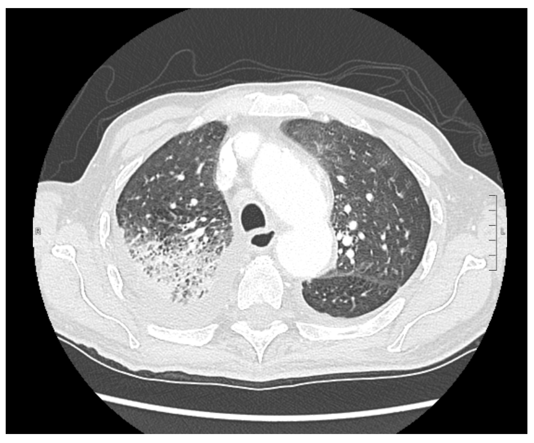
An 83-year-old male was admitted to the Emergency Department due to fever (up to 38.5 °C), cough, severe asthenia, and the appearance of an ulcerative lesion on the nose. The patient reported a story of losing around 5 kg in the previous 30 days. On admission, the nasopharyngeal swab tested positive for SARS-CoV-2 and the patient was transferred to the Infectious Diseases Unit. His medical history showed gastroesophageal reflux disease, paroxysmal supraventricular tachycardia, and obliterans chronic arteriopathy. He took betablockers, cilostazol, and proton pump inhibitors. The patient was fully vaccinated (three doses) against SARS-CoV-2. On admission, he was febrile (T: 38 °C), blood pressure was 130/80 mmHg, heart rate was 70 bpm, oxygen saturation in room air was 91%, and respiratory rate was 18 breaths/min. Glasgow Coma Scale (GCS) was 14. Arterial blood gas analysis showed hypoxemia and a Venturi Mask 10 lt/FiO2 40% was positioned. Physical examination displayed decreased vesicular breath sounds in the right thorax and an ulcerative lesion on the left side of the nose (Figure 1), which were analyzed by biopsy. A chest X-ray and subsequent thorax CT scan highlighted right medio-basal lung consolidation, perivascular interstitial thickening, and bilateral pleural effusion (Figure 2). In addition, diffuse mediastinic lymphadenopathy and ascending aorta aneurysm (4.8 cm) was highlighted. The abdomen and sinus CT scans did not show abnormal findings.
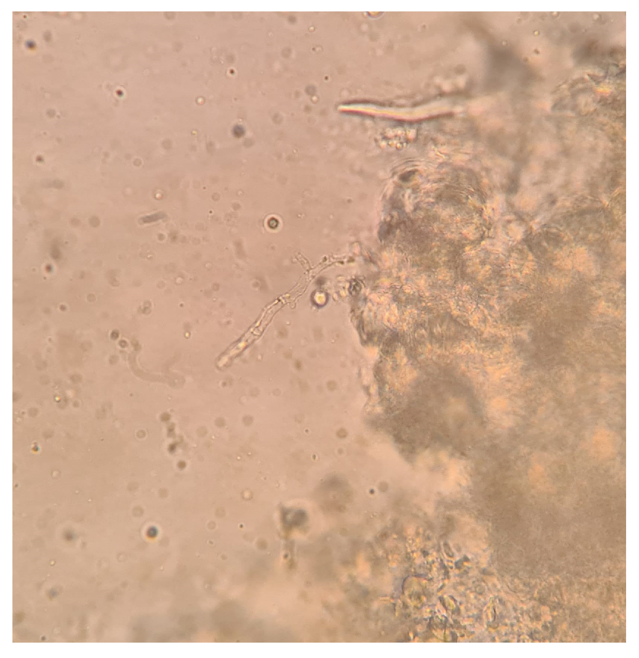
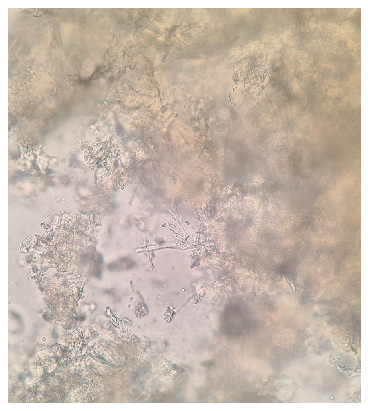
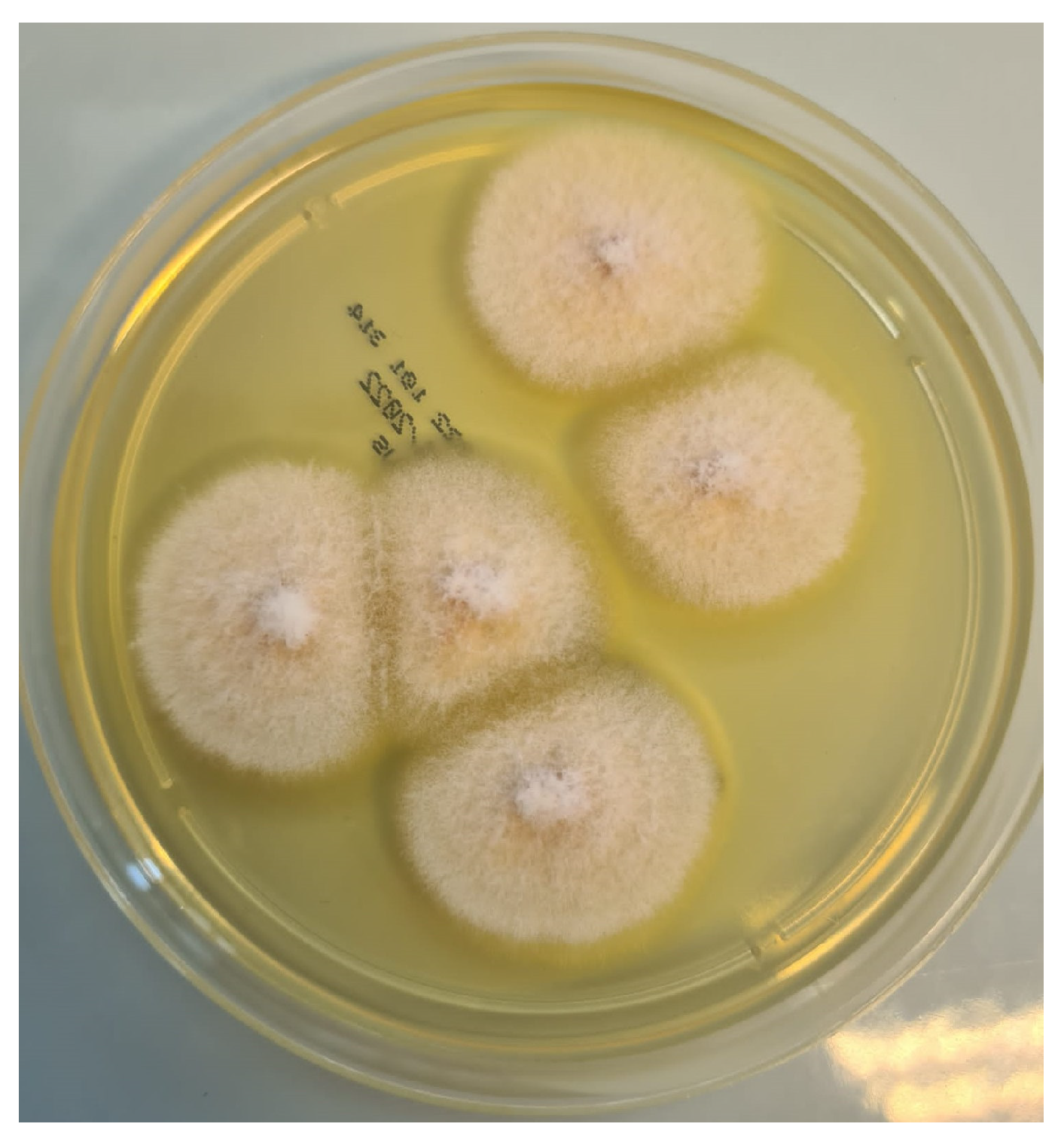
Blood tests revealed pancytopenia with a low white blood cell count (WBC 0.9 × 10³/mm3, Neutrophils 1.6%, Lymphocytes 56.2%, Monocytes 42%), anemia (Hb 8.76 g/dL), and a low platelets count (41 × 103/mm3). Inflammatory markers were elevated: C-Reactive Protein (CRP) was 26.42 mg/dL (normal range < 0.5 mg/dL), Erythrocyte sedimentation rate (ESR) was 97 mm/h (normal range < 10 mm/h). Procalcitonin was 2.6 mcg/L (normal value < 0.1 mcg/L). Transaminases and bilirubin levels were normal, as well as coagulation parameters. Creatinine was 0.9 mg/dL with an eGFR of 79 mL/min. The patient also presented with hypoalbuminemia (2.24 g/dL) and high ferritin levels (2000 ng/mL). Glucose levels were normal. HIV, HBV, and HCV serology tested negative. Legionella and pneumococcal urinary antigens also tested negative. Blood cultures and urine cultures tested negative, as well as serum EBV- and CMV-DNA. Serum beta-glucan was 294 pg/mL (normal values < 60 pg/mL), and serum galactomannan was negative. Empiric antibiotic therapy was started with intravenous (IV) piperacillin/tazobactam 4.5 gr three times daily and teicoplanin 400 mg daily (after loading dose); empirical antifungal therapy was administered with IV liposomal amphotericin B at a dosage of 5 mg/kg. In addition, supportive COVID-19 therapy was started with corticosteroids. A biopsy and a bone marrow aspirate were performed, and acute myeloid leukemia was diagnosed. In addition, microscopic examination of the ulcerated nasal lesion revealed the presence of septate and non-septate hyaline hyphae (Figure 3 and Figure 4). Septate hyphae were attributed to Fusarium (F.) solanii, whose colonies were revealed after culture exams (Figure 5). Non-septate hyphae were suggestive of possible zygomycete infections. Considering the low culture sensitivity in the case of zygomycosis, a molecular assay was performed to further investigate the microscopic records. The multiplex real-time PCR assay (MucorGenius®, PathoNostics, Maastricht, The Netherlands), which targets the Mucorales 18S rDNA, showed a positive result. Antimicrobial susceptibility testing was provided only for the F. solanii strain by using broth microdilution (SensititreYeastOne® method; Thermo Fisher Scientific, Cleveland, OH, USA). The test revealed a significant increase in MIC values for all of the tested antifungal drugs. Specifically, the following MIC values were recorded: >8 mg/L for anidulafungin, caspofungin, micafungin, amphotericin, posaconazole and voriconazole; >256 mg/L for fluconazole; >64 mg/L for flucytosine; >16 mg/L for itraconazole. Intravenous antibiotic therapy was administered for 18 days, obtaining a moderate decrease in inflammatory marker levels along with pulmonary imaging improvement. However, clinical conditions continued to deteriorate due to leukemia. In addition, serum Beta-D-glucan levels were decreased. According to microbiological reports, liposomal amphotericin B was maintained at the same dose combined with oral posaconazole 300 mg/day (with previous loading dose). Prophylactic cotrimoxazole was added. Furthermore, corticosteroids, as suggested by hematologists, were maintained. After 18 days, SARS-CoV-2 tested negative on a nasopharyngeal swab and the patient was transferred to the Hematology Unit, where he died after 20 days due to cardiac arrest.

3. Discussion

IFIs are associated with high morbidity and mortality in patients with hematological malignancies, especially for those affected by acute leukemias, with prolonged neutropenia being a major risk factor [3].
Here, we described a patient suffering from acute myeloid leukemia who developed a mixed fungal infection from Mucorales/Fusarium, both recovered from skin biopsy. Furthermore, clinical conditions were compromised by SARS-CoV-2 infection and concomitant pneumoniae impacting a dysregulated immune system.
Among Fusarium species, F. solanii is the most frequent microorganism associated with human diseases, followed by F. oxysporum and F. verticillioides [4]. Those fungi, ubiquitous in the environment, are considered serious opportunistic pathogens for immunocompromised patients with a mortality rate of up to 70% [5].
Although the airways represent Fusarium’s primary way of entry by inhalation of airborne conidia, leading to severe sinusitis or pneumoniae, skin is the second most frequent entrance, resulting in localized or disseminated clinical forms, especially among immunocompromised patients [4]. As discussed by Nucci et al. [6], among neutropenic subjects, both localized and disseminated Fusarium skin localizations are characterized by high mortality rates. Different cutaneous manifestations of Fusarium have been described, mostly depending on the host immune status, varying from disseminated disease with painful nodular or papular ecthyma-like lesions at different stages of evolution, to localized target forms developing in any site with rapid evolution to necrotic forms.
The management of Fusarium infections may be significantly challenging, due to antifungal resistance determined by drug efflux mechanisms, biofilm formation, and target alterations [7]. Azole intrinsic resistance is a major concern, mainly caused by the wide use of azole in plant protection [8]. Furthermore, echinocandins show less efficiency against Fusarium species, complicating patients’ therapeutic management [9].
Regarding the treatment of cutaneous fusariosis, guidelines suggest surgical debridement along with antifungal therapy. Although different species could be susceptible to different drugs, liposomal amphotericin B is the preferred option. Studies have reported combination options with azoles (voriconazole or posaconazole), with no conclusive results [4]. Undoubtedly, therapy response strictly correlates with hematological disease resolution and/or neutrophil recovery [10].
On the other hand, cutaneous mucormycosis is the third most typical clinical manifestation of Mucorales infections, following rhino-cerebral and pulmonary forms. Skin involvement could be localized, extended (muscles, bones), or disseminated to noncontiguous organs [11]. Imitating Fusarium, Mucorales is ubiquitous in the environment and its spores could be recovered in compost piles, soil, fruits, and decaying vegetation. Hematological conditions along with immunosuppressive diseases represent key risk factors for the development of mucormycosis [12,13].
Cutaneous mucormycosis usually develops as a single, indurated, painful area of cellulitis evolving in ecthyma-like lesions. Dissemination and deep tissue involvement are uncommon complications. Intravenous liposomal amphotericin B is the drug of choice to treat Mucorales, whereas posaconazole and voriconazole could be used as a salvage therapy for those patients who do not tolerate amphotericin [14]. According to current guidelines, mucormycosis diagnostic assessments should be performed by microscopic examination and fungal culture on tissue biopsy (in case of suspected cutaneous forms) or sinus washing in case of rhinocerebral disease [15]. Unluckily, these conventional methods suffer from low sensitivity due to the intrinsic fragility of zygomycetes coenocytic hyphae. Real-time PCR assays have been recently validated for zygomycetes detection in clinical samples such as tissue biopsies, bronchoalveolar-lavage fluids, and serum samples. These molecular technologies offer the possibility to improve sensitivity yield, providing fast results for severe and time-dependent infections. Mucorales species involved in human infections can be correctly detected through easy-to-use PCR assays, which should be integrated into all diagnostic workflows performed for mucormycosis clinical suspicion [14,16]. Mucorales do not share 1,3-Beta-D-glucan and galactomannan as cell wall components and these tests are negative in patients with mucormycosis.
Regarding Fusarium spp., although more studies are needed, Beta-D-glucan could be helpful to rule out fusariosis, due to its high negative predictive value (99%). However, it should not be used to decide treatment initiation due to a very low positive predictive value (7%) and low specificity (54%) [17].
A Galactomannan assay performed to diagnose Aspergillosis could cross-react in patients with Fusarium infections. Neither the number of positive tests nor galactomannan value is useful to discriminate between invasive aspergillosis and invasive fusariosis, especially in regions where fusariosis is uncommon [18]. Although not statistically significant, the patient we described had positive serum beta-D-glucan and a negative value for serum galactomannan.
Several authors remarked on the importance of integrating rapid polymerase chain reaction-based methods into fungal infection laboratory diagnosis [19,20,21]. According to these literary data, in critically ill patients, fungal DNA detection is a promising method to rapidly detect pathogens. Sensitivity rates can reach 43 to 100%, while specificities ranges are approximately 64 to 100%. Fluctuations are justifiable depending on the microorganisms count or clinical sample interferences. Otherwise, the sensitivity percentages of culture-based methods reach no more than 25–50% [16,21].
Among bacterial and viral superinfections [22,23,24], COVID-19 has been widely associated with mucormycosis through several pathological patterns such as immune system impairment due to cytokine storm [25], impaired phagocytosis, and endothelitis [26,27]. In addition, immunosuppressive therapies such as corticosteroids and immunomodulatory therapies such as anti-IL6 antibodies favor the development of COVID-19 associated mucormycosis (CAM) [28,29,30,31].
The patient we described had acute leukemia along with COVID-19 pneumonia. In addition to the two major risk factors for fungal diseases such as severe neutropenia and SARS-CoV-2 infections, the patient was also aged and hospitalized.
Based on worsening clinical conditions and laboratory results, empirical antifungal therapy was started before the results of the skin biopsy examination. Due to the poor general status of the patient, surgical debridement of the skin lesions was not performed. Bronchoscopy with bronchoalveolar lavage was also not performed, as it was challenging to fulfill because of the patient’s severe respiratory conditions. A Fusarium resistant pattern would not have allowed any therapeutical choice, although liposomal amphotericin B was confirmed and posaconazole was added; the same combination could have been appropriate for Mucorales infection, as reported by guidelines. Although pharmacologic therapies were used, insufficient immune recovery due to the rapid progression of leukemia contributed to the patient’s unfortunate outcome.
There are few reports in scientific literature about Mucorales/Fusarium co-infection, and little data about its clinical management and therapeutic approach. Based on treatment guidelines about these infections alone, our clinical point of view about co-infection suggests that liposomal amphotericin B may represent a valid empirical choice, due to its wide fungal coverage and safety. However, both novel and classic diagnostic methods, along with precise drug susceptibility tests, should be performed as soon as possible. The rationale should be to promptly provide the correct diagnosis, avoiding clinical and therapeutical delays.

4. Conclusions

Among the general population, the incidence of IFIs is gradually rising. This trend could be explained by both the increasing prevalence of immunocompromised patients and the significative improvement in pathogenic fungi diagnosis. Mucorales and Fusarium infections represent both an example of difficult to diagnose pathogens and a paradigm of difficult-to treat germs, becoming challenging clinical conditions to deal with [32], especially considering mixed infections, which are uncommon, especially in our region. Microscopic and culture-based investigations still represent the gold standard in fungal diseases work-up, allowing for definitive diagnosis and an antifungal susceptibility test. The limitations of these methods, such as the time-to-result parameter, could be fixed by more sensitive and faster molecular techniques which should be integrated into fungal infection laboratory diagnosis [19,20,21,33]. As a final consideration, the rapid progression of severe fungal infections should be faced through both a prompt clinical suspicion and recognition along with a timely diagnostic protocol. These diagnostic tips could play a key role in assuring optimal clinical and therapeutical patient management.

Author Contributions

Conceptualization, A.M. and M.C. (Maddalena Calvo); methodology, U.C.; investigation, L.T.; data curation, M.G.; writing—original draft preparation, A.M.; writing—review and editing, M.C. (Manuela Ceccarelli); supervision, G.S., G.N. and B.C. All authors have read and agreed to the published version of the manuscript.

Funding

This research received no external funding.

Institutional Review Board Statement

Not applicable.

Informed Consent Statement

Written informed consent has been obtained from the patient to publish this paper.

Data Availability Statement

Not applicable.

Conflicts of Interest

The authors declare no conflict of interest.

References

  1. Ceccarelli, M.; Marino, A.; Pulvirenti, S.; Coco, V.; Busà, B.; Nunnari, G.; Cacopardo, B.S. Bacterial and Fungal Co-Infections and Superinfections in a Cohort of COVID-19 Patients: Real-Life Data from an Italian Third Level Hospital. Infect. Dis. Rep. 2022, 14, 372–382. [Google Scholar] [CrossRef]
  2. Casalini, G.; Giacomelli, A.; Ridolfo, A.; Gervasoni, C.; Antinori, S. Invasive fungal infections complicating COVID-19: A narrative review. J. Fungi 2021, 7, 921. [Google Scholar] [CrossRef]
  3. Bellanger, A.P.; Rocchi, S.; Berceanu, A.; Scherer, E.; Larosa, F.; Millon, L. Positive quantitative PCR detecting Fusarium solani in a case of mixed invasive fungal disease due to Mucorales and Fusarium solani. Bone Marrow Transplant. 2020, 55, 873–876. [Google Scholar] [CrossRef] [PubMed]
  4. Nucci, M.; Anaissie, E. Fusarium infections in immunocompromised patients. Clin. Microbiol. Rev. 2007, 20, 695–704. [Google Scholar] [CrossRef]
  5. Durand-Joly, I.; Alfandari, S.; Benchikh, Z.; Rodrigue, M.; Espinel-Ingroff, A.; Catteau, B.; Cordevant, C.; Camus, D.; Dei-Cas, E.; Bauters, F.; et al. Successful outcome of disseminated Fusarium infection with skin localization treated with voriconazole and amphotericin B-lipid complex in a patient with acute leukemia. J. Clin. Microbiol. 2003, 41, 4898–4900. [Google Scholar] [CrossRef]
  6. Anaissie, E.; Nucci, M. Cutaneous infection by Fusarium species in healthy and immunocompromised hosts: Implications for diagnosis and management. Clin. Infect. Dis. 2002, 35, 909–920. [Google Scholar] [CrossRef]
  7. Zhao, B.; He, D.; Wang, L. Advances in Fusarium drug resistance research. J. Glob. Antimicrob. Resist. 2021, 24, 215–219. [Google Scholar] [CrossRef]
  8. Al-Hatmi, A.M.S.; Meis, J.F.; de Hoog, G.S. Fusarium: Molecular Diversity and Intrinsic Drug Resistance. PLoS Pathog. 2016, 12. [Google Scholar] [CrossRef]
  9. Walker, L.A.; Gow, N.A.R.; Munro, C.A. Fungal echinocandin resistance. Fungal Genet. Biol. 2010, 47, 117–126. [Google Scholar] [CrossRef] [PubMed]
  10. Letscher-Bru, V.; Campos, F.; Waller, J.; Randriamahazaka, R.; Candolfi, E.; Herbrecht, R. Successful outcome of treatment of a disseminated infection due to Fusarium dimerum in a leukemia patient. J. Clin. Microbiol. 2002, 40, 1100–1102. [Google Scholar] [CrossRef] [Green Version]
  11. Skiada, A.; Drogari-Apiranthitou, M.; Pavleas, I.; Daikou, E.; Petrikkos, G. Global Cutaneous Mucormycosis: A Systematic Review. J. Fungi 2022, 8, 194. [Google Scholar] [CrossRef] [PubMed]
  12. Marino, A.; Zafarana, G.; Ceccarelli, M.; Cosentino, F.; Moscatt, V.; Bruno, G.; Bruno, R.; Benanti, F.; Cacopardo, B.; Celesia, B.M. Immunological and Clinical Impact of DAA-Mediated HCV Eradication in a Cohort of HIV/HCV Coinfected Patients: Monocentric Italian Experience. Diagnostics 2021, 11, 2336. [Google Scholar] [CrossRef]
  13. Micali, C.; Russotto, Y.; Facciolà, A.; Marino, A.; Celesia, B.M.; Pistarà, E.; Caci, G.; Nunnari, G.; Pellicanò, G.F.; Rullo, E.V. Pulmonary Kaposi Sarcoma without Respiratory Symptoms and Skin Lesions in an HIV-Naïve Patient: A Case Report and Literature Review. Infect. Dis. Rep. 2022, 14, 228–242. [Google Scholar] [CrossRef]
  14. Guegan, H.; Iriart, X.; Bougnoux, M.E.; Berry, A.; Robert-Gangneux, F.; Gangneux, J.P. Evaluation of MucorGenius® mucorales PCR assay for the diagnosis of pulmonary mucormycosis. J. Infect. 2020, 81, 311–317. [Google Scholar] [CrossRef] [PubMed]
  15. Diagnosis and Testing of Mucormycosis | Mucormycosis | CDC. Available online: https://www.cdc.gov/fungal/diseases/mucormycosis/diagnosis.html (accessed on 21 November 2022).
  16. Normand, A.C.; Imbert, S.; Brun, S.; Al-Hatmi, A.M.S.; Chryssanthou, E.; Cassaing, S.; Schuttler, C.; Hasseine, L.; Mahinc, C.; Costa, D.; et al. Clinical Origin and Species Distribution of Fusarium spp. Isolates Identified by Molecular Sequencing and Mass Spectrometry: A European Multicenter Hospital Prospective Study. J. Fungi 2021, 7, 246. [Google Scholar] [CrossRef]
  17. Nucci, M.; Barreiros, G.; Reis, H.; Paixão, M.; Akiti, T.; Nouér, S.A. Performance of 1,3-beta-D-glucan in the diagnosis and monitoring of invasive fusariosis. Mycoses 2019, 62, 570–575. [Google Scholar] [CrossRef] [PubMed]
  18. Nucci, M.; Carlesse, F.; Cappellano, P.; Varon, A.G.; Seber, A.; Garnica, M.; Nouér, S.A.; Colombo, A.L. Earlier diagnosis of invasive fusariosis with Aspergillus serum galactomannan testing. PLoS ONE 2014, 9, e87784. [Google Scholar] [CrossRef]
  19. Trovato, L.; Betta, P.; Romeo, M.G.; Oliveri, S. Detection of fungal DNA in lysis-centrifugation blood culture for the diagnosis of invasive candidiasis in neonatal patients. Clin. Microbiol. Infect. 2012, 18, E63–E65. [Google Scholar] [CrossRef]
  20. Trovato, L.; Marino, A.; Pizzo, G.; Oliveri, S. Case Report: Molecular Diagnosis of Fungal Keratitis Associated With Contact Lenses Caused by Fusarium solani. Front. Med. 2021, 8, 249. [Google Scholar] [CrossRef]
  21. Arvanitis, M.; Anagnostou, T.; Fuchs, B.B.; Caliendo, A.M.; Mylonakis, E. Molecular and nonmolecular diagnostic methods for invasive fungal infections. Clin. Microbiol. Rev. 2014, 27, 490–526. [Google Scholar] [CrossRef] [Green Version]
  22. El-Sokkary, R.; Uysal, S.; Erdem, H.; Kullar, R.; Pekok, A.U.; Amer, F.; Grgić, S.; Carevic, B.; El-Kholy, A.; Liskova, A.; et al. Profiles of multidrug-resistant organisms among patients with bacteremia in intensive care units: An international ID-IRI survey. Eur. J. Clin. Microbiol. Infect. Dis. 2021, 40, 2323–2334. [Google Scholar] [CrossRef] [PubMed]
  23. Erdem, H.; Hargreaves, S.; Ankarali, H.; Caskurlu, H.; Ceviker, S.A.; Bahar-Kacmaz, A.; Meric-Koc, M.; Altindis, M.; Yildiz-Kirazaldi, Y.; Kizilates, F.; et al. Managing adult patients with infectious diseases in emergency departments: International ID-IRI study. J. Chemother. 2021, 33, 302–318. [Google Scholar] [CrossRef]
  24. Cosentino, F.; Moscatt, V.; Marino, A.; Pampaloni, A.; Scuderi, D.; Ceccarelli, M.; Benanti, F.; Gussio, M.; Larocca, L.; Boscia, V.; et al. Clinical characteristics and predictors of death among hospitalized patients infected with SARS-CoV-2 in Sicily, Italy: A retrospective observational study. Biomed. Rep. 2022, 16, 34. [Google Scholar] [CrossRef]
  25. Marino, A.; Munafò, A.; Augello, E.; Bellanca, C.M.; Bonomo, C.; Ceccarelli, M.; Musso, N.; Cantarella, G.; Cacopardo, B.; Bernardini, R. Sarilumab Administration in COVID-19 Patients: Literature Review and Considerations. Infect. Dis. Rep. 2022, 14, 360–371. [Google Scholar] [CrossRef] [PubMed]
  26. Marino, A.; Pampaloni, A.; Scuderi, D.; Cosentino, F.; Moscatt, V.; Ceccarelli, M.; Gussio, M.; Celesia, B.M.; Bruno, R.; Borraccino, S.; et al. High-flow nasal cannula oxygenation and tocilizumab administration in patients critically ill with COVID-19: A report of three cases and a literature review. World Acad. Sci. J. 2020, 2, 23. [Google Scholar] [CrossRef]
  27. Campanella, E.; Marino, A.; Ceccarelli, M.; Gussio, M.; Cosentino, F.; Moscatt, V.; Micali, C.; Nunnari, G.; Celesia, B.M.; Cacopardo, B. Pain crisis management in a patient with sickle cell disease during SARS-CoV-2 infection: A case report and literature review. World Acad. Sci. J. 2022, 4, 14. [Google Scholar] [CrossRef]
  28. Marino, A.; Campanella, E.; Ceccarelli, M.; Larocca, L.; Bonomo, C.; Micali, C.; Munafò, A.; Celesia, B.M.; Nunnari, G.; Cacopardo, B. Sarilumab administration in patients with severe COVID-19: A report of four cases and a literature review. World Acad. Sci. J. 2022, 4, 24. [Google Scholar] [CrossRef]
  29. Pal, R.; Singh, B.; Bhadada, S.K.; Banerjee, M.; Bhogal, R.S.; Hage, N.; Kumar, A. COVID-19-associated mucormycosis: An updated systematic review of literature. Mycoses 2021, 64, 1452–1459. [Google Scholar] [CrossRef]
  30. Chiurlo, M.; Mastrangelo, A.; Ripa, M.; Scarpellini, P. Invasive fungal infections in patients with COVID-19: A review on pathogenesis, epidemiology, clinical features, treatment, and outcomes. New Microbiol. 2021, 44, 71–83. [Google Scholar] [PubMed]
  31. Trovato, L.; Calvo, M.; Migliorisi, G.; Astuto, M.; Oliveri, F.; Oliveri, S. Fatal VAP-related pulmonary aspergillosis by Aspergillus niger in a positive COVID-19 patient. Respir. Med. Case Rep. 2021, 32, 101367. [Google Scholar] [CrossRef]
  32. Wiederhold, N.P. Antifungal resistance: Current trends and future strategies to combat. Infect. Drug Resist. 2017, 10, 249–259. [Google Scholar] [CrossRef] [PubMed]
  33. Antinori, S.; Corbellino, M.; Parravicini, C. Challenges in the Diagnosis of Invasive Fungal Infections in Immunocompromised Hosts. Curr. Fungal Infect. Rep. 2018, 12, 12–22. [Google Scholar] [CrossRef] [PubMed]
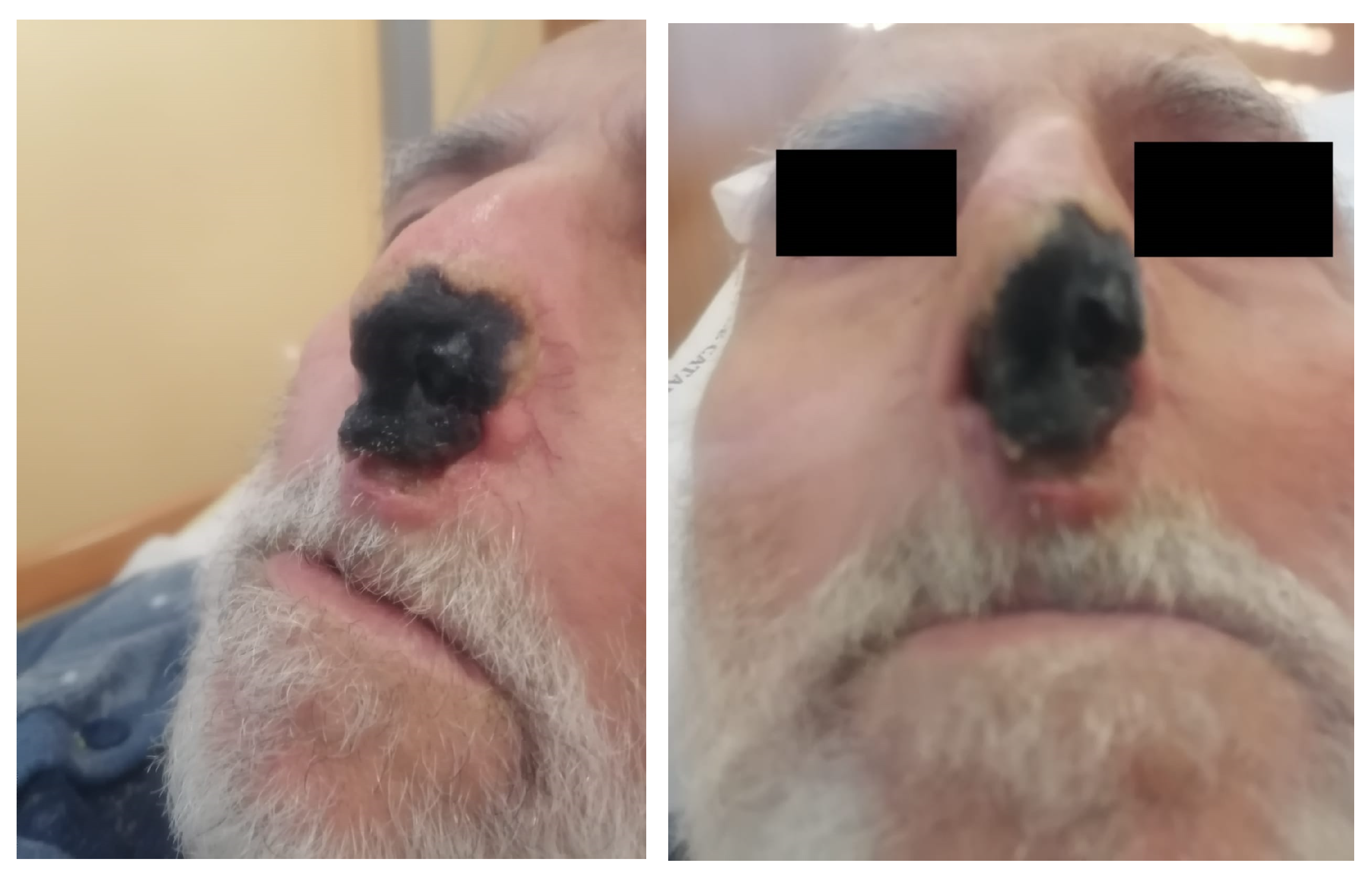
Figure 1. Echtyma-like necrotic-ulcerative lesions.
Pathogens 12 00304 g001
Figure 2. Thorax CT scan showing right medio basal consolidation, pleural effusion, and ascending aorta aneurysm.
Pathogens 12 00304 g002
Figure 3. Septate hyphae from microscopic investigation on biopsy samples (40× magnification).
Pathogens 12 00304 g003
Figure 4. Non-septate hyphae from microscopic investigations on biopsy samples (40× magnification).
Pathogens 12 00304 g004
Figure 5. Fusarium solanii colonies on Sabouraud culture examination on biopsy samples.
Pathogens 12 00304 g005
Disclaimer/Publisher’s Note: The statements, opinions and data contained in all publications are solely those of the individual author(s) and contributor(s) and not of MDPI and/or the editor(s). MDPI and/or the editor(s) disclaim responsibility for any injury to people or property resulting from any ideas, methods, instructions or products referred to in the content.