Abstract
The Eurasian badger (Meles meles) is widespread in Italy and occupies different habitats. The occurrence and species of gastrointestinal parasites were evaluated in a free-ranging badger population living in a highly anthropic area in central Italy. A total of 43 fecal samples were examined using the flotation test, the Mini-FLOTAC and Baermann techniques, and a rapid immunoassay for the detection of Giardia duodenalis and Cryptosporidium spp. fecal antigens. Molecular investigations were also performed that aimed at identifying Giardia genotypes. Overall, 37/43 samples (86%) were found positive. Specifically, 48.8% (21 samples) were positive for G. duodenalis, 23.2% (10/43) for Cryptosporidium spp., and 7% (3/43) for coccidian oocysts. Strongyloides sp. nematode larvae were detected in 3/43 samples (7%). Ascarid (1/43, 2.3%), capillariid (1/43, 2.3%), and strongyle-type eggs (76.7%, 33/43) were also identified. Among the 11 readable sequences of samples that were positive for G. duodenalis by end-point PCR (18/21), the zoonotic assemblage A sub-assemblage AII and mixed assemblage A and B were identified. This is the first report of zoonotic G. duodenalis genotypes in the Eurasian badger. Moreover, most of identified parasites have zoonotic potential and/or potential impact on the population health of wild badgers and other wild and domestic animals.
1. Introduction
The Eurasian badger (Meles meles) is an opportunistic omnivorous meso-carnivore of the Mustelidae family, mainly feeding on a wide variety of plants and animal foods (i.e., earthworms, large insects, small mammals, carrion, cereals, and tubers) [1,2,3]. Badgers are social, and in a high-density population this mustelid species lives in a mixed-sex clan composed of up to 12 individuals, while it adopts a more solitary lifestyle in a low-density population [3,4]. Badgers are semi-fossorial and mainly nocturnal mammals and use burrow systems (i.e., settlements) as main sites for overwintering, breeding, and sleeping during daylight hours [4,5,6]. They usually share the same settlement at different times with other semi-fossorial mammals, such as the crested porcupine (Hystrix cristata) and red fox (Vulpes vulpes), and occasionally can cohabit with the crested porcupine [7]. Except for the Italian islands, the Eurasian badger is widespread throughout all mainland Italy, where it can occupy different habitats, such as mountainous areas and the Mediterranean coastal zone but also suburban, cultivated, and anthropized areas (i.e., riverbanks, gardens, and city parks) [8,9,10,11]. Although the species is widespread in Italy, few data are available on the gastrointestinal parasites of badger populations in the country [12,13,14]. In Italy, the nematodes Physaloptera sibirica, Uncinaria criniformis, Aonchotheca putorii, and Molineus patens and the cestode species Mesocestoides melesi have been identified in badgers [12,13,14]. However, in other European countries, several gastrointestinal parasite species have been identified in badger populations [15,16,17,18,19,20,21], including parasite species that can also infect humans and/or other wild and domestic animal species, such as Baylisascaris melis described for the first time in badgers in Belgium [22], Cryptosporidium spp., and Giardia duodenalis [17,18]. Nevertheless, data on these badger parasite infections are completely lacking in Italy. In Italy, G. duodenalis infection has been detected in several wild animal species, such as the wolf (Canis lupus italicus), wild boar (Sus scrofa), Alpine chamois (Rupicapra rupicapra rupicapra), Apennine chamois (Rupicapra pyrenaica ornata), and crested porcupine (H. cristata) [23,24,25,26]. Moreover, potentially zoonotic assemblages and sub-assemblages have been identified in some of these animal species, such as R. r. rupicapra, R. p. ornata, and crested porcupines, living in both wild and anthropized areas of central Italy [23,25,26], where the Eurasian badger is also present. Therefore, this study was primarily designed to update data on gastrointestinal parasites infecting the Eurasian badger in Italy and to also evaluate the occurrence and genotypes of G. duodenalis in a Eurasian badger population living in an anthropized area of central Italy.
2. Results
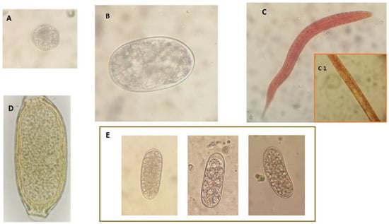
Overall, 37 out of 43 (86%) samples were positive for gastrointestinal parasites at the parasitological analysis performed. Eight different parasite taxa were identified (Table 1). At the immunoassay, 48.8% (21/43) of the analyzed fecal samples were positive for G. duodenalis and 23.2% (10/43) for Cryptosporidium spp. Based on morphology, two types of coccidian oocysts (7%, 3/43, 15–55 oocysts per gram of feces (OPG)) were evidenced by the flotation and the Mini-FLOTAC techniques: the first type of oocyst was ovoidal, about 32 µm × 26 µm in size (Table 1, Figure 1A), which was detected in two samples, while in the third positive sample, oocysts were oval/elliptical and measured about 20 µm × 16 µm. At the flotation test and the Mini-FLOTAC technique, positivity for gastrointestinal strongyle-type eggs (76.7%, 33/43) (Table 1, Figure 1B,E) with a mean number of 84.8 eggs per gram of feces (EPG; range 10–800 EPG), capillariid eggs (2.3%, 1/43, 10 EPG) (Table 1, Figure 1D), and ascarid eggs (1/43, 2.3%, 10 EPG) was also found. Among the strongyle-type eggs, Uncinaria criniformis was identified in all samples (Table 1, Figure 1B). However, unidentified strongyle-type eggs were also detected in three badger fecal samples (3/43, 7%), which were concurrently positive for U. criniformis (Table 1, Figure 1E). Finally, Strongyloides spp. larvae were detected using the Baermann test in 3 out of 43 (7%) examined samples (Table 1, Figure 1C).

Table 1.
Gastrointestinal parasites detected in 43 fecal samples of a European badger (Meles meles) population living in an anthropized area in central Italy.

Figure 1.
Digestive tract parasites identified in fecal samples from a Eurasian badger population in Tuscany, central Italy. (A) Isospora melis unsporulated oocyst; (B) Uncinaria criniformis egg; (C) Strongyloides sp. first stage larva particularly of the rhabditoid esophagus (C1); (D) Aonchoteca putorii egg; (E) unidentified strongyle-type nematode eggs.
Multiple parasites were found in 14/37 positive samples (37.8%), most of which were coinfections between Giardia plus other parasites (11/43, 25.6%). More specifically, Giardia + strongyle-type eggs and Giardia + strongyle-type eggs + Strongyloides sp. larvae were detected in eight and two Giardia-positive samples, respectively. In the remaining Giardia-positive sample, gastrointestinal strongyle-type eggs + Strongyloides sp. + coccidia were identified.
Out of 21 samples that resulted Giardia-positive using the immunoassay, 18 were positive at end-point PCR and 11 readable sequences were obtained. In detail, at the bg locus, six samples were assigned to assemblage A sub-assemblage AII. Moreover, the presence of double peaks in diagnostic positions allowed us to identify a mixed assemblages A and B infection in one sample. As for the tpi locus, two samples were assigned to assemblage A sub-assemblage AII. Finally, one sample was successfully amplified for both genes and identified as assemblage A sub-assemblage AII at the bg locus and as mixed assemblages A and B infection at the tpi locus. Raw data (FASTA sequences) are reported in Supplementary file S1.
3. Discussion
Gastrointestinal parasites may influence mustelid population size by reducing host fitness; lowering host body condition and fecundity, causing mortality; and changing host behavior or vulnerability to predation [27,28]. In a study performed in Switzerland on the causes of mortality and morbidity in free-ranging mustelids [16], gastrointestinal parasites were found to be associated with pathological changes of the digestive system in about 10% of the infected animals. The significance of the negative impact of gastrointestinal parasites on mustelid populations may be related to specific parasite species and/or infection intensity [28,29]. Nonetheless, in Italy, the Eurasian badger is not an endangered species, and parasites may also represent an important factor limiting the population growth of this wild animal. However, even if some gastrointestinal parasites are specific to a particular mustelid host and their circulation is therefore related to the dynamics of that host population [28,30], some mustelid gastrointestinal parasite species can infect multiple mustelids and other animal host species [22,31,32] including humans, thus having zoonotic significance [18,22,32]. In an anthropized environment, such as the area selected in this study, the risk of transmission of parasites from wild animals to humans and domestic animals and vice versa can be high [33], and this possibility strongly increases the relevance of the Eurasian badger population here examined in the epidemiology of potentially zoonotic parasites.
Among the protozoa, G. duodenalis infection was prevalent in the examined badger population, considering that about 50% of the examined samples (21/43) were positive for G. duodenalis according to the results of the rapid immunoassay. Moreover, the results of molecular analysis confirmed the positivity for this species in 18/21 samples. Only a previous study reported G. duodenalis identification in badgers, and this protozoan enteropathogen was suspected to be responsible for poor conditions and chronic diarrhea in rescued Eurasian badger cubs in the U.K. [19]. In free-ranging badgers, however, no positivity for G. duodenalis was found both in a single badger examined in Poland [34] and 70 badgers examined in Spain [18]. In the badger cub found positive in the U.K. [19], the results of molecular analysis revealed G. duodenalis assemblage E, while in the present study, genotyping of 11 PCR positive samples revealed potentially zoonotic assemblages (A and B) and sub-assemblages (A-II). In fact, molecular studies showed that G. duodenalis includes eight distinct genotypes, also called assemblages, identified with the alphabetic letters from A to H [35]. Assemblages A and B are more frequently found in humans, but they can also infect animals and are considered potentially zoonotic [36]. Assemblages A and B have been further divided into sub-assemblages, some of which are more common in humans and some in animals. Sub-assemblage A-II is considered human-specific, although it has also been detected in animals [36,37]. Assemblage B is genetically more polymorphic than assemblage A, and this makes assignment to specific genotypes more difficult [36,38]. Therefore, this study shows for the first time that Eurasian badgers can be infected with zoonotic genotypes of G. duodenalis. Consequently, this wild animal species may play a role in the environmental contamination with G. duodenalis zoonotic genotypes that can be responsible for infections in other wild and domestic animals and humans, thus extending the public health relevance of this mustelid species to G. duodenalis. The badger population examined in this study lives in a highly anthropized area in burrows located near gardens, vegetable gardens, etc. In developed countries, human infections caused by zoonotic G. duodenalis genotypes are mainly waterborne infections [39]. Therefore, although defecation of badgers takes place in latrines, some atmospheric events, among which are mainly the torrential rainfalls being observed in Italy in recent years [40,41], can easily cause the spread of cysts of these zoonotic G. duodenalis assemblages in the surrounding environment and, consequently, the easy contamination of vegetable gardens, gardens, and surface water used for human consumption [39]. Furthermore, a recent study performed in this area [7] reported that badgers cohabited or shared the same burrows with crested porcupines and foxes. More specifically, they cohabited with porcupines in the same burrow system in 43% of the cases, and interactions between adult badgers and porcupettes were also recorded [7]. Consequently, G. duodenalis cross-infections between crested porcupines, foxes, and badgers can be very likely in this area. Crested porcupines and foxes may be susceptible to infections with zoonotic assemblages of G. duodenalis [26,42] and, compared with badgers, have a habit of being closer to human settlements, as they frequently visit vegetable gardens, gardens, henhouses, etc., and they can easily contaminate these areas with zoonotic G. duodenalis genotypes, causing the passage of these zoonotic assemblages from the wild to the domestic environment. Moreover, in the examined area, crested porcupines (H. cristata) were also found to be infected by potentially zoonotic G. duodenalis genotypes [26].
In previous European studies in wild mustelids [18,43], Cryptosporidium infection was reported in American minks (Neovison vison) in Denmark and badgers in Switzerland and Spain [16,18], with a 3% prevalence found in the badgers examined in Spain [18]. In the present study, 23.2% of badger fecal samples were positive for Cryptosporidium spp. at the immunoassay results. Although it is likely that samples from the same badgers may have been collected and examined more than one time during the study period, results obtained in this study may be indicative of a high frequency of Cryptosporidium infection in the badger population here examined with respect to that reported in previous studies in other European areas. In Spain [18], Cryptosporidium parvum and Cryptosporidium hominis zoonotic species were identified after a molecular analysis was performed in positive badgers. Therefore, further molecular studies should be performed on Cryptosporidium-positive badger samples in this study to evaluate the zoonotic significance of this finding and the potential role of the badger population here examined in human Cryptosporidium infections.
In the case of coccidia, the Eurasian badger can be infected by the species Eimeria melis and Isospora melis [21,44]. E. melis infection occurs at higher intensity and prevalence in cubs than in adults, and it is considered a significant cause of mortality in cubs, although of less pathological significance in adults [21,44]. I. melis seems to have limited pathological significance both in adults and cubs, but coinfections between E. melis and I. melis are frequently observed in adult badgers, and a direct relationship between the intensity of the two species has been also evidenced [21,44]. Based on morphological features, coccidian oocysts here detected in the fecal samples of two coccidia-infected badgers were identified with I. melis, while the third positive badger was infected by E. melis. In Europe, both these coccidian species have been previously reported in badgers only in England [21,44].
Gastrointestinal nematodes previously reported in European badgers include the species U. criniformis, Placoconus lotoris, Spirocerca lupi, Mastophorus muris, Aonchotheca putorii, Molineus patens, Physaloptera sibirica, Vigisospirura potekhina hugoti, Baylisascaris melis, and Strongyloides sp. [13,14,15,17,20,45]. Among these nematode infections, positivity for gastrointestinal strongylid eggs were prevalent in the examined badger population, as 76.7% of badger fecal samples here examined were positive for these nematodes, although with a low mean EPG number (84.8 EPG). Based on egg morphology and size, U. criniformis was identified in all positive samples, confirming the data of previous reports about the frequent occurrence of this nematode species in the Eurasian badger [45,46,47]. U. criniformis is a blood-feeding nematode species belonging to the Ancylostomatidae family, which is primarily found in badgers and closely related to Uncinaria stenocephala, a pathogenic species of wild and domestic canids and felids [15,20,46]. However, three samples were concurrently positive for strongylid eggs that were morphologically different from those of U. criniformis (Figure 1B,E) and probably belonging to M. patens, a gastrointestinal strongyle species previously reported in Italian badgers [13,14].
Two examined badger fecal samples were positive for the presence of eggs of other nematode taxa. More specifically, a sample was positive for 10 EPG of ascarid eggs and a further sample for 10 EPG of capillariid eggs. Capillariid eggs were 59–63 µm in length and 22–26 µm in width and showed a striated shell with fine net-like ridges on the outer surface and two protruding plugs. Based on these features, the detected eggs were identified as A. putorii, a capillariid species that is mainly found in the stomach and less frequently in the small intestine of wild and domestic carnivore hosts, and that is considered a potential cause of gastritis in pet carnivores [31]. In mustelids, this capillariid species may have a negative impact by reducing body condition and probably affecting population sizes [28]. In previous studies, this species was reported in European badger populations in Spain, Poland, and northwestern Italy [14,45,47].
In the case of the ascarid-positive badger sample, B. melis is the only ascarid species known to infect European badgers, and it was first described in Belgium [22]. Baylisascaris is a nematode genus infecting the small intestine of carnivores, omnivores, and herbivores, but a wide range of other animal species may act as paratenic hosts, in which these nematodes may cause visceral, ocular, and neural larva migrans [22,32,48]. However, no confirmed cases of natural larva migrans caused by B. melis have been reported, although this species was found to cause visceral, ocular, and neural larva migrans in experimentally infected rodents [22]. Nonetheless, this species is included among the potential causes of baylisascariasis in humans, along with other Baylisascaris species [45]. Among these, Baylisascaris procyonis infecting raccoons (Procyon lotor) is considered the most important, as this species may cause severe neurologic disease in humans and numerous animal species [22]. B. procyonis has been reported in raccoons in the USA, Canada, and many European countries [30,49,50,51,52,53,54], including an area of central Italy very near to that of the badger population examined in this study [55]. Further molecular and epidemiological studies are needed to establish if the ascarid found in this single badger sample is effectively B. melis and to evaluate the frequency of occurrence and diffusion of B. melis infection in the examined badger population.
Although Strongyloides infections have been frequently reported in badgers, Strongyloides infecting European badger populations have never been identified at the species level [17,20]. In Europe, the species Strongyloides procyonis, Strongyloides martis, Strongyloides mustelorum, and Strongyloides lutrae have been reported in other mustelid species [30,56,57,58]. The pathogenicity of Strongyloides species infecting mustelids is still unknown, although it is assumed that respiratory distress can result from migration of Strongyloides spp. larvae through the lungs [59]. Interestingly, S. procyonis was demonstrated to cause experimental creeping eruption and a short-lived intestinal infection in inoculated human volunteers [33]. Moreover, the morphological parameters of Strongyloides sp. found in the Japanese badger (Meles anakuma) seemed to be comparable to those of S. procyonis and S. martis, but data from a phylogenetic study suggested that it was a species separate from S. procyonis [60].
4. Materials and Methods
4.1. Study Area and Sampling
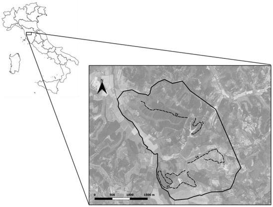
Sampling was performed in a hilly area of 714 ha located in Crespina-Lorenzana and Lari-Casciana Terme (10.56815° N–43.56796° E) in the province of Pisa (Tuscany, central Italy) (Figure 2). The study area included 18 towns, with an average human density of 134.08 people/km2. The area was characterized by a highly anthropic fragmented agroecosystem characterized by small woody areas interspersed with agricultural and urban zones and interconnected by a dense network of ecological paths [61]. A wide variety of wild mammals lived in the study area, such as crested porcupines (H. cristata), wild boars (Sus scrofa), roe deer (Capreolus capreolus), pine martens (Martes martes), stone martens (Martes foina), skunks (Mustela putorius), badgers (Meles meles), hares (Lepus europeus), eastern cottontails (Sylvilagus floridanus), wild rabbits (Oryctolagus cuniculus), red foxes (V. vulpes), wolves (Canis lupus), and the introduced invasive coypu (Myocastor coypus) [61].

Figure 2.
Map of Italy: the inset detail of the study area in Crespina-Lorenzana and Lari-Casciana Terme, Tuscany, central Italy (black line border), and the four transects (dashed black lines) in which badger fecal samples were collected from latrines.
Badger fecal sampling was performed from September 2020 to April 2021 from latrines. Badger latrine detection was performed along 4 transects (2 SD 1.2 km), randomly chosen within the study area, for a total length of 8 km (Figure 2). Every transect was covered once per week, and from each detected latrine, two aliquots of each fresh fecal sample present were collected and pooled. From each latrine, only samples judged fresher were collected for parasitological analysis, avoiding collecting dry or moldy fecal samples. The freshness state of fecal samples was assigned based on external appearance and local weather conditions. Badger fecal samples were discriminated according to their deposition site (i.e., latrines), size, shape, and composition (i.e., amorphous, part of small invertebrates, and seeds) [62,63]. Collected fecal samples were stored at 4 °C and analyzed within 24 h.
4.2. Parasitological Analysis
Fecal samples were examined by using a commercial rapid immunoassay to search for Giardia and Cryptosporidium spp. fecal antigens (Rida Quick® Cryptosporidium/Giardia Combi, R-Biopharm, Darmstadt, Germany). Moreover, for the detection and quantification of gastrointestinal nematode eggs and coccidian oocysts, all the fecal samples were examined by the flotation test and Mini-FLOTAC technique on 2 g of feces using saturated sodium chloride as the flotation solution (NaCl, specific gravity 1.2) [64]. Results are expressed as the arithmetic mean number of eggs/(oo)cysts per gram (EPG/OPG) of feces [64]. The Baermann test [65] was used for the detection of Strongyloides sp. larvae. Magnifications of 100× and 400× were used to identify nematode eggs/larvae and protozoan oocysts, which were measured under an optical microscope by using a micrometric eyepiece. Microscopic parasite identification was based on morphological and metric data on badger gastrointestinal nematodes and protozoa reported in previous studies [15,20,21,28,30,31,44,45,46,47].
4.3. Molecular Analysis
In the case of Giardia-positive samples using the immunoassay, molecular analysis was performed to identify Giardia species and genotypes. For DNA extraction, samples were processed using a commercial kit (QIAamp DNA Stool Mini Kit, QIAGEN, Valencia, CA, USA). PCR protocols were applied to amplify fragments of the small subunit ribosomal RNA (SSU rRNA, 130 bp) [66], ß-giardin (bg, 384 bp) [67], and triose phosphate isomerase (tpi, 530 bp) genes [68]. Positive amplicons were purified using the mi-PCR Purification Kit, Metabion International AG (Planegg/Steinkirchen, Munich, Germany). Amplification products were sent to an external laboratory for sequencing (Bio-Fab Research, Rome, Italy). Forward and reverse sequences were manually checked using Finch TV 1.4 software (Geospiza, Inc., Seattle, WA, USA). The obtained consensus sequences were then compared to those available in the GenBank database using the Standard Nucleotide BLAST search.
5. Conclusions
The data obtained in this study broaden our knowledge of gastrointestinal parasites of the Eurasian badger in Europe, and this is the first report of zoonotic G. duodenalis genotypes in this wild animal species. Moreover, G. duodenalis, Cryptosporidium spp., coccidia, Strongyloides sp., and ascarids were recorded for the first time in the Eurasian badger in Italy. Several of the parasites identified in this study have zoonotic potential and/or potential impact on the population health of wild badgers and other wild and domestic animals, thus highlighting the public and animal health relevance of this mustelid species.
Supplementary Materials
The following supporting information can be downloaded at: www.mdpi.com/article/10.3390/pathogens11080906/s1, Supplementary File S1: Raw FASTA sequences.
Author Contributions
Conceptualization, S.P. and A.F.; investigation, all authors; resources, S.P., F.B. and A.F.; data curation, S.P., F.B. and A.F.; writing—original draft preparation, S.P.; writing—review and editing, all authors; visualization, all authors; supervision, S.P., F.B. and A.F.; project administration, S.P. All authors have read and agreed to the published version of the manuscript.
Funding
This research received no external funding.
Institutional Review Board Statement
Not applicable.
Informed Consent Statement
Not applicable.
Data Availability Statement
Not applicable.
Conflicts of Interest
The authors declare no conflict of interest.
References
- Melis, C.; Cagnacci, F.; Bargagli, L. Food habits of the Eurasian badger in a rural Mediterranean area. Z. Jagdwiss 2002, 48, 236–246. [Google Scholar] [CrossRef]
- Balestrieri, A.; Remonti, L.; Prigioni, C. Diet of the Eurasian badger (Meles meles) in an agricultural riverine habitat (North-West Italy). Hystrix It. J. Mamm. 2004, 15. [Google Scholar] [CrossRef]
- Roper, T.J. Badger, Book 114; Collins New Naturalist Library, Collins: London, UK, 2010. [Google Scholar]
- Kruuk, H. Spatial organization and territorial behaviour of the European badger Meles meles. J. Zool. Lond. 1978, 184, 1–19. [Google Scholar] [CrossRef]
- Roper, T.J. The structure and function of badger setts. Zool. Lond. 1992, 227, 691–698. [Google Scholar] [CrossRef]
- Roper, T.J.; Ostler, J.R.; Schmid, T.K.; Christian, S.F. Sett use in European badgers Meles meles. Behaviour 2001, 138, 173–187. [Google Scholar] [CrossRef]
- Coppola, F.; Dari, C.; Vecchio, G.; Scarselli, D.; Felicioli, A. Cohabitation of settlements among crested porcupine (Hystrix cristata), red fox (Vulpes vulpes) and European badger (Meles meles). Curr. Sci. 2020, 119, 817–822. [Google Scholar] [CrossRef]
- Rosalino, L.M.; Loureiro, F.; Macdonald, D.W.; Santon-Reis, M. Dietary shifts of the badger (Meles meles) in Mediterranean woodlands: An opportunistic forager with seasonal specialisms. Mamm. Biol. 2005, 70, 12–23. [Google Scholar] [CrossRef]
- Prigioni, C.; Deflorian, M.C. Sett-site selection by the Eurasian badger (Meles meles) in an Italian Alpine area. Ital. J. Zool. 2005, 72, 43–48. [Google Scholar] [CrossRef]
- Remonti, L.; Balestrieri, A.; Prigioni, C. Range of the Eurasian badger (Meles meles) in an agricultural area of northern Italy. Ethol. Ecol. Evol. 2006, 18, 61–67. [Google Scholar] [CrossRef]
- Fabrizio, M.; Di Febbraro, M.; D’Amico, M.; Frate, L.; Roscioni, F.; Loy, A. Habitat suitability vs. landscape connectivity determining roadkill risk at a regional scale: A case study on European badger (Meles meles). Eur. J. Wildl. Res. 2019, 65, 7. [Google Scholar] [CrossRef]
- Ferroglio, E.; Ragagli, C.; Trisciuoglio, A. Physaloptera sibirica in foxes and badgers from the Western Alps (Italy). Vet. Parasitol. 2009, 163, 164–166. [Google Scholar] [CrossRef] [PubMed]
- Magi, M.; Banchi, C.; Barchetti, A.; Guberti, V. The parasites of the badger (Meles meles) in the north of Mugello (Florence, Italy). Parassitologia 1999, 41, 533–536. [Google Scholar] [PubMed]
- Di Cerbo, A.R.; Manfredi, M.T.; Bregoli, M.; Ferro Milone, N.; Cova, M. Wild carnivores as source of zoonotic helminths in north-eastern Italy. Helminthologia 2008, 45, 13–19. [Google Scholar] [CrossRef]
- Rosalino, L.M.; Torres, J.; Santos-Reis, M.A. Survey of helminth infection in Eurasian badgers (Meles meles) in relation to their foraging behaviour in a Mediterranean environment in southwest Portugal. Eur. J. Wildl. Res. 2006, 52, 202–206. [Google Scholar] [CrossRef]
- Akdesir, E.; Origgi, F.C.; Wimmershoff, J.; Frey, J.; Frey, C.F.; Ryser-Degiorgis, M.P. Causes of mortality and morbidity in free-ranging mustelids in Switzerland: Necropsy data from over 50 years of general health surveillance. BMC Vet. Res. 2018, 14, 195. [Google Scholar] [CrossRef]
- Byrne, R.L.; Fogarty, U.; Mooney, A.; Harris, E.; Good, M.; Marples, N.M.; Holland, C.V. The helminth parasite community of European badgers (Meles meles) in Ireland. J. Helminthol. 2019, 94, e37. [Google Scholar] [CrossRef]
- Mateo, M.; de Mingo, M.H.; de Lucio, A.; Morales, L.; Balseiro, A.; Espí, A.; Barral, M.; Lima Barbero, J.F.; Habela, M.Á.; Fernández-García, J.L.; et al. Occurrence and molecular genotyping of Giardia duodenalis and Cryptosporidium spp. in wild mesocarnivores in Spain. Vet. Parasitol. 2017, 235, 86–93. [Google Scholar] [CrossRef]
- Barlow, A.M.; Mullineaux, E.; Wood, R.; Taweenan, W.; Wastling, J.M. Giardiosis in Eurasian badgers (Meles meles). Vet. Rec. 2010, 167, 1017. [Google Scholar] [CrossRef]
- Millán, J.; Sevilla, I.; Gerrikagoitia, X.; García-Pérez, A.L.; Barral, M. Helminth parasites of the Eurasian badger (Meles meles L.) in the Basque Country (Spain). Eur J. Wildl. Res. 2004, 50, 37–40. [Google Scholar] [CrossRef]
- Anwar, M.A.; Newman, C.; MacDonald, D.W.; Woolhouse, M.E.; Kelly, D.W. Coccidiosis in the European badger (Meles meles) from England, an epidemiological study. Parasitology 2000, 120, 255–260. [Google Scholar] [CrossRef]
- Sapp, S.G.H.; Gupta, P.; Martin, M.K.; Murray, M.H.; Niedringhaus, K.D.; Pfaff, M.A.; Yabsley, M.J. Beyond the raccoon roundworm: The natural history of non-raccoon Baylisascaris species in the New World. Int. J. Parasitol. Parasites Wildl. 2017, 6, 85–99. [Google Scholar] [CrossRef]
- De Liberato, C.; Berrilli, F.; Marangi, M.; Santoro, M.; Trogu, T.; Putignani, L.; Lanfranchi, P.; Ferretti, F.; D’Amelio, S.; Giangaspero, A. Giardia duodenalis in Alpine (Rupicapra rupicapra rupicapra) and Apennine (Rupicapra pyrenaica ornata) chamois. Parasit. Vectors. 2015, 8, 650. [Google Scholar] [CrossRef]
- Di Francesco, C.E.; Smoglica, C.; Paoletti, B.; Angelucci, S.; Innocenti, M.; Antonucci, A.; Di Domenico, G.; Marsilio, F. Detection of selected pathogens in Apennine wolf (Canis lupus italicus) by a non-invasive GPS-based telemetry sampling of two packs from Majella National Park, Italy. Eur. J. Wildl. Res. 2019, 65, 84. [Google Scholar] [CrossRef]
- Guadano Procesi, I.; Montalbano Di Filippo, M.; De Liberato, C.; Lombardo, A.; Brocherel, G.; Perrucci, S.; Di Cave, D.; Berrilli, F. Giardia duodenalis in Wildlife: Exploring Genotype Diversity in Italy and Across Europe. Pathogens 2022, 11, 105. [Google Scholar] [CrossRef]
- Coppola, F.; Maestrini, M.; Berrilli, F.; Procesi, I.G.; Felicioli, A.; Perrucci, S. First report of Giardia duodenalis infection in the crested porcupine (Hystrix cristata L., 1758). Int. J. Parasitol. Parasites Wildl. 2020, 11, 108–113. [Google Scholar] [CrossRef]
- Kołodziej-Sobocińska, M.; Tokarska, M.; Zalewska, H.; Popiołek, M.; Zalewski, A. Digestive tract nematode infections in non-native invasive American mink with the first molecular identification of Molineus patens. Int. J. Parasitol. Parasites Wildl. 2020, 14, 48–52. [Google Scholar] [CrossRef]
- Zalewski, A.; Kołodziej-Sobocińska, M.; Bartoń, K.A. A tale of two nematodes: Climate mediates mustelid infection by nematodes across the geographical range. Int. J. Parasitol. Parasites. Wildl. 2022, 17, 218–224. [Google Scholar] [CrossRef]
- Ramírez-Pizarro, F.; Silva-de la Fuente, C.; Hernández-Orellana, C.; López, J.; Madrid, V.; Fernández, Í.; Martín, N.; González-Acuña, D.; Sandoval, D.; Ortega, R.; et al. Zoonotic Pathogens in the American mink in its southernmost distribution. Vector Borne Zoonotic Dis. 2019, 19, 908–914. [Google Scholar] [CrossRef]
- Popiołek, M.; Szczęsna-Staśkiewicz, J.; Bartoszewicz, M.; Okarma, H.; Smalec, B.; Zalewski, A. Helminth parasites of an introduced invasive carnivore species, the raccoon (Procyon lotor L.), from the Warta Mouth National Park (Poland). J. Parasitol. 2011, 97, 357–360. [Google Scholar] [CrossRef]
- Bowman, D.D.; Hendrix, C.M.; Lindsay, D.S.; Barr, S.C. Feline Clinical Parasitology; Iowa State University Press: Ames, IW, USA, 2002. [Google Scholar] [CrossRef]
- Bauer, C. Baylisascariosis-infections of animals and humans with ‘unusual’ roundworms. Vet. Parasitol. 2013, 193, 404–412. [Google Scholar] [CrossRef]
- Otranto, D.; Deplazes, P. Zoonotic nematodes of wild carnivores. Int. J. Parasitol Parasites Wildl. 2019, 9, 370–383. [Google Scholar] [CrossRef]
- Stojecki, K.; Sroka, J.; Cacciò, S.M.; Cencek, T.; Dutkiewicz, J.; Kusyk, P. Prevalence and molecular typing of Giardia duodenalis in wildlife from eastern Poland. Folia Parasitol. 2015, 62, 042. [Google Scholar] [CrossRef]
- Ryan, U.; Cacciò, S.M. Zoonotic potential of Giardia. Int. J. Parasitol. 2013, 43, 943–956. [Google Scholar] [CrossRef]
- Cai, W.; Ryan, U.; Xiao, L.; Feng, Y. Zoonotic giardiasis: An update. Parasitol. Res. 2021, 120, 4199–4218. [Google Scholar] [CrossRef]
- Feng, Y.; Xiao, L. Zoonotic potential and molecular epidemiology of Giardia species and giardiasis. Clin. Microbiol. Rev. 2011, 24, 110–140. [Google Scholar] [CrossRef]
- Šoba, B.; Islamovi’c, S.; Skvarˇc, M.; Cacciò, S.M. Multilocus genotyping of Giardia duodenalis (Lambl, 1859) from symptomatic human infections in Slovenia. Folia Parasitol. 2015, 62, 10–14411. [Google Scholar] [CrossRef]
- Ryan, U.M.; Feng, Y.; Fayer, R.; Xiao, L. Taxonomy and molecular epidemiology of Cryptosporidium and Giardia—A 50 year perspective (1971–2021). Int. J. Parasitol. 2021, 51, 1099–1119. [Google Scholar] [CrossRef]
- La Repubblica. Available online: https://www.repubblica.it/argomenti/maltempo (accessed on 19 July 2022).
- La Nazione. Available online: https://www.lanazione.it/cronaca/maltempo-toscana-1.6848480 (accessed on 19 July 2022).
- Hamnes, I.S.; Gjerde, B.K.; Forberg, T.; Robertson, L.J. Occurrence of Giardia and Cryptosporidium in Norwegian red foxes (Vulpes vulpes). Vet. Parasitol. 2007, 143, 347–353. [Google Scholar] [CrossRef]
- Sengupta, M.E.; Pagh, S.; Stensgaard, A.S.; Chriel, M.; Petersen, H.H. Prevalence of Toxoplasma gondii and Cryptosporidium in Feral and Farmed American Mink (Neovison vison) in Denmark. Acta Parasitol. 2021, 66, 1285–1291. [Google Scholar] [CrossRef]
- Newman, C.; Macdonald, D.W.; Anwar, M.A. Coccidiosis in the European badger, Meles meles in Wytham Woods: Infection and consequences for growth and survival. Parasitology 2001, 123, 133–142. [Google Scholar] [CrossRef]
- Górski, P.; Zalewski, A.; Lakomy, M. Parasites of carnivorous mammals in Białowieza Primeval Forest. Wiad Parazytol. 2006, 52, 49–53. [Google Scholar]
- Seguel, M.; Gottdenker, N. The diversity and impact of hookworm infections in wildlife. Int J. Parasitol. Parasites Wildl. 2017, 6, 177–194. [Google Scholar] [CrossRef]
- Torres, J.; Miquel, J.; Motjé, M. Helminth parasites of the Eurasian badger (Meles meles L.) in Spain: A biogeographic approach. Parasitol Res. 2001, 87, 259–263. [Google Scholar] [CrossRef]
- Sharifdini, M.; Heckmann, R.A.; Mikaeili, F. The morphological and molecular characterization of Baylisascaris devosi Sprent, 1952 (Ascaridoidea, Nematoda), collected from Pine marten (Martes martes) in Iran. Parasit Vectors. 2021, 14, 33. [Google Scholar] [CrossRef] [PubMed]
- Al-Sabi, M.N.S.; Chriél, M.; Hansen, M.S.; Enemark, H.L. Baylisascaris procyonis in wild raccoons (Procyon lotor) in Denmark. Vet. Parasitol. Reg. Stud. Rep. 2015, 1, 55–58. [Google Scholar] [CrossRef] [PubMed]
- Rentería-Solís, Z.; Birka, S.; Schmäschke, R.; Król, N.; Obiegala, A. First detection of Baylisascaris procyonis in wild raccoons (Procyon lotor) from Leipzig, Saxony, Eastern Germany. Parasitol. Res. 2018, 117, 3289–3292. [Google Scholar] [CrossRef] [PubMed]
- Rentería-Solís, Z.; Meyer-Kayser, E.; Obiegala, A.; Ackermann, F.; Król, N.; Birka, S. Cryptosporidium sp. skunk genotype in wild raccoons (Procyon lotor) naturally infected with Baylisascaris procyonis from Central Germany. Parasitol. Int. 2020, 79, 102159. [Google Scholar] [CrossRef]
- Heddergott, M.; Steinbach, P.; Schwarz, S.; Anheyer-Behmenburg, H.E.; Sutor, A.; Schliephake, A.; Jeschke, D.; Striese, M.; Müller, F.; Meyer-Kayser, E.; et al. Geographic Distribution of Raccoon Roundworm, Baylisascaris procyonis, Germany and Luxembourg. Emerg Infect. Dis. 2020, 26, 821–823. [Google Scholar] [CrossRef]
- Maas, M.; Tatem-Dokter, R.; Rijks, J.M.; Dam-Deisz, C.; Franssen, F.; van Bolhuis, H.; Heddergott, M.; Schleimer, A.; Schockert, V.; Lambinet, C.; et al. Population genetics, invasion pathways and public health risks of the raccoon and its roundworm Baylisascaris procyonis in northwestern Europe. Transbound Emerg. Dis. 2021, 69, 2191–2200. [Google Scholar] [CrossRef]
- Duscher, G.G.; Frantz, A.C.; Kuebber-Heiss, A.; Fuehrer, H.P.; Heddergott, M. A potential zoonotic threat: First detection of Baylisascaris procyonis in a wild raccoon from Austria. Transbound Emerg. Dis. 2021, 68, 3034–3037. [Google Scholar] [CrossRef]
- Lombardo, A.; Brocherel, G.; Donnini, C.; Fichi, G.; Mariacher, A.; Diaconu, E.L.; Carfora, V.; Battisti, A.; Cappai, N.; Mattioli, L.; et al. First report of the zoonotic nematode Baylisascaris procyonis in non-native raccoons (Procyon lotor) from Italy. Parasit. Vectors 2022, 15, 24. [Google Scholar] [CrossRef] [PubMed]
- Romeo, C.; Cafiso, A.; Fesce, E.; Martínez-Rondán, F.J.; Panzeri, M.; Martinoli, A.; Cappai, N.; Defilippis, G.; Ferrari, N. Lost and found: Helminths infecting invasive raccoons introduced to Italy. Parasitol. Int. 2021, 83, 102354. [Google Scholar] [CrossRef] [PubMed]
- Torres, J.; Feliu, C.; Fernández-Morán, J.; Ruíz-Olmo, J.; Rosoux, R.; Santos-Reis, M.; Miquel, J.; Fons, R. Helminth parasites of the Eurasian otter Lutra lutra in southwest Europe. J. Helminthol. 2004, 78, 353–359. [Google Scholar] [CrossRef]
- Torres, J.; Miquel, J.; Fournier, P.; Fournier-Chambrillon, C.; Liberge, M.; Fons, R.; Feliu, C. Helminth communities of the autochthonous mustelids Mustela lutreola and M. putorius and the introduced Mustela vison in south-western France. J. Helminthol. 2008, 82, 349–355. [Google Scholar] [CrossRef] [PubMed]
- Spriggs, M.C.; Kaloustian, L.L.; Gerhold, R.W. Endoparasites of American marten (Martes americana): Review of the literature and parasite survey of reintroduced American marten in Michigan. Int J. Parasitol. Parasites Wildl. 2016, 5, 240–248. [Google Scholar] [CrossRef] [PubMed][Green Version]
- Ko, P.P.; Suzuki, K.; Canales-Ramos, M.; Aung, M.P.P.T.H.H.; Htike, W.W.; Yoshida, A.; Montes, M.; Morishita, K.; Gotuzzo, E.; Maruyama, H.; et al. Phylogenetic relationships of Strongyloides species in carnivore hosts. Parasitol. Int. 2020, 78, 102151. [Google Scholar] [CrossRef] [PubMed]
- Macchioni, F.; Coppola, F.; Furzi, F.; Gabrielli, S.; Baldanti, S.; Boni, C.B.; Felicioli, A. Taeniid cestodes in a wolf pack living in a highly anthropic hilly agro-ecosystem. Parasite 2021, 28, 10. [Google Scholar] [CrossRef] [PubMed]
- Lang, A. Tracce di Animali. Impronte, Escrementi, Tracce di Pasti, Borre, Tane e Nidi; Zanichelli: Bologna, Italy, 1989; p. 127. [Google Scholar]
- Chame, M. Terrestrial mammal feces: A morphometric summary and description. Mem. Inst. Oswaldo Cruz. 2003, 98 (Suppl. 1), 71–94. [Google Scholar] [CrossRef] [PubMed]
- Cringoli, G.; Maurelli, M.P.; Levecke, B.; Bosco, A.; Vercruysse, J.; Utzinger, J.; Rinaldi, L. The Mini-FLOTAC technique for the diagnosis of helminth and protozoan infections in humans and animals. Nat. Protoc. 2017, 12, 1723–1732. [Google Scholar] [CrossRef]
- Bowman, D.D. Georgis’ Parasitology for Veterinarians, 6th ed.; W. B. Saunders Company: Philadelphia, PA, USA, 1995; pp. 295–296. [Google Scholar]
- Read, C.; Walters, J.; Robertson, I.D.; Thompson, R.C.A. Correlation between genotype of Giardia duodenalis and diarrhoea. Int. J. Parasitol. 2002, 32, 229–231. [Google Scholar] [CrossRef]
- Cacciò, S.M.; De Giacomo, M.; Pozio, E. Sequence analysis of the ß-giardin gene and development of a polymerase chain reac-tion-restriction fragment length polymorphism assay to genotype Giardia duodenalis cysts from human faecal samples. Int. J. Parasitol. 2002, 32, 1023–1030. [Google Scholar] [CrossRef]
- Sulaiman, I.M.; Fayer, R.; Bern, C.; Gilman, R.H.; Trout, J.M.; Schantz, P.M.; Das, P.; Lal, A.A.; Xiao, L. Triosephosphate isomerase gene characterization and potential zoonotic transmission of Giardia duodenalis. Emerg. Infect. Dis. 2003, 9, 1444–1452. [Google Scholar] [CrossRef] [PubMed]
Publisher’s Note: MDPI stays neutral with regard to jurisdictional claims in published maps and institutional affiliations. |
© 2022 by the authors. Licensee MDPI, Basel, Switzerland. This article is an open access article distributed under the terms and conditions of the Creative Commons Attribution (CC BY) license (https://creativecommons.org/licenses/by/4.0/).