Article

Helicobacter pylori CagA Protein Regulating the Biological Characteristics of Gastric Cancer through the miR-155-5p/SMAD2/SP1 axis

1
Education Ministry Key Laboratory for Environmental Medicine Engineering, Department of Occupation and Environment Health, School of Public Health, Southeast University, Nanjing 210009, China
2
Department of Epidemiology and Health Statistics, School of Public Health, Southeast University, Nanjing 210009, China
*
Author to whom correspondence should be addressed.
These authors contributed equally to this work.
Pathogens 2022, 11(8), 846; https://doi.org/10.3390/pathogens11080846
Submission received: 15 June 2022 / Revised: 25 July 2022 / Accepted: 26 July 2022 / Published: 28 July 2022
(This article belongs to the Section Bacterial Pathogens)

Abstract

:
Helicobacter pylori (Hp) is a grade Ι carcinogen of gastric cancer (GC), and its high infection rate seriously affects human health. Cytotoxin-associated gene A (CagA) plays a key role in the carcinogenesis of Hp as one of its main virulence factors. miR-155-5p is abnormally expressed in patients with GC, associated with the occurrence and development of cancer. However, little is known about the association between CagA and miR-155-5p. (1) Background: This study explored the association and mechanism of CagA and miR-155-5p in GC. (2) Methods: The CagA sequence was obtained from the NCBI. After sequence optimization, it was connected to the pcDNA3.1 vector to construct a CagA eukaryotic expression plasmid (pcDNA-CagA). Quantitative real-time polymerase chain reaction (qRT-PCR) was used to investigate the expression of miR-155-5p and CagA in GC cells. The function of CagA on GC cells was detected by CCK8, wound healing, and Transwell assays. Similarly, the function of miR-155-5p was also studied through the above functional experiments after the miR-155-5p overexpression and knockdown models had successfully been constructed. The associations among CagA, miR-155-5p, and SMAD2/SP1 were evaluated using RNA immunoprecipitation (RIP) and rescue experiments. (3) Results: The expression of miR-155-5p was significantly reduced in GC cells, and the expression of miR-155-5p was further reduced after CagA induction. Both overexpressed CagA and knockdown miR-155-5p cell models enhanced malignant transformation, whereas overexpressed miR-155-5p inhibited malignant transformation in vitro. The function of miR-155-5p on GC cells could be influenced by CagA. We also found that the influence of miR-155-5p on SMAD2 and SP1 could be regulated by CagA. (4) Conclusions: CagA potentially regulates the biological function of GC cells through the miR-155-5p/SMAD2/SP1 axis. miR-155-5p could be a therapeutic target for GC related to CagA.

1. Introduction

Helicobacter pylori (Hp) is a Gram-negative micro-aerobic spiral bacterium, which can colonize the human body for a long time, promote the inflammatory reaction of gastric mucosa, trigger further progression, and lead to cancer. It is a grade I carcinogen [1]. The global infection rate of Hp is about 50% [2,3], 10–15% of infected people develop ulcers, and nearly 1% develop gastric adenocarcinoma [4]. However, about 90% of noncardiac gastric cancer (GC) in the world is caused by Hp [5], and the infection rate of Hp in China is as high as 80% [6]. It has been confirmed that eradicating Hp is related to reducing the incidence rate of GC. A study in Taiwan found that the average incidence of GC decreased from 40.3/100,000 person years to 30.4/100,000 person years after eradicating Hp, indicating that eradicating Hp could help prevent the occurrence of GC [7].
Hp has a variety of virulence factors, and each of them plays different role in the carcinogenesis of Hp. As one of the main virulence factors of Hp, cytotoxin-associated gene A (CagA) plays a key role in the occurrence of GC caused by Hp infection. It was found that infection with CagA-positive Hp is related to the severity and poor clinical outcome of patients with GC [8], which was also considered to be a bacterial oncoprotein. Therefore, inactivating CagA may be an effective method for the treatment of GC [8,9].
miRNA, as a small endogenous functional molecule with 19–22 nucleotides, can target mRNA molecules, regulate their expression by degrading the target mRNA or inhibiting their translation level, and participate in a variety of biological processes in all stages of cancer, such as cell proliferation, autophagy, and apoptosis [10,11]. miR-155-5p is downregulated in GC [12,13], and the degree of downregulation was related to the grading and metastasis of advanced gastric tumors [13]. In addition, Zhu et al. found that miR-155-5p inhibition promotes the transformation of bone marrow mesenchymal stem cells into GC-tissue-derived MSC-like cells via nuclear factor kappa B p65 activation [14]. As a tumor suppressor, it plays an important role in the development of GC [15]. However, the carcinogenic mechanism of miR-155-5p in GC is still unclear.
CagA plays an important role in the progression of GC, and miR-155-5p is also involved in the development of GC. Therefore, our study raises the question: can the carcinogenic impact of miR-155-5p can be regulated by CagA? In order to prove the association between CagA and miR-155-5p, we constructed a CagA eukaryotic expression plasmid and designed functional experiments, further exploring the potential mechanism between miR-155-5p and CagA. In conclusion, our study aimed to explore the potential mechanism of CagA and provide a theoretical basis for the targeted treatment of CagA-related GC.

2. Materials and Methods

2.1. Construction of CagA Eukaryotic Expression Plasmid

The eukaryotic expression plasmid pcDNA3.1 (+) used in the experiment was purchased from the Jie Rui Biological Company (Shanghai, China). Firstly, the empty plasmid vector and the full-length CagA gene were digested with EcoRI-XhoI. Secondly, the treated vector and gene were recovered, and the full-length CagA gene/EcoRI-XhoI 4 μL, pcDNA3.1/EcoRI-XhoI 2 μL, T4 DNA ligase 1 μL, 10 × T4 buffer 2 μL, and 11 μL water were taken to configure a 20 μL ligation reaction system, incuvate them at 22 °C for 30 min, and preliminarily synthesize the CagA eukaryotic expression plasmid. Then, this was transformed into XL10-Gold competent cells, and the plasmid was identified by colony screening and Sanger sequencing; this is shown in File S1 and File S2. After the plasmid construction was proven to be successful, the plasmid was extracted using the plasmid small amount extraction kit GK2004 and the GoldHi EndoFree Plasmid Midi Kit CW2581S for subsequent experiments.

2.2. Cell Culture and Transfection

The normal gastric mucosal epithelial cells GES-1 and GC cells HGC-27 were purchased from Saiku Biological Company (Guangzhou, China) and human gastric adenocarcinoma cells AGS from Wobixin Biological Company (Nanjing, China). All cells were cultured in RPMI 1640 (Gibico, Waltham, MA, USA), containing 10% FBS (BI, Kibbutz Beit Haemek, Israel), with100 IU/mL penicillin and 100 IU/mL streptomycin (Gibico, Waltham, MA, USA), incubated at 37 °C with 5% CO2.
The sequences of miR-155-5p mimics, miR-155-5p mimics NC (normal control), miR-155-5p inhibitors, and miR-155-5p inhibitors NC are shown in Table S1. Cells were plated into 6-well, 24-well, or 96-well plates and transfected using Lipofectamine 2000 (Invitrogen, Carlsbad, CA, USA), according to the manufacturer’s instructions.

2.3. Quantitative Real-Time Polymerase Chain Reaction (qRT-PCR)

Total RNA was isolated from cell lines with TRIGene reagent (Genestar, Beijing, China) by strictly following the manufacturer’s instructions. RNA was reverse-transcribed into cDNA according to the protocol of the PrimeScript RT Regent Kit (TaKaRa, Tokyo, Japan) and miRNA First Strand cDNA Synthesis (Tailing Reaction) (Sangon, Shanghai, China), respectively. qRT-PCR was used to detect the expression levels of CagA, miR-155-5p, SMAD2 and SP1 using the SYBR Green Master Mixture (Yisheng, Shanghai, China) reagent in QuantStudio 7 (ThermoFisher, Waltham, MA, USA). The CagA and mRNA levels were normalized with GAPDH. The miRNA levels were normalized with U6. The qRT-PCR primer sequences were synthesized by the JieRui Company (Shanghai, China) or Sangon Company (Shanghai, China), and are listed in Table S2. The 2−ΔΔCt method was used to calculate the relative expression.

2.4. Western Blot

Cells were lysed in RIPA buffer (Beyotime Biotechnology, Shanghai, China). A BCA assay was used to detect the protein concentration. Total protein samples were separated by SDS-PAGE polyacrylamide gel and transferred onto a PVDF membrane (Millipore, Billerica, MA, USA). The membranes were incubated overnight at 4 °C with primary antibodies. Their concentrations are as follows: CagA (1:500; Santa Cruz, Santa Cruz Biotechnology (Shanghai) Co., Ltd, Shanghai, China); SMAD2 (1:1000, ABclonal, Wuhan, China); SP1 (1:1000, ABclonal, Wuhan, China); GAPDH (1:1000; ABclonal, Wuhan, China). PVDF membranes were washed with TBST 3 times, then incubated with secondary antibody (HRP-conjugated goat/rabbit IgG (1:2000; Transgene, Strasbourg, France) for 1 h at room temperature. Lastly, the protein bands were detected after washing with TBST 5 times. Protein expression was normalized with GAPDH levels. Image J (Version 1.53, National Institutes of Health, Bethesda, MD, USA) was used to quantify the density of each band.

2.5. RNA Immunoprecipitation (RIP)

The RIP assay was carried out using an Immunoprecipitation Kit (Millipore, Billerica, MA, USA), according to the manufacturer’s instructions. AGS cells were harvested and lysed in RIP lysis buffer with a ribonuclease inhibitor and protease inhibitor cocktail. Then, magnetic beads were configured for immunoprecipitation, and protein antibodies SMAD2/SP1/IgG were added for a further 30 min incubation at room temperature. Then, anti-SMAD2/SP1 or anti-IgG magnetic bead antibodies were added to the supernatant of the AGS cell lysate after centrifugation, which were incubated overnight at 4 °C. After the beads were washed with wash buffer, immunocomplexes of SMAD2/SP1/IgG and RNAs were de-crosslinked with protease K buffer at 55 °C for 30 min. The immunoprecipitated RNAs were then purified using TRIzol and ethanol precipitation and subjected to qRT-PCR analysis.

2.6. CCK8 Assay

The CCK8 assay was performed to measure the proliferative capacity of the GC cells. After cells were seeded in a 96-well plate, we transfected AGS cells with pcDNA-CagA, pcDNA-NC (pcDNA3.1, empty vector), miR-155-5p mimics, miR-155-5p mimics NC, miR-155-5p inhibitors, or miR-155-5p inhibitors NC. The original medium was removed from each well and they were treated with 90 μL new medium and 10 μL CCK-8 regents after 10, 22, 46 and 70 h incubation at 37 °C with 5% CO2, followed by incubation at 37 °C for 2 h. The results were measured at a 450 nm OD value using a microplate reader (Bio-Rad, Hercules, CA, USA). All experiments were performed in triplicate.

2.7. Wound Healing Assay

AGS cells were seeded in 6-well plates. Cell layers in serum-free medium were scratched with a sterile 20 μL pipette tip. After scratching, the debris was removed with PBS. After 48 h incubation, 3 fields were randomly visualized to assess migration. The total wound area was analyzed using ImageJ (Version 1.53, National Institutes of Health, USA) software to assess the cell migration capacity. All experiments had 3 parallel sets.

2.8. Transwell Assays

We assessed cell migrative and invasive abilities. We filled the lower chamber with 500 μL of medium containing 10% FBS, and GC AGS cells were seeded on Matrigel-coated Transwell with 500 μL of serum-free medium. After incubation for 24 h, the cells were fixed with methanol for 15 min, and stained with crystal violet for 10 min. Finally, we took photographs of the cells for quantification. Similarly, the migration procedure of the GC cells was the same as that of invasion experiment, but there was no matrix gel.

2.9. Statistical Analysis

Data analysis was performed using SPSS 22.0, and the results are presented as mean ± standard deviation; data were analyzed with two independent sample t-tests or one-way ANOVA, and further pairwise comparisons were performed using the least significant difference (LSD) method. The significant difference was p < 0.05.

3. Results

3.1. CagA Promoted the Proliferation, Migration, and Invasion of GC Cells

We transfected AGS, HGC-27 and GES-1 with pcDNA-CagA and pcDNA-NC. Then, detected the expression of CagA by qRT-PCR. It was found that CagA was overexpressed nearly 1000-fold compared with the pcDNA-NC control group (p < 0.001, Figure 1a). AGS cells were used in subsequent experiments for CagA, having the highest transfection efficiency. The results of the Western blot experiment also showed that CagA protein was successfully expressed in AGS (Figure 1b).
Therefore, we further explored the function of CagA overexpression on GC AGS cells. A CCK-8 assay, wound healing assay, and Transwell assay were used to detect the effects of CagA on cell proliferation, migration, and invasion. The CCK8 assay results showed that CagA overexpression significantly increased cell viability at 48 h and 72 h as compared with NC (p < 0.001, Figure 1c). According to the results of wound healing experiments, the overexpression of CagA increased cell migration, (p < 0.01, Figure 1d). Similarly, the overexpression of CagA significantly increased the invasion ability of AGS (Figure 1e).

3.2. miR-155-Inhibited the Proliferation, Migration, and Invasion of GC Cells

Compared with GES-1, miR-155-5p was significantly downregulated in AGS, and the overexpression of CagA further downregulated the expression of miR-155-5p compared with NC (p < 0.001, Figure 2a), which indicated that miR-155-5p may be involved in the carcinogenic process of CagA.
Then, we constructed miR-155-5p overexpression and knockdown models (p < 0.001, Figure 2b). Gain- and loss-of-function experiments were used to evaluate the effects of miR-155-5p on AGS cells. The results indicated that miR-155-5p overexpression decreased cell viability (p < 0.05, Figure 2c); in contrast, miR-155-5p depletion significantly increased cell viability at 48 h and 72 h compared with the respective NC (p < 0.05, Figure 2d). According to the results of the wound healing experiments, the overexpression of miR-155-5p in AGS cells reduced migration, whereas knocking down miR-155-5p in AGS cells increased migration (p < 0.05, Figure 2e). The results of the Transwell migration experiment were consistent with the wound healing experiment; similarly, the overexpression of miR-155-5p significantly reduced the invasion ability in AGS cells, whereas downregulating miR-155-5p in AGS cells significantly increased the invasion ability (Figure 2f).

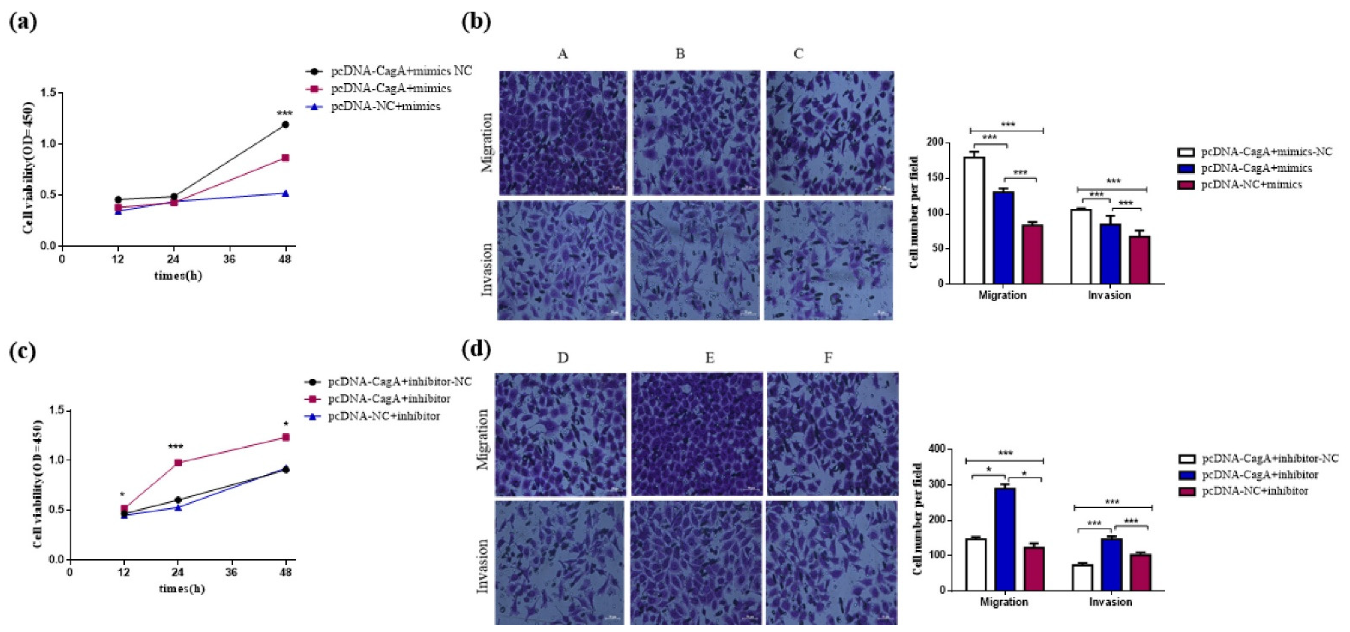
3.3. CagA Promoted the Proliferation, Migration, and Invasion of GC Cells by Regulating miR-155-5p

We designed rescue experiments to explore whether the role of miR-155-5p on AGS could be regulated by CagA. pcDNA-CagA + miR-155-5p mimics/inhibitors NC, pcDNA-CagA + miR-155-5p mimics/inhibitors and pcDNA-NC + miR-155-5p mimics/inhibitors were co-transfected into GC AGS cells. The results showed that the proliferation, migration, and invasion abilities of the pcDNA-CagA + miR-155-5p mimics NC group was significantly higher than those of the pcDNA-CagA + miR-155-5p mimics group, and these abilities of the pcDNA-CagA + miR-155-5p mimics group were significantly higher than those of the pcDNA-NC + miR-155-5p mimics group (Figure 3a,b). When miR-155-5p inhibitor was used, the opposite result was produced (Figure 3c,d). These results indicate that the function of miR-155-5p on GC cells could be influenced by CagA, and the carcinogenic effects of CagA could be assessed using miR-155-5p.

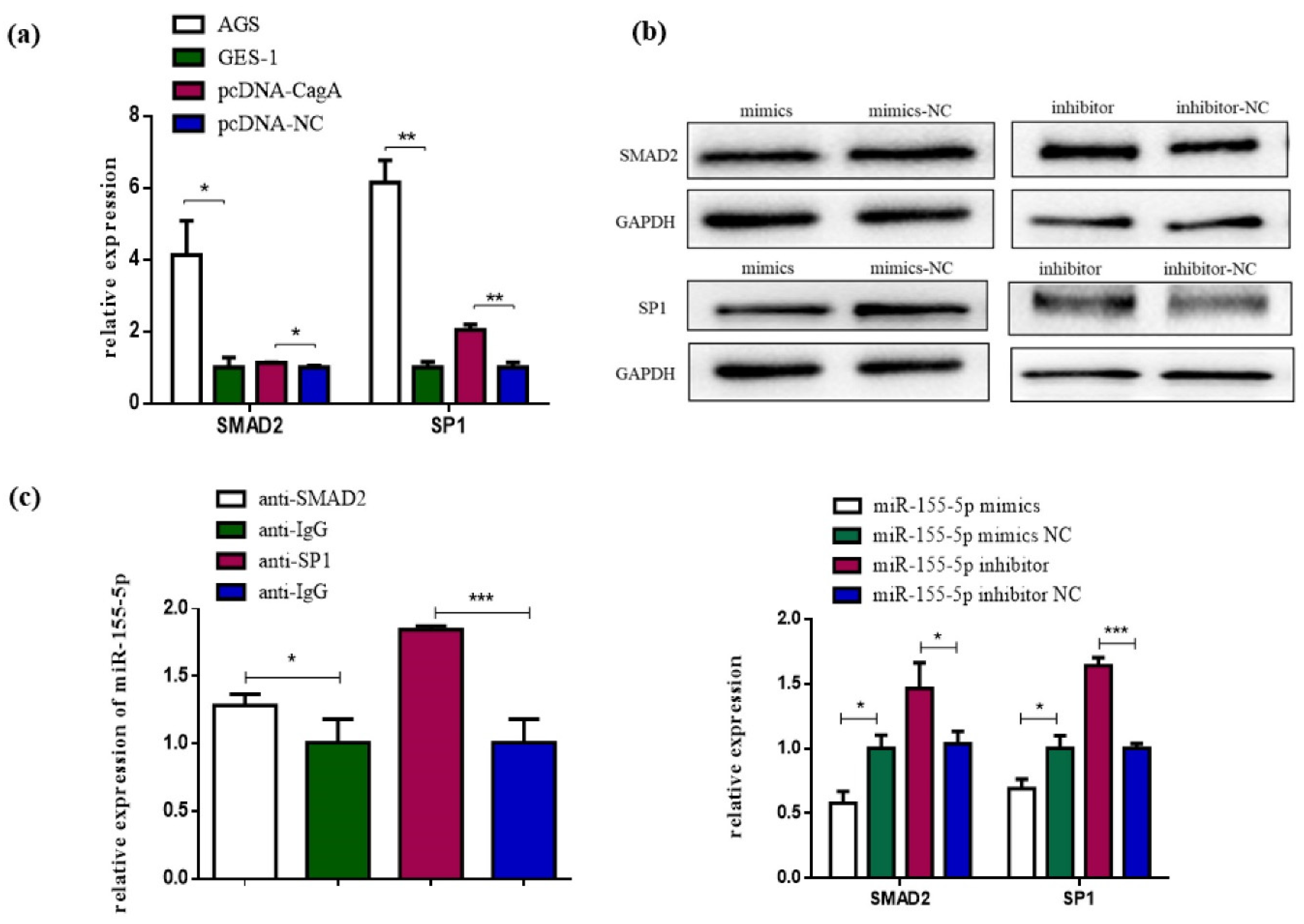
3.4. SP1 and SMAD2 Were Targets of miR-155-5p

The downstream target genes of miR-155-5p were predicted by Targetscan 8.0 [16] (https://www.targetscan.org/vert_80/) (accessed on 1 October 2021), miRDB 6.0 [17] (http://mirdb.org/index.html) (accessed on 1 October 2021). and miRTarbase 9.0 [18](https://mirtarbase.cuhk.edu.cn/) (accessed on 1 October 2021). RAC1, MITF, EGFR, FOS, ETS1, SMAD2, and SP1 were selected for further investigation based on the literature. Further analysis showed that SMAD2 and SP1 were upregulated in AGS cells compared with GES-1 cells, which is in contrast to the expression of miR-155-5p, and only the expression levels of SMAD2 and SP1 were decreased after miR-155-5p overexpression and increased after miR-155-5p knockdown (Figure 4a). Therefore, SMAD2 and SP1 were selected for the next Western blot and RIP experiments.
Using Western blots, we found that the expression levels of SMAD2 and SP1 protein in the miR-155-5p mimics group decreased compared with the miR-155-5p mimics NC group; the expression levels of SMAD2 and SP1 protein in the miR-155-5p inhibitors group were increased compared with the miR-155-5p inhibitors NC group (Figure 4b).
Then, SMAD2/SP1 antibodies were used to perform RIP experiments in order to analyze whether miR-155-5p could interact with SMAD2/SP1 directly. Results showed that the expression levels of miR-155-5p in the SMAD2 and SP1 immunoprecipitation complex were 1.27 and 1.85 times higher than negative control group (anti-IgG), respectively (Figure 4c), which proved that miR-155-5p could directly bind to SMAD2/SP1. Taken together, our results have demonstrated that miR-155-5p regulates SMAD2 and SP1 expression at both the post-transcriptional and protein levels.

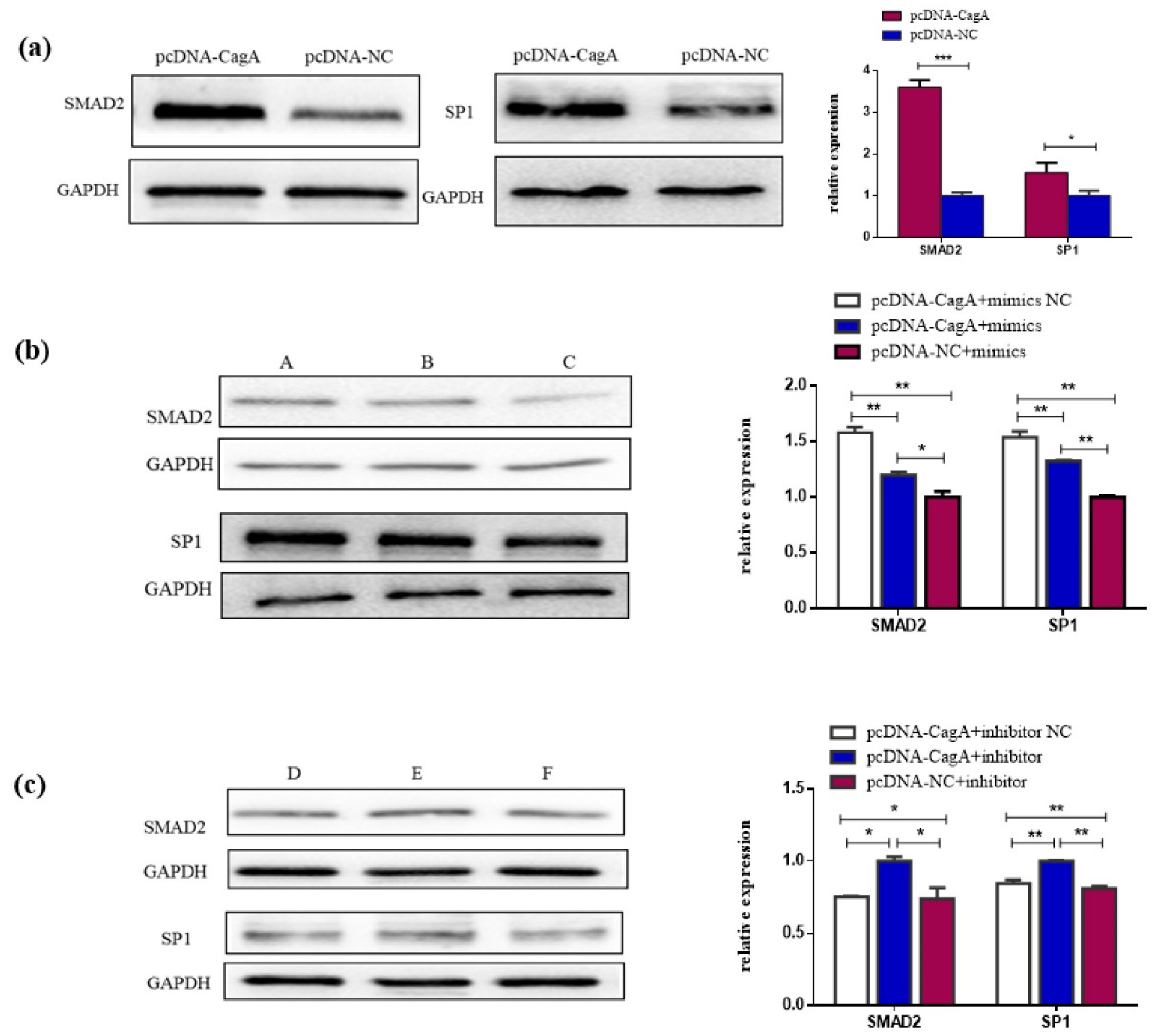
3.5. CagA Promotes the Proliferation, Migration, and Invasion of GC Cells through the miR-155-5p-SP1/SMAD2 axis

Our study found that the overexpression of CagA could increase the expression of SMAD2 and SP1 protein (Figure 5a). Thus, we designed a rescue experiment to explore whether the effects of miR-155-5p on SMAD2 and SP1 were regulated by CagA. A Western blot showed that the expression levels of SMAD2 and SP1 protein in the pcDNA-CagA + miR-155-5p mimics group were significantly decreased compared with the pcDNA-CagA + miR-155-5p mimics NC group (Figure 5b); the expression levels of SMAD2 and SP1 protein in the pcDNA-CagA + miR-155-5p mimics group were significantly higher compared with the pcDNA-NC + miR-155-5p mimics group (Figure 5b). However, when the inhibitor was used, the results were the opposite

4. Discussion

In the process of exploring the carcinogenic ability of Hp, have researchers focused their attention on the virulence gene of Hp, and they agreed that the CagA virulence gene plays an important role in the carcinogenic ability of Hp [19,20,21,22]. miRNA participates in the occurrence and development of GC through interaction with oncogenes, tumor suppressor genes, and other cancer-related genes [23,24].
Our research focused on the relationship between CagA and miRNA and explored the mechanism of carcinogenic effects of CagA through miRNA so as to provide some new ideas for the treatment of patients infected with CagA-positive Hp. The results showed that CagA can promote the cell proliferation, migration, and invasion of GC AGS cells, which indicated that CagA could promote the malignant phenotype of AGS cells. Previous research reported that CagA-positive Hp infection was associated with a worse disease stage and clinical outcome [19,20,21,25], and our study demonstrates the carcinogenic ability of CagA at the cellular level, which was consistent with previous studies.
Then, we found the expression of miR-155-5p was downregulated in GC AGS cells and further downregulated under the action of CagA, which means that miR-155-5p may be a key molecule in the carcinogenic pathway of CagA. Thus, we next verified the function of miR-155-5p. After the overexpression and knockdown of miR-155-5p, it was found that the overexpression of miR-155-5p inhibited the proliferation, migration, and invasion of GC AGS cells, whereas the knockdown of miR-155-5p enhanced the proliferation, migration and invasion of cells, indicating that miR-155-5p may play an important role in promoting cancer progression as an important functional molecule. However, the expression of miR-155-5p is tissue-specific, upregulated in kidney cancer, bladder cancer, and cervical cancer [26,27], and downregulated in liver cancer, atherosclerosis, and GC [12,13,15,28,29], which is consistent with our study, suggesting that miR-155-5p is a molecule which warrants further exploration.
Yang et al. found that CagA could upregulate the expression of miR-223-3p and participate in the progression of GC by reducing the expression of ARID1A [30]. In this study, we found that CagA could cause \further downregulation of miR-155-5p in AGS cells. We also verified the functional role of miR-155-5p on AGS. In order to prove whether CagA plays a carcinogenic role through miR-155-5p, rescue experiments were designed. Results showed that the overexpression of miR-155-5p could inhibit the proliferation, migration, and invasion of AGS cells, which could be restored by the overexpression of CagA; similarly, the knockdown of miR-155-5p could enhance the proliferation, migration, and invasion of AGS cells, which could be enhanced by the overexpression of CagA, which indicates that the function of miR-155-5p on AGS cells was regulated by CagA. Therefore, we speculate that cells being affected by CagA further lead to the downregulation of miR-155-5p, which may promote the occurrence and development of cancer.
The abnormal expression of miRNA could further cause the abnormal expression of downstream target mRNA molecules and promote cancer [31]. In this study, SMAD2 and SP1 were identified as two targets of miR-155-5p by qRT-PCR, RIP, and Western blot experiments. SMAD2 could be activated by the TGF-β1 signaling pathway, which, in turn, promotes KLF8 accumulation, leading to cancer progression in breast cancer [32]. In GC, it was found that the downregulation of the phosphorylation level of SMAD2 significantly inhibited the proliferation and migration ability of GC cells [33]. In addition, Niu et al. found that SMAD2 was a target of miR-155-5p and played an oncogenic role in the progression of cancer [34], which also supports the results of our study and proves its carcinogenic effects. SP1 was highly expressed in GC, and its activation may serve as a biomarker for poor prognosis and contribute to the development of GC [35]. Lei et al. found that transcription factor SP1 could activate PCAT19, promoting GC progression and triggering poor prognosis [36]. It could also activate p62 to decrease autophagy flux and promote cell proliferation in GC cells [37]. In addition, a meta-analysis related to SP1 showed that SP1 expression was significantly higher in GC tissues than in normal gastric mucosa, and was positively correlated with the invasion depth and TNM stage of GC [38]. The above studies showed that SMAD2 and SP1 could induce carcinogenic effects in GC.
In our study, we verified the association of CagA with SMAD2 and SP1 proteins at the protein level via Western blot and proved that miR-155-5p can directly bind SMAD2 and SP1 via an RIP assay. The results showed that the overexpression of miR-155-5p caused the downregulation of SMAD2 protein and SP1 protein levels, which could be partially reverted after the overexpression of CagA. Additionally, the knockdown of miR-155-5p caused the upregulation of expression levels of SMAD2 and SP1 protein, which were enhanced after the overexpression of CagA. These results indicated that CagA could cause the downregulation of miR-155-5p and further upregulation of SMAD2 and SP1 expression, thereby exerting its effect on the proliferation, migration, and invasion ability of GC cells. It is well illustrated that CagA can exert its effects on the proliferation, migration, and invasion abilities of GC cells through the CagA/miR-155-5p/SMAD2/SP1 axis.

5. Conclusions

In summary, this study explored a novel pathway in which CagA caused changes to the function of GC cells. CagA influenced the expression of SMAD2/SP1 protein through miR-155-5p, which, in turn, promoted cell proliferation, migration, and invasion (Scheme 1)
This study associated CagA and miRNA molecules with the function of the development and progression in GC, which is innovation. It is intended to provide a certain theoretical basis for research on the treatment of CagA-associated GC, and also provide a basis for research on the current relevant aspects in the field of GC. However, this study is still far from achieving this goal. There are several shortcomings of this study, which need to be improved in depth. Our study was only conducted based on cell experiments and did not validate the findings at the population level, which is required for the study to achieve greater value. Future research will be performed at the population level to further illustrate this conclusion.

Supplementary Materials

The following supporting information can be downloaded at: https://www.mdpi.com/article/10.3390/pathogens11080846/s1, File S1: Plasmid Synthesis Report; File S2: Sanger sequencing alignment file; Table S1: miR-155-5p interference sequences; Table S2: Sequence-specific primers.

Author Contributions

Conceptualization, F.J. and L.W.; methodology, L.W.; validation, F.J. and L.W.; formal analysis, L.W.; investigation, L.W.; resources, L.W.; data curation, L.W.; writing—original draft preparation, L.W.; writing—review and editing, L.W. and F.J.; visualization, L.W.; supervision, L.W. and F.J.; funding acquisition, X.S. All authors have read and agreed to the published version of the manuscript.

Funding

This research received no external funding.

Institutional Review Board Statement

Not applicable.

Informed Consent Statement

Not applicable.

Data Availability Statement

Not applicable.

Acknowledgments

Thanks to Shen from the Laboratory of Environment and Hygiene of Southeast University for their experimental ideas and technical assistance. Thanks for the financial and emotional support provided by the research group of Shen family class.

Conflicts of Interest

The authors declare no conflict of interest.

References

  1. Ishaq, S.; Nunn, L. Helicobacter pylori and gastric cancer: A state of the art review. Gastroenterol. Hepatol. Bed Bench 2015, 8, 6–14. [Google Scholar]
  2. Plummer, M.; Franceschi, S.; Vignat, J.; Forman, D.; de Martel, C. Global burden of gastric cancer attributable to Helicobacter pylori. Int. J. Cancer 2015, 136, 487–490. [Google Scholar] [CrossRef]
  3. Backert, S.; Tegtmeyer, N. Type IV Secretion and Signal Transduction of Helicobacter pylori CagA through Interactions with Host Cell Receptors. Toxins 2017, 9, 115. [Google Scholar] [CrossRef] [PubMed] [Green Version]
  4. Bravo, D.; Hoare, A.; Soto, C.; Valenzuela, M.A.; Quest, A.F. Helicobacter pylori in human health and disease: Mechanisms for local gastric and systemic effects. World J. Gastroenterol. 2018, 24, 3071–3089. [Google Scholar] [CrossRef] [PubMed]
  5. Plummer, M.; de Martel, C.; Vignat, J.; Ferlay, J.; Bray, F.; Franceschi, S. Global burden of cancers attributable to infections in 2012: A synthetic analysis. Lancet Glob. Health 2016, 4, 609–616. [Google Scholar] [CrossRef] [Green Version]
  6. Zhang, R.G.; Duan, G.C.; Fan, Q.T.; Chen, S.Y. Role of Helicobacter pylori infection in pathogenesis of gastric carcinoma. World J. Gastrointest. Pathophysiol. 2016, 7, 97–107. [Google Scholar] [CrossRef]
  7. Lee, Y.C.; Chen, T.H.; Chiu, H.M.; Shun, C.T.; Chiang, H.; Liu, T.Y.; Wu, M.S.; Lin, J.T. The benefit of mass eradication of Helicobacter pylori infection: A community-based study of gastric cancer prevention. Gut 2013, 62, 676–682. [Google Scholar] [CrossRef] [Green Version]
  8. Baj, J.; Forma, A.; Sitarz, M.; Portincasa, P.; Garruti, G.; Krasowska, D.; Maciejewski, R. Helicobacter pylori Virulence Factors-Mechanisms of Bacterial Pathogenicity in the Gastric Microenvironment. Cells 2020, 10, 27. [Google Scholar] [CrossRef]
  9. Alipour, M. Molecular Mechanism of Helicobacter pylori-Induced Gastric Cancer. J. Gastrointest. Cancer 2021, 52, 23–30. [Google Scholar] [CrossRef]
  10. Xu, Z.; Li, Z.; Wang, W.; Xia, Y.; He, Z.; Li, B.; Wang, S.; Huang, X.; Sun, G.; Xu, J.; et al. MIR-1265 regulates cellular proliferation and apoptosis by targeting calcium binding protein 39 in gastric cancer and, thereby, impairing oncogenic autophagy. Cancer Lett. 2019, 449, 226–236. [Google Scholar] [CrossRef]
  11. Konishi, H.; Sato, H.; Takahashi, K.; Fujiya, M. Tumor-Progressive Mechanisms Mediating miRNA-Protein Interaction. Int. J. Mol. Sci. 2021, 22, 12303. [Google Scholar] [CrossRef] [PubMed]
  12. Li, H.; Xie, S.; Liu, M.; Chen, Z.; Liu, X.; Wang, L.; Li, D.; Zhou, Y. The clinical significance of downregulation of mir-124-3p, mir-146a-5p, mir-155-5p and mir-335-5p in gastric cancer tumorigenesis. Int. J. Oncol. 2014, 45, 197–208. [Google Scholar] [CrossRef] [PubMed] [Green Version]
  13. Zare, A.; Alipoor, B.; Omrani, M.D.; Zali, M.R.; Malekpour Alamdari, N.; Ghaedi, H. Decreased miR-155-5p, miR-15a, and miR-186 Expression in Gastric Cancer Is Associated with Advanced Tumor Grade and Metastasis. Iran. Biomed. J. 2019, 23, 338–343. [Google Scholar] [CrossRef] [Green Version]
  14. Zhu, M.; Wang, M.; Yang, F.; Tian, Y.; Cai, J.; Yang, H.; Fu, H.; Mao, F.; Zhu, W.; Qian, H.; et al. miR-155-5p inhibition promotes the transition of bone marrow mesenchymal stem cells to gastric cancer tissue derived MSC-like cells via NF-κB p65 activation. Oncotarget 2016, 7, 16567–16580. [Google Scholar] [CrossRef] [Green Version]
  15. Li, S.; Zhang, T.; Zhou, X.; Du, Z.; Chen, F.; Luo, J.; Liu, Q. The tumor suppressor role of miR-155-5p in gastric cancer. Oncol. Lett. 2018, 16, 2709–2714. [Google Scholar] [CrossRef] [Green Version]
  16. McGeary, S.E.; Lin, K.S.; Shi, C.Y.; Pham, T.M.; Bisaria, N.; Kelley, G.M.; Bartel, D.P. The biochemical basis of microRNA targeting efficacy. Science 2019, 366, eaav1741. [Google Scholar] [CrossRef] [PubMed]
  17. Chen, Y.; Wang, X. miRDB: An online database for prediction of functional microRNA targets. Nucleic Acids Res. 2020, 48, 127–131. [Google Scholar] [CrossRef] [PubMed] [Green Version]
  18. Huang, H.Y.; Lin, Y.C.; Cui, S.; Huang, Y.; Tang, Y.; Xu, J.; Bao, J.; Li, Y.; Wen, J.; Zuo, H.; et al. miRTarBase update 2022: An informative resource for experimentally validated miRNA-target interactions. Nucleic Acids Res. 2022, 50, 222–230. [Google Scholar] [CrossRef]
  19. Ansari, S.; Yamaoka, Y. Helicobacter pylori Virulence Factor Cytotoxin-Associated Gene A (CagA)-Mediated Gastric Pathogenicity. Int. J. Mol. Sci. 2020, 21, 7430. [Google Scholar] [CrossRef]
  20. He, H.; Liu, J.; Li, L.; Qian, G.; Hao, D.; Li, M.; Zhang, Y.; Hong, X.; Xu, J.; Yan, D. Helicobacter pylori CagA Interacts with SHP-1 to Suppress the Immune Response by Targeting TRAF6 for K63-Linked Ubiquitination. J. Immunol. 2021, 206, 1161–1170. [Google Scholar] [CrossRef]
  21. Satoshi, I.; Takuya, O.; Naoko, M.K.; Daisuke, K.; Kamrunnesa, T.; Weida, W.; Atsushi, T.K.; Takaya, K.C.; Akiko, K.; Nobumi, S.; et al. Helicobacter pylori CagA elicits BRCAness to induce genome instability that may underlie bacterial gastric carcinogenesis. Cell Host Microbe 2021, 29, 941–958. [Google Scholar]
  22. Lin, L.; Wei, H.; Yi, J.; Xie, B.; Chen, J.; Zhou, C.; Wang, L.; Yang, Y. Chronic CagA-positive Helicobacter pylori infection with MNNG stimulation synergistically induces mesenchymal and cancer stem cell-like properties in gastric mucosal epithelial cells. J. Cell Biochem. 2019, 120, 17635–17649. [Google Scholar] [CrossRef] [PubMed]
  23. Li, W.; Li, J.; Mu, H.; Guo, M.; Deng, H. MiR-503 suppresses cell proliferation and invasion of gastric cancer by targeting HMGA2 and inactivating WNT signaling pathway. Cancer Cell Int. 2019, 19, 164. [Google Scholar] [CrossRef] [Green Version]
  24. Shi, Y.; Huang, X.; Chen, G.; Wang, Y.; Liu, Y.; Xu, W.; Tang, S.; Guleng, B.; Liu, J.; Ren, J. miR-632 promotes gastric cancer progression by accelerating angiogenesis in a TFF1-dependent manner. BMC Cancer 2019, 19, 14. [Google Scholar] [CrossRef]
  25. Nicole, T.; Aileen, H.; Klemens, R.; Steffen, B. Helicobacter pylori CagA Induces Cortactin Y-470 Phosphorylation-Dependent Gastric Epithelial Cell Scattering via Abl, Vav2 and Rac1 Activation. Cancers 2021, 13, 4241. [Google Scholar]
  26. Lei, Q.Q.; Huang, Y.; Li, B.; Han, L.; Lv, C. MiR-155-5p promotes metastasis and epithelial-mesenchymal transition of renal cell carcinoma by targeting apoptosis-inducing factor. Int. J. Biol. Markers 2021, 36, 20–27. [Google Scholar] [CrossRef] [PubMed]
  27. Li, N.; Cui, T.; Guo, W.; Wang, D.; Mao, L. MiR-155-5p accelerates the metastasis of cervical cancer cell via targeting TP53INP1. Onco Targets Ther. 2019, 12, 3181–3196. [Google Scholar]
  28. Wang, J.; Che, J. CircTP63 promotes hepatocellular carcinoma progression by sponging miR-155-5p and upregulating ZBTB18. Cancer Cell Int. 2021, 21, 156. [Google Scholar] [CrossRef] [PubMed]
  29. Chen, L.; Zheng, S.Y.; Yang, C.Q.; Ma, B.M.; Jiang, D. MiR-155-5p inhibits the proliferation and migration of VSMCs and HUVECs in atherosclerosis by targeting AKT1. Eur. Rev. Med. Pharmacol. Sci. 2019, 23, 2223–2233. [Google Scholar] [PubMed]
  30. Yang, F.; Xu, Y.; Liu, C.; Ma, C.; Zou, S.; Xu, X.; Jia, J.; Liu, Z. NF-κB/miR-223-3p/ARID1A axis is involved in Helicobacter pylori CagA-induced gastric carcinogenesis and progression. Cell Death Dis. 2018, 9, 12. [Google Scholar] [CrossRef] [Green Version]
  31. Chen, C.Z. MicroRNAs as oncogenes and tumor suppressors. N. Engl. J. Med. 2005, 353, 1768–1771. [Google Scholar] [CrossRef] [PubMed] [Green Version]
  32. Cherukunnath, A.; Davargaon, R.S.; Ashraf, R.; Kamdar, U.; Srivastava, A.K.; Tripathi, P.P.; Chatterjee, N.; Kumar, S. KLF8 is activated by TGF-β1 via Smad2 and contributes to ovarian cancer progression. J. Cell. Biochem. 2022, 123, 921–934. [Google Scholar] [CrossRef] [PubMed]
  33. Zhou, Z.; Gao, W.; Yuan, B.; Zhang, S.; Wang, K.; Du, T. TRIM22 inhibits the proliferation of gastric cancer cells through the Smad2 protein. Cell Death Discov. 2021, 7, 234. [Google Scholar] [CrossRef] [PubMed]
  34. Niu, Q.; Dong, Z.; Liang, M.; Luo, Y.; Lin, H.; Lin, M.; Zhong, X.; Yao, W.; Weng, J.; Zhou, X. Circular RNA hsa_circ_0001829 promotes gastric cancer progression through miR-155-5p/SMAD2 axis. J. Exp. Clin. Cancer Res. 2020, 39, 280. [Google Scholar] [CrossRef] [PubMed]
  35. Zhang, X.; Yang, H.; Jia, Y.; Xu, Z.; Zhang, L.; Sun, M.; Fu, J. circRNA_0005529 facilitates growth and metastasis of gastric cancer via regulating miR-527/Sp1 axis. BMC Mol. Cell Biol. 2021, 22, 6. [Google Scholar] [CrossRef] [PubMed]
  36. Xiao, L.; Yuan, W.; Huang, C.; Luo, Q.; Xiao, R.; Chen, Z.H. LncRNA PCAT19 induced by SP1 and acted as oncogene in gastric cancer competitively binding to miR429 and upregulating DHX9. J. Cancer 2022, 13, 102–111. [Google Scholar] [CrossRef]
  37. Xu, X.W.; Pan, C.W.; Yang, X.M.; Zhou, L.; Zheng, Z.Q.; Li, D.C. SP1 reduces autophagic flux through activating p62 in gastric cancer cells. Mol. Med. Rep. 2018, 17, 4633–4638. [Google Scholar] [CrossRef] [Green Version]
  38. Shi, S.; Zhang, Z.G. Role of Sp1 expression in gastric cancer: A meta-analysis and bioinformatics analysis. Oncol. Lett. 2019, 18, 4126–4135. [Google Scholar] [CrossRef] [Green Version]
Figure 1. The expression of CagA in AGS cells and the effects of CagA overexpression on the biological characteristics of GC AGS cells. (a) qRT-PCR analysis of CagA mRNA expression after treatment with pcDNA-CagA and pcDNA-NC. (b) Western blot analysis of CagA. (c) Assessment of the proliferation of AGS cells transfected with pcDNA-CagA and pcDNA-NC by CCK-8 assay. (d) Wound healing assay to detect the effect of CagA on cell migration. (e) CagA overexpression regulated the migration and invasion of AGS cells. ** p < 0.01; *** p < 0.001; **** p < 0.0001.
Pathogens 11 00846 g001
Figure 2. The expression of miR-155-5p in AGS cells and the effects of overexpression and knockdown of miR-155-5p on the biological characteristics of AGS. (a,b) qRT-PCR analysis of miR-155-5p mRNA expression after treatment with pcDNA-CagA, miR-155-5p mimics and inhibitor regents. (c,d) Assessment of the proliferation of AGS cells transfected with miR-155-5p mimics and inhibitors by CCK-8 assay. (e) Wound healing assay to detect the effect of miR-155-5p on cell migration. (f) miR-155-5p overexpression and knockdown regulated the migration and invasion of AGS cells. * p < 0.05; ** p < 0.01; *** p < 0.001; **** p < 0.0001.
Pathogens 11 00846 g002
Figure 3. CagA regulates malignant transformation through miR-155-5p in vitro. (a,b) Co-transfection of miR-155-5p mimics and pcDNA-CagA to investigate malignant transformation of cells with CCK-8 and Transwell assays in the AGS cell line. (c,d) Co-transfection of miR-155-5p inhibitor and pcDNA-CagA to investigate malignant transformation of cells with CCK-8 and Transwell assays in the AGS cell line A: pcDNA-CagA + miR-155-5p mimics NC; B: pcDNA-CagA + miR-155-5p mimics; C: pcDNA-NC + miR-155-5p mimics; D: pcDNA-CagA + miR-155-5p inhibitors NC; E: pcDNA-CagA + miR-155-5p inhibitors; F: pcDNA-NC + miR-155-5p inhibitors; * p < 0.05; *** p < 0.001.
Pathogens 11 00846 g003
Figure 4. SMAD2 and SP1 were targets of miR-155-5p. (a) qRT-PCR of SMAD2 and SP1 expression in AGS cells transfected with miR-155-5p mimics and inhibitor. (b) RIP assays with anti-SMAD2, anti-SP1, and anti-IgG antibodies in AGS cell lines were used to detect miR-155-5p mRNA levels. (c) Western blot analysis of SMAD2 and SP1 transfected with miR-155-5p mimics and inhibitor. * p < 0.05; ** p < 0.01; *** p < 0.001.
Pathogens 11 00846 g004
Figure 5. CagA regulates malignant transformation through the miR-155-5p/SMAD2/SP1 axis in vitro. (a) Western blot analysis of SMAD2 and SP1 transfected with pcDNA-CagA. (b) Co-transfection of miR-155-5p mimics and pcDNA-CagA to investigate the level of SMAD2 and SP1 expression with Western blots in the AGS cell line. (c). Co-transfection of miR-155-5p inhibitors and pcDNA-CagA to investigate the levels of SMAD2 and SP1 expression with Western blots in the AGS cell line A: pcDNA-CagA + miR-155-5p mimics NC; B: pcDNA-CagA + miR-155-5p mimics; C: pcDNA-NC + miR-155-5p mimics; D: pcDNA-CagA + miR-155-5p inhibitors NC; E: pcDNA-CagA + miR-155-5p inhibitors; F: pcDNA-NC + miR-155-5p inhibitor; * p < 0.05; ** p < 0.01; *** p < 0.001.
Pathogens 11 00846 g005
Scheme 1. General diagram summarizing the discovery and pathway.
Pathogens 11 00846 sch001
Publisher’s Note: MDPI stays neutral with regard to jurisdictional claims in published maps and institutional affiliations.

