Article

Cobalt (II) Chloride Regulates the Invasion and Survival of Brucella abortus 544 in RAW 264.7 Cells and B6 Mice

1
Institute of Applied Sciences, HUTECH University, 475A Dien Bien Phu St., Ward 25, Binh Thanh District, Ho Chi Minh City 72300, Vietnam
2
Institute of Animal Medicine, College of Veterinary Medicine, Gyeongsang National University, Jinju 52828, Korea
3
Department of Veterinary Paraclinical Sciences, College of Veterinary Medicine, University of the Philippines Los Baños, College, Laguna 4031, Philippines
4
College of Veterinary Medicine, Chonbuk National University, Iksan 54596, Korea
*
Author to whom correspondence should be addressed.
Pathogens 2022, 11(5), 596; https://doi.org/10.3390/pathogens11050596
Submission received: 18 April 2022 / Revised: 14 May 2022 / Accepted: 15 May 2022 / Published: 18 May 2022

Abstract

:
The effects of Cobalt (II) chloride (CoCl2) in the context of Brucella abortus (B. abortus) infection have not been evaluated so far. Firstly, we found that CoCl2 treatment inhibited the phagocytosis of B. abortus into RAW 264.7 cells. The inhibition of bacterial invasion was regulated by F-actin formation and associated with a reduction in the phosphorylation of ERK1/2 and HIF-1α expression. Secondly, the activation of trafficking regulators LAMP1, LAMP2, and lysosomal enzyme GLA at the transcriptional level activated immune responses, weakening the B. abortus growth at 4 h post-infection (pi). The silencing of HIF-1α increased bacterial survival at 24 h pi. At the same time, CoCl2 treatment showed a significant increase in the transcripts of lysosomal enzyme HEXB and cytokine TNF-α and an attenuation of the bacterial survival. Moreover, the enhancement at the protein level of HIF-1α was induced in the CoCl2 treatment at both 4 and 24 h pi. Finally, our results demonstrated that CoCl2 administration induced the production of serum cytokines IFN-γ and IL-6, which is accompanied by dampened Brucella proliferation in the spleen and liver of treated mice, and reduced the splenomegaly and hepatomegaly. Altogether, CoCl2 treatment contributed to host resistance against B. abortus infection with immunomodulatory effects.

1. Introduction

Brucella abortus (B. abortus) is one of the twelve currently recognized Brucella species and causes disease in cattle. Additionally, the other Brucella species also have different preferential host specificities, including B. melitensis (goat and sheep); B. suis (pig, hare, reindeer, caribou, and rodent); B. ovis (sheep); B. neotomae (desert rat); B. canis (dog); B. ceti (dolphins); B. pinnipedialis (seals); B. microti (wild voles); B. inopinata (human); B. papionis (baboons); and B. vulpis (red foxes) [1]. They are the causative agents of brucellosis, a common zoonosis in the world. Among them, B. inopinata, B. abortus, B. suis, and B. melitensis are the most pathogenic species in humans. Both human and animal brucellosis have caused negatively severe medical and economic impacts [2,3]. These impacts are due to the stealthy strategies of Brucella to provoke a systemic infection, resulting in the dissemination of bacteria to different organs, tissues, and cells. Moreover, the most crucial virulence determinant, the Type IV secretion system (T4SS), facilitates the successful evasion of the host bactericidal activity and the establishment of an intracellular replicative niche, leading to chronic infection. For these reasons, the diagnosis, prevention, and treatment of brucellosis have become complicated and challenging [4,5].
Cobalt is well-known as a core element of vitamin B12 and an essential trace element in the human diet. It plays a crucial role in forming enzymes such as methionine aminopeptidase in bacteria and yeast and nitrile hydratase in actinomycetes and bacteria [6,7]. In addition to causing harmful impacts such as lipemia, cardiomyopathy, and hard metal asthma of long-term exposure to a high concentration, Cobalt (II) chloride (CoCl2) can display advantageous effects in anemia treatment, fracture healing in mice, and anti-Helicobacter pylori (H. pylori) activity [8,9,10]. Recently, many studies have reported the stabilization of transcription factor HIF-1α induced by CoCl2 by a proven mechanism to mimic hypoxia. Notably, HIF-1α functions as a central regulator of oxygen and energy homeostasis, cellular metabolism, and host immunity [11,12]. A study by Zenk et al. [13] demonstrated that a stabilized HIF-1α decreased the intracellular growth of Mycobacterium tuberculosis. Furthermore, another study by Hwang et al. [14] showed an enhancement of antiviral activity associated with CoCl2-stabilized HIF-1α in a T98G cell line.
Briefly, the prevention and treatment of brucellosis have been based on vaccines and antibiotics. Herein, the antibiotic therapy is often empiric and relies on the choice of treatment regimen and duration of various antibiotics such as doxycycline, aminoglycosides, rifampicin, minocycline, trimethoprim, sulfamethoxazole, and quinolone [15]. Brucella are intracellular bacteria and reside in intracellular compartments that require cell-penetrating antibiotics to eliminate. However, this kind of antibiotic remains challenging, including retention and accumulation in host cells and penetration into bacteria-containing compartments [16]. Additionally, although live attenuated vaccine strains, including B. abortus S19, B. abortus RB51, and B. melitensis Rev.1, are effective against brucellosis in cattle and small ruminants, they still have some drawbacks [17]. Recently, immunotherapy has emerged as a potential alternative for bacterial infection treatment, especially for intracellular bacteria [18]. Delving deeper into understanding the defensive mechanisms of host immunity and cellular effectors is a promising approach in immunotherapy. Recently, CoCl2 has emerged as a promising therapeutic agent for the prevention and treatment of various cancers, as well as inflammatory diseases [19,20,21]. In this study, we carried out in vitro and in vivo experiments in RAW 264.7 cells and B6 mice, respectively, using CoCl2 as a stimulator of the host immune responses against B. abortus infection.

2. Results

2.1. CoCl2 Treatment Inhibited the Invasion of B. abortus into RAW 264.7 Macrophage Cells and Affected Brucella survival in Axenic Culture

The 3-(4,5-dimethylthiazol-2yl)-2,5-diphenyl-2H-tetrazolium bromide (MTT) assay was performed to evaluate the cytotoxic effect of CoCl2 on RAW 264.7 cells. The 10 mg/mL, 1 mg/mL, and 100 µg/mL concentrations of CoCl2 significantly induced cytotoxicity in the cells, whereas all lower concentrations had no cytotoxic effect compared to the control cells (Figure 1A). Therefore, the highest noncytotoxic concentration of CoCl2 (10 µg/mL) was used for the subsequent experiments. The result of the bacterial internalization assay showed that, at the early stage of infection, the number of bacteria in the CoCl2-treated cells was lower than in the control cells but not significant. However, at both 30 and 45 min after infection, this reduction became significant (Figure 1B). At the same time, the immunoblotting analysis demonstrated that CoCl2 treatment led to a decrease in the F-actin formation and phosphorylation of ERK1/2 in B. abortus-infected cells compared to the nontreated infected control cells at 30 min post-infection (pi). Notably, CoCl2 treatment induced a marked expression of HIF-1α compared to the untreated cells. In addition, Brucella infection reduced the HIF-1α expression in CoCl2-treated cells compared to uninfected cells (Figure 1C,D). Moreover, in an axenic culture condition containing sufficient nutrients to maintain Brucella viability during the experimental period, the survival rates of Brucella decreased significantly at all concentrations of CoCl2 at 24 h and in higher concentrations (10, 100, and 1000 µg/mL) at 48 h post-incubation (Figure 1E). This result demonstrated the direct bactericidal activity of CoCl2 against Brucella.

2.2. CoCl2 Treatment Affected the Survival of B. abortus in RAW 264.7 Macrophage Cells at 4 h pi

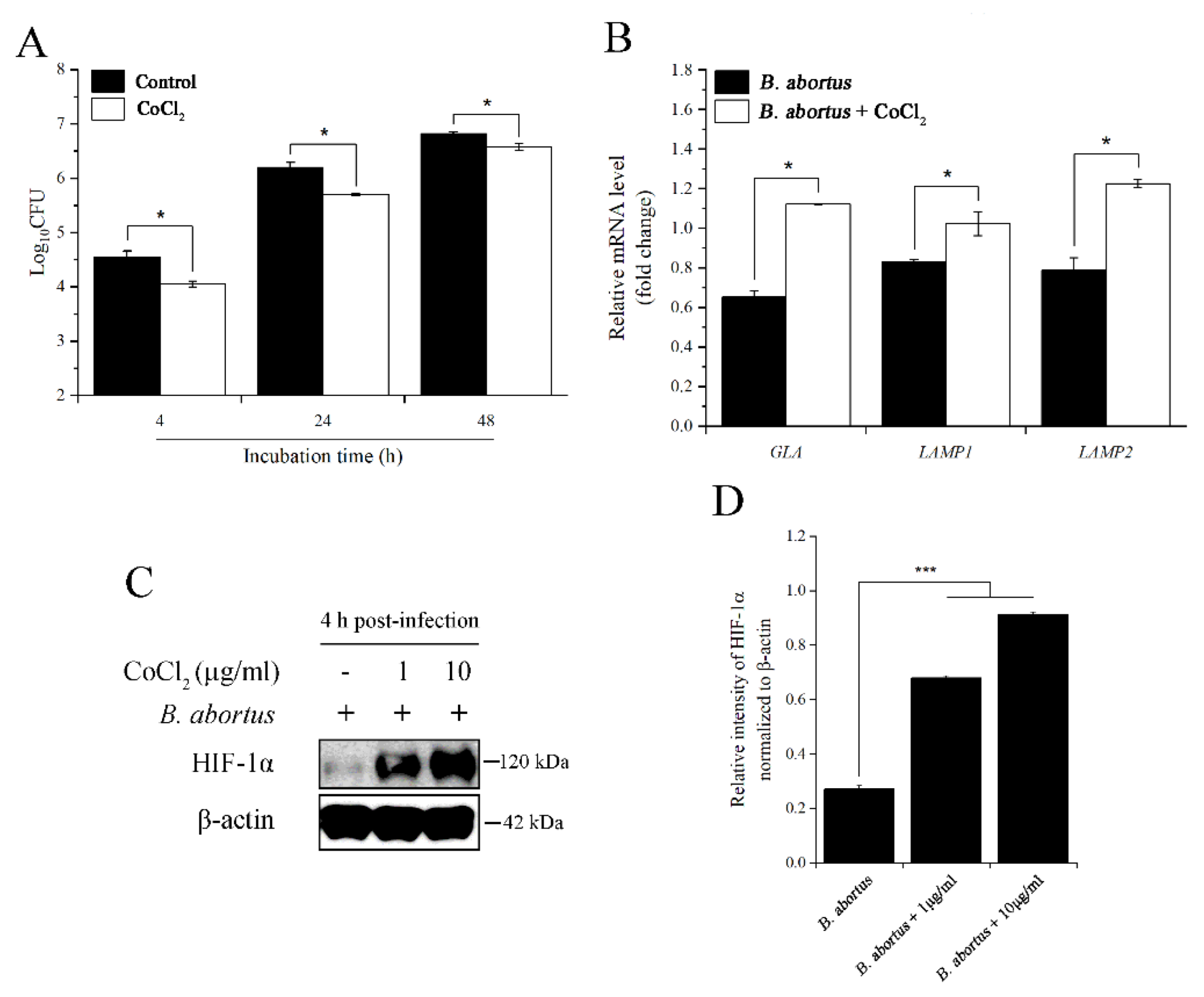
The bacterial intracellular growth assay result displayed a significant reduction in bacterial survival at 4, 24, and 48 h pi in CoCl2-treated cells compared to the control cells (Figure 2A). To clarify this potential role of CoCl2 in macrophage cells at the early stage of infection, we utilized quantitative real-time PCR (qRT-PCR) to analyze the expression of the genes related to phagolysosome fusion, which is considered a central effector in brucellacidal activity [22]. CoCl2 treatment elevated the expression of two trafficking regulators, LAMP1 and LAMP2, and lysosomal enzyme GLA upon B. abortus infection (Figure 2B). Additionally, the results showed that CoCl2 treatment increased the expression of the HIF-1α protein at concentrations of 1 and 10 µg/mL (Figure 2C,D). This transcription factor has been associated with a wide range of diseases and host immunity [23].

2.3. CoCl2 and HIF-1α Affected the Survival of B. abortus in RAW 264.7 Macrophage Cells at 24 h pi

As shown in Figure 2A, CoCl2 treatment decreased the intracellular growth of B. abortus at all three examined time points. Thus, we next checked how CoCl2 induced host immune responses to eliminate B. abortus at 24 h pi. Proinflammatory cytokine TNF-α and lysosomal enzyme HEXB effectively contribute to bactericidal activity at this time point of infection [24,25]. Herein, the transcriptional profiles of TNF-α and HEXB were evaluated using qRT-PCR. CoCl2 treatment upregulated the expression of these genes during B. abortus infection compared to the untreated control (Figure 3A). Furthermore, similar to the early time point (4 h) of infection, CoCl2 stabilized the HIF-1α protein, proven by immunoblotting assay in both concentrations of CoCl2-treated cells (Figure 3B,C). Interestingly, even when the cells were not treated with CoCl2, the HIF-1α protein was slightly activated upon B. abortus infection. This transcription factor was probably involved in the antimicrobial effect of macrophage cells. To clarify this, we knocked down this gene in RAW 264.7 cells by using a siRNA transfection. After 48 h post-transfection with HIF-1α siRNA, the results displayed an effective reduction in the relative mRNA level of this gene compared to the PBS and negative siRNA control (Figure 3D). After an effective silencing, we then checked its effect on the intracellular growth of B. abortus in macrophage cells. HIF-1α-silenced cells slightly increased the number of bacteria at 24 h pi compared to both control cells, while there was no difference in the number of bacteria at 4 h pi upon HIF-1α knockdown (Figure 3E).

2.4. CoCl2 Administration Mediated Host Immunity against B. abortus Infection in Mice

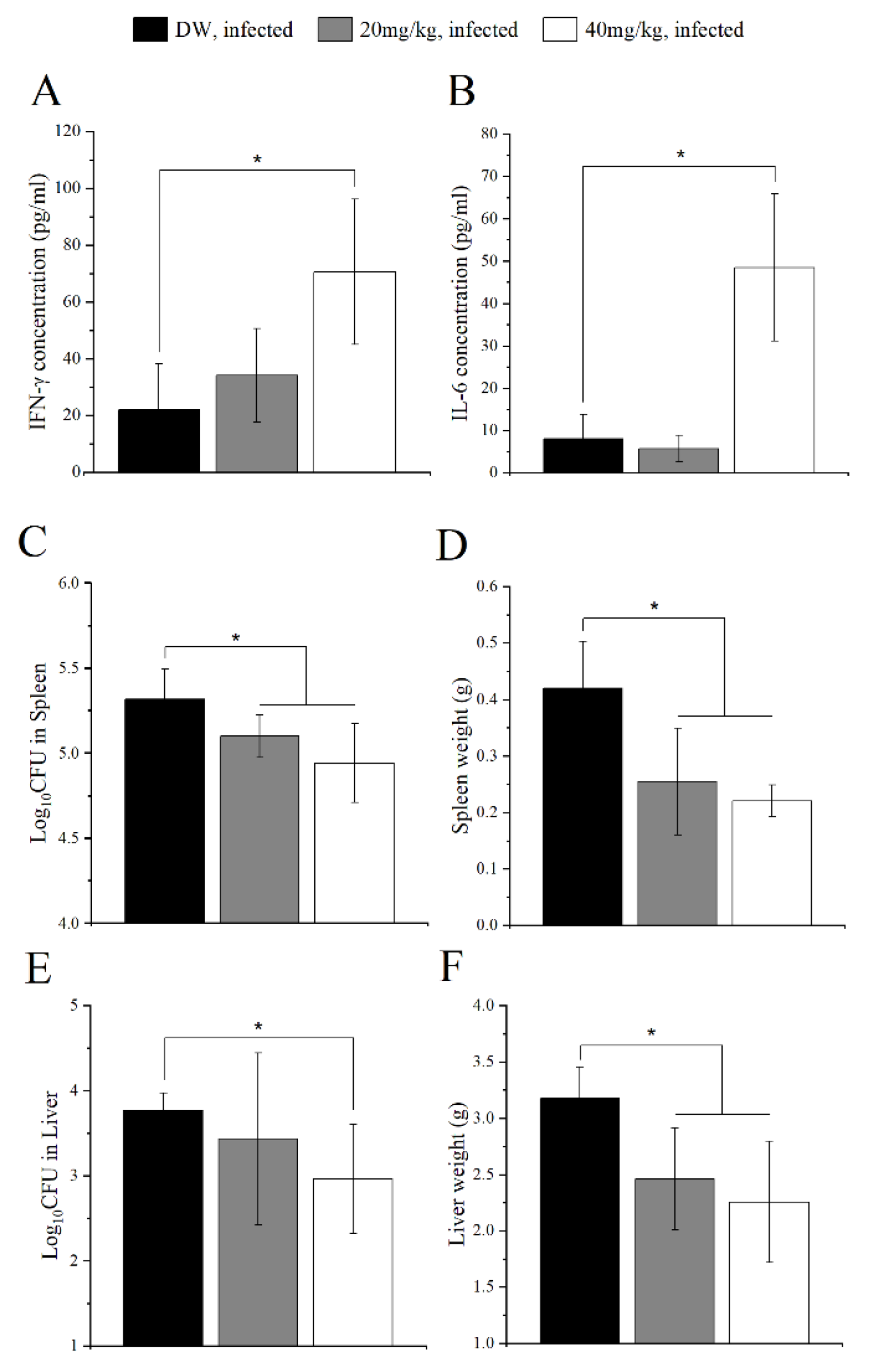
Following promising results in in vitro experiments, it is necessary to further clarify the role of CoCl2 in an in vivo examination using a mouse model in the context of B. abortus infection. Mice were administered with two concentrations of CoCl2 (20 and 40 mg/kg/day). On day 14 after infection, the serum cytokines measurement was processed for evaluating the host immune responses. Cytokines IFN-γ and IL-6 play essential roles in killing Brucella. Our results showed a significant increase in the production of IFN-γ and IL-6 in higher concentrations of the CoCl2-treated group (Figure 4A,B). Moreover, both treatment groups displayed a lower bacterial load in the spleen and the spleen weight than the control group (Figure 4C,D). CoCl2 treatment at a high concentration also decreased the bacterial load in the liver and liver weight compared to the control group. These results indicated the effect of CoCl2 on mediating the host immunity and protecting against B. abortus infection.

3. Discussion

Macrophage is known as the first line of innate immunity. An outstanding function of macrophages is phagocytosis, by which they can ingest foreign particles or pathogenic microorganisms. Brucella are intracellular bacteria, and the invasion into macrophages is considered a critical determinant of their intracellular growth fate [26]. This is initiated by the interaction of Brucella with macrophage membranes through lipid rafts via TLR4. Furthermore, the intracellular circulation of Brucella has entirely fulfilled in autophagic Brucella-containing vacuoles as the end point where it can be released to continue infecting other host cells [27]. This current study showed that CoCl2 treatment reduced the phagocytosis of B. abortus into RAW 264.7 macrophage cells. To investigate how CoCl2 could mediate this event, we evaluated the expression of regulators related to macrophage phagocytosis, including F-actin and ERK1/2 MAPK. The TLR4/ERK1/2 MAPK/F-actin axis is necessary for Brucella internalization and has been exploited in many previous studies, even in the infection of other bacteria [28,29,30]. A study done by Murata et al. [31] displayed the impact of CoCl2 treatment on the disruption of F-actin in both HepG2 and PLC/PRF/5 cell lines. Similarly, our data showed a reduction in F-actin expression and the phosphorylation of ERK1/2 at 30 min pi. Notably, although CoCl2 stabilizes the transcription factor HIF-1α in normoxic conditions, we found that the expression of HIF-1α was decreased in CoCl2-treated cells during Brucella infection at the same examined time point. HIF-1α is linked to macrophage phagocytosis via p38 MAPK activation and the activated TLR4/ERK1/2 MAPK/NF-ĸB pathway [32,33]. In addition, a study by Shweta et al. [34] showed that CoCl2 treatment reduced the expression of TLR4, which is the crucial receptor for Brucella invasion into macrophages. Therefore, our results suggested that CoCl2 reduced Brucella uptake through the MAPK signaling pathway, which is probably related to the stabilization of HIF-1α. A comprehensive evaluation using gene silencing or other molecular methods would provide further insight into the probable role of HIF-1α in Brucella invasion into macrophages.
After successful internalization, the phagosomes containing Brucella fuse with lysosomes. The phagolysosome fusion is one of the macrophage brucellacidal activities that contributes to a 90% reduction of Brucella viability at the early stage of infection. However, this is a struggle for survival; hence, Brucella has acquired crafty evasive strategies to prevent this event that facilitates its intracellular growth [35]. The T4SS system of Brucella successfully accomplishes its role as a critical virulence factor by releasing a variety of effectors that facilitate trafficking and arrival at the endoplasmic reticulum and prevents the host killing mechanism by phagolysosome fusion [36]. Strikingly, CoCl2 treatment decreased the intracellular growth of B. abortus at all examined time points. As mentioned above, phagolysosome fusion plays a vital role in Brucella elimination. Trafficking regulators LAMP1 and LAMP2, as well as lysosomal enzymes GLA and HEXB, are critical markers for this event [25,37]. In addition to these potential weapons, macrophages can secrete cytokines, especially TNF-α, to regulate various bactericidal effectors [24]. For this reason, we sought to evaluate their expression in CoCl2 treatment during B. abortus infection. Our results displayed an increase in the transcripts of GLA, LAMP1, and LAMP2 at an early time point, as well as HEXB and TNF-α at 24 h pi. These findings agree with the previous studies that showed an enhancement of LAMP1 colocalization in HepG2 cells and TNF-α production in BV2 cells [38,39]. As proven earlier, HIF-1α was considered to relate to macrophage phagocytosis. Thus, we next elucidate the involvement of HIF-1α expression in B. abortus intracellular growth. HIF-1α has emerged as a potential key to improving host immune responses against intracellular bacterial, fungal, and protozoan pathogens. In particular, HIF-1α-deficient mice enhanced the survival of Listeria monocytogenes (L. monocytogenes) and Mycobacterium avium (M. avium) in the liver [40]. Moreover, a study done by Li et al. [41] showed that HIF-1α promoted a macrophage inflammatory response by producing cytokine TNF-α during L. monocytogenes infection, and CoCl2 treatment stabilized HIF-1α expression, as well as induced TNF-α production, upon Candida albicans yeast infection. This is consistent with our results of the CoCl2 treatment at 24 h pi. In addition, HIF-1α silencing slightly increased B. abortus survival at this tested time point. On the other hand, our data about the expression of the HIF-1α protein is notable. CoCl2 treatment increased the HIF-1α proteins at 4 and 24 h during B. abortus infection. CoCl2 is well-known to stabilize HIF-1α at the post-translational level by inhibiting the propyl hydroxylase, which is mainly responsible for HIF-1α degradation in the presence of oxygen [42]. CoCl2 was also proven to enhance the NF-ĸB activity, leading to increasing the expression of proinflammatory mediators such as IL-6 and iNOS, which are beneficial in controlling Brucella abortus [43]. Moreover, NF-ĸB was demonstrated to be a direct modulator of HIF-1α transcription [44]. These results strengthened the role of CoCl2 and the distribution of HIF-1α in the macrophage defense against intracellular bacterial infection. This effect of CoCl2 is probably related to the transcription factor NF-ĸB.
On the other hand, Cobalt (II) has been considered a potential ligand to form complexes with that have antimicrobial properties. The novel Cobalt (II) complexes with various antibiotics displayed an extensive range of antimicrobial activity against many pathogens [45,46]. Moreover, CoCl2 was examined directly as a specific antimicrobial agent against H. pylori through competition with nickel ions [10]. Likewise, we first reported that CoCl2 displayed a direct inhibitory effect against B. abortus. This is possibly due to the inhibition of the synthesis of cyanide-sensitive oxidative enzymes [47]. This result raised the question of whether CoCl2 could be a potential alternative strategy to control animal brucellosis. Together with obtaining the other promising in vitro results, we next treated mice with CoCl2 at concentrations of 20 and 40 mg/kg/day, demonstrating the acquisition of a hypoxia-like condition [48]. CoCl2 was involved in the induction of interferon and IL-6 production. More evidence has proposed a functional role in activating the host immunity of proinflammatory cytokines IFN-γ and IL-6 in the control of Brucella infection [49,50,51,52]. In this study, CoCl2 administration in mice elevated the production of IFN-γ and IL-6, leading to reducing the bacterial load in the spleen and liver. In addition, splenomegaly and hepatomegaly observed in Brucella-infected mice were reduced in CoCl2-treated mice. Moreover, HIF-1α has been implicated in the protection against M. avium and L. monocytogenes in a mouse model through the activation of immune effectors, which is beneficial in controlling intracellular bacteria [40,41].

4. Materials and Methods

4.1. Cell Culture and Bacterial Growth Condition

Murine macrophage RAW 264.7 cells (ATCC, TIB-71) were cultured at 37 °C in 5% CO2 atmosphere in RPMI 1640 medium (Gibco, 11875119) containing 10% (v/v) heat-inactivated fetal bovine serum (FBS) (Gibco, 1600-044) with or without 1% antibiotics (Gibco, 15140122). For the following assays, the cells were seeded at concentrations of 3 × 10⁴ and 6 × 10⁵ cells per well in 96-well and 6-well cell culture plates, respectively, in a culture medium containing RPMI plus 10% FBS. The smooth, virulent, wild-type B. abortus 544 biovar 1 strain (ATCC 23448) was cultured in Brucella broth (BBL BD, USA) at 37 °C until the stationary phase or Brucella broth containing 1.5% agar, followed by three days incubation for colony-forming unit (CFU) counting.

4.2. CoCl2 Solution Preparation and Cell Viability Assessment Assay

CoCl2 (Sigma, C8661) was dissolved in distilled water (DW) to make a concentration of 10 mg/mL as a stock solution. The stock solution was sterilized by filtration using a 0.22-µm-pore size membrane.
Cell viability assessment was performed using the MTT assay. Cells were subcultured in a 96-well plate. After overnight incubation, cells were treated with different concentrations of CoCl2 (10 mg/mL, 1 mg/mL, 100 µg/mL, 10 µg/mL, 1 µg/mL, 100 ng/mL, and 10 ng/mL) in 100 µL of RPMI plus 10% FBS for 72 h. Afterward, the medium was changed to a new medium containing 5 mg/mL of MTT solution (Sigma, M5655) and incubated for 4 h. DMSO was added to each well to dissolve the formazan crystals, then measured the absorbance at an optical density of 540 nm using a spectrophotometer (Thermo Labsystems Multiskan, Chantilly, VA, USA).

4.3. Bacterial Invasion and Intracellular Growth Assay

For the invasion assay, after the overnight culture, the cells were treated with 10 µg/mL of CoCl2 for 6 h, while the control cells were treated with DW. Treated cells were infected with B. abortus at a multiplicity of infection of 20 by centrifuging at 150× g for 10 min at room temperature (RT). The infected cells were then incubated for 15, 30, and 45 min at 37 °C. At each time point, the culture medium was changed to a new medium containing 50 µg/mL of gentamicin (Gibco, 15710-064) and incubated for 30 min to kill the extracellular bacteria. The cells were then washed twice with PBS and lysed with DW, followed by serial dilution, and plated on an agar plate to enumerate the number of invaded bacteria.
The same procedure was performed as that of the invasion assay for the intracellular replication assay, with some modifications. Briefly, at one hour pi, the cells were treated the same as the pretreatment protocol in a new medium containing 50 µg/mL of gentamicin and incubated for 4, 24, and 48 h. At the indicated time point, the washing, lysing, and plating procedures were done the same way as the invasion assay. After three days of incubation, the number of CFU was counted, and a base-10 logarithm was calculated.

4.4. Western Blot

The immunoblotting analysis was performed as previously described [53]. Cells were subcultured in a 6-well plate and incubated overnight. After that, the cells were treated as mentioned above in the invasion and intracellular growth assays. At the indicated time points, the cells were lysed in 200 µL of RIPA lysis buffer (Pierce, 89900) containing 1% of protease inhibitor cocktail (Promega, G6521, Madison, WI, USA) to collect the total cellular proteins, followed by BCA protein quantification. The protein samples were subjected to SDS-PAGE and then transferred onto the immobilon-P membrane (Millipore, Burlington, MA, USA). The membrane was blocked with blocking buffer (5% skim milk (BD, 232100) in TBS-Tween 20) for 30 min at RT. Afterward, the membrane was incubated overnight at 4 °C with different primary antibodies that were diluted in blocking buffer, including F-actin (1:400; Bioss, BS-1571R, Woburn, MA, USA), phospho-ERK1/2 (1:500; Cell signaling, 4377S, Danvers, MA, USA), pan-ERK1/2 (1:500; Cell signaling, 4695S), HIF-1α (1:2000; Invitrogen, Carlsbad, CA, USA, PA1-16601 or 1:1000; Cell signaling, 14179S), and β-actin (1:2000; Cell signaling, 4967S). Following binding with the primary antibodies, the membrane was washed three times with TBS-Tween 20 for 20 min and then incubated with a secondary antibody (1:2000; Cell signaling, 7074S). Finally, the membrane was washed three times with the same washing buffer for 10 min, and the protein bands were visualized using EzWestLumi chemiluminescent substrate (Atto, WSE-7120L, Tokyo, Japan).

4.5. Bactericidal Effect Assay

The CoCl2 stock solution was diluted by using RPMI medium and added to 96-well plate to reach various concentrations (1 µg/mL, 10 µg/mL, 100 µg/mL, and 1000 µg/mL). After that, Brucella at a density of 1 × 10⁴ CFU was added to each well containing different concentrations of CoCl2 and 90 μL of PBS. The bactericidal effect of CoCl2 was evaluated at 37 °C for 0, 6, 24, and 48 h. At specific time points, each diluent was plated onto Brucella agar and cultured for three days to determine the CFUs.

4.6. Ribonucleic Acid (RNA) Isolation and Quantitative Real-Time PCR

To investigate the expression of genes that related to the host immune responses against B. abortus infection, the overnight cultured macrophage cells in a 6-well plate were treated as mentioned in the bacterial intracellular growth assay. At the indicated time points, the total RNA was extracted using a RNeasy mini kit (Qiagen, 74104, Hilden, Germany). The RNA concentration was measured and equalized for the complementary DNA (cDNA) synthesis, which was performed by the QuantiTech Reverse Transcription Kit (Qiagen, 205311). The synthesized cDNA was used as the template for SYBR Green-based qRT-PCR (Promega, A6002). The thermal cycle conditions were 95 °C for 10 min, 39 cycles with 95 °C for 15 s and 60 °C for 1 min, followed by a melting curve analysis from 65 °C to 95 °C with an increment of 1 °C each 5 s using the CFX96 Touch Real-Time PCR Detection System (Bio-Rad, Contra Costa County, CA, USA). The results were analyzed with Bio-Rad CFX Manager software, version 3.1, and the relative fold change of the mRNA level was calculated using the 2∆∆CT method. All kits were used according to the manufacturer’s protocol, and the primers for all the related genes are listed in Table 1.

4.7. siRNA Knockdown

Cells were transfected with HIF-1α siRNA (Santa Cruz, sc-35562), as previously reported [54]. Briefly, cells were grown at concentrations of 4 × 10⁵ cells per well in a 6-well plate and 104 cells per well in a 96-well plate for cDNA template preparation and the bacterial intracellular survival assay, respectively. After that, siRNA transfection into the cells was accomplished by using Lipofectamine RNAiMAX (Invitrogen, 13778150) with 60 pmoL per well in a 6-well plate and 3 pmoL per well in a 96-well plate. At 48 h post-transfection, qRT-PCR was utilized to assess the silencing efficiency. The intracellular growth assay mentioned above was performed to evaluate the role of HIF-1α in B. abortus infection. The negative siRNA control (Santa Cruz, sc-37007) was used at the concentrations of 60 pmoL per well in a 6-well plate and 2 pmoL per well in a 96-well plate.

4.8. Mice Treatment with CoCl2 and Protection Experiment

Fifteen twelve-week-old female B6 mice (Samtako, Korea) were first acclimatized for one week and then distributed into three groups of five mice each. Two groups were orally pretreated with 20 mg/kg/day and 40 mg/kg/day of CoCl2, while the control group was given with DW in a total volume of 100 µL for three days prior to infection. All mice were then intraperitoneally (IP) infected with 2 × 10⁵ CFUs of Brucella in 100 µL of PBS per mouse and started on an additional 14-day treatment regimen. After that, all mice were sacrificed by cervical dislocation, and the spleens and livers were aseptically removed. The bacterial load in 0.05 g of homogenized spleen and liver was determined by serial dilution on a Brucella agar plate. In addition, the spleen and liver weights were measured to evaluate the splenomegaly and hepatomegaly. Finally, a base-10 logarithm of the number of CFU was calculated to evaluate the protective effect of CoCl2 against Brucella infection in vivo.

4.9. Serum Cytokine Level Measurement

The level of IFN-γ and IL-6 cytokines in serum reflects the host immune responses against Brucella infection. Hence, the peripheral blood samples were collected via tail vein at two weeks pi, followed by centrifugation at 2000× g at 4 °C for 10 min, to collect the serum samples. After that, the concentration of these cytokines in the serum samples was determined by Cytometric Bead Array (BD CBA Mouse Inflammation Kit, 552364) and analyzed using BD FACSVerse flow cytometry.

4.10. Statistical Analysis

The data were expressed as the means ± the standard deviations (SD). Statistical analysis was performed with GraphPad InStat using an unpaired Student’s t-test. The results with *, p < 0.05; **, p < 0.01; and ***, p < 0.001 were considered statistically significant.

5. Conclusions

In conclusion, our study contributes to a better understanding of CoCl2 effects on host immunity against B. abortus infection. In particular, CoCl2 treatment reduced the phagocytosis of B. abortus into RAW 264.7 macrophage cells, possibly through the F-actin/ERK1/2 MAPK signaling pathway. In controlling the intracellular replication of B. abortus, CoCl2 treatment activated effectors that promoted host immune responses both in vitro and in vivo. Meanwhile, CoCl2 is well-known to not only stabilize HIF-1α but also activate the NF-ĸB signaling pathway. Therefore, further, deeper investigations should be performed to clarify the reciprocal regulation of the pivotal transcription factors HIF-1α and NF-ĸB in the context of the invasion and intracellular growth of B. abortus in a cell line or animal model.

Author Contributions

T.X.N.H.: Investigation, Methodology, Formal analysis, and Writing—original draft. A.W.B.R. and T.T.N.: Writing—review and editing. T.T.N. and H.K.: Methodology. W.M., H.J.L. and J.H.L.: Conceptualization and Resources. S.K.: Data curation, Funding acquisition, Writing—review and editing, and Project administration. All authors have read and agreed to the published version of the manuscript.

Funding

This research was supported by the Basic Science Research Program through the National Research Foundation of Korea (NRF) funded by the Ministry of Education (2018R1D1A3B05048283).

Institutional Review Board Statement

The method of animal handling and sacrifice conducted in this experiment was in accordance with established federal guidelines and institutional policies approved by the Animal Ethical Committee of Chonbuk National University (Authorization Number CBNU-2018-101).

Informed Consent Statement

Not applicable.

Data Availability Statement

The data described in this study are available for sharing.

Conflicts of Interest

The authors declare no conflict of interest.

References

  1. El-Sayed, A.; Awad, W. Brucellosis: Evolution and expected comeback. Int. J. Vet. Sci. Med. 2018, 6, S31–S35. [Google Scholar] [CrossRef] [PubMed] [Green Version]
  2. He, Y. Analyses of Brucella pathogenesis, host immunity, and vaccine targets using systems biology and bioinformatics. Front. Cell. Infect. Microbiol. 2012, 2, 2. [Google Scholar] [CrossRef] [PubMed] [Green Version]
  3. Addis, M. Public health and economic importance of Brucellosis: A review. Public. Policy. Adm. Res. 2015, 5, 68–84. [Google Scholar]
  4. Gonzalez-Espinoza, G.; Arce-Gorvel, V.; Memet, S.; Gorvel, J. Brucella: Reservoirs and niches in animals and humans. Pathogens 2021, 10, 186. [Google Scholar] [CrossRef]
  5. Roop, R.M., II; Barton, I.S.; Hopersberger, D.; Martin, D.W. Uncovering the hidden credentials of Brucella virulence. Microbiol. Mol. Biol. Rev. 2021, 85, e00021-19. [Google Scholar] [CrossRef]
  6. Simonsen, L.O.; Harbak, H.; Bennekou, P. Cobalt metabolism and toxicology-A brief update. Sci. Total. Environ. 2012, 432, 210–215. [Google Scholar] [CrossRef]
  7. Kobayashi, M.; Shimizu, S. Cobalt proteins. Eur. J. Biochem. 1999, 261, 1–9. [Google Scholar] [CrossRef]
  8. Barceloux, D.G.; Barceloux, D.D. Cobalt. J. Toxicol. Clin. Toxicol. 1999, 37, 201–216. [Google Scholar] [CrossRef]
  9. Huang, J.; Liu, L.; Feng, M.; An, S.; Zhou, M.; Li, Z.; Qi, J.; Shen, H. Effect of CoCl2 on fracture repair in a rat model of bone fracture. Mol. Med. Rep. 2015, 12, 5951–5956. [Google Scholar] [CrossRef] [Green Version]
  10. Bruggraber, S.F.A.; French, G.; Thompson, R.P.H.; Powell, J.J. Selective and effective bactericidal activity of the cobalt (II) cation against Helicobacter pylori. Helicobacter 2004, 9, 422–428. [Google Scholar] [CrossRef]
  11. Munoz-Sanchez, J.; Chanez-Cardenas, M.E. The use of cobalt chloride as a chemical hypoxia model. J. Appl. Toxicol. 2018, 39, 556–570. [Google Scholar] [CrossRef] [PubMed]
  12. Zinkernagel, A.S.; Johnson, R.S.; Nizet, V. Hypoxia inducible factor (HIF) function in innate immunity and infection. J. Mol. Med. 2007, 85, 1339–1346. [Google Scholar] [CrossRef] [PubMed] [Green Version]
  13. Zenk, S.F.; Hauck, S.; Mayer, D.; Grieshober, M.; Stenger, S. Stabilization of Hypoxia-inducible factor promotes antimicrobial activity of human macrophages against Mycobacterium tuberculosis. Front. Immunol. 2021, 12, 678354. [Google Scholar] [CrossRef]
  14. Hwang, I.I.L.; Watson, I.R.; Der, S.D.; Ohh, M. Loss of VHL confers Hypoxia-inducible factor (HIF)-dependent resistance to vesicular stomatitis virus: Role of HIF in antiviral response. J. Virol. 2006, 80, 10712–10723. [Google Scholar] [CrossRef] [PubMed] [Green Version]
  15. Solera, J. Update on brucellosis: Therapeutic challenges. Int. J. Antimicrob. Agents. 2010, 36, S18–S20. [Google Scholar] [CrossRef] [Green Version]
  16. Kamaruzzaman, N.F.; Kendall, S.; Good, L. Targeting the hard to reach: Challenges and novel strategies in the treatment of intracellular bacterial infections. Br. J. Pharmacol. 2017, 174, 2225–2236. [Google Scholar] [CrossRef]
  17. Goodwin, Z.I.; Pascual, D.W. Brucellosis vaccines for livestock. Vet. Immunol. Immunopathol. 2016, 181, 51–58. [Google Scholar] [CrossRef] [Green Version]
  18. McCulloch, T.R.; Wells, T.J.; Souza-Fonseca-Guimaraes, F. Towards efficient immunotherapy for bacterial infection. Trends. Microbiol. 2021, 30, 158–169. [Google Scholar] [CrossRef]
  19. Mahey, S.; Kumar, R.; Arora, R.; Mahajan, J.; Arora, S.; Bhardwaj, R.; Thukral, A.K. Effect of cobalt(II) chloride hexahydrate on some human cancer cell lines. SpringerPlus 2016, 5, 930. [Google Scholar] [CrossRef] [Green Version]
  20. Li, Q.; Ma, R.; Zhang, M. CoCl2 increased the expression of hypoxic markers HIF-1α, VEGF and CXCR4 in breast cancer MCF-7 cells. Oncol. Lett. 2018, 15, 1119–1124. [Google Scholar] [CrossRef] [Green Version]
  21. Kwak, J.; Choi, S.J.; Oh, W.; Yang, Y.S.; Jeon, H.B.; Jeon, E.S. Cobalt chloride enhances the anti-inflammatory potency of human umbilical cord blood-derived mesenchymal stem cells through the ERK-HIF-1α-MicroRNA-146a-mediated signaling pathway. Stem. Cells. Int. 2018, 2018, 4978763. [Google Scholar] [CrossRef] [PubMed] [Green Version]
  22. Celli, J.; de Chastellier, C.; Franchini, D.M.; Pizarro-Cerda, J.; Moreno, E.; Gorvel, J.P. Brucella evades macrophage killing via VirB-dependent sustained interactions with the endoplasmic reticulum. J. Exp. Med. 2003, 198, 545–556. [Google Scholar] [CrossRef] [PubMed]
  23. dos Santos, S.A.; de Andrade Junior, D.R. HIF-1alpha and infectious diseases: A new frontier for the development of new therapies. Rev. Inst. Med. Trop. S. Paulo. 2017, 59, e93. [Google Scholar] [CrossRef] [PubMed]
  24. Hop, H.T.; Reyes, A.W.B.; Huy, T.X.N.; Arayan, L.T.; Min, W.; Lee, H.J.; Rhee, M.H.; Chang, H.H.; Kim, S. Activation of NF-ĸB-mediated TNF-induced antimicrobial immunity is required for the efficient Brucella abortus clearance in RAW 264.7 cells. Front. Cell. Infect. Microbiol. 2017, 7, 437. [Google Scholar] [CrossRef] [PubMed]
  25. Koo, I.C.; Ohol, Y.M.; Wu, P.; Morisaki, J.H.; Cox, J.S.; Brown, E.J. Role for lysosomal enzyme β-hexosaminidase in the control of mycobacteria infection. Proc. Natl. Acad. Sci. USA 2008, 105, 710–715. [Google Scholar] [CrossRef] [Green Version]
  26. Celli, J. Surviving inside a macrophage: The many ways of Brucella. Res. Microbiol. 2006, 157, 93–98. [Google Scholar] [CrossRef]
  27. Jiao, H.; Zhou, Z.; Li, B.; Xiao, Y.; Li, M.; Zeng, H.; Guo, X.; Gu, G. The mechanism of facultative intracellular parasitism of Brucella. Int. J. Mol. Sci. 2021, 22, 3673. [Google Scholar] [CrossRef]
  28. Lee, J.J.; Kim, D.H.; Kim, D.G.; Lee, H.J.; Min, W.; Rhee, M.H.; Cho, J.Y.; Watarai, M.; Kim, S. Toll-like receptor 4-linked Janus Kinase 2 signaling contributes to internalization of Brucella abortus by macrophages. Infect. Immun. 2013, 81, 2448–2458. [Google Scholar] [CrossRef] [Green Version]
  29. de Bagues, M.P.J.; Gross, A.; Terraza, A.; Dornand, J. Regulation of the mitogen-activated protein kinases by Brucella spp. expressing a smooth and rough phenotype: Relationship to pathogen invasiveness. Infect. Immun. 2005, 73, 3178–3183. [Google Scholar] [CrossRef] [Green Version]
  30. Rossi, A.; Lord, J. Adiponectin inhibits Neutrophil phagocytosis of Escherichia coli by inhibition of PKB and ERK 1/2 MAPK signalling and Mac-1 activation. PLoS ONE 2013, 8, e69108. [Google Scholar] [CrossRef] [Green Version]
  31. Murata, K.; Suzuki, H.; Okano, H.; Oyamada, T.; Yasuda, Y.; Sakamoto, A. Hypoxia-induced des-γ-carboxy prothrombin production in hepatocellular carcinoma. Int. J. Oncol. 2010, 36, 161–170. [Google Scholar] [CrossRef] [PubMed] [Green Version]
  32. Anand, R.J.; Gribar, S.C.; Li, J.; Kohler, J.W.; Branca, M.F.; Dubowski, T.; Sodhi, C.P.; Hackam, D.J. Hypoxia causes an increase in phagocytosis by macrophages in a HIF-1α-dependent manner. J. Leukoc. Biol. 2007, 82, 1257–1265. [Google Scholar] [CrossRef] [PubMed] [Green Version]
  33. Lei, R.; Li, J.; Liu, F.; Li, W.; Zhang, S.; Wang, Y.; Chu, X.; Xu, J. HIF-1α promotes the keloid development through the activation of TGF-β/Smad and TLR4/MyD88/NF-κB pathways. Cell Cycle 2019, 18, 3239–3250. [Google Scholar] [CrossRef] [PubMed]
  34. Shweta; Mishra, K.P.; Chanda, S.; Singh, S.B.; Ganju, L. A comparative immunological analysis of CoCl2 treated cells with in vitro hypoxic exposure. Biometals 2015, 28, 175–185. [Google Scholar] [CrossRef] [PubMed]
  35. Smith, J.A.; Khan, M.; Magnani, D.D.; Harms, J.S.; Durward, M.; Radhakrishnan, G.K.; Liu, Y.; Splitter, G.A. Brucella induces an unfolded protein response via TcpB that supports intracellular replication in macrophages. PLoS Pathog. 2013, 9, e1003785. [Google Scholar] [CrossRef] [Green Version]
  36. Xiong, X.; Li, B.; Zhou, Z.; Gu, G.; Li, M.; Liu, J.; Jiao, H. The VirB system plays a crucial role in Brucella intracellular infection. Int. J. Mol. Sci. 2021, 22, 13637. [Google Scholar] [CrossRef]
  37. Gutierrez, M.G.; Mishra, B.B.; Jordao, L.; Elliott, E.; Anes, E.; Griffiths, G. NF-ĸB activation controls phagolysosome fusion-mediated killing of Mycobacteria by macrophages. J. Immunol. 2008, 181, 2651–2663. [Google Scholar] [CrossRef] [Green Version]
  38. Owada, S.; Endo, H.; Shida, Y.; Okada, C.; Ito, K.; Nezu, T.; Tatemichi, M. Autophagy-mediated adaptation of hepatocellular carcinoma cells to hypoxia-mimicking conditions constitutes an attractive therapeutic target. Oncol. Rep. 2018, 39, 1805–1812. [Google Scholar] [CrossRef]
  39. Lu, Y.; Gu, Y.; Ding, X.; Wang, J.; Chen, J.; Miao, C. Intracellular Ca2+ homeostasis and JAK1/STAT3 pathway are involved in the protective effect of propofol on BV2 microglia against hypoxia-induced inflammation and apoptosis. PLoS ONE 2017, 12, e0178098. [Google Scholar] [CrossRef] [Green Version]
  40. Knight, M.; Stanley, S. HIF-1α as a central mediator of cellular resistance to intracellular pathogens. Curr. Opin. Immunol. 2019, 60, 111–116. [Google Scholar] [CrossRef]
  41. Li, C.; Wang, Y.; Li, Y.; Yu, Q.; Jin, X.; Wang, X.; Jia, A.; Hu, Y.; Han, L.; Wang, J.; et al. HIF1α-dependent glycolysis promotes macrophage functional activities in protecting against bacterial and fungal infection. Sci. Rep. 2018, 8, 3603. [Google Scholar] [CrossRef] [PubMed]
  42. Siegert, I.; Schodel, J.; Nairz, M.; Schatz, V.; Dettmer, K.; Dick, C.; Kalucka, J.; Franke, K.; Ehrenschwender, M.; Schley, G.; et al. Ferritin-mediated iron sequestration stabilizes Hypoxia-inducible factor-1α upon LPS activation in the presence of ample oxygen. Cell. Rep. 2015, 13, 2048–2055. [Google Scholar] [CrossRef] [PubMed] [Green Version]
  43. Kim, S.; Seong, K.; Kim, W.; Jung, J. Epigallocatechin gallate protects against hypoxia-induced inflammation in microglia via NF-ĸB suppression and Nrf-2/HO-1 activation. Int. J. Mol. Sci. 2022, 23, 4004. [Google Scholar] [CrossRef] [PubMed]
  44. Rius, J.; Guma, M.; Schachtrup, C.; Akassoglou, K.; Zinkernagel, A.S.; Nizet, V.; Johnson, R.S.; Haddad, G.G.; Karin, M. NF-ĸB links innate immunity to the hypoxic response through transcriptional regulation of HIF-1α. Nature 2008, 453, 807–811. [Google Scholar] [CrossRef] [Green Version]
  45. Chohan, Z.H.; Supuran, C.T. In-vitro antibacterial and cytotoxic activity of cobalt (ii), copper (ii), nickel (ii) and zinc (ii) complexes of the antibiotic drug cephalothin (Keflin). J. Enzyme. Inhib. Med. Chem. 2005, 20, 463–468. [Google Scholar] [CrossRef]
  46. Chohan, Z.H.; Pervez, H.; Rauf, A.; Khan, K.M.; Supuran, C.T. Antibacterial cobalt (II), copper (II), nickel (II) and zinc (II) complexes of mercaptothiadiazole-derived furanyl, thienyl, pyrrolyl, salicylyl and pyridinyl Schiff bases. J. Enzyme. Inhib. Med. Chem. 2006, 21, 193–201. [Google Scholar] [CrossRef]
  47. Altenbern, R.A.; Williams, D.R.; Ginoza, H.S. Effect of cobalt on population changes in Brucella abortus. J. Bacteriol. 1959, 77, 509. [Google Scholar] [CrossRef] [Green Version]
  48. Rani, A.; Prasad, S. CoCl2-induced biochemical hypoxia down regulates activities and expression of super oxide dismutase and catalase in cerebral cortex of mice. Neurochem. Res. 2014, 39, 1787–1796. [Google Scholar] [CrossRef]
  49. Matsumoto, K.; Fujishiro, H.; Satoh, M.; Himeno, S. DNA microarray analysis of the liver of mice treated with cobalt chloride. J. Toxicol. Sci. 2010, 35, 935–939. [Google Scholar] [CrossRef] [Green Version]
  50. Yang, C.; Ling, H.; Zhang, M.; Yang, Z.; Wang, X.; Zeng, F.; Wang, C.; Feng, J. Oxidative stress mediates chemical hypoxia-induced injury and inflammation by activating NF-κB-COX-2 pathway in HaCaT Cells. Mol. Cells. 2011, 31, 531–538. [Google Scholar] [CrossRef] [Green Version]
  51. Dorneles, E.M.S.; Teixeira-Carvalho, A.; Araujo, M.S.S.; Sriranganathan, N.; Lage, A.P. Immune response triggered by Brucella abortus following infection or vaccination. Vaccine 2015, 33, 3659–3666. [Google Scholar] [CrossRef]
  52. Hop, H.T.; Huy, T.X.N.; Reyes, A.W.B.; Arayan, L.T.; Vu, S.H.; Min, W.; Lee, H.J.; Kang, C.K.; Kim, D.H.; Tark, D.S.; et al. Interleukin 6 promotes Brucella abortus clearance by controlling bactericidal activity of macrophages and CD8+ T cell differentiation. Infect. Immun. 2019, 87, e00431-19. [Google Scholar] [CrossRef] [Green Version]
  53. Huy, T.X.N.; Reyes, A.W.B.; Hop, H.T.; Arayan, L.T.; Min, W.; Lee, H.J.; Rhee, M.H.; Chang, H.H.; Kim, S. Intracellular trafficking modulation by Ginsenoside Rg3 inhibits Brucella abortus uptake and intracellular survival within RAW 264.7 cells. J. Microbiol. Biotechnol. 2017, 27, 616–623. [Google Scholar] [CrossRef] [Green Version]
  54. Hop, H.T.; Reyes, A.W.B.; Huy, T.X.N.; Arayan, L.T.; Min, W.; Lee, H.J.; Rhee, M.H.; Chang, H.H.; Kim, S. Interleukin 10 suppresses lysosome-mediated killing of Brucella abortus in cultured macrophages. J. Biol. Chem. 2018, 293, 3134–3144. [Google Scholar] [CrossRef] [Green Version]
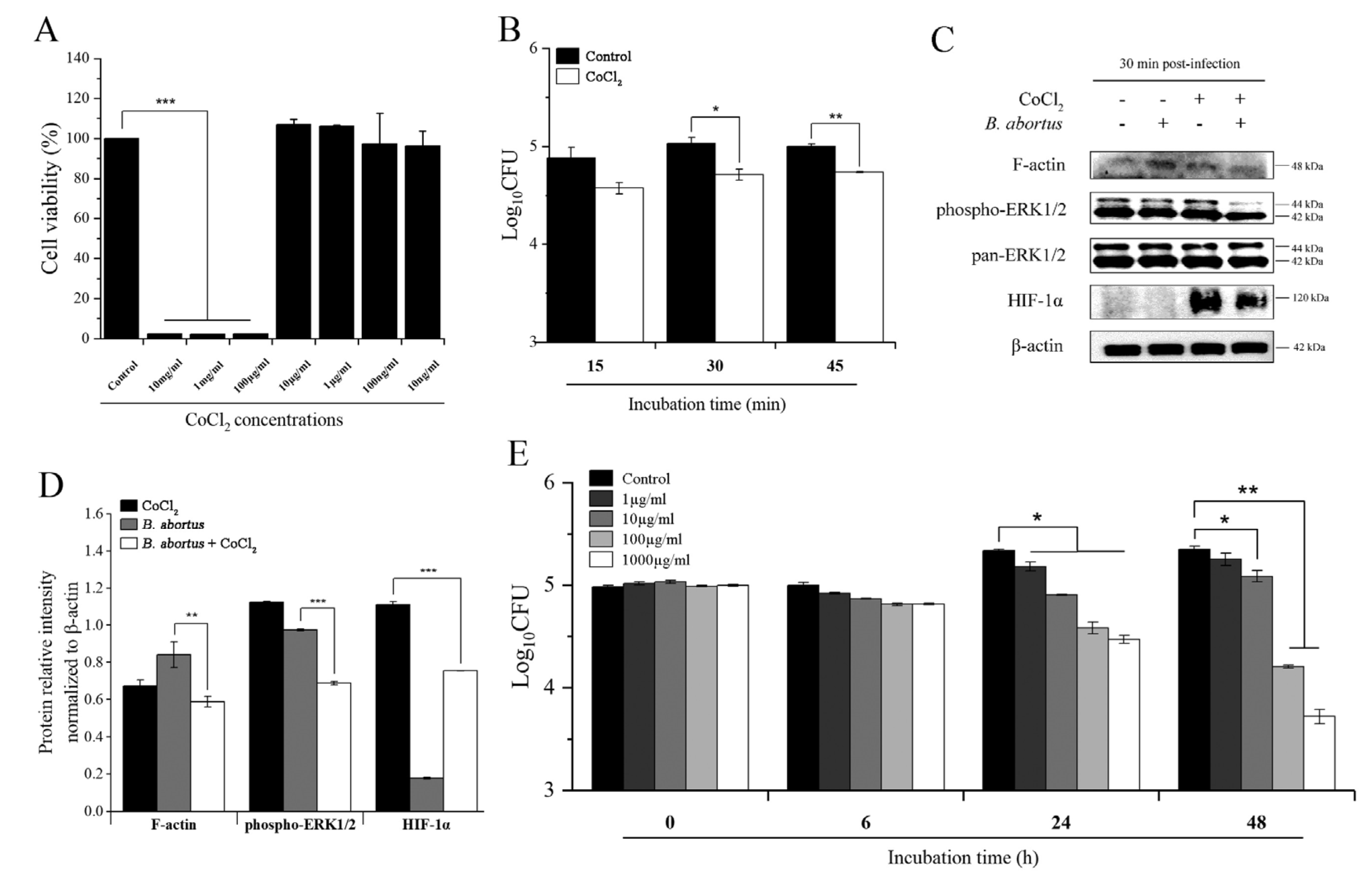
Figure 1. The effect of CoCl2 on RAW 264.7 cells and B. abortus viability and B. abortus internalization into macrophage cells. RAW 264.7 cells were pretreated with different concentrations of CoCl2 for 72 h, and the cell viability was evaluated using the MTT assay (A). Cells were pretreated with 10 µg/mL of CoCl2 for 6 h, and the number of invaded Brucella was determined at 15, 30, and 45 min pi (B). The involvement of F-actin, ERK1/2, and HIF-1α proteins in the phagocytosis signaling pathway at 30 min pi was determined using an immunoblotting assay (C). Protein intensity from the Western blot bands was analyzed by ImageJ software and normalized relative to β-actin (D). The direct bactericidal effect of CoCl2 on Brucella survival was evaluated for 0, 6, 24, and 48 h (E). The data are represented as the mean ± SD of duplicate samples from at least two independent experiments. Statistically significant differences relative to the control group are indicated by an asterisk (* p < 0.05, ** p < 0.01, and *** p < 0.001).
Pathogens 11 00596 g001
Figure 2. The effects of CoCl2 on the intracellular growth of B. abortus at 4 h pi. Cells were treated with 10 µg/mL of CoCl2, and the number of intracellular bacteria was determined at 4, 24, and 48 h pi (A). At 4 h pi, RNA extraction and cDNA synthesis were performed, followed by qRT-PCR, to check the transcripts of GLA, LAMP1, and LAMP2 genes (B). At the same time, the immunoblotting assay was used to check the expression of the HIF-1α protein using two concentrations of 1 and 10 µg/mL of CoCl2 (C), and the relative protein intensity normalized to β-actin was carried out by ImageJ software (D). The data are represented as the mean ± SD of duplicate samples from at least two independent experiments. Statistically significant differences relative to the control group are indicated by an asterisk (* p < 0.05 and *** p < 0.001).
Pathogens 11 00596 g002
Figure 3. The effects of CoCl2 and HIF-1α on the intracellular growth of B. abortus at 24 h pi. The transcripts of TNF-α and HEXB were determined using qRT-PCR at 24 h pi (A). At the same time, the immunoblotting assay was used to check the expression of the HIF-1α protein using two concentrations of CoCl2 (1 and 10 µg/mL) (B), and the relative protein intensity normalized to β-actin was carried out by ImageJ software (C). Cells were transfected with HIF-1α siRNA for 48 h, and qRT-PCR was utilized to evaluate the transfection efficacy (D). The successful knockdown of the HIF-1α gene was used to determine the bacterial intracellular growth at 4 and 24 h pi (E). The data are represented as the mean ± SD of duplicate samples from at least two independent experiments. Statistically significant differences relative to the control group are indicated by an asterisk (* p < 0.05 and ** p < 0.01).
Pathogens 11 00596 g003
Figure 4. Protection against B. abortus in B6 mice treated with CoCl2. B6 mice were orally administered with 20 or 40 mg/kg/day of CoCl2 for three days prior to infection. After that, the mice were IP challenged with B. abortus, followed by a continuous 14-day treatment regimen. At day 14 pi, the serum was collected to evaluate the production of cytokines IFN-γ (A) and IL-6 (B). At the same time, mice were sacrificed, the spleens and livers were collected and homogenized, and the number of CFU in each spleen and liver was counted (C,E). The weights of the spleen and liver were evaluated. (D,F) The data are represented as the mean ± SD of each group of five mice. Asterisks indicate statistically significant differences (* p < 0.05).
Pathogens 11 00596 g004
Table 1. Primer sequences used for qRT-PCR.
GeneDescriptionForward PrimerReverse Primer
GLAGalactosidase, alpha5′-GGC CAT GAA GCT TTT GAG CAG AGA TAC-3′5′-AGT CAA GGT TGC ACA TGA AAC GTT CCC-3′
LAMP1Lysosomal-associated membrane protein 15′-GGC CGC TGC TCC TGC TGC TGC TGG CAG-3′5′-ATA TCC TCT TCC AAA AGT AAT TGT GAG-3′
LAMP2Lysosomal-associated membrane protein 25′-AGG GTA CTT GCC TTT ATG CAG AAT GGG-3′5′-GTG TCG CCT TGT CAG GTA CTG AAT GG-3′
HIF-1αHypoxia inducible factor 1, alpha subunit5′-TCC CAT ACA AGG CAG CAG AA-3′5′-GTG CAG TAT TGT AGC CAG GC-3′
TNF-αTumor necrosis factor, alpha5′-CAG GTT CTG TCC CTT TCA CTC ACT-3′5′-GTT CAG TAG ACA GAA GAG CGT GGT-3′
HEXBHexosaminidase B5′-CCC GGG CTG CTG CTG CTG CAG GCG CTG-3′5′-GTG GAA TTG GGA CTG TGG TCG ATG CTG-3′
β-ACTINActin, beta5′-CGC CAC CAG TTC GCC ATG GA-3′5′- TAC AGC CC GGG GAG CAT CGT-3′
Publisher’s Note: MDPI stays neutral with regard to jurisdictional claims in published maps and institutional affiliations.